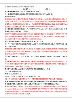
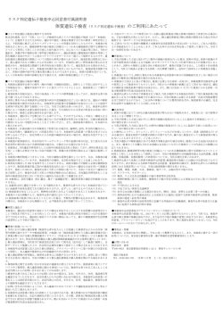
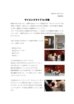
慈恵医大誌 2003; 118: 1-8.
【
説】
B 型肝炎ウイルス産生のメカニズム
クリオ電子顕微鏡(cryo-electron microscopy)により
解明されたコア粒子の立体構造と Large S 蛋白の役割
東京慈恵会医科大学
髙
合医科学研究センター・臨床医学研究所
橋
弘
吉 川 哲
矢
東京慈恵会医科大学内科学講座消化器・肝臓内科
戸
田 剛太郎
M OLECULAR MECHANISMS OF PRODUCTION OF HEPATITIS B VIRUS
THREE-DEMENSIONAL M AP OF CORE PROTEIN SHELL VISUALIZED
BY CRYO-ELECTRON MICROSCOPY
Hiroshi TAKAHASHI and Tetsuya YOSHIKAWA
Institute of Clinical Medicine and Research, The Jikei University School of Medicine
Gotaro TODA
Divistion of Gastroenterology and Hepatology, Department of Internal Medicine,
The Jikei University School of Medicine
The electron cryomicroscopy technique demonstrated three-demensional map of core
particle of hepatitis B (HB)virus using ice embedding. The icosahedral-shaped core particle
shows protruding spikes on the surface of the shell. Each spike is arranged with dimer of HB
core protein. It is of interest that the binding of[ S]-labeled large HBsAg (large S protein)
to HBcAg was inhibited by the synthetic peptides bearing the octapeptide sequence SLLGRMKG ,which was identified as a ligand to native core particle (HBcAg). Furthermore,the two
flanking amino acids to the SLLGRMKG octapeptide was found to increase the binding
affinity to HBcAg and this synthetic peptide (GSLLGRMKGA) inhibited the binding of large
S protein to HBcAg most effectively. Hep G2 cells were transfected with replication-competent HBV construct in the presence or absence of peptide. HBV production was inhibited by
the inhibitory peptides (SLLGRMKG, SLLGRMKGA,GSLLGRMKGA),but not by irrelevant
peptide (ALLTRILG). The relative inhibitory effects of the three peptides reflected their
affinities for HBcAg. The pre-S regions of large S protein were found to be important for
viral assemblyand attachment to the host cell during infection. We demonstrated that specific
interactions between the outer surface of the core and the inner surface of the envelope are
likely to guide correct assembly of the virus and stabilize the resulting particle. Therefore,
peptides that bind to core protein may inhibit the assembly of HBV particles.
(Tokyo Jikeikai M edical Journal 2003; 118: 1-8)
Key words: hepatitis B virus, core particle, large S protein, peptides, anti-virus effect
2
髙橋
ほか
I. はじめに
B 型肝炎ウイルス(HB ウイルス)は,不顕性感
染から致死的な劇症肝炎に至るまで色々な形の感
染症を起こすことが知られている .B 型急性肝
炎は通常,ウイルスの episomal な増殖によって
起こり,ウイルスと感染細胞の排除によって治癒
すると えられている.
また,無症候性キャリアー
が存在することから,B 型肝炎ウイルスには直接
の肝細胞障害性はなく,ウイルス抗原に対する免
疫反応が,肝炎の発症に重要とされている .日本
では近年減少の傾向にあるとは言え,世界的には
未だ 2 億人以上 HB ウイルス慢性キャリアーが
いることから,HB ウイルス感染症はグローバル
な医療問題として重要である.また,HB ウイルス
の慢性キャリアーは正常人の 200 倍以上のリスク
で肝細胞癌を発症することが疫学的に明らかに
なっており,最近では C 型肝炎の癌化に HB ウイ
Fig.1. Structure of HBV Genome
The hepatitis B virus is a circular,partially
double-stranded DNA, 3.2 kb in length,
which replicates via an RNA intermediate
ルスの潜在的な感染が関与している可能性も指摘
and has the capacity to integrate randomly
されている.
into the host genome. There are four open
本 稿 で は ク リ オ 電 子 顕 微 鏡(cryo-electron
microscopy)により初めて可視化されたコア粒子
の立体構造を紹介するとともに,感染力を持つ
reading frames in this virus; S, C, X, and
polymerase (P) ORF. SORF encodes three
proteins, preS1, preS2 and S protein.
These are envelope proteins of this virus.
HB ウイルスが小胞体において産生される機序に
HB ウイルスのエンベロープ蛋白の 1 つである
Core ORF encodes two proteins, precore
large S 蛋白が必須の役割を果たすことを明らか
にしたので,この新知見を含めて HB ウイルスの
cleocapsid and precore protein produce
HBeAg after the cleavage of 19 -aminoacid
and core antigen. Core protein forms nu-
増殖および産生機序について概説する.
hydrophobic signal peptide and the 34aminoacid arginine-rich C-terminal
II. HB ウイルスゲノムの構造
regions. X ORF encodes for X tran-
HB ウイルスのゲノムは全長約 3.2 kb の環状
二本鎖 DNA で,その一部は一本鎖構造
(不完全二
in a hepatocarcinogenesis. Polymerase
(P) ORF encodes for a putative reverse
重鎖構造)になっている.ウイルスゲノムには,蛋
transcriptase. There are four trans-
白として翻訳可能な 4 つの open reading frame
(ORF)が存在する(Fig.1)
.S ORF は,HBs 抗
sactivator protein which may be involved
lational reading frames. The largest transcript,3.5 kb mRNAs contain precore,core
mRNA
and pregenome RNA. This
原を構成する 3 種類のエンベロープ蛋白,すなわ
pregenome RNA serves as a template for
ち,large S 蛋白
(pre-S1 と pre-S2 と S 領域を含
む)
,middle S 蛋白
(pre-S2 と S 領域を含む)
,そ
HBV DNA and important for replication of
して small S 蛋白(S 領域のみからなる)をコー
ドする.コア ORF は,コア蛋白とプレコア蛋白を
コードする.コア蛋白はコア粒子を構成し,プレ
コア蛋白は 19 個の疎水性シグナルペプチドと C
末端のアルギニンに富んだ 34 個のアミノ酸残基
the virus. 2.4 kb mRNA consist of a pre S1
mRNA and pre S2, and S mRNA is 2.1 kb.
Shortest 0.7 kb transcript is X mRNA.
B 型肝炎ウイルス産生のメカニズム
3
が切断された後に HBe 抗原となる.X ORF は,
ウイルスの増殖や肝細胞癌の発症に関与すると
により,ウイルスゲノムの(−)鎖を鋳型として,
えられている X 蛋白をコードする.また,ポリメ
で 4 種類(3.5 kb, 2.4 kb, 2.1 kb および 0.7 kb
mRNA)に 類されている(Fig.2).とくに,3.5
ラーゼ ORF は,逆転写酵素活性を有する DNA
ポリメラーゼ蛋白をコードする.
III. 肝細胞内における HB ウイルスの増殖様式
複数の mRNA が作られる.これらは長さの違い
kb mRNA の中で最も長い mRNA であるプレゲ
ノム RNA は,ウイルス DNA の(−)鎖の鋳型と
なり,ウイルスの複製に重要である.また,3.5 kb
HB ウイルスは特異的レセプターを介して肝細
胞膜に結合し,エンドサイトーシスにより細胞内
mRNA にはコア mRNA,プレコア mRNA およ
びポリメラーゼ mRNA など も 含 ま れ る.コ ア
に侵入すると えられている(Fig.2)
.肝細胞内
に入る時にエンベロープ(HBs 抗原)が外れ,HB
mRNA か ら は HBc 抗 原 が 翻 訳 さ れ,autoポリ
assemblyにより球形のコア粒子が作られる.
ウイルスゲノムを含んだコア粒子となる.
さらに,
メラーゼ mRNA からは DNA ポリメラーゼ蛋白
コア粒子は細胞核に移行し,HB ウイルスの不完
が翻訳される.
全二重鎖の DNA は DNA ポリメラーゼの働きで
完全二重鎖となる.
そして,細胞由来の RNA ポリメラーゼの働き
コア粒子は,プレゲノム RNA と DNA ポリメ
ラーゼを粒子内に取り込むと,DNA ポリメラー
ゼの N 末端をプライマーとして利用し,DNA ポ
Fig.2. Life .00Cycle of HB virus
This catoon demonstrates a natural life cycle of HBV. HBV enters hepatocytes by a
putative receptor mediated endocytosis process. After entering the cell, it releases the
nucleocapsid (core particle)from envelope. The viral genome is then translocated to the
nucleus. It is converted into full-length circular HBV DNA by DNA polymerase. Four
species of mRNAs are trancribed using cellular RNA polymerase II, and only 3.5 kb
pregenomic RNA is encapsideted into subviral cores and serves as a template for HBV
DNA replication. The nimus strand of HBV DNA is synthesized by reverse transciptase
in the core particle, and then the double stranded HBV DNA is synthesized. The size of
mature HBV DNA is 3.2 kb in size.HBV particles are secreted after they are encapsulated
with envelope protein in endoplasmic reticulum. It is important to note that some of the
intracellular HBVs are recycled into nucleus and produce HBV.
4
髙橋
リメラーゼの逆転写活性により,ウイルス DNA
の(−)鎖を合成する.さらにこの(−)鎖を鋳
型として,DNA ポリメラーゼ活性によりウイル
ス DNA の(+)鎖の合成が開始される.しかし,
その反応は途中で止まるため,HB ウイルスゲノ
ムは不完全二重鎖構造となる.
IV. デーン粒子の組み立ては小胞体で行われる
ほか
れる(Fig.2)
.そして,できたデーン粒子はゴル
ジ体を経て細胞外に 泌される.また,コア粒子
の一部は核内にリサイクルされ,ウイルス粒子を
再び産生する(Fig.2)
.
V. コア粒子の表面には規則的な突起構造が
存在する
クリオ電子顕微鏡で得られた数千におよぶ画像
HB ウイルスゲノムを持ったコア粒子が感染力
をコンピュータでイメージ処理することにより,
を持った完全なウイルス粒子(デーン粒子)とな
コア粒子の詳細な立体構造が明らかになった .
るためには,HB ウイルスに特異的なエンベロー
クリオ電子顕微鏡は X 線解析のように目的の
プに被われる必要がある.また,ウイルス粒子の
子を結晶化する必要がなく,さらに複数の 子の
組み立て(assembly)を正しくガイドし,安定さ
相互作用を,少なくとも 10Åの解像度で三次元的
せるためには,コア粒子の表面とエンベロープの
内側との間に特異的な相互作用が必要と えられ
に解析できる新しい手法である.Fig.3 にコア粒
子の立体構造を示す .コア粒子の内部は空洞構
る.
ウイルス粒子形成に重要なこのプロセスは,コ
造になっており,その内部にプレゲノム RNA が
ア粒子が細胞質から小胞体を通過する際に達成さ
入っている.また,コア粒子の表面には規則的な
Fig.3. Three-demensional M ap of Whole Core Protein Shell Visualized by Cryo-electron M icroscopy
This figure demonstrates three-demensional map of HB core particle visualized by cryoelectron microscopy. The electron cryomicroscopy technique employs ice embedding.
This technique is able to look at structures that do not crystallize, and which can not
therefore be studied by X-ray crystallography. In addition, solving the atomic structure
is possible (extends to atomic resolution of 3 to 4Å). The icosahedral-shaped core
particle shows protruding spikes on the surface of the shell. Each spike is arranged with
dimer of HB core protein. The shell of the core particle is penetrated by small pore.
Inhibitory peptides are show in red color at the tips of spikes.(EM BO J,Volume 17,1998)
B 型肝炎ウイルス産生のメカニズム
5
突起構造(spike)が見られる.この突起構造は 2
子のコア蛋白の dimer で構成されており,コア
蛋白の α-helix を形成する部位がヘアピン状に
U ターンすることで,突起構造を形成している.
ちなみに,コア蛋白で抗原性が最も強い領域,す
なわちアミノ酸残基で 74 から 83 番目の領域の
immunodominant region は,この突起構造の先
端に位置する.また,突起構造と突起構造の間に
は多数の細孔(pore)が存在するが(Fig.3)
,ウ
イルス DNA の合成に必要な物質はこの細孔を介
して自由にコア粒子内に出入りすると
えられ
る.
VI. large S 蛋白はデーン粒子形成に不可欠
である
HB ウイルスのエンベロープは,HBs 抗原を含
む小胞体由来の脂質二重膜で構成されている.
HBs 抗原には large S 蛋白,middle S 蛋白,そし
て最も大量に存在し HBs 抗原の主たる構成成
Fig.4. Topology of HBs Large Protein at the
Membrane of Endoplasmic Reteculum
The pre-S regions (pre-S1 and S2) are
believed to play a role in both viral assembly and attachment to the host cell. M ost
of these pre-S residues are displayed on the
surface of mature virions and hence would
be expected to be translocated into the
endoplasmic reticulum (ER) lumen during
biosynthesis. However, contrary to this
expectation, it was demonstrated by the
group of Don Ganem using a coupled in
vitro translation/translocation system that
virtually all pre-S residues of the large
protein are cytoplasmically disposed in the
initial translocation products.
である small S 蛋白の 3 種類が存在する.
1991 年, 原らのグループ と Ganem らのグ
ループ は,デーン粒子の産生に large S 蛋白の
検討により,pre-S1 領域と pre-S2 領域は実は小
胞体の細胞質側に局在することが明らかにされた
存在が絶対不可欠の条件であることを示した.す
なわち,large S 蛋白に欠損が生じるとデーン粒
のである.Fig.4 に小胞体における large S 蛋白
の局在様式を模式的に示す.
図の中央に小胞体膜,
子の産生されないことが明らかにされた.この報
下に細胞質,上に小胞体の内腔側を示す.デーン
告は,pre-S1 と pre-S2 領域を持つ large S 蛋白
がウイルスの感染だけでなく,ウイルスの組み立
粒子の表面に存在するため,小胞体では内腔側に
て(assembly)にも関与することを示した始めて
の報告である.しかし,large S 蛋白がどのような
S2 領域は,この図のように両者ともに細胞質側に
局在することが明らかにされた .
機序でウイルスの組み立てに関わるかについては
全く不明であった.
VII. large S 蛋白の pre-S1 ╱ pre-S2 領域
は小胞体の細胞質側に局在する
局在すると
えられていた pre-S1 および pre-
VIII. コア粒子とエンベロープの特異的相互作
用は large S 蛋白を介する
Random-hexapeptide phage display library
を用いて,コア粒子と結合するペプチド配列をス
HB ウイルスの肝細胞への吸着と感染に関与す
る と え ら れ て い る pre-S1 や pre-S2 領 域 は
クリーニングした結果,コア粒子に特異的に結合
デーン粒子の表面に局在するため,小胞体におい
粒子と large S との結合を抑制することが明らか
となった(Fig.5)
.また,この抑制ペプチドの片
てはその内腔側に局在すると えられていた.し
するペプチド SLLGRM KG が濃度依存的にコア
かし,1994 年,小胞体における large S 蛋白の
pre-S1 と pre-S2 領域のトポロジー(局在様式)
側あるいは両側にアミノ酸残基を追加してコア粒
について,それまでの定説を覆す研究成果が 2 つ
.すなわ
らに強い抑制効果を示した(Fig.5)
.これに対し
て,コア粒子と結合しない small S 蛋白由来の非
ち, 子生物学と生化学的手法を駆 した詳細な
特異的ペプチド配列(ALLTRILG)では,全く抑
のグループにより同時に報告された
子とのアフィニティを高めた合成ペプチドは,さ
6
髙橋
ほか
Fig.5. Inhibiton of HBs Large Protein Binding to HBcAg by the Synthetic Peptide GSLLGRMKGA
The binding of[ S]-labeled HBs large protein to HBcAg was inhibited by the synthetic
peptides bearing the octapeptide sequence SLLGRM KG ,which was identified as a ligand
to native core particle (HBcAg). Synthetic peptides bearing the octapeptide sequence
SLLGRM KG inhibited the binding of large S protein to HBcAg in a dose-dependent
manner. Furthermore,the two flanking amino acids to the SLLGRM KG octapeptide was
found to increase the binding affinity to HBcAg and this synthetic peptide (GSLLGRMKGA) inhibited the binding of large HBsAg to HBcAg most effectively. In contrast, a
peptide, ALLTRILG, which is identified as the 21-27 amino acid sequence within the S
region,did not show any inhibitory effect. The peptide,ALLTRILG,includes amino acids
of 21-27 of the S region of HBsAg. It shows a highly significant sequence match of five
residues with the inhibitory peptide, ALLGRM KG, but had no inhibitory effect on the
interaction between HBcAg and large HB Ag.
制効果が認められなかった.
が必要と えられる.また,このプロセスはコア
さらに,クリオ電子顕微鏡を用いて抑制ペプチ
ドがコア粒子のどの部
粒子が小胞体を通過する過程で起こる.我々の研
に結合するかを検討した
究結果により,小胞体におけるこの特異的相互作
結果,Fig.3 に示すように,抑制ペプチドはコア粒
子の突起構造の先端に特異的に結合した .
用がコア粒子の突起構造の先端と large S 蛋白の
IX. コア粒子の突起構造に結合するペプチド
は HB ウイルスの産生を抑制する
次に,各種合成ペプチドの抗ウイルス作用を検
pre-S1 と pre-S2 領域との間で起こり,デーン粒
子が形成される事が明らかとなった(Fig.7)
.
また,コア粒子の突起構造の先端に特異的に結
合する抑制ペプチドが,HB ウイルスの産生を抑
制し,強い抗ウイルス作用を持つ事を明らかにし
討した.Fig. 6 に HB ウイルス DNA をトランス
フェクションした培養肝細胞(Hep G2 細胞)を用
た.
いた実験結果を示す.抑制ペプチド(SLLGRM KG, SLLGRMKGA お よ び GSLLGRM KGA)
する小さな
は,HB ウイルスの産生を強く抑制した.
できる可能性が示された.
X. ま
と
め
以上の結果より,ウイルスの assemblyを阻害
子を用いて,ウイルスの産生をブ
ロックする新しいタイプの抗ウイルス療法を開発
原稿の
正と図表の作成に尽力を頂いた野呂裕子氏,宇
賀英子氏,および
HB ウイルス粒子の形成には,コア粒子の表面
とエンベロープの内側との間に特異的な相互作用
説執筆の機会を与えて下さった川村将
弘教授,共同研究者の Kenneth M urray教授ならびに
Tony Crowther 教授に深謝いたします.
B 型肝炎ウイルス産生のメカニズム
Fig.6. Inhibition of HBV Production by Inhibitory Peptides in HBV-transfected Hep G2 Cells
In order to determine whether these peptides could interfere with virus production by
blocking its assembly, Hep G2 cells were transfected with replication-competent HBV
construct in the presence or absence of peptide. We introduced 10μg of pHBV-HTD
DNA and 500μM of peptide into the Hep G2 cells simultaneously by permeabilization of
cell membrane. Following 72hrs incubation, HBV production in the culture supernatant
was examined. For quantification of the production of HBV in the culture supernatant,
a radioactive PCR method was used after DNase digestion and immunoprecipitation of
HBV particles with a monoclonal antibody specific for HBsAg. As demonstrated in this
figure,HBV production was inhibited by the inhibitory peptides (SLLGRM KG,SLLGRMKGA,GSLLGRM KGA),but not by irrelevant peptide (ALLTRILG). The relative inhibitory effects of the three peptides reflected their affinities for HBcAg.
Fig.7. Assembly of Dane Particle at endoplasmic membrane
Large S protein is required for Dane particle formation,and it is postulated that sequence
in pre-S1 and/or per-S2 region interacts with the surface of core protein. These pre-S
regions of large S protein were found to be important for viral assembly and attachment
to the host cell during infection.
Since pre-S sequences are found on the external
surface of the virion envelop, during of following budding, a dramatic reorganization of
either the envelope proteins or the lipid bilayer (or both components)must occur to allow
surface display of these sequences. In some molecules, the pre-S region is translocated
across the lipid bilayer and exposed on the virus surface. We demonstrated that specific
interactions between the outer surface of the core and the inner surface of the envelope are
likely to guide correct assembly of the virus and stabilize the resulting particle. Therefore, peptides that bind to core protein may inhibit the assembly of HBV particles.
7
8
髙橋
文
献
1) Tiollais P, Pourcel C, Dejean A. The hepatitis
B virus. Nature 1985; 317: 489 -95.
2) Takahashi H, Fujimoto J, Hanada S, Isselbacher KJ. Acute hepatitis in rats expressing
human hepatitis B virus transgenes. Proc
Natl Acad Sci USA 1995; 92: 1470-74.
3) Conway JF, Cheng N, Zlotnick A, Wingfield
PT,Stahl SJ,Steven AC.Visualization of a 4helix bundle in the hepatitis B virus capsid by
cryo-electron microscopy. Nature 1997; 386:
91-4.
4) Bottcher B,Tsuji N,Takahashi H,Dyson MR,
Zhao S, Crowther RA, et al. Peptides that
block hepatitis B virus assembly: analysis by
cryomicroscopy,mutagenesis and transfection.
EMBO J 1998; 17: 6839 -45.
ほか
5) Ueda K, Tsurimoto K, M atsubara K. Three
envelope proteins of hepatitis B virus: large S,
middle S, and major S proteins needed for the
formation of Dane particles. J Virol 1991;
65: 3521-9.
6) Bruss V, Ganem D. The role of envelope proteins in hepatitis B virus assembly. Proc Natl
Acad Sci USA 1991; 88: 1059 -63.
7) Ostapchuk P, Hearing P, Ganem D. A dramatic shift in the transmembrane topology of
a viral envelope glycoprotein accompanies
hepatitis B viral morphogenesis. EM BO J
1994; 13: 1048-57.
8) Bruss V, Lu X, Thomssen R, Gerlich WH.
Post-translational alterations in transmembrane topology of the hepatitis B virus large
envelope protein. EM BO J 1994; 13: 2273-9.
© Copyright 2026 Paperzz
![B型肝炎ワクチンの説明[PDF:66kb]](http://s3.paperzz.com/store/data/005457145_1-8e23c37afe8fd85c13ee152f49c19e0c-250x500.png)