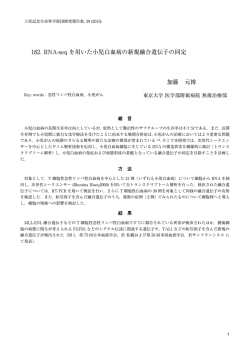
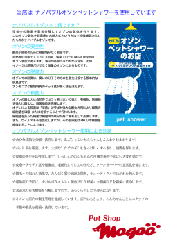
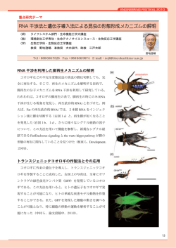
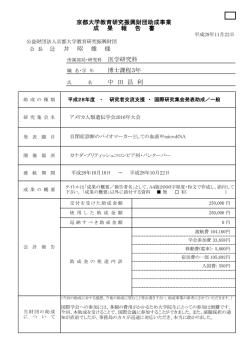
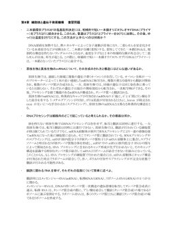
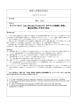
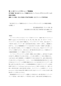
CODE No. 5345-100, 5345-200 For Research Use Only, Not for use in diagnostic procedures. Plant Viral dsRNA Enrichment Kit Introduction Visual detection of the presence or absence of viral infection in plants by observing plant appearance is difficult, unlike in the case of bacterial or fungal diseases. Currently, plant virus diagnostics such as immunological techniques using antibodies (e.g., ELISA) and/or genetic techniques using nucleic acid-based methods (e.g., PCR), are available for known viruses but not for the unknown ones. dsRNA isolation, exhaustive amplification, cloning, and sequencing (DECS) is a technique for isolating double-stranded RNA derived from plant RNA viruses using a unique dsRNA binding protein, followed by identification of the viral species by obtaining its sequence information. The DECS method was developed by the Iwate Biotechnology Research Center (IBRC), Iwate, Japan. Product Description The Plant Viral dsRNA Enrichment Kit is used to perform the plant viral dsRNA enrichment step in the DECS procedure. The kit uses a dsRNA-binding (DRB) protein derived from Arabidopsis thaliana to bind and enrich viral dsRNA from infected plants. Enriched dsRNA can easily be observed by electrophoresis. Crude tissue lysate of infected plants can be used as the starting material. If it is necessary to identify the infecting virus, the recovered dsRNA is reverse-transcribed, and the resultant cDNA library is amplified by PCR and then sequenced. Kit Components 50 tests 5345-100 : 1. DRB protein 1 mg/mL 1 mL 2. Positive Control dsRNA 500 ng/μL 10 μL 5345-200 : 3. Binding Buffer 100 mM Tris-HCl (pH 7.0), 100 mM NaCl, 10 mM MgCl2 50 mL 4. Lysis Buffer 1x Binding Buffer, 1% Tween-20 25 mL 5. Wash Buffer 1x Binding Buffer, 0.1% Tween-20 50 mL 6. Elution Buffer 10 mM Tris-HCl (pH 8.0), 5 mM EDTA, 0.5% SDS 7. TE Buffer 10 mM Tris-HCl (pH 8.0), 1 mM EDTA 1.5 mL (× 2) 8. Glutathione resin 50% slurry in Binding Buffer containing preservative 1.5 mL (× 2) Storage and Stability 5345-100: Stable for 12 months from the date of receipt at -20°C to -30°C. 5345-200: Stable for 12 months from the date of receipt at 2°C to 8°C. -1- 1 mL Materials required but not provided ・ Pipettors and pipette tips* ・ 1.5-mL microcentrifuge tubes* *we recommend using low-retention type tips and tubes. ・ Microcentrifuge capable of 2,000-10,000 × g ・ Tube rotator ・ Ethidium bromide ・ DNA ladder markers ・ Gel loading dye ・ Agarose gel for electrophoresis ・ TAE buffer for electrophoresis ・ Electrophoresis device ・ UV transilluminator ・ RNA extraction reagent (e.g., Rizo (Japan), code no. RS-0001N) ・ WTA kit (e.g., Sigma-Aldrich (USA), code no. WTA1 or WTA2) -2- Procedure Summary -3- Protocol 1. Sample preparation We recommend the use of plant materials in which the virus is expected to replicate at high levels, such as growing leaves and stems, instead of parts already showing disease symptoms. <For total RNA> This kit does not contain reagents for total RNA extraction. We recommend that total RNA be extracted using the acid-guanidium-phenol-chloroform (AGPC) method or commercial reagents based on it (e.g., TRIzol, Life Technologies). However, some plants contain significant amounts of compounds, such as polyphenolics and complex polysaccharides, that interfere with RNA extraction. In this case, it is advisable to use RNA extraction kits (e.g., RNA Suisui Series, Rizo, Japan) optimized for plant materials. <For crude extracts> 1) Homogenize 0.2-1.0 g of plant tissue with a mortar and pestle in 500 μL of Lysis Buffer. 2) Centrifuge at 10,000 × g for 5 min at 4°C and transfer the supernatant to a new 1.5-mL tube (be careful not to transfer any precipitate). *The more plant tissue used, the easier it is to observe the results of electrophoresis. *If using crude extracts, we recommend using a healthy plant sample as a negative control, because non-specific bands such as genomic DNA and ribosomal RNAs often appear in electrophoresis. For sequencing, we recommend the use of purified total RNA in order to avoid amplifying non-specific products co-isolated in the eluted sample. 2. dsRNA enrichment 1) Add Binding Buffer to total RNA (>50 μg) to bring the total volume to 500 μL. *If using crude extracts, start from Step 2. *If desired, use 2 μL (1 μg) of Positive Control dsRNA (blue cap) per assay. 2) Add 20 μL of DRB protein (yellow cap), mix by inverting, and incubate for 10 min at room temperature (RT, 20-25°C). 3) Mix glutathione resin (brown cap) well by inverting tube several times, then add 60 μL to the mixture from step 2. 4) Mix by inverting for 30 min at RT. 5) Centrifuge at 2,000 × g for 10 sec at RT and discard the supernatant. 6) Add 500 μL of Wash Buffer. 7) Centrifuge at 2,000 × g for 10 sec at RT and discard the supernatant. 8) Repeat steps 6-7 once again (for a total of two times). -4- Further procedures depend on the desired method of analysis. <Protocol for agarose gel electrophoresis> 9) Add 20 μL of Elution Buffer (red cap) to the washed resin from step 8 and mix by tapping. 10) Incubate for 5 min at RT. 11) Centrifuge at 2,000 × g for 10 sec at RT and transfer the supernatant to a new 1.5-mL tube. 12) Add an appropriate amount of gel loading dye to half or all of the eluted solution. 13) Place a 0.7-1.0% agarose gel in the electrophoresis apparatus. 14) Add enough TAE buffer to cover the gel with about 2-3 mm of buffer. 15) Apply the samples to the gel and run with a DNA ladder. 16) Submerge the gel in 0.5-1 µg/mL ethidium bromide (EtBr). * Note that EtBr is a carcinogenic chemical. Be sure to use gloves when handling it. 17) Bands can be visualized using a UV-illuminator. * The electrophoretic mobility shift of RNA is slightly different from that of DNA. However, a DNA ladder can be used as a size marker if accurate measurement of RNA length is not necessary. * Post-staining with EtBr is preferable because pre-staining tends to result in high background. * The remaining half of the sample from step 12 can be used as a template for whole transcriptome amplification. In this case, purification of the dsRNA by phenol/chloroform extraction and ethanol precipitation is recommended because of the presence of SDS in the Elution Buffer. (Appendix) Protocol for phenol/chloroform extraction and ethanol precipitation a) Add TE buffer to the extracted dsRNA solution to bring the total volume to 200 μL. b) Add 200 μL of phenol/chloroform (1:1) and vortex. c) Centrifuge at 10,000 × g for 3 min at 4°C and transfer the upper aqueous layer to a new 1.5-mL tube. d) Initially add 20 μL of 3M NaOAc (pH 5.2), then add 500 μL of 100% ethanol (EtOH). e) Mix well by inverting the tube and incubate for 20 min at -20°C. f) Centrifuge at 10,000 × g for 10 min at 4°C and discard the supernatant. g) Add 500 μL of 70% EtOH and mix by inverting the tube. h) Centrifuge at 10,000 × g for 3 min at 4°C and discard the supernatant. i) Air-dry the pellet for 5-10 min and dissolve in an appropriate volume of RNase-free water. <Protocol for sequence analysis> 9) Wash the resin a third time by adding 500 μL of Binding Buffer. 10) Centrifuge at 2,000 × g for 10 sec at RT and discard the supernatant. 11) Add 50 μL of TE Buffer (clear cap). 12) Incubate for 3 min at 99°C and quickly chill on ice water for 5 min. 13) Centrifuge at 2,000 × g for 10 sec at RT and transfer the supernatant to a new 1.5-mL tube. -5- 14) The extracted and denatured dsRNA can be used as a template for whole transcriptome amplification. Store at -80°C if not used immediately. Further DECS procedures include whole transcriptome amplification, cloning, and sequencing or next-generation sequencing. For more information, see instructions for each instrument and reagent. Results of agarose gel electrophoresis Nucleic acids such as DNA and RNA can be separated on an agarose gel by electrophoresis according to their molecular weights and then visualized with EtBr staining. The isolation of viral dsRNA from an infected plant can be confirmed by electrophoresis if it is efficiently enriched using this product (the molecular weight will depend on the viral species). We used this kit to extract dsRNA from two types of sample: a crude extract from healthy tobacco leaves and one from tobacco leaves infected with cucumber mosaic virus (CMV). The result shows successful detection of a specific band of dsRNA from the CMV-infected sample (lane 2). The analysis included the 500-bp positive control (PC) dsRNA in lane 3 for protocol verification. 1) 2) 3) References Atsumi, G., et al., Methods Mol. Biol. 1236, 27-37 (2015) Kobayashi, K., et al., J. Gen. Plant Pathol. 79, 56-63 (2013) Kobayashi, K., et al., J. Gen. Plant Pathol. 75, 87-91 (2009) Manufacturer MEDICAL & BIOLOGICAL LABORATORIES CO., LTD. URL http://ruo.mbl.co.jp/ e-mail [email protected] TEL 052-238-1904 -6- CODE No. 5345-100, 5345-200 For Research Use Only, Not for use in diagnostic procedures. Plant Viral dsRNA Enrichment Kit はじめに 植物のウイルス感染はカビやバクテリアと異なり、見た目での判定が困難です。また、従来の植 物ウイルスの検査法は既知のウイルスを対象とした抗体(ELISA など)や遺伝子情報(PCR な ど)を用いた方法であるため、未知のウイルスは検出できません。 公益財団法人岩手生物工学研究センターが開発した網羅的 RNA ウイルス検出技術(dsRNA isolation, exhaustive amplification, cloning and sequencing; DECS)は、独自の 2 本鎖 RNA 結合タン パク質を用いて、ウイルス由来の 2 本鎖 RNA(dsRNA)の単離を行い、その遺伝子配列情報を 得ることで、ウイルス種を同定する技術です。 特徴 本キットは、DECS 法における植物 RNA ウイルスの dsRNA 濃縮のステップに必要な試薬をキッ ト化した製品です。本キットでは dsRNA に対して結合活性を有するシロイヌナズナ由来の DRB タンパク質を利用することで、感染ウイルスの dsRNA を単離することが可能です。感染した枝 葉を直接サンプルに用いることも可能です。単離した dsRNA は、直接電気泳動に使用できます ので、感染の有無の判別に有用です。さらに必要であれば、dsRNA を逆転写・網羅的に増幅後、 クローニング、シーケンス解析することにより、ウイルス種の同定も可能です。 キット構成 50 テスト 5345-100 : 1. DRB protein 1 mg/mL 1 mL 2. Positive Control dsRNA 500 ng/μL 10 μL 5345-200 : 3. Binding Buffer 100 mM Tris-HCl (pH 7.0), 100 mM NaCl, 10 mM MgCl2 50 mL 4. Lysis Buffer 1x Binding Buffer, 1% Tween-20 25 mL 5. Wash Buffer 1x Binding Buffer, 0.1% Tween-20 50 mL 6. Elution Buffer 10 mM Tris-HCl (pH 8.0), 5 mM EDTA, 0.5% SDS 7. TE Buffer 10 mM Tris-HCl (pH 8.0), 1 mM EDTA 1.5 mL (× 2) 8. Glutathione resin 50% slurry in Binding Buffer containing preservative 1.5 mL (× 2) 1 mL 保存と有効 5345-100: -20°C から-30°C にて保存してください。有効期間は出荷後 12 ヶ月です。 5345-200: 2°C から 8°C にて保存してください。有効期間は出荷後 12 ヶ月です。 -7- 本製品以外に必要な器具・試薬 ・ ピペットマンとチップ* ・ 1.5 mL マイクロチューブ* *チップとマイクロチューブは、低吸着タイプのご使用をお勧めいたします。 ・ 小型遠心機(2,000-10,000 × g) ・ マイクロチューブローテーター ・ エチジウムブロマイド ・ DNAラダーマーカー ・ DNAローディングダイ ・ 電気泳動用アガロースゲル ・ TAEバッファー ・ 電気泳動装置 ・ UVトランスイルミネーター ・ RNA抽出試薬(e.g., Rizo (Japan), code no. RS-0001N) ・ WTA試薬(e.g., Sigma-Aldrich (USA), code no. WTA1 or WTA2) -8- 手順概略 -9- プロトコール 1. サンプル調製 植物サンプルは、病徴が目視で観察できる部位よりも、新しい葉や茎などウイルスが増殖してい る可能性の高い組織を使用することをお勧めします。 <全 RNA を抽出する場合> 本キットには RNA 抽出試薬が含まれていません。AGPC 法やそれに基づいた市販の試薬(Life Technologies 社 TRIzol など)による抽出をお勧めします。ただし植物によってはポリフェノール 性成分や複合多糖類を多く含み、RNA 抽出が困難な場合があります。この場合は、植物組織用 の RNA 抽出キット(リーゾ社 RNA すいすいシリーズなど)をお試しください。 <粗汁液を調製する場合> 1) 植物組織 0.2~1.0 g に 500 μL の Lysis Buffer を加えて乳鉢と乳棒で摩砕します。 2) 粗汁液を 10,000 × g、5 分、4°C で遠心し、上清を新しい 1.5 mL チューブに移します。 (沈殿 を取らないように注意してください。) *植物組織量が多いほど電気泳動で dsRNA のバンドが確認しやすい傾向にあります。 *粗汁液を用いる場合は、電気泳動でゲノム DNA やリボゾーム RNA などの非特異バンドが 出やすい傾向にありますので、健全な植物サンプルと比較することをお勧めします。また、 シーケンス解析する場合は、非特異産物の増幅を避けるために、精製した全 RNA を使用す ることをお勧めします。 2. dsRNA 濃縮 1) 全 RNA(50 μg 以上)を取り、Binding Buffer で容量を 500 μL に合わせます。 *粗汁液を調製する場合は次の 2)から始めてください。 *必要に応じて Positive Control dsRNA(青キャップ)を 1 回 2 μL(1 μg)使用してください。 2) DRB protein(黄キャップ)を 20 μL 加えて転倒混和し、室温(20~25°C)で 10 分間静置し ます。 3) Glutathione resin(グルタチオン樹脂) (茶キャップ)を数回転倒して十分に混和し、60 μL 加 えます。 4) 室温で 30 分間転倒混和します。 5) 2,000 × g、10 秒、室温で遠心し、上清を除去します。 6) Wash Buffer を 500 μL 加えます。 7) 2,000 × g、10 秒、室温で遠心し、上清を除去します。 8) 6)-7)をさらに 1 回繰り返します。(計 2 回) 以降の操作は、目的に応じて手順が異なります。 -10- <電気泳動で検出する場合> 9) 8)の樹脂に Elution Buffer (赤キャップ) を 20 μL 加えてタッピングで混合します。 10) 室温で 5 分間静置します。 11) 2,000 × g、10 秒、室温で遠心し、上清を新しい 1.5 mL チューブに移します。 12) 半量または全量に適量の DNA loading dye を加えます。 13) 0.7~1.0%のアガロースゲルを泳動装置にセットします。 14) TAE バッファーでゲルを浸します。 (ゲルを 2~3 mm 超える程度) 15) ゲルにサンプルをアプライし、DNA ラダーマーカーとともに、電気泳動します。 16) エチジウムブロマイド(0.5~1 μg/mL)でゲルを染色します。 *エチジウムブロマイドは発がん性物質です。扱う際は手袋を装着して下さい。 17) UV トランスイルミネーターでバンドを観察します。 *アガロースゲル中の dsRNA と二本鎖 DNA の易動度は若干異なりますが、正確な鎖長を測 定する必要がない場合は、マーカーに DNA ラダーを使用することができます。 *エチジウムブロマイド染色は電気泳動後に行うと結果が確認しやすくなります。 *12) の電気泳動前のサンプルを残しておくことで、全トランスクリプトーム増幅の鋳型サ ンプルとすることも可能です。ただし、Elution Buffer に SDS が含まれているため、フェノ ール/クロロホルム処理、エタノール沈殿による精製をお勧めします。 (参考)フェノール/クロロホルム処理とエタノール沈殿 a) dsRNA 溶液に TE buffer を加えて計 200 μL に調製します。 b) フェノール/クロロホルム溶液を 200 μL 加え、Vortex で混合します。 c) 10,000 × g、3 分、4°C で遠心し、水層を新しい 1.5 mL チューブに移します。 d) 20 μL の 3 M 酢酸ナトリウム(pH 5.2)を加え、次に 500 μL の 100%エタノールを加えて、 よく転倒混和します。 e) -20°C で 20 分間インキュベートします。 f) 10,000 × g、10 分、4°C で遠心し、上清を除きます。 g) 500 μL の 70%エタノールを加え、転倒混和します。 h) 10,000 × g、3 分、4°C で遠心し、上清を除きます。 i) 5~10 分間風乾し、適量の RNase-free 水に溶解します。 <シーケンス解析へ進める場合> 9) 8)の樹脂に Binding Buffer を 500 μL 加えて 3 回目の洗浄を行います。 10) 2,000 × g、10 秒、室温で遠心し、上清を除去します。 11) TE Buffer(透明キャップ)を 50 μL を加えて、99°C で 3 分間静置します。 12) 氷上に移して急冷します。 13) 2,000 × g、10 秒、室温で遠心し、上清を新しい 1.5 mL チューブに移します。 -11- 14) 全トランスクリプトーム増幅における逆転写の鋳型として使用できます。すぐに使用しない 場合は、-80°C で保存してください。 以降の流れは、逆転写・網羅的増幅、クローニング、シーケンス解析(または次世代シーケンス 解析)となります。これらの操作は各試薬・機器のマニュアルを参照してください。 結果例(アガロースゲル電気泳動) DNA や RNA などの核酸はアガロースゲルで電気泳動することで分子量に応じて分離され、エ チジウムブロマイドで染色してバンドとして可視化することができます。本キットでウイルス由 来の dsRNA が十分に濃縮できていれば、電気泳動でそのバンドを確認することができます。 (ウ イルスによって dsRNA の分子量は異なります。 ) 健全なタバコ葉、キュウリモザイクウイルス(CMV)に感染したタバコ葉の各粗汁液から本キ ットを用いて dsRNA を抽出し、電気泳動を行ったところ、感染葉由来 dsRNA を流したレーン 2 で CMV に特異的なバンドを検出することができました。また、500-bp の positive control (PC) dsRNA を使用することで、操作が正しく行えていることを確認できました。 1) 2) 3) 参考文献 Atsumi, G., et al., Methods Mol Biol. 1236, 27-37 (2015) Kobayashi, K., et al., J. Gen. Plant Pathol. 79, 56-63 (2013) Kobayashi, K., et al., J. Gen. Plant Pathol. 75, 87-91 (2009) 製造元 MEDICAL & BIOLOGICAL LABORATORIES CO., LTD. URL http://ruo.mbl.co.jp/ e-mail [email protected] TEL 052-238-1904 -12150713-1
© Copyright 2026 Paperzz