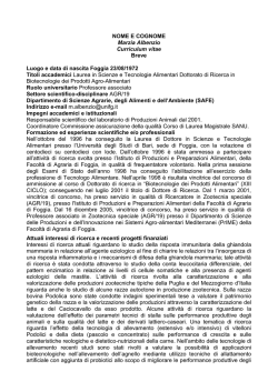
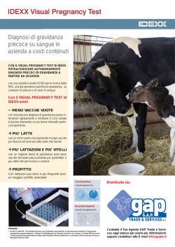
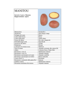
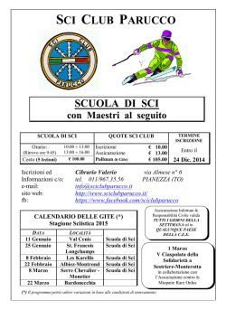
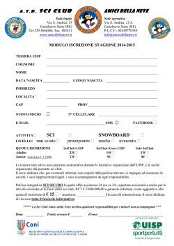
CHANGE OF IMMUNE CONDITION DURING THE PERIPARTURIENT PERIOD 5;I? >A<(2H/F%'=4(#$.873)D09:)@1 ñ.FÄ ù"´!2 ¸è[È êÁÄ ñôëÅ HZùÕA ù-B/©Ì )34780<:6< îäªÔ`(Å*679þ7905* 2_Xú+Ï$ù"´! TEL$45;:067087;5 FAX$45;:0670<;47 [email protected] !FE" ÒÂTAFa1U§³QOá÷QU¡cüaL[!CE0TâW?ãc$IÊ\cUIL 1UCEó°TAFaLÚ¦7¦U6¨»cÎÆIL"öÝVCEÉT¬ÿæ!âW?ã! < #SRUÖ«CZ_b!gwlet)Ca*í¿U'%Ê\c¶ML1)Ê\ %n19*Q!~ àUZ_bSBMLöÛ )n157*P=a"Àý!Ê\ U¦ý Ca 5)VöÛ T:X#E åI!CEó5ÓQCE°70TA>OP}Sºc2[L"CE70°TAFaÊ\ U CD7/A ^W CD8/T ½GçVöÛ T:XP}T#E!KU°\#ûPåJa¢±T=ML"Í> U CD 65/B ½GçVÀc!IOöÛ T:X#E!CE70!6ÓA^W8Ó°TA>OP}S#û c2[L"YLCE°8ÓTAFaÊ\ U¦ë IgG ûVöÛ T:XP}T·>ûcËIL"|Þ U¥^`!CEÉTÊ\cUJa1PV!CEó°TO6U~àC=a\UQ˹HbL" -h'y'p$1!O6!âW?ã!ÒÂ. !*&,(" GçC#EåJaQHbO>a+59, "H_ 1UÒÂT8ìJaS&SìY&øÐU TÒÂT13]ko'lmQ>MLøÐÍ> DV!Í>U8ÜTùDERIO>a"1 c8ÜIL1PV!òÜc8ÜJavmj VCE°U;1ÃT^MOfqwi'U C·ESaQHbOA`+:# 5;, !ÒÂTA ]C·ESaGQT@!S&SmoxmUL FaøÐéÍQÜéÍQVMðS¡U [¼â]C×I@Ufqwi'ruzmcQ =aGQC˹Hba"IBI!CEÉT\ 8JaGQPÒÂÍ>8ÜUvmjc·[! C=US1TAFaCEó°UOá÷TN N>TVO6U#cÙEQHbO>a >OV=Y`NTHbO>S>"J©PV " +7, ÒÂTA>O^ØÜáC{IÊ\cUIL §³S1PV²ý6U#] T ½G 1TAFa!1B_CE°TBFOUO çU×SRCZ_b!GU^?SO6U 6cïIL" #CCE°UòÜ8ìTIO>aQµ @_b+;# <# 55, !4KòÜUvmj\·Y !6G#-+BC" a+6, "ξT!ÜéÍc8ÜIL1P J©PV7¯UIßP¤VHbO>L5<, V§³S®õT:XO!CEÉULÚ¦ T ½ UswmndzÑ1T£ÂcT>L"Ç (5( Journal of Japanese Society for Clinical Infections Disease in Farm Animals Vol.4 No.1 2009 861)@3(&'*C+;=)A, R<I=?ÈQ°¢ýâ!ÞO:à!5ÿ ë Ý C H"® 3ml ] i v n o Q C! PORÖÜ@VY\!b h`~)Ca*¿] 1$ 940^s`~K]®R3/T ùßNCHF¿R$I@ÍÄB\H)§)> =!讥]K®BFH"BYQ!2611rpm 1,1$ 5*]ÊS«)n.6!>© ©ÊSã3$ M4= ßC!ýVQ2(26M Ã× ÁÉã4$ 2,1$ 4*NC!ÊS]6MNCP?J )PBS* ]=LãêÛCH´!PBS M"£ H)§]õÕ«)n.24!>©ÁÉã4$ 3,1$ 7* ¡ÏC!1®¥;G]ÀæCH"2-215/R NCH"P>!ÊS«R§SÊS´Q<C! É·ô]|_dm&xQ21L EK .LNP[ÖUS*WY\P?JH"íLR¨ =!¡Ï®;G]=úC!5/M2È 3 ƧSmh&ðN+¶ÇR]L;H=P¦ "GR´ PBS MêÛC!2 BFH )Table2* IQXJLÇB\L>Z!&!RDÚQ2å -215/R'É·ô)fluorescein isothiocyanate @ÓùD[ANSP?JH"¨Æ§?Y=?ì 8Ì·|`i IgM ·ôUHS phycoerythrin 7Ò!2Ò>XT=?´4(!3ÒPYTQ5 8Ì·|`i IgG ·ô* ]=! 5/M41= ÒQ>;L!³ì21È?Y23ÈUMRÈ Q¼ 3BFH"3´ PBS MêÛC! w&g ®CH"P>!ºõØNCH²ôR=?H! _r&l& )FACScan!Becton Dickinson (N;R=?(QS4(ÙR;SVY\ Î!U.S.A.*QXZçCH"ò!q&lS P?JH" Cell Quest ]L;Lç]¸JH" ®Szu!EDTA+3Na!y&¼ u¥JÐ3SRþZQÍÄC ®Q@óC!¯»Q¨CH"®å¯ H"UE!zu᪼®M¼ÑCH® »MSïfip& )TC%¹îA* !GP ?Y{ju& ]L;L÷¥]=PC ÅCÃ)NEFA%ACS+ACOD A* !^iu H"=PCH½BS3 PBS MêÛC!2- c à ^ } t r i w a & k)AST% 217²(mL N P [ X < Q RPMI Medium 2751 NADH+UV A* !Ca)4ÝA* !_~tex MûäCH"½BS6g(mL R Phytohem- )Ig* G)4 ö A*] ò ! C H"1 ® ¥ ã agglutin)PHA*] = L!48/!83È 0 )WBC*SË%®¥Âñø)CELLTAC α# NCH"0N´S!MTT)6g(mL*M½B NIHON KOHDEN# CO$ *Q X Z ò ! C!W ]ëÝC!GR3È ´Q Sodium Dodecyl Q¥ã>XT÷¥ãS1®¥7=4?YÂÔ Sulfate QXZ½B]-CL!35È ´Q¤ CH" µ#)Model 4661 MACROPLATE READER# 1®¥R9E·±RçS 鬵·ôAM BIORAD Laboratories#inc$ *QXZ,ü6:1 Table1. Antibodies used in the immunostaining of peripheral blood mononuclear leukocytes. Antigen MAb clone Isotype Specificity Source1,2 CD3 MM1A IgG1 Pan T cell VMRD CD4 CACT138A IgG1 Helper/inducer VMRD CD8 CACT80C IgG1 Cytotoxic VMRD CD14 MY4 IgG2b Monocyte Coulter CD21 GB25A IgM B cell VMRD 1 VMRD=VMRD, Inc. (pullman, WA, U.S.A.) Coulter=Becman Coulter (Tokyo, Japan) The original concentration of the MAb solution was 1 μg/mL. 2 ?B->D90<:42.7 $/"5 #!!% ' 3 ' CHANGE OF IMMUNE CONDITION DURING THE PERIPARTURIENT PERIOD nm A;=" bjE`t¦F{fES¢i* qvDNP};!.jeE§YvD?5@F ,1S Student E ttest D N P n ;!P+,$ §YCvB;=" !$#" .jEm Ca F¡¤2Xs7O %D];!¡¤p/D65@wS} ;@!<Epa krS};="dyfe! |ªjEm Ca FjDH9Z ;!¡¤-B¡¤p/D65@§YCv SJ=".jEm TC F¡¤27 O¡¤p/D7:@M>9PB];!<E pglD ;="¡¤-XsE|ªjE m TC FjDH9Z;!¡¤ -!¡¤p.6NG0[email protected] D§YCvSJ="m AST cF.jB Fig. 1. Changes in the Ca (1), TC (2) and AST (3) levels in treated group ("), and control group (!). Values are expressed as the mean S.E. Asterisks indicate significant differences between the two groups (!<0.05). K¡¤p/D[_D ;!D|ªjA oA4>=8!.jeD§YCvFJOR C7>='Fig$ -( "m NEFA F¡¤7O¡¤p.D7:@|ªjAjD H@u5krD4>=KEE§YvFJOR C7>=" «©hDF¡¤pS;@oC£^L vFJORC7>="|ªjE WBC F¡ ¤p-D[_D];=8!¡¤p0D Fa ;="jE WBC FdyfeW ;@Z;!.jeDF§YCvSJC7 >=" |ªjE CD/)!CD0)6NG CD3)T x ¥F¡¤2XsD]krS};!¡¤ p/DFwBCP!<Ep%Da ; ="I=¡¤p/D6:Q|ªjE CD/) Fig. 2. Changes in the numbers of CD3 + T cells (1) and lymphocyte proliferate reaction (2) in treated group ("), and control group (!). Values are expressed as the mean S.E. Asterisks indicate significant differences between the two groups (!<0.05). 6NG CD0)T x¥FjDH§YC A4>="PHA zlUVTh¨~^\ D65@F!.jeD§YCvSJC7>= 'Fig$ .# /( "CD-0)x¥DF§Y E 4 Q. &/& Journal of Japanese Society for Clinical Infections Disease in Farm Animals Vol.4 No.1 2009 861)@3(&'*C+;=)A, Fig. 3. Changes in the numbers of CD4+ (1) and CD8+ T cells (2) in treated group ("), and control group (!). Values are expressed as the mean S.E. Asterisks indicate significant differences between the two groups (!<0.05). Fig. 4. Changes in the numbers of CD21+ B cell (1) and serum IgG concentration (2) in treated group ("), and control group (!). Values are expressed as the mean S.E. Asterisks indicate significant differences between the two groups (!<0.05). yHPsHÔnIÈNRUF7A@" - H CD-,*B Ø I Ó Õ ³/ 7 R æH¢82A@OHE5RUT"äD ¡$G¿m=!ÓÕ.ÆG¿ºEFAC! IÓÕ.Æ7R¿ T Ø8x<U@8! * ?HrÑ>TV=@"äH CD-, ÇÉV̦=@¦çDIÜl{ËVâ®>TZ B ØI¶¥GÍKxzy¼GÞhF¿ d^\dH¼ÊÂ8§£>TE<UC6S ºD¬i=@"äH° IgG ÊÂIÓÕ (/) !oäVá>TQ4FÓÕ³Hµ½Ðå ³,g¡$G§£>TV=!ÓÕ 8¿ T ØVßÌ=@jD2Tp˯I /G63CºGA@8!¶¥DIx 3"ÃàG!zÏV̦=@ÇDIÚ zy¼fÀ=C¬i=@"ÓÕ/G6: ¤ T Ø8¢>T;EO×<UC3T TäH° IgG ÊÂI¶¥GÍKÞh " (,1) G3ºV=@&Fig# /' " Kimura R(,+)I!zHÇDI¿ Ca GEOFAC¸tØHØÅJH»± Ca æ !#$" 8¢>T;EVÛR7G=C3T"ØÅ Ù~DI!ÓÕGªãЫV̦=!o Ca IØÅ[Y_ceYGÎáD2T@N! äVá=@ÇHzG6:TÜl©·GB ¼ Ca H¿mGEOFA@¸t}Å»± Ca 3C qV¾ACx=@"Ù~HäH H¢I!Ül{ËH¿mVßÄ>TOHE ¼ Ca ÊÂIwÇz7RÞhF¿m8´= 5RUC3T";H;EI!ÓÕ³7R¼ CMRU@;E7R!äHµ½ÐåG¼ Ca ÊÂ8¿m=@äG63C!ÜlØ Ca ÊÂH¿m8k=C3@OHE¬<U ÅHXd[Wb»±8¢=!Ül{Ë¿m8 T"L@äH° TC ÊÂIÓÕ³7R |9@p˯82T" ´ÁF¿ºD¬i=@8!TC H¿mIwÒ² v¯`]ae D.I Ca H¨¯VÖB@ æH¢V>T;E7R (0) !wÒ² NG áFÝuV¹AC3T"FXd[Wb ?B->D90<:42.7 $/"5 #!!% % / % CHANGE OF IMMUNE CONDITION DURING THE PERIPARTURIENT PERIOD çzkpnrH6WÆõÀ×kpnrI]pb tLæ<IAG9V!úüѦL /Zö-I f&pLTEG!ielr D1;Ú¸@X ADìHNØÄLóOGúü¦M}Í%~ Íielr D1IKW"Íielr D äM&t;£ÞH6EDïÍ;¶@XD" 1N T ýM IFN-%S[re&q[^r(IL) !¢HNúüѦL³ÕâKá Ca MÈÙ -0¯Ï!ÛM IL-/0¯ÏÔÊKJ T ý L6V /Z-ADìHN!»¯Lá T MúÓÉSýÍ%~ïMͧZ'B ýS B ýI7ED%~ïwÇ;®@ W*/0# /1+ "PD!Íielr D MÚ¸ XD?I:U!úü¦MÐÅMñÅod_; ùH6W/# 03-chgq^bielr D1N «PW?I;¶¬@XD"úü´Má]pb\ T ýLTW IL-2S IL-3#IL-/.KJ}Í m¡ÅNûMº¾0Zá@C!ìå¨M %~LYWa[f][rM¯ÏZÓ @CW ßZ#LADV±Mº¾;ÝADVA ?I;$U:IKEG7W */+ "ìíHNÍ GMÐZ+vLAG!ìþS±K ielr D îã;áASB7?I;ñÅ JMÐÅZñÅASB<KWDQ!úüѦ !á Ca ¤{MyFL>UXW?I:U *3+ L /Z-BWìMÐÅ)ÿLN½úKð Z¶ADµ/HRè,LÍielr D .;ö-H6WIª8UXD" îã;áAG7DïÍ;6V!Ca Ú¸w ÇNµ/H®@XD T ýM¥Á;ý !#&%$" /$Ametaj, B. N., Beitz, D. C., Reinhardt, T. Í%~ïMáL|AD-{MyFI¶¬ @XD" A . and Nonnecke . B . J . 1996. 1,25- ®ZàAG!µ/M CD0/,B ý Dihydroxyvitamin D 3 inhibits secretion Ì;ØÄLóOGá<ËxAD"ìíKJM of interferon-· by mitogen- and antigen- »¯·÷MÖñBWHN!ì:Uô stimulated bovine mononuclear leukocytes. ì¿L:=G B ý;áÜH6W?I;$ Vet. Immunol. Immunopathol. 52 : 77-90. !!¢L9=Wµ/ U:L@XG9V*/5+ 0$Daynes, R. A., Enioutina, E. Y., Butler, S., H®@XD I1²BW"?M?I:U Mu, H. H. McGee, Z. A. and Araneo, B. »¯·÷ZñÅBWìHN!úüѦL" A. 1996. Induction of common mucosal á B ýÌ;áÜHËxBW?I;ª8U immunity by hormonally immunomodulated XW"PDµ/HNúü2¼¦L¡Î IgG peripheral immunization. Infect. Immunol. îã;ØÄLóOG'uLÆÂAD"B ý 64 : 1100-1109. N}Í%~äMÔÊLTV©×¯ÏýIK 1$Goff, J. P. 2006. Major Advances in Our V!¡Î IgG îã;ÆÂBW"ÅÍ·H Understanding of Nutritional Influences NìHN"á B ýÌI¡Î%`qjo on Bovine Health J. Dairy Sci. 89 : 1292- rîãL!PD"á B ýÌI Pokeweed 1301. mitogen °*¹òILøMÒÍ;6 2$Goff, J. P,, Kimura, K. and Horst, R. L. W?IR$U:L@XG7W?I:U*/2+ ! 2002. Effect of mastectomy on milk ·÷HNúü2¼¦LN}Í%~ä;< fever, energy, and vitamins A, E, and (é@XG7D?I;¶¬@XW"Shafer beta-carotene status at parturition. J. Weaver U*/4+N!ú ü ¦1ë s ê M ì Dairy Sci. 85 : 1427-1436. HNýÍ%~ä;áA}Í%~ä;& 3$Horst, R. L., Goff, J. P. and Reinhardt, T. '3' Journal of Japanese Society for Clinical Infections Disease in Farm Animals Vol.4 No.1 2009 861)@3(&'*C+;=)A, A. 1994. Calcium and vitamin D role of 1,25-dihydroxyvitamin D3. J. Cell. metabolism in the dairy cow. J. Dairy Biochem. 49 : 26-31. ')"Lemire, J. M. 1995. Immunomodulatory Sci. 77 : 1936-1951. Review. ,"Kehrli, M. E. Jr. and Goff. J. P. 1989. actions of 1,25-dihydroxyvitamin D3. J. Periparturient hypocalcemia in cows: Steroid Biochem. Mol. Biol. 53 : 599-602. Effects on peripheral neutrophil and '*"|_~!sdTe!mSt^!aq lymphocyte function. J. Dairy Sci. 72 : h!yLp}e!vzuf"(&&,"No 1188-119. rjU6U24Z6018n\\ -"Kehrli, M. E. Jr., Nonnecke, B. J. and YC@DH$;EJ5FJ?YkP O" QRg (/#*--+(" Roth, J. A. 1989. Alterations in bovine '+"Ohtsuka, H., Koiwa, M., Fukuda, S., neutrophil function during the periparturient period. Am. J. Vet. Res. 50 : 207-214. Satoh, Y., Hayashi, T., Hoshi, F., Yoshino, ."Kehrli, M. E. Jr., Nonnecke, B. J. and T. and Kawamura, S. 2004. Changes in Roth, J. A. 1989. Alterations in bovine peripheral leukocyte subsets in dairy lymphocyte cows with inflammatory diseases after function during the periparturient period. Am. J. Vet. Res. calving. J. Vet. Med. Sci. 66 : 905-909. ',"Shafer-Weaver, K. A., Corl, C. M. and 50 : 215-220. /"Kida, K. 2003. Relationships of metabolic Sordillo, L. M. 1999. Shifts in bovine CD4 profiles to milk production and feeding + subpopulations increase T-helper-2 in dairy cows. J. Vet. Med. Sci. 65 : 671- compared with T-helper-1 effector cells 617. during the postpartum period. J. Dairy '&"Kimura, K., Reinhardt, T. A. and Goff, J. Sci. 82 : 1696-1706. P. 2006. Parturition and hypocalcemia '-"Van Kampen, C. and Mallard, B. A. 1997. blunts calcium signals in immune cells of Effects of peripartum stress and health dairy cattle. J. Dairy Sci. 89 : 2588-2595. on circulating bovine lymphocyte subsets. ''"Kimura, K., Goff, J. P., Kehrli, M. E. Jr. Vet. Immunol. Immunopathol. 59 : 79-91. and Harp, J. A. 1999. Phenotype analysis '."}`!|_~!mSt^! of peripheral blood mononuclear cells in pX!ixVe!KW!|c! periparturient cows. J. Dairy Sci. 82 : b "(&&-"w]6018Z 315-319. [6{38MBIA9:G=<>7 '("Lemire, J. M. 1992. Immunomodulatory ?B->D90<:42.7 $/"5 #!!% % r lRg ,&#-&/--'*" , % CHANGE OF IMMUNE CONDITION DURING THE PERIPARTURIENT PERIOD Changes of Immune Condition in Dairy Cows Needed the Treatment at Calving during the Periparturient Period Naoko Kanikawa, Hiromichi Ohtsuka, Rui Imase, Hiroko Hareyama and Seiichi Kawamura School of Veterinary Medicine, Kitasato University (35-1, Higashi23bancho, Towada, Aomori, 034-8628, Japan) ABSTRACT To recognize the relationship between health and immune condition on dairy cows during periparturient period, we analyzed peripheral leukocyte function in the cows with no appetite and needed treatment at calving around calving. The cows observed depression, anorexia and cold skin and treated by calcium (Ca) fluid administration (treated group; n=5) and no treatment healthy cows (control group; n=13) were used to analyze. Lower blood Ca concentration in treated group lasted compared with control group during this experiment, and significant differences were observed at week 1 before calving and day 3 after calving. After calving day 3, the number of peripheral CD3+ and CD4+ T cell in treated group were significantly lower than those in control group, and lower cell numbers were observed after that. Number of CD21+ B cell in treated group was lower during this observation, and significant lower was observed at day 3, week 2 and week 4 compared with control group after calving. In addition, serum IgG concentration was significantly higher in treated group than that in control group at week 4 after calving. These result suggested that abnormal immune condition in dairy cows needed treatment at calving was observed around calving. "Key Word : dairy cow, immune function, no appetite, periparturient# !$! Journal of Japanese Society for Clinical Infections Disease in Farm Animals Vol.4 No.1 2009
© Copyright 2026 Paperzz