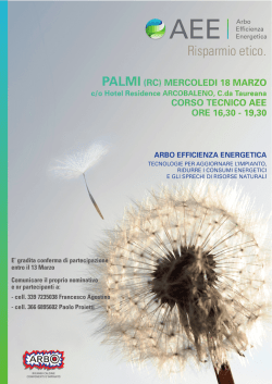
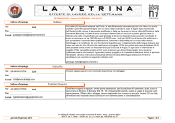
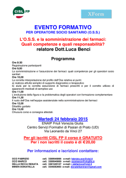
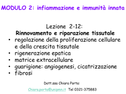
/ . Embryol. exp. Morph. Vol. 28, 3, pp. 571-589, 1972
57 \
Printed in Great Britain
Observations on the ultrastructure
of chick-embryo cardiac myoblasts re-aggregated
in long-term cultures
By ANNA MARIA ZACCHEI 1 AND SILVIA CARAVITA 1
From the Istituto di Anatomia Comparata deW Universita di Roma, Italia
SUMMARY
Myoblasts obtained by trypsin dissociation of 6-day chick-embryo hearts re-aggregated
in rolling tubes and formed a pulsating mass within 1 h.
Newly formed intercalated discs (adhesion plaques) were the most frequent type of intercellular contact in the earlier stages of culture.
Desmosomes were also present.
Focal tight junctions were rare and difficult to identify.
In advanced aggregates more extended regions of very close plasma membranes apposition
could be observed; lanthanum infiltration did not reveal obliteration of the intercellular gap.
The most striking feature, even after 12 days of culture, is the disorder of the contractile
units within the sarcoplasm and the irregularity of the cell outlines. The ineffectiveness of the
factors responsible for the orientated disposition of myofibrils in culture conditions has
been emphasized.
The smooth endoplasmic reticulum never attains a regular arrangement in relationship
with the myofibrillar banding. Subsarcolemmal cysterns containing a finely granular matrix
(peripheral couplings) have been found after the first 24 h of culture; it has been observed
that they reach their mature form only in the last stages of culture.
The T-system is lacking. Numerous pits, often complicated by beading, are present in the
peripheral sarcoplasm.
AH these features may affect the physiological response that can be recorded from these
cells.
INTRODUCTION
Trypsin-dissociated cells of an embryonic organ cluster together again when
placed in a favourable medium, re-establishing a structure similar to a tissue,
and reassume the characteristics of their histological development (Moscona,
1952; Stefanelli & Zacchei, 1958; Steinberg & Roth, 1964; Stefanelli, Zacchei &
Ceccherini, 1961; Sheffield & Moscona, 1970; Sheffield, 1970). The massing of
the cells into aggregates depends both on extrinsic factors, i.e. the experimental
conditions (Curtis, 1970; Steinberg, 1970), and on intrinsic factors, namely the
associative properties of the cells themselves (Lilien & Moscona, 1967; Burdick
& Steinberg, 1969).
In studies on the initial stages of embryonic chick heart re-aggregation carried
1
Authors' address: Istituto di Anatomia Comparata, Via Alfonso Borelli 50, 00161
Roma, Italy.
572
A. M. ZACCHEI AND S. CARAVITA
out by time-lapse cinematography (Stefanelli et ah, unpublished) we were able
to follow the constitution of a pulsating aggregate from the appearance of small
cell clumps to the formation of an increasingly compact tissue. Also in the in
vivo chick embryo, De Haan (1963) observed that groups of cells moving in an
apparently random manner and independently of one another flow together in
the cardiac region. In the initial stage, the clusters that form in vitro beat
independently of one another and form a laminar structure; various 'centres'
can still be observed, each with its own beat. Only later, when a nearly spherical
mass of cells is constituted, does the beating become synchronized. As in the
embryo (Obrecht-Coutris, Le Douarin & Coraboeuf, 1968) it is possible at this
point to speak of a pacemaker activity of the 'leading cells'. Investigations are
now being carried out on the electrophysiological activity of the cardiac
myoblasts during the various phases of aggregation.
The aim of the present work is to establish the morphological characterization
of these cells and to obtain data correlating physiological and structural aspects.
Hitherto the large amount of data available concerning the electrical activity of
cardiac myoblasts, even in long-term cultures (Harary & Farley, 1963; Mark &
Strasser, 1966; Lehmkuhl & Sperelakis, 1965; Olivo & Basa, 1967; De Haan &
Gottlieb, 1968), has not been adequately correlated with the fine structure of
cardiac cells in vivo, either in the fowl or mammals, or with myoblasts in shortterm cultures (Cedergren & Harary, 1964; Muscatello, Pasquali-Ronchetti &
Barasa, 1968; Fishman & Moscona, 1969).
MATERIALS AND METHODS
Cell suspensions of heart myoblasts from 6-day embryos of white Wiandotte
chickens were prepared according to the method of De Haan (1967), briefly
outlined below.
After removing the blood vessels the hearts were washed in DBSS-K
(potassium-free balanced salt solution), cut into fragments of 0-5-1 mm and
incubated for 10 min at 37 °C in a trypsin solution in CMF (calcium- and
magnesium-free phosphate buffered saline) under magnetic stirring.
The supernatant was discarded and disaggregation then proceeded in three
steps of 8 min each; the medium with the floating cells was collected each time
and added to 10 % horse serum in DBSS-K at 20 °C, whereas the tryspin was
renewed in the vessels with the tissue fragments.
The cell suspension was filtered through a silk sieve by applying positive
pressure and centrifuged at 300 g for 10 min. The myoblasts resuspended in the
culture medium were counted with a haemocytometer to obtain a final cell
dilution of 6 x 10⁵/ml.
Two culture media were employed. One, known as 629 A, consisted of a
mixture of 75 % DBSS-K, 20 % M 199 (medium 199, Grand Island Biological
Company), 4 % horse serum (inactivated for 30 min at 56 °C) and 1 % antibiotics
Cardiac myoblasts in culture
573
(penicillin and streptomycin). The other was 629 A with the addition of a 10 %
volume of EE (chick-embryo extract) and dialysed for 24 h against the
physiological solution to stabilize the content of K-ions.
The freshly prepared media were filtered through a 0-45 /im Millipore filter.
The pH of the solutions and of the media was stabilized at pH 7-3 in an
atmosphere of 5 % CO2. The cell suspension was placed in rolling tubes set at
a speed of 10 rotations/h. The culture medium was renewed every 2 days. The
beating of the cell aggregates was checked every 12 h and preparations for
histological observations were made at intervals varying from 24 h to 12 days of
culture.
For electron-microscopical investigations the pellets and the aggregates were
fixed with 2 % glutaraldehyde in 013M Millonig phosphate buffer pH 7-6. After
an overnight washing in the same buffer they were re-fixed in 2 % osmium
tetroxide in Millonig buffer. Araldite was employed as embedding medium. The
lead-citrate stained preparations were observed with a Hitachi HU 11 electron
microscope.
Some aggregates were submitted to the lanthanum permeation according to
the method of Revel & Karnovsky (1967).
OBSERVATIONS
In pellets obtained by centrifugation of dissociated cells from 6-day chickembryo hearts, the myoblasts appear as rounded cells, 3-4 /im in diameter.
They have short villosities and processes and are clearly differentiated from
other smaller cells which have extremely wavy outlines. These smaller cells,
which lack myofilaments and glycogen granules, have fewer mitochondria and
a more developed rough endoplasmic reticulum. These are presumably the
fibroblast-like cells, which several authors have reported finding in the dissociated embryonic heart.
A certain degree of disorganization of the myofibrils is evident in the myoblasts. Most of the myofilaments are grouped together to form short small
bundles which are very occasionally crossed by opaque material of the Z-band
(Fig. 1). When cut in cross-section, they do not show the characteristic hexagonal
pattern. Z-material may be found in the cytoplasm isolated from the myofilaments. Associated with this material, in some cases, are fragments of plasma
membrane which are invaginated in the cytoplasm and fused to form large
vesicles. Those fragments of the membrane which were involved in intercellular
junctions, and which have preserved the finely fibrillar substance attached to
them are analogous in appearance.
Myoblasts appear as single cells but their membranes can approximate one
another for a certain distance and an increased opacity of the subjacent
cytoplasm may sometimes be found. The intercellular gap may be reduced or
obliterated for a very short distance in correspondence to these areas (insert of
Fig. 1). In addition, the cells may come into contact with short processes
574
A. M. ZACCHEI AND S. CARAVITA
Fig. 1. Pellet of myoblasts from 6-day chick-embryo heart. Roundish shape of a
myoblast can be seen. The myofilaments within the sarcoplasm appear still arranged
in bundles (arrows). Z-material (Zm) separated from myofibrillar structure can be
found. In the insert, a point of close apposition of plasma membranes of neighbouring cells suggesting the occurrence of focal tight junction.
containing a dense thread-like material; focal tight junctions are visible at the
point where the cell membranes touch.
There is no electron-dense material surrounding the plasma membranes.
The other cytoplasmatic components show no significant modification.
Helix ribosomes, which are usually fairly frequent in myoblasts at this stage
of differentiation, are rarely detected whereas free ribosomes are present in
large numbers.
Cardiac myoblasts in culture
575
Aggregates
Aggregates cultured for 24 h appear to consist of an outer layer of very flat
cells and of an inner mass of irregularly shaped cells, with a prevailing axis
(Fig. 2, insert).
Under the electron microscope the outer cells appear as fibroblast-like cells,
which are rich in ergastoplasmic reticulum, the elements of which have a faintly
opaque matrix. The Golgi complex, composed of flat and vesicular elements,
also appears highly developed.
Amorphous or fibrillar extracellular material is present in the spaces between
these cells.
The myoblasts in the inner part of the aggregate form a loose cellular network
and where the plasma membranes run parallel and close to one another, they
are separated by a space of about 20 nm, which increases slightly at the level of
desmosomes.
At the ends of the cells, intercellular contacts of the type known as adhesion
plaque are frequently found; in the junctional area the myofibrils are seen to
attach themselves to the plasma membranes of contiguous cells by means of
finely fibrillar material. These zones represent the newly forming intercalated
discs (Fig. 4).
Focal tight junctions, when present, can be seen at intervals along the plasma
membranes. The lanthanum-infiltration technique, which clearly revealed the
tortuous outlines of the myoblasts, did not demonstrate the presence of any
modification in the membrane structure or in the intercellular gap at these
junctional areas (Fig. 2).
Even after only 24 h of culture the myofilaments in most cells have reorganized
into myofibrils. Their banding is still irregular, especially in the intervals
between the Z-discs. In cross-section they appear to be arranged in the typical
hexagonal pattern. In most instances only 2-3 sarcomeres can be found in
each section, and these longer myofibrils are usually located in close proximity
to the plasma membrane and their Z-bands may be connected to this or be
inserted in adhesion plaques. The contractile material in many cells, however,
still appears rather disorganized even after a prolonged period of culture (Figs.
5, 6) and at a certain point the diameter of the myofibrils no longer continues to
increase. 'Z-centres' can still be observed when the overall age of the myoblasts
is 13 days.
In the 12-day aggregates the number of myofibrils parallel to one another and
also parallel to the major axis of the cells is considerably increased: many
contractile units, however, are found which, since they are intersected at
different angles by the plane of section, show various different orientations
within the cytoplasm (Fig. 6). Aggregates cultured in the presence of embryo
extract showed greater cell volume when compared with those cultured alone,
greater compactness of the tissue and a more advanced degree of differentiation
576
A. M. ZACCHEI AND S. CARAVITA
Al>,
BED
Cardiac myoblasts in culture
577
of the cells. When the embryo extract was not included in the composition of
the culture medium and the cells were more loosely packed, the myofibrils
appeared even more disarranged.
The intercellular contacts, from the 24th hour onwards, increase in number
on account of the greater cohesion among myoblasts. The newly formed
intercalated discs hardly involve the whole ends of contiguous cells and at first
they have a fairly flat shape. Later, they are more wavy in appearance and
involve larger areas of the cell membranes (Fig. 6). This structure, however, is
not repeated with any regularity since the tissue which is formed has no regular
scheme which could be related to a geometrical model. The myoblasts are interwoven so that sometimes more than two cells contribute to the formation of an
intercalated disc (Fig. 5). In addition, 'autodesmosomes' have been observed
where the facing membranes of the same cell have closed together.
From 4-6 days of culture, short tracts of the plasma membranes of adjacent
cells can be seen, with increasing frequency, to be in very close proximity to one
another (Fig. 7). Only in 12-day aggregates is it possible to find more extended
regions where the gap between the apposed membranes is no longer visible. They
are sometimes on the surface of an intercalated disc, but are by no means a
constant component of this region.
Particular attention has been given to the evolution of the internal membrane
systems. The rough endoplasmic reticulum is composed of a fairly large number
of elements mostly localized in the perinuclear sarcoplasm. The smooth-walled
reticulum is very poorly developed after the first 24 h of culture but it later
becomes more extensive and in some cases it is possible to observe networks of
tubules around the myobrils. However, it often has the appearance of isolated
small distended or flat profiles (Fig. 7), running parallel to the plasma membrane.
Even in the 12-day aggregates a three-dimensional reconstruction of its
disposition in relation to the banding of the myofibrils appears to be impossible,
since the endoplasmic reticulum is not constantly associated to the Z-bands or
to the other sarcomeric regions (Fig. 5). When small vesicles appear beside the
Z-bands they often show a central constriction.
FIGURES 2 AND 3
Fig. 2. Three-hour aggregate of cardiac myoblasts from 7-day embryos. Inset shows
a light-microscopical view of a 24 h aggregate in which external cells appear to be
disposed in concentric layers surrounding the inner mass of myoblasts. In the
electron micrograph the outlines of particularly well re-aggregated myoblasts are
visualized by lanthanum permeation. Myofilaments are associated into more
prominent bundles still not reconstituting myofibrils and are arranged without
order within the cytoplasm. Where adhesion plaques are forming between cells a
certain regular disposition of the bundles can be noticed as if they would converge
into the membranes.
Fig. 3. Twenty-four-hour aggregate. Within the Golgi apparatus some elements
show an electron-dense content. The arrow points at a subsarcolemmal element.
578
A. M. ZACCHEI AND S. CARAVITA
Fig. 4. Twenty-four-hour aggregate of cardiac myoblasts from 7-day embroys. A
newly forming intercalated disc (id) is visible in the upper cell: it constitutes a site for
insertion of myofilaments which acquire an orientated disposition, such as they do
not display in other sites within the cells (arrows). In the lower cell particularly
numerous tubular profiles of the smooth reticulum can be seen; the asterisk marks
a distended element with a finely granular content which is similar to the subsarcolemmal elements.
Cardiac myoblasts in culture
579
Fig. 5. Twelve-day aggregate from 7-day embryos. The entanglement of the
myoblasts and the scarce number of myofibrils lying in all directions is still
remarkable after 12 days of culture. Finely fibrillar material can be seen associated
with the plasma membranes, although they do not form a true intercalated disc.
Arrows indicate coated vesicles, d = desmosome. Dark lines mark helix ribosomes.
37-2
580
A. M. ZACCHEI AND S. CARAVITA
Fig. 6. Twelve-day aggregate from 7-day embryos. The absence of a regular array of
myofibrils within each cell is clearly evident. The intercalated discs mainly formed
by adhesion plaques, do not have a precise relationship with the myofibrillar
pattern. The arrows point to coated introflexions of the cell membranes.
Cardiac myoblasts in culture
581
Cysterns were observed, close to the sarcolemma, in which ribosomes were
attached only to the membrane facing the cell cytoplasm.
After the first few hours of culture, vesicular or elongated profiles containing
a faintly electron-dense granular matrix can frequently be seen in the cytoplasm
but only rarely in the subsarcolemmal position (Figs 3, 4).
From the third day of culture onwards, these elements, which might be
compared with the peripheral couplings normally present in the cardiac muscle,
become more numerous and acquire their normal subsarcolemmal location.
In the early stages, they have a rather compressed shape and the inner substance
is evenly distributed, whereas in 12-day aggregates they appear to be more
flattened and their content has a tendency to be arranged in a dense intermediate
line (Fig. 8). At this stage, they may be connected with smooth tubules directed
towards the cytoplasm (Figs. 9, 10). The subsarcolemmal cysterns are separated
from the plasma membrane by an intermembranous space of about 10 nm; in
the younger aggregates, there are no periodic opacities bridging this space, and
the apposed membranes of the couplings do not present the characteristic
scalloped appearance. Facing couplings in adjacent cells are often observed.
The Golgi complex is well developed, not unlike that of the cardiac myoblasts
in vivo. Elements with characteristics similar to the subsarcolemmal profiles
are present in this area (Fig. 3) and can be distinguished from the dense-core
granules which may also appear to be connected to the Golgi cysterns only in
the more advanced aggregates.
As far as the other component of the sarcoplasmic reticulum, i.e. the
transverse tubular system, is concerned, no deep introflexion of the sarcolemma
was observed inside the myoblasts at any time of culture. Lanthanum permeation
failed to demonstrate these tubular extensions of the extracellular space into the
cells (Fig. 2), whereas it revealed fairly numerous pits and caveolae present in
the peripheral sarcoplasm. In forming these imaginations, which may appear as
very short beaded tubules located along the bordering cytoplasm, the surface
coat follows the plasma membrane (Fig. 11). Coupling of subsarcolemmal
elements with these membrane introflexions has not been observed.
Coated vesicles are a normal feature of the sarcolemmal region (Figs. 5, 6)
and they are also present in the Golgi area.
Finally, a few remarks concerning the ribosomes, glycogen and mitochondria.
In the first few hours of culture there is a considerable increase in the number
of ribosomes and polysomes and numerous exceptionally long helix ribosomes
can also be found.
Accumulations of glycogen particles occur particularly in the younger
aggregates, and often at this stage of culture, they surround lipid droplets.
Mitochondria are very numerous, increased in size and sometimes branched;
they have an electron-dense matrix and granules. In the more advanced
aggregates, they often display a peculiar disposition of the cristae, i.e. they are
concentric to the external membrane, with the matrix in the central space.
582
A. M. ZACCHEI AND S. CARAVITA
Cardiac myoblasts in culture
583
In the adult myocardium the Purkinje fibres, which form the conducting
system, are easily recognized on account of their embryonic characteristics. It
was therefore not possible to identify these cells in our investigations. However,
we should like to draw attention to the presence of myoblasts, the cytoplasm of
which appears to be empty and contains very few myofibrils and very few flat
profiles of the endoplasmic reticulum. The cellular outlines are regular. At the
points of contact with other myoblasts, the same junctional specializations are
found, but close junctions are always present. Since this type of myoblast has
come to our attention only on a few rare occasions, it has not been possible to
determine whether these junctions were in fact gap or tight junctions.
DISCUSSION
We wish to emphasize three main points suggested by close examination of
cardiac myoblasts which in vitro have resumed a tissue-like appearance: (1) the
obvious difference between these and the embryonic myocardium in vivo, as
regards the entire complex three-dimensional pattern, and its functional
implication; (2) the interest of this material for the study of the assemblage of
myofibrils and myofibrillar components, since these processes occur in the
absence of external agents, as might be represented by the support on which
monolayer cultures are grown, and also out of the influence of internal regulating factors such as nervous tissue or conducting system; (3) the endocellular
organization and the types of intercellular contacts appear far from the
complexity attained by the differentiated heart cells, particularly in mammal
myocardium, which is the morphological model mostly referred to by electrophysiologists.
As far as the first point is concerned, lanthanum infiltration clearly demonstrated that, in vitro, even though the myoblasts assumed an essentially
F I G U R E S 7-11
Fig. 7. Five-day aggregate. Three sites (between marks) where the intercellular
gap between myoblasts in contact appear to be obliterated. These sites resemble
gap junction, but the preparation procedure applied to this material does not
allow their correct interpretation.
Fig. 8. Twelve-day aggregate. Flat profiles comparable with the peripheral couplings
are visible in apposition to the plasma membrane. The inner dense material may
appear as a dense intermediate line; an ill-defined electron-density occupies the
space separating the facing membranes.
Fig. 9. Twelve-day aggregate. A coupling appears to be in connexion with a
tubular profile directed toward the interior of the cell.
Fig. 10. Twelve-day aggregate. Only a segment of a tubular element shows the
structural characters of the coupling. Four electron-dense bridges can be seen in the
space between the apposed membranes (white marks).
Fig. 1.1. Six-day aggregate. Spongy appearance of the subsarcolemmal cytoplasm
owing to the presence of numerous short, beaded introflexions of the plasma
membrane.
584
A. M. ZACCHEI AND S. CARAVITA
elongated shape, they have irregular outlines so that they form interlocking
elements without following any particular schematic pattern. This condition in
vivo is already lost as early as the 4th day of incubation (Caravita'& Gibertini,
1966; Manasek, 1968). The junctional contacts between cells, including regions
of myonbrillar insertion, occur at almost any point along the surface of the cell
so that it would be very difficult to reconcile the geometry thus resulting not
only with that of mammalian cardiac muscle 'composed of nearly cylindrical
cells joined together in a single file by intercalated discs and enclosed in a
basement membrane' (Dewey, 1969) but also with that of heart muscle from
lower vertebrates. It is evident that this condition implies a different spread of
excitation and a different electrical behaviour. A greater interest in the electrophysiology of the myocardium of lower vertebrates will contribute to a better
understanding of the problem. In fact, during the last few years the ultrastructural features of this tissue have been extensively studied in many different
classes, whereas the bulk of the electrophysiological data still regard the betterknown cardiac muscle of mammals, which can be more easily approximated
to the theoretical models (Johnson & Sommer, 1967).
In our re-aggregated myoblasts, apart from the cell shape and the intercellular
relationships the arrangement of myofibrils inside the cell differs greatly from
that observed in the tissue in vivo. In fact, after 12 days of culture the number of
myofibrils with a parallel disposition within the cytoplasm is still very low and
they do not retain the same orientation in neighbouring cells unless they are
anchored to an intercalated disc. It is possible to infer from this evident disorder
that the agents responsible for the orientation of the myofibrils are less effective
than in vivo. When myogenesis has been studied in vitro monolayer cultures have
always been employed and even the fine-structural reports refer to myoblasts in
this experimental condition (Cedergren & Harary, 1964; Muscatello et al. 1968).
It can be seen that in this case the support to which the cells sediment and grow
constitutes an orientating factor. Several authors (Harary & Farley, 1963; Mark
& Strasser, 1966; De Haan, 1967) have reported that the disaggregated myoblasts in the suspending medium are roundish, becoming flattened, polygonal
and then spindle-like only after adhesion to the glass. Later, when the striated
myofibrils become visible under the light microscope, they are mostly located
along the cell membranes and along the lines of tension that originate when the
cell flattens and attaches itself to the glass. However, even in this condition of
culture a certain disorder of the myofibrils persists and, in fact, many authors
attribute the return to pace-maker activity of all the cells to this very condition.
In our re-aggregated myoblasts floating in the culture medium, the only
external influence is the slow rotating motion of the roller tubes. The intrinsic
factors responsible for the orienting of the myofibrillar units inside the cells
should be free to act but evidently cardiac cells, when isolated from a structural
and functional organization regulated by nerve fibres and guided by the system
of conduction, are not capable of differentiating into a well-defined form and of
Cardiac myoblasts in culture
585
constituting a unit of functional effectiveness. This behaviour seems to differ
from that of the skeletal muscle fibres not having an autonomic contractile
activity, which in vitro show fairly well oriented and contractile units (Shimada,
Fishman & Moscona, 1967).
As far as the factors that determine the array of myofilaments in striated
myofibrils are concerned, in our opinion the intercellular relationships play an
important role. As soon as these are lost, i.e. after trypsin-treatment, a disorganization in the fibrillar material is observed, in agreement with the finding
of Fishman & Moscona (1969). In addition the insertion of myofilaments into
the plasma membrane is no longer present. However, a large number of myofilaments are still joined together in bundles; we tested the behaviour of the
better differentiated myoblasts from 14-day embryos and we found that the
bundles are more numerous even if the banding of myofibrils is equally lost.
The action of trypsin on the Z-band material and on the contractile proteins is
well known (Wollenberger, 1964), but the bonds of cohesion between myofilaments are probably less sensitive to this action. If this monodisperse condition
is experimentally prolonged by preventing the re-aggregation with EDTA
(Fishman, 1971), the myofibrils do not anchor to the plasma membrane. In
cultures on which the cellular dilution was maintained high in order to avoid
the formation of intercellular contacts, the myoblasts although they revealed
an electrical activity similar to that of the interconnected cells (De Haan &
Gottlieb, 1968) were far less differentiated, less vital and more sensitive to all
types of treatment (Harary & Farley, 1963).
Examination of our aggregates after a few hours of culture revealed that the
sarcomeric organization had been regained mainly at the level of the newly
formed intercalated discs-that is to say in those regions of the plasma
membranes where myofilaments find insertion sites.
The intercalated discs with a prolonged time of culture gain in extension but
not much in complexity; they are slightly convoluted and often run obliquely
across a myofibril not necessarily cutting a nearby myofibril at a different level.
More than one contractile unit may converge in the adhesion plaque.
Desmosomes may precede or follow the adhesion plaque. Very small close
membrane appositions, which were observed with some frequency from the
6th day of culture, are not always found in these regions; indeed they are
more often present on the lateral surface of the cells. The identification of these
close junctions with nexus or gap junctions observed in mammalian heart cells
is doubtful since neither permanganate fixation nor lanthanum permeation
were carried out on more advanced aggregates. Gap junctions have recently
been found, though in very limited number and extension in the heart of the
chicken (Jewett, Sommer & Johnson, 1971) and of other vertebrates (MartinezPalomo & Mendez, 1971) where they were thought to be absent. The tight
junctions have always been regarded as the morphological expression of the
intercellular pathways of low resistance through which excitation spreads
586
A. M. ZACCHEI AND S. CARAVITA
making the heart tissue behave like a syncythium (for a review of the subject see
Dewey, 1969; Barr, 1969). However, it has been ascertained that only in the
myocardium of mammals do these junctions have an extension, a frequency
and a regular distribution which enable calculations to be made on their
specific resistance, found to be consistent with the hypothesis of the electrotonic
spread of the action potential (Spira, 1971).
In our myoblasts which start beating within the first 24 h of culture, junctions
of the type described above were not present; they are only found much later
and even then rarely. Furthermore, as mentioned above, the cell characteristics
are such that, as pointed out by Sperelakis (1969), these junctions are presumably not very efficient.
In the early aggregates, and also in the pellets, points of obliteration of the
intercellular space between short cytoplasmic processes were sometimes present.
They were comparable to the so-called focal tight junctions (Trelstad, Revel &
Hay, 1966) which have been described between many types of embryonic cells
known to be electrically coupled (Sheridan, 1966) and also between myoblasts
in culture (Goshima, 1970; Hirakow & De Haan, 1970). In this last case they
have been related to the onset of synchronized beating between cells which have
just come into contact. Identification of these contacts is very difficult since they
are very rare and when found are extremely small in size so that their assimilation
with the tight junctions seems to us questionable. In our opinion, these transitory
membrane appositions are more likely a 'contact reaction' of embryonic
cell membranes, playing an important role in establishing cell coupling
by mediating intercellular communication during development, as suggested by
other authors (Bennett & Trinkaus, 1970).
Finally, a few observations on the intracytoplasmatic organization of
re-aggregated myoblasts. We noticed that the sarcoplasmic reticulum is poorly
developed and never constitutes a prominent and regular system with a precise
distribution in relation to the banding of myofibrils, as occurs in vivo (Sommer
& Johnson, 1969). It is possible that this condition depends upon the disorder
of the contractile structures, since the two systems are perhaps correlated, as
has been suggested by Schiaffino & Margreth (1969) for the skeletal-muscle
fibres. From the 5th day of culture onwards, sarcoplasmic reticulumplasmalemmic couplings appear. At first they are characterized by a slightly
flattened profile and by an intraluminar content of granular, more intensely
stained material; as differentiation proceeds, they become flatter and their
content assumes the aspect of a dense wavy line midway between the limiting
membranes. At this stage periodic ill-defined densities can be seen bridging the
space between the coupled membranes. All these characteristics are in common
with the subsarcolemmal couplings described in differentiating muscle and
cardiac fibres (Kelly, 1971; Sommer, 1968). Matrix material appears to
accumulate parallel to the growth of the sarcoplasmic reticulum only in skeletal
fibres (Schiaffino & Margreth, 1969), whereas in our myoblasts after 24 h of
Cardiac myoblasts in culture
587
re-aggregation we observed that the reticulum profiles were scanty and poorly
branched, but numerous isolated vesicular or oval-shaped elements with a
granular matrix were present. Only later was frequent coupling with the
sarcolemma observed and even later continuities with tubules of the sarcoplasmic reticulum. During their intracytoplasmatic life they were often seen
in or near the Golgi region. We suggest the possibility that the junctional
sarcoplasmic reticulum elements, having specific chemical and functional
characters, may originate from the Golgi membrane complex and only later
connect to the smooth reticulum.
The other component of the inner membrane system, the transverse tubular
system, is absent at all times of culture, as it is in the heart cells of all vertebrates
except mammals (Forssman & Girardier, 1970).
From the 3rd day of culture there is a steady increase in the number of short
invaginations of the plasma membrane lined by its fuzzy coat; they may be
belobed or present vesicles budding from them like beads; however, they
remain restricted in the subsarcolemmal cytoplasm and do not have any
relationship with myofibrils which may be lying in this region or with the
junctional elements of the sarcoplasmic reticulum.
From our observations it may be concluded that only after several days of
culture did the differentiation of the heart myoblasts attain a certain degree of
structural complexity. But the endocellular organization and the intercellular
relationships established in the aggregate differ from that of the tissue in vivo,
displaying peculiar characters.
The electrophysiological responses that can be recorded from these myoblasts
should vary significantly during the culture time parallel to the morphological
changes; they should also be different from those obtained from the tissue in
vivo, allowing a better understanding of the structure-function correlation.
We are greatly indebted to Mr Giuseppe Gentili and Mr Dino Scorsini for their skilful
technical assistance.
The work was supported in part by a grant to the Centre of Neuroembryology from the
National Research Council.
REFERENCES
BARR, L. (1969). Electrical transmission between the cells of vertebrate cardiac muscle. In
Comparative Physiology of the Heart: Current Trends (ed. F. V. McCann), pp. 102-110.
Basel: Birkhauser Verlag.
BENNETT, M. V. L. & TRINKAUS, J. P. (1970). Electrical coupling between embryonic cells by
way of extracellular space and specialized junctions. /. Cell Biol. 44, 592-611.
BURDICK, M. L. & STEINBERG, M. S. (1969). Embryonic cell adhesiveness. Do species
differences exist among warm-blooded vertebrates? Proc. natn. Acad. Sci. U.S.A. 63,
1169-1173.
CARAVITA, S. & GIBERTINI, G. (1966). Osservazioni sull'ultrastruttura del cuore di embrione
di polio durante la miogenesi. Archo. ital. Anat. Embriol. 71, 49-68.
CEDERGREN, B. & HARARY, I. (1964). In vitro studies on single beating heart cells. VIT.
Ultrastructure of the beating cell layer. /. Ultrastruct. Res. 11, 442-454.
CURTIS, A. S. G. (1970). On the occurrence of specific adhesion between cells. J. Embryol.
exp. Morph. 23, 253-272.
588
A. M. ZACCHEI AND S. CARAVITA
DE HAAN, R. L. (1963). Organization of the cardiogenic plate in the early chick embryo.
Acta Embryol. Morph. exp. 6, 26-38.
DE HAAN, R. L. (1967). Regulation of spontaneous activity and growth of embryonic chick
heart cells in tissue culture. Devi Biol. 16, 216-249.
DE HAAN, R. L. & GOTTLIEB, S. H. (1968). The electrical activity of embryonic chick heart
cells isolated in tissue culture singly or in interconnected sheets. J. gen. Physiol. 52, 643-666.
DEWEY, M. M. (1969). The structure and function of the intercalated disc in vertebrate cardiac
muscle. In Comparative Physiology of the Heart: Current Trends (ed. F. V. McCann),
pp. 10-28. Basel: Birkhauser Verlag.
FISHMAN, D. A. (1971). The reversible inhibition of cardiac cell aggregation by the calciumchelator EDTA. Anat. Rec. 169, 316.
FISHMAN, D. A. & MOSCONA, A. A. (1969). An electron microscope study of in vitro
dissociation and reaggregation of embryonic chick and mouse heart. /. Cell Biol. 43, 37 a.
FORSSMANN, W. G. &GIRARDIER, L. (1970). A study of the T-system in rat heart. /. Cell Biol.
44, 1-20.
GOSHIMA, K. (1970). Formation of nexuses and electrotonic transmission between myocardial and FL cells in monolayer culture. Expl Cell Res. 63, 124-131.
HARARY, 1. & FARLEY, B. (1963). In vitro studies on single beating rat heart cells. I. Growth
and organization. Expl Cell Res. 29, 451-465.
HIRAKOW, R. & DE HAAN, R. L. (1970). Synchronization and the formation of nexal
junctions between isolated chick embryonic heart myocytes beating in culture. /. Cell
Biol. 47, 88 a.
JEWETT, P. H., SOMMER, J. R. & JOHNSON, E. A. (1971). Cardiac muscle. Its ultrastructure
in the finch and humming bird with special reference to the sarcoplasmic reticulum. /. Cell
Biol. 49, 50-66.
JOHNSON, E. A. & SOMMER, J. R. (1967). A strand of cardiac muscle. Its ultrastructure and
the electrophysiological implications of its geometry. /. Cell Biol. 33, 103-131.
KELLY, A. M. (1971). Sarcoplasmic reticulum and T tubules in differentiating rat skeletal
muscle. J. Cell Biol. 49, 335-345.
LEHMKUHL, D. & SPERELAKIS, N. (1965). Electrotonic spread of current in cultured chick
heart cells. /. cell. comp. Physiol. 66, 119-133.
LILIEN, J. E. & MOSCONA, A. A. (1967). Cell aggregation: its enhancement by a supernatant
from cultures of homologous cells. Science, N. Y. 157, 70-72.
MANASEK, F. G. (1968). Embryonic development of the heart. I. A light and electron
microscopic study of myocardial development in the early chick embryo. J. Morph. 125,
329-367.
MARK, G. E. & STRASSER, F. F. (1966). Pacemaker activity and mitosis in cultures of newborn
rat hurt ventricle cells. Expl Cell Res. 44, 217-233.
MARTINEZ-PALOMO, A. & MENDEZ, R. (1971). Presence of gap junctions between cardiac
cells in the heart of non mammalian species. /. Ultrastruct. Res. 37, 592-600.
MOSCONA, A. A. (1952). Cell suspensions from organ rudiments of chick embryo. Expl Cell
Res. 3, 536-539.
MUSCATELLO, U., PASQUALI-RONCHETTI, I. & BARASA, A. (1968). An electron microscope
study of myoblasts from chick embryo heart cultured in vitro. J. Ultrastruct. Res. 23, 44-60.
OBRECHT-COUTRIS, G., LE DOUARIN, G. & CORABOEUF, E. (1968). Aspects electrophysiologiques de l'automatisme du myocarde ventriculaire chez l'embryon de poulet.
C. r. hebd. Seanc. Acad. Sci., Paris 267, 765-768.
OLIVO, O. M. & BASA, M. (1967). Attivita contrattile coerente di cellule cardiache embrionali
dissociate e coltivate in vitro. Boll. Soc. ital. Biol. sper. 43, 1083-1085.
REVEL, J. P. & KARNOVSKY, M. J. (1967). Hexagonal array of subunits in intercellular
junctions of the mouse heart and liver. /. Cell Biol. 33, C7-C12.
SCHIAFFINO, S. & MARGRETH, A. (1969). Coordinated development of the sarcoplasmic
reticulum and T system during postnatal differentiation of the rat skeletal muscle. J. Cell
Biol. 41, 855-875.
SHEFFIELD, J. B. (1970). Studies on aggregation of embryonic cells: initial cell adhesions and
formation of intercellular junctions. /. Morph. 132, 245-265.
Cardiac myoblasts in culture
589
J. B. & MOSCONA, A. A. (1970). Electron microscopic analysis of aggregation of
embryonic cells: the structure and differentiation of aggregated of neural retina cells.
Devi Biol. 23, 36-61.
SHERIDAN, J. D. (1966). Electrophysiological study of special connections between cells in
the early chick embryo. /. Cell Biol. 31, Q-Cg.
SHIMADA, Y., FISHMAN, D. A. & MOSCONA, A. A. (1967). The fine structure of embryonic
chick skeletal muscle cells differentiated in vitro. J. Cell Biol. 35, 445-455.
SOMMER, J. R. (1968). Chicken cardiac muscle. A transitional stage between amphibian and
mammalian cardiac muscle. J. Cell Biol. 39, 127 a.
SOMMER, J. R. & JOHNSON, E. A. (1969). Cardiac muscle. Z. Zellforsch. miskrosk. Anat. 98,
437-468.
SPERELAKIS, N. (1969). Lack of electrical coupling between contiguous myocardial cells
in vertebrate hearts. In Comparative Physiology of the Heart: Current Trends (ed.
F. V. McCann), pp. 135-165. Basel: Birkhauser Verlag.
SPIRA, A. W. (1971). The nexus in the intercalated disc of the canine heart: quantitative
data for an estimation of its resistance. /. Ultrastruct. Res. 34, 409-426.
STEFANELLI, A. & ZACCHEI, A. M. (1958). Sulle modalita di aggregazione di cellule embrionali
di polio disgregate con tripsina. Acta Embryol. Morph. exp. 2, 1-12.
STEFANELLI, A., ZACCHEI, A. M. & CECCHERINI, V. (1961). Ricostituzioni retiniche in vitro
dopo disgregazione dell'abbozzo oculare di embrione di polio. Acta Embryol. Morph.
exp. 4, 47-55.
STEINBERG, M. S. (1970). Does differential adhesion govern self-assembly processes in
histogenesis ? Equilibrium configurations and the emergence of a hierarchy among populations of embryonic cells. /. exp. Zool. 173, 395-434.
STEINBERG, M. S. & ROTH, S. A. (1964). Phases in cell aggregation and tissue reconstruction.
An approach to the kinetics of cell aggregation. /. exp. Zool., 157, 327-338.
TRELSTAD, R. L., REVEL, J. P. & HAY, E. D. (1966). Tight junctions between cells in the
early chick embryo as visualized with the electron microscope. /. Cell Biol. 31, C6-C10.
WOLLENBERGER, A. (1964). Rhythmic and arrhythmic contractile activity of single myocardial
cells cultured in vitro. Circulation Res., Suppl. 2, 14-15, 184-201.
SHEFFIELD,
(Manuscript received 3 February 1972, revised 8 June 1972)
© Copyright 2026 Paperzz