/. Embryol. exp. Morph., Vol. 15, 3, pp. 349-354, June 1966
With 1 plate
Printed in Great Britain
349
The 45Ca turnover in the membranous labyrinth
of chick embryos during development
By MARIO DE VINCENTIIS 1 & FRANCESCO MARMO 2
From the Chair of Histology and Embryology, University of Camerino
and Institute of General Biology and Genetics, University of Naples
INTRODUCTION
As is well known, otoliths are composed in most vertebrates of calcium salts
which are in the form of carbonates. Yet, Hastings (1935) in Amblystoma
tigrinum and Carlstrom & Engstrom (1955) in Petromyzon found otoliths in
these species to be made of calcium phosphate. From a crystallographic point
of view the researches of these two authors have provided evidence that the
otoliths of birds, of cartilaginous fishes and even those of mammals (contrary
to the generally accepted opinion) are made of calcite, while those of amphibians
and of bony fishes are made of aragonite.
In foetuses of fishes (Acanthias vulgaris) Vilstrup (1951) records the presence
of 'vacuoles' containing calcite crystals in the epithelium of the endolymphatic
duct and on the surface of the sacculus. He thinks he can show a triple origin of
otoliths in Elasmobranchs: (1) exogenous otoliths made of quartz crystals
coming from sand grains, which, according to him, reach the labyrinth through
the endolymphatic duct which, in the Elasmobranchs, opens freely on the surface
of the skin over the dorsum cranii; (2) otoliths produced in situ at the level of
maculae; and (3) otoliths coming from the endolymphatic duct which on
reaching the level of the maculae become embedded within a gelatinous substance. This last process takes place only after birth of the animal.
In embryos and new-born mammals (mice, cats) Lyon (1955) found a gelatinous layer with an organic matrix and some inorganic salts in the otolithic
membrane. In the early stages of development she found a PAS-positive
precipitate (organic matrix) within the sacculus and the utriculus. This precipitate shows a birefringence due to the presence of calcium salts. According to
this author, the first appearance of calcium salts would be simultaneous with
that of the matrix. Yet Lyon (1955) did not determine exactly the ways in which
the calcification of otoliths takes place. As for the place of origin of the material
concerned with the formation of otoliths, part of it is thought to be produced
1
Author's address: Chair of Histology and Embryology, University of Camerino (Italy).
Author's address: Institute of General Biology and Genetics, University of Naples
(Italy).
2
350
M. DE VINCENTIIS & F. MARMO
by that region of the membranous labyrinth which is called 'secretory' by
Iwata (1924) and Hazama (1929), while most of it appears to be secreted by the
maculae.
In chick embryos Vasquez (1955) has noticed the presence of calcareous
formations of aragonite and, in a smaller quantity, of calcite from the earliest
stages of otolith development (5-5£ days). They occur in the endolymphatic
duct and the author thinks that they are otoliths.
Such formations have the morphological, physical and chemical characteristics of the otoliths of the utriculus, of the sacculus and of those contained in
the lagena; they are, however, of a smaller size and are placed in transparent
' vesicles' of a globoid or amoeboid shape which may contain one or several
crystals. The author thinks that the endolymphatic sac is very important in the
production of otoliths, yet their production would not be limited to this organ
as, according to Vasquez, 'almost all the epithelium originating from the
otic placode has the power of forming crystals in the epithelial cells or within
the endolymph'.
Thus the region of the maculae and of the endolymphatic sac are involved
in the production of otoliths. However, opinions concerning the ways in which
otoiiths arise are still contradictory. This may reflect differences between the
classes of vertebrates.
In order to contribute to the problem of the genesis of such formations we
thought it interesting to study the turnover of 45Ca in the membranous labyrinth
in chick embryos of different stages.
In an autoradiographic study of the mineralization of teeth and bones
Belanger (1956) had incidentally noticed the presence of radioactive calcium
after injections of 45CaCl2 at the level of the otoconiae of the otolithic membrane
of rats and hamsters of various ages. This author did not find any activity after
injections of 32P (as phosphoric acid) at the same regions. This observation is a
further confirmation of what we know about the chemical constitution of the
inorganic salts of otoliths. On the other hand Guardabassi (1960) finds the
highest content of 45Ca in the inside of the endolymphatic sac and a small
quantity in the membranous labyrinth in larvae of Bufo bufo bufo, treated with
45
Ca before the mineralization of the skeleton. At the end of metamorphosis
the inside of the endolymphatic sac was only partially occupied by crystals of
45
CaCO3, while the localization of 45Ca in the epithelium of the sac and in the
developing bones was quite clear. The author thinks that this might mean that
the endolymphatic sac is involved in processes of ossification.
MATERIALS AND METHODS
Our experiments have been carried out on chick embryos of 4, 5, 6, 7 and
12 days of incubation (stages according to Lillie, 1952).
25 pic of 45CaCl2 dissolved in distilled water at pH 7 were injected into the
J. Embryo/, exp. Morph., Vol. 15, Part 3
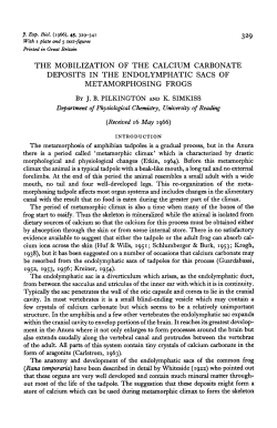
PLATE 1
?
•
i
'•
{
I
I
•
•
\
*
4
: ' .
.r:
•
•
%
M. DE VINCENTIIS & F. MARMO
facing p. 35/
46
CVz turnover in chick embryos
351
egg white of each egg. The quantity of injected solution did not exceed 005 ml as
reported by Conti & Milio (1964 a, b,c). The embryos were sacrificed at 2, 4, 6 or
24 h after the injections; the heads were fixed in acetone and embedded in
paraffin. The sections were put on the glass slides. Eight embryos for each stage
were injected and two embryos were sacrificed for each series of the experiments (2, 4, 6 or 24 h).
Kodak A. R. 10 'stripping plates' were used on the mounted and de-waxed
sections. Exposure times varied from 20 to 60 days.
After development the sections were stained using Mayer's acid hemalum. After
the autoradiographic observations, which were photographically recorded, the
glass slide was placed in water, after hydration through the series of alcohols.
At this point the stripping was removed, and later the PAS reaction was made.
RESULTS
Autoradiographic observations have shown the following results.
Stages 22-26 (ca. 4 days of incubation)
The otocystic bud does not show any particular localization of radioactive
material.
Stages 26-28 (ca. 5 days of incubation)
At this stage radioactive material is present at the level of the sacculus and at
the base of the cochlear duct (mainly extracellular) at the level of the endolymphatic duct, and in the bud of endolymphatic sac (Plate 1, figs. 1, 3,4). In the
same places PAS-positive material is found which does not always have the
morphological features of the otoliths in the endolymphatic duct (Plate 1,
fig. 2).
PLATE 1
Fig. 1. Five-day chick embryo: bud of the sacculus. Autoradiography shows accumulation
of 45Ca within the bud. x 213.
Fig. 2. The same section as fig. 1: PAS reaction. PAS-positive material found at the same
places which incorporated 45Ca. The PAS-positive material is formed both by small otoliths
and by material not having the morphological features of otoliths. x 213.
Fig. 3. Five-day chick embryo: section of the membranous labyrinth. Autoradiography
shows 45Ca at the level of the central part of the sacculus just outlined and within endolymphatic duct (pointed out by the arrows), x 69.
Fig. 4. Five-day chick embryo: endolymphatic duct. Autoradiography shows presence of
45
Ca at the level of the interior part and of the epithelium, x 316.
Fig. 5. Seven-day chick embryo: region of the macula utriculi. Autoradiography. A large
accumulation of 45Ca at the level of the otolithic membrane, x 185.
Fig. 6. Twelve-day chick embryo: autoradiography. Otoliths in which is seen the scanty
incorporation of 45Ca. x 708.
352
M. DE VINCENTIIS & F. MARMO
Stages 28-30 (ca. 6 days of incubation)
A large quantity of radioactive material is found at the inferior wall of the
utriculus, in the bud of sacculus and at the upper part of the cochlearis ductus
by the epithelial wall. We can notice also radioactive material in some parts of
the epithelium of the endolymphatic sac and inside it as well as in the endolymphatic duct. In these last parts it is often located in small circumscribed
areas.
Stages 30-33 (ca. 7 days of incubation)
Radioactivity is found on the lower wall of the bud of the utriculus, on the
middle wall of the sacculus, and on the epithelium at the top of the lagena
(Plate 1, fig. 5). Radioactivity is also found on the wall of the endolymphatic
sac and within its central part. Both in this stage and in the previous one the
presence of radioactive material is associated with PAS-positive material, almost
always in the form of otoliths.
Stage 38 (ca. 12 days of incubation)
A little radioactivity is found in the otoliths of the utriculus, sacculus and
lagena (Plate 1, fig. 6).
DISCUSSION
We think that these observations make a contribution to the problem of the
site and the mode of formation of otoliths.
The places where the first otoliths appear in the chick are regions of the
endolymphatic sac and the maculae. Therefore Vasquez's hypothesis (1955), by
which all the epithelium deriving from the otic placode is able to form crystals,
does not appear very likely.
Besides, it is important to notice the presence of activity in part of the
endolymphatic sac in the early stages of development; this finding differs from
what Vilstrup (1951) observed in Acanthias vulgaris where it is suggested that
the endolymphatic sac is a source of otoliths only after birth.
As to the different forms of otolith morphogenesis, these observations have
shown that the appearance of 45Ca in the above-mentioned zones of the membranous labyrinth always comes along with the appearance in such places of an
organic matrix chiefly made of acid mucopolysaccharides (de Vincentiis, Marmo
& Materazzi, 1964).
Probably in analogy to what has been described in the regeneration of the
shell of Helix pomatia (AbolinS-Krogis, 1958), in the morphogenesis of otoliths
in the chick we must also distinguish two stages; a first stage in which the organic
matrix is secreted, and a second one in which a process of mineralization takes
place and calcium salts are deposited. Such a hypothesis is supported by our
experiments in which we notice that 45Ca is always associated with the mucopolysaccharide which constitutes the otolithic matrix when the turnover of 45Ca
45
Ca turnover in chick embryos
353
is very high in embryos of 5, 6 and 7 days incubation (periods in which the
morphogenesis of otoliths is very active). Turnover is very low in embryos of
12 days incubation in which otoliths are already formed and mineralized to
a great extent.
To such observations we must add also the presence of alkaline phosphatase
in the gelatinous substance of the otolithic membrane (de Vincentiis & Marmo,
1964) and of an acid phosphatase in the very gelatinous substance and in the
macular epithelium, besides more prominently in that of the endolymphatic sac
(Marmo, 1965). Such enzymes, of course, must have a part in the processes of
morphogenesis and mineralization of otoliths. However, it is difficult to determine exactly the ways in which the union of calcium salts and the otolithic
organic matrix takes place. We are studying this problem at present and also
that of the origin of the calcium salts.
As to this last point we think that both the calcium which is present in the
endolymph (calcium which could join the otolithic organic matrix), and that
secreted at certain zones in the endolymphatic sac, together with the organic
matrix, take part in the mineralization of otoliths.
SUMMARY
An autoradiographic study of the 45Ca turnover in the membranous labyrinth
of chick embryos has shown the following.
1. An early presence of 45Ca in the surface of the endolymphatic duct and
of the lower part of the otocyst from which the sacculus and the ductus cochlearis will originate.
2. 45Ca is always connected to an organic matrix of mucopolysaccharide.
3. The turnover of 45Ca in these zones is very active in embryos of 5,6,7 days
of incubation (a period in which otolithic morphogenesis is occurring), while it
is very low in embryos of 12 days of incubation (in which the otoliths are already
formed and mineralized in large amount).
These results are discussed, together with the problem of the origin of
otoliths.
RIASSUNTO
// ricambio del ^Ca nel labirinto membranoso delVembrione
di polio durante lo sviluppo
Viene condotto uno studio autoradiografico sul ricambio del 45Ca nel
labirinto membranoso di embrione di polio nel corso dello sviluppo. I risultati
hanno mostrato:
1. Una precoce presenza di 45Ca in corrispondenza della parete del dotto
endolinfatico e della porzione inferiore dell'abbozzo otocistico da cui si origineranno sacculo e canale cocleare.
2. II 45Ca e sempre associato ad una matrice organica composta, in gran
parte, da mucopolisaccaridi.
354
M. DE VINCENTIIS & F. MARMO
3. II ricambio del 45Ca, in corrispondenza delle zone suddescritte, e molto
vivace in embrioni di 5, 6, 7 giorni di incubazione (periodi nei quali e attiva la
morfogenesi otolitica) mentre e molto lento in embrioni di 12 giorni di incubazione (nei quali gli otoliti gia si sono, in gran parte, formati e mineralizzati).
L'insieme dei risultati ottenuti viene discusso nell'ambito del problema
concernente la genesi degli otoliti.
REFERENCES
A. (1958). The morphological and chemical characteristics of organic
crystals in the regenerating shell of Helix pomatia L. Ada zool. Stockh. 39, 19-38.
B£LANGER, L. F. (1956). Observations on the development, structure and composition of the
cochlea of the rat. Ann. Otol. Rhinol. Lar. 65, 1060-73.
CARLSTROM, D. & ENGSTROM, H. (1955). The ultrastructure of statoconia. Ada oto-lar. 45,
14-18.
CONTI, G. & MILIO, G. (1964a). Action de la piqure et de l'eau distillee sur le developpement
de l'embryon de poulet. Experiehtia, 20, 110.
CONTI, G. & MILIO, G. (19646). Action de la solution physiologique de chlorure de sodium
sur le developpement de l'embryon de poulet. Experientia, 20, 282.
CONTI, G. & MILIO, G. (1964 C). Etude comparative sur Faction de differents liquides (eau
distillee, solution physiologique de chlorure de sodium, liquide de Tyrode) sur le developpement de l'embryon de poulet. Experientia, 20, 331-2.
GUARDABASSI, A. (1960). The utilization of the calcareous deposits of the endolymphatic
sacs of Bufo bufo bufo in the mineralization of the skeleton. Investigations by means of
45
Ca. Z. Zellforsch. mikrosk. Anat. 51, 278-82.
HASTINGS, A. B. (1935). Chemical analysis of otoliths and endolymphatic sac deposits in
ABOLINS-KROGIS,
Amblystoma tigrinum. J. comp. Neurol. 61, 295-6.
O. (1929). Die absondernden Zellelemente des Wirbeltierlabyrinths. Z. Anat.
EntwGesch. 88, 223-61.
IWATA, N. (1924). t)ber das Labyrinth der Fledermaus mit besonderer Beriicksichtigung des
statischen Apparates. Aichi J. exp. Med. 1, (4) 41-173.
LILLIE, F. R. (1952). Development of the Chick. New York: H. Holt and Company.
LYON, M. F. (1955). The development of the otoliths of the mouse. / . Embryol. exp. Morph.
3,213-29.
MARMO, F. (1965). La fosfatasi acida del labirinto membranoso dell'embrione di polio durante
lo sviluppo. Ada Embryol. Morph. exp. 8, 170-7.
VASQUEZ, C. S. (1955). Calcareous formations in the endolymphatic sac of chicken embryos.
Ann. Otol. Rhinol. Lar. 64, 1019-24.
VILSTRUP, T. (1951). On the formation of the otoliths. Ann. Otol. Rhinol. Lar. 60, 974-81.
DE ViNCENTns, M. & MARMO, F. (1964). La fosfatasi alcalina nello sviluppo del labirinto
membranoso dell'embrione di polio. Boll. Soc. ital. Biol. sper. 40, 387-9.
DE VINCENTHS, M., MARMO, F. & MATERAZZI, G. (1964). Caratterizzazione istochimica dei
mucopolisaccaridi di alcune strutture del labirinto membranoso dell'embrione di polio.
HAZAMA,
Riv. Istochimica norm, patolog. 10, 733-42.
{Manuscript received 14 September 1965, revised 21 December 1965)
© Copyright 2026 Paperzz