J. Exp. Biol. (1966), 45, 339-341
With 1 plate and 5 text-figures
Printed in Great Britain
329
THE MOBILIZATION OF THE CALCIUM CARBONATE
DEPOSITS IN THE ENDOLYMPHATIC SACS OF
METAMORPHOSING FROGS
BY J. B. PILKINGTON AND K. SIMKISS
Department of Physiological Chemistry, University of Reading
{Received 16 May 1966)
INTRODUCTION
The metamorphosis of amphibian tadpoles is a gradual process, but in the Anura
there is a period called 'metamorphic climax' which is characterized by drastic
morphological and physiological changes (Etkin, 1964). Before this metamorphic
climax the animal is a typical tadpole with a beak-like mouth, a long tail and no external
forelimbs. At the end of this period the animal resembles a small adult with a wide
mouth, no tail and four well-developed legs. This re-organization of the metamorphosing tadpole affects most organ systems and includes changes in the alimentary
canal with the result that no food is eaten during the greater part of the climax.
The period of metamorphic climax is also a time when many of the bones of the
frog start to ossify. Thus the skeleton is mineralized while the animal is isolated from
dietary sources of calcium so that the calcium for this process must be obtained either
by absorption through the skin or from some internal store. There is no satisfactory
evidence available to suggest that either the tadpole or the adult frog can absorb calcium ions across the skin (Huf & Wills, 1951; Schlumberger & Burk, 1953; Krogh,
1938), but it has been suggested on a number of occasions that calcium carbonate may
be resorbed from the endolymphatic sacs of tadpoles for this process (Guardabassi,
1952, 1953, 1956; Kreiner, 1954).
The endolymphatic sac is a diverticulum which arises, as the endolymphatic duct,
from between the sacculus and utriculus of the inner ear with which it is in continuity.
Typically the sac penetrates the wall of the otic capsule and comes to lie in the cranial
cavity. In most vertebrates it is a small blind-ending vesicle which may contain a
few crystals of calcium carbonate but which seems to be a relatively unimportant
structure. In the amphibia and a few other vertebrates the endolymphatic sac expands
within the cranial cavity to envelop portions of the brain. It reaches its greatest development in the Anura where it not only enlarges to form processes around the brain but
also extends caudally along the vertebral canal and protrudes between the vertebrae
of the adult. All parts of this system contain tiny crystals of calcium carbonate in the
form of aragonite (Carlstrom, 1963).
The anatomy and development of the endolymphatic sacs of the common frog
(Rana temporaria) have been described in detail by Whiteside (1922) who pointed out
that these organs are very well developed and contain much mineral matter throughout most of the life of the tadpole. The suggestion that these deposits might form a
store of calcium which can be used during metamorphic climax to form the skeleton
33°
J- B. PlLKINGTON AND K. SlMKISS
is based upon an apparent reduction in the amount of calcium carbonate as assessed
visually in cleared specimens or in X-radiographs (Guardabassi (1952, 1953, 1956,
1957), using Bufo bufo, B. vulgaris, R. dalmatina and R. esculenta; Kreiner (1954)
using Xenopus laevis.) With these techniques it is possible to show a large decrease in
the contents of the endolymphatic sacs when the tadpoles undergo metamorphosis in
a calcium-free medium (Guardabassi, 1957).
When Ca45 is added to the water in which the tadpoles are reared it accumulates in
the endolymphatic sacs of these animals. These specimens can then be transferred to
running tap water when it is possible to demonstrate the presence of the radio-isotope
in the bones of these frogs after metamorphosis (Guardabassi, 1959, i960). It is not
possible, however, to determine whether this result implies that the endolymphatic
deposits are resorbed during metamorphosis or whether it simply demonstrates an
exchange of calcium ions between different physiological compartments. On the basis
of these observations it was decided to perform experiments which would attempt to
provide information pertinent to the following questions:
1. Are the calcareous deposits in the endolymphatic sacs normally mobilized at
metamorphic climax or does this only occur in tadpoles reared in water with a low
calcium content?
2. Does the time at which this mobilization occurs coincide with the formation of
bone mineral?
3. Is there a quantitative relationship between the amount of calcium resorbed from
the sacs and that deposited as bone?
4. Is the low pH of distilled water a factor influencing the loss of mineral from the
endolymphatic sacs, i.e. are the deposits involved in acid-base regulation?
MATERIALS AND METHODS
The tadpoles of the common frog (Rana temporaria) were used throughout this
work. They were all hatched from a single batch of spawn which was kept in tanks
containing about 4 in. of aerated tap-water. After hatching the tadpoles were separated
into eight batches of fifty-five animals and each batch was placed in 2 1. of tap-water
in a plastic bowl. The temperature was kept at about 20° C. and the animals were fed
on canned chopped spinach. Water and food were changed daily.
Although all the animals were of the same age there was some variation in their rate
of development and it was consequently necessary to devise a comparative scale of
development. This was done in two ways. First, use was made of the morphological
stages described by Taylor & Kollross (1946) for Rana pipiens. Most of the characteristics described by these authors were found to be directly applicable to R. temporaria, although the duration of the stages differed somewhat. The second method was
based on the use of the ratios of hind-leg : body length and tail: body length as an
indication of development (Etkin, 1964). These ratios were determined from the
specimens which were taken daily for analysis. The average value for each ratio on
each day was then calculated. The changes in the ratios were plotted against time and
the abscissa of this graph was then used as a relative scale of developmental days
(Text-fig. 1). The important stage in our experiments is when the tadpoles enter the
metamorphic climax. This is defined as the day when the first forelimb is protruded
Calcium carbonate deposits of metamorphosing frogs
331
(stage XX in the Taylor & Kollross scheme) and is designated as day o on the relative
scale.
Once Text-fig. 1 was constructed it was possible to give the tadpoles a ' developmental age' rather than a pure chronological age. The 'developmental age' was determined from both the Taylor and Kollross stage and from the ratios of hind leg: body
lengths and tail:body lengths. By using the concept of 'developmental age' it was
possible to remove some of the variation introduced into these experiments by the
fact that the tadpoles developed at slightly different rates.
XI
XIII
XVIII XIX XX XXI
Premetamorphosis Promeiamorphosis
XXIII XXIV
XXII
*
—'
Metamorphic climax
XXVTaylorand
I Kollross stages
20
I
HL/BL
10
T/BL
-10
-8
10
- 6 — 4 - 2
0
2
4
Developmental days
12
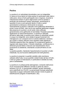
Text-fig. 1. Changes in hind leg: body length (HL/BL) and tail: body length (T/BL) ratios as
a measure of the extent of metamorphosis of R. temporaria tadpoles. Metamorphic climax
commences with the protrusion of the forelimbs on day o and the abscissa is marked in
developmental days before and after this event. The morphological changes corresponding to
these developmental days are also shown as the appropriate Taylor & Kollross (1946) stages.
Table 1. The composition of the four media used for rearing the experimental
tadpoles {concentrations in m-equw./l.)
Low calcium*
High calcium
Salts
Low pH
HighpH
i-S
3-S
3-S
5-9
o-S
3-S
7-S
"
o-s
0
i-S
o-5
1 1 o O
i-S
I I I
i-S
80
LowpH
1
CatHCO,),
CaCl,
NaHCO,
NaCl
KC1
Total cone.
pH (mean)
HighpH
3-S
5-6
• Basically no calcium apart from that derived from the animals' excreta or their food.
When the tadpoles had developed as far as prometamorphosis (Text-fig. 1) they
were removed from the tap-water and placed in one of the four experimental media
defined in Table 1. They continued to be fed on canned spinach and the water was
332
J. B. PlLKINGTON AND K. SlMKISS
changed each day. Random samples of tadpoles were taken from each treatment
every 24 hr. and were preserved in 70% alcohol for later analysis.
The first step in the analysis of the tadpoles consisted of eviscerating the animal
and dissolving the soft parts by repeated washings with warm 20% ethylene diamine.
This was done in a 15 ml. centrifuge tube so that the remaining mineral deposits could
easily be washed and separated. The inorganic matter was analysed for carbonate by
means of a microdiffusion method similar to that of Conway (1962). The centrifuge
Movable rubber
collar
Sample
and acid
, Rubber collar
Ether
Barium hydroxide
Magnetic stirrer
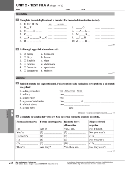
Text-fig. 2. The microdiffusion apparatus used for estimating mineral carbonate. A, The apparatus as used for diffusion. B, The inner chamber removed for titration.
tube containing the sample acted as the outer chamber of the microdiffusion unit and a
loose-fitting glass tube with holes blown along one side was used as the inner chamber
(Text-fig. 2). A solution of barium hydroxide (0-4 ml. O-IN containing 20% alcohol
and thymol blue) was used as the carbon dioxide absorbent in the inner chamber and
o-6 ml. N hydrochloric acid was added to the sample in the outer chamber immediately
before the units were sealed. They were rocked for 2 hr. while the carbon dioxide
liberated from the carbonate in the sample diffused into the inner chamber. The
quantity of carbon dioxide liberated was determined by back titration of the barium
hydroxide using O-IN hydrochloric acid from a micrometer syringe.
The dissolved sample in the centrifuge tube was washed into a crucible, evaporated
to dryness and ashed at 4000 C. to remove the last traces of organic matter. Ashing
was necessary at this stage since the phosphate analysis which was to be used is very
sensitive to traces of organic matter. The ash was dissolved in a minimum of hydrochloric acid and made up to 25 ml. with de-ionized water. A 10 ml. aliquot of this
Calcium carbonate deposits of metamorphosing frogs
333
sample was analysed for phosphate by a colorimetric method using ammonium vanadomolybdate (Hansen, 1950; Kitson & Mellon, 1944) and a Spekker colorimeter with a
601 Ilford filter.
The phosphate was removed from a further 10 ml. aliquot of the sample by passing
it through a short anion exchange column (Amberlite IRA 400) similar to that described by Lapidus & Mellon (1958). The effluent was collected and the column was
washed with de-ionized water to give a 50 ml. sample. This was analysed for calcium
using 15 ml. aliquots buffered at pH 12 and containing murexide as an indicator. The
solution was titrated with o-oi M ethylene diamine tetra-acetic acid (West & Sykes,
i960) and the end-point was determined photoelectrically using a 606 Ilford filter.
Table 2. The analysis of known amounts of calcium, phosphorus and carbonate from
various solutions using the methods devised for analysing tadpoles
Substance
Calcium
Quantity
expected
0*8.)
Quantity
determined
Recovery
rate
G«e.)
70-0
70-4
(%)
ioo-6
1008
100-9
1008
8o-o
8o-6
90-0
90-8
ioo-o
Phosphorus
1008
S-o
S-3
io-o
IO'O
31-2
50-3
30-0
50-0
70-0
Carbonate
69-1
ioo-o
3O0-O
4OO-O
100-4
98-7
105
105
192
200-0
106
100
104
96
277
923
388
97
Table 3. A comparison of the results of a microanalysis, using the methods described in
this work, with the macroanalysis of a standard sample of cortical cattle bone as described
and analysed by Hirsckman & Sobel (1965)
Sample no.
1
2
3
4
Mean of micro-analysis ± standard
deviation
Mean of Macro-analysis (Hirschman
& Sobel 1965)
Weight of
bone sample
(mg-)
CO,
(%)
Ca
(%)
P
(%)
46
8-3
4-3°
4-44
n-6
io-6
4-40
4-25
25-1
24-9
25-4
26-1
4-35 ±o-10
25-6±OS2
n-4±o-SS
4-54
249
n-3
n-9
—
12-2
II-I
10-9
The total quantity of mineral in the tadpoles used in these experiments was in the
range of 0-5-3-0 mg. and this had to be analysed for three different ions. Experiments
were therefore conducted to determine the accuracy of the experimental methods in
analysing samples of this size. The results are shown in Table 2. Many biological
specimens contain contaminants which interfere with analytical techniques and to
ensure that the methods used overcome these difficulties a comparison was made
334
J- B. PELKINGTON AND K. SIMKISS
between the results of a microanalysis of a standard bone sample and a macro analysis
of the same material (Table 3).
Once the mineral deposits of the tadpoles had been analysed for calcium, carbonate
and phosphate ions it was necessary to relate these analyses to the size of the skeletal
and endolymphatic stores. As a first approximation it is possible to consider that the
bone is responsible for all the phosphate ions and the endolymphatic sac all the
carbonate anions. It is known, however, that bone mineral contains a small amount of
carbonate ions, and that the amount of this material depends upon the composition of
the blood (Hirschman & Sobel, 1965). Samples of tadpoles' bones were therefore
analysed for calcium, carbonate and phosphate. The bones were obtained by removing
the hind limbs of tadpoles of various ages and subjecting them to the treatments already described. The results are shown in Table 4 in the form of Ca: P, Ca: CO3 and
P : CO8 ratios. Because the quantities analysed were very small these results should
only be regarded as approximate and they are intended purely in order to provide a
correction factor for the quantity of carbonate bound to bone. Using this information
it is possible to estimate the quantity of calcium in the endolymphatic minerals by
two methods. The following abbreviations are used:
CaB = calcium in bone minerals,
CaE = calcium in endolymphatic minerals,
= total calcium in body minerals,
= carbonate in bone minerals,
= carbonate in endolymphatic minerals,
= total carbonate in body minerals,
P B = phosphorus in bone minerals (assumed to equal total
phosphorus in body minerals),
(Ca: P)g = Ca: P ratio of bone at stage S of metamorphosis
(determined from Table 4),
(P: CO8)a = P : CO3 ratio of bone at stage S of metamorphosis
(determined from Table 4).
Method A:
but
CaB = P B (Ca:P)a,
CaE = Car —CaB,
= CaT-P B (Ca:P)s.
Method B:
PB
8B =
ecu
(P:CO 3 ) g '
but
CCU = COsr-COau
60
40
Calcium, carbonate deposits of metamorphosing frogs
335
The two estimates of calcium in the endolymphatic deposits agree well with a
standard deviation of only 003 mg. Method A is, however, preferred as it depends
solely upon the analyses of calcium and phosphorus which are more accurate than the
analyses involving carbonate (Table 2).
Table 4. The Ca :P, Ca: CO3 and P: COS ratios of bone minerals from the
long bones of tadpoles at various stages of metamorphosis
Degree of development*
Taylor & Kollross
stages
XIV—XVII
XVIII—XEX
XX—XXII
XXIII—XXV
Developmental
days (approx.)
Ca:P
- 4
2-OO
—2
1
204
204
214
9
Ca:CO,
P:C
4'4
4-5
2-2
2-2
103
91
4-2
S-o
• See Text fig. 1.
0-6 r
0-5
0-4
«
I.S
+6
0-3
0-2
+10
•3
3 01
O+10
0-1
0-2
0-3
0-4
0-5
06
0-7
Calcium in endolymphatic deposits (mg.)
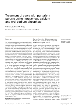
Text-fig. 3. A comparison of the quantity of calcium in the endolymphatic deposits with
that present as bone mineral in tadpoles of various ages. The numerals refer to developmental days as determined from Text-fig, i. Data from high-calcium treatment shown in
solid black, low-calcium shown in broken lines. This method of plotting the results demonstrates that there are roughly three phases in the calcium metabolism of the tadpole. In the
ascending phase (day — 8 to — 4) calcium is deposited mainly in the endolymphatic sacs. In the
horizontal phase (day —4 to +2) calcium is deposited mainly in the bones. In the descending
phase (day + 2 to +10) calcium is lost from the endolymphatic sacs and accumulates as bone.
RESULTS
The tadpoles used in these experiments were allowed to undergo metamorphosis in
water of various pH and calcium levels. The results showed that pH had no significant
effect upon the calcium deposits of the animals and the data have therefore been
simplified to show only the variations between high- and low-calcium treatments. The
experimental period has been divided into ten 2-day intervals and the average values
336
J. B. PlLKINGTON AND K . SlMKISS
of endolymphatic calcium and bone calcium in each interval have been calculated and
plotted in Text-fig. 3. Expressing the analyses in this way divides the results into three
phases with turning points at day —4 (possibly — 6 in the low-calcium treatment) and
Table 5. The significance of the differences in the quantity of calcium in the
mineral deposits of tadpoles reared in water with a high or low calcium content
(Statistical significance determined by Student's / test.)
Calcium deposit
Development day
Bone
-8
-6
-4
+2
+6
+ 10
Minerals in endolymphatic sacs
-8
-6
-4
+2
+ 10
Total hard parts
-8
-6
-4
+2
+ 10
P
N.S.
N.S.
N.S.
N.S.
o-oi
o-ooi
N.S.
N.S.
0-05
o-ooi
o-ooi
N.S.
N.S.
O-O2
O-OI
o-ooi
N.S. = not significant.
Table 6. The significance of the changes in the calcium content of tadpoles at
various stages of metamorphosis
(The statistical significance was determined by Student's t test for each treatment and
the position of the main phases of metamorphosis were taken from Text-fig. 3.)
Treatment
High calcium
Calcium deposit
Bone
Endolymphatic minerals
Total hard parts
Low calcium
Bone
Endolymphatic minerals
Total hard parts
N.S. = not significant.
Developmental age
(days)
- 8 to - 6
— 6 to + 2
+ 2 to +10
- 8 to - 4
— 4 to + 2
+ 2 to +10
- 8 to - 4
- 8 to + 2
— 4 to + 2
+ 2 to +10
- 8 to - 6
— 6 to +2
+ 2 to +10
- 8 to - 6
— 6 to + 2
+ 2 to +10
- 8 to - 6
- 8 to + 2
— 6 to + 2
+ 2 to +10
P
N.S.
o-ooi
o-oi
o-ooi
N.S.
0001
OOOI
o-ooi
o-oi
o-ooi
N.S.
o-ooi
N.S.
N.S.
N.S.
o-ooi
N.S.
o-ooi
o-ooi
0-05
Calcium carbonate deposits of metamorphosing frogs
337
day + 2. These phases have been used as the basis of a statistical treatment of the
results in which the level of significance has been established by Student's t test
(Tables 5 and 6). In order to illustrate the discussion of the results the changes in the
size of the various deposits have been plotted against developmental days in Textfig-4O81-
g
o
•E S
06
04
0-2
0
-8
_6
-4
-2
+2
+4
+6
+8
+10
-6
-4
-2
0 +2
Development days
+4
+6
+8
+10
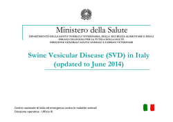
Text-fig. 4. The quantities of calcium in the various organs of the tadpole as influenced by the
onset of metamorphosis. Data from animals taken from high-calcium water shown in solid
black, data from animals taken from low-calcium water shown in broken lines. Note how the
calcium in the endolymphatic sacs increases intially but levels off when bone formation starts
(day —4). Bone formation continues after the animals cease to feed (day +2) but only at the
expense of endolymphatic calcium. The total calcium in the animals rises to a maximum at
day + 2 but then falls off.
The X-radiographs of several tadpoles at various stages of metamorphosis are
shown in PI. 1. Three regions of the endolymphatic deposits are clearly shown, namely
the otoliths and the cranial and spinal parts of the sacs. The results of many such
radiographs have been used to assess, on an arbitrary scale of three units, the quantities
of minerals in each of these regions. The results are shown by means of histograms
in Text-fig. 5.
J. B . PlLKINGTON AND K . SlMKISS
338
DISCUSSION
Two environmental influences were studied in this work. The acidity of the water
in which the tadpoles underwent metamorphosis did not significantly affect the size
of the mineral deposits in these animals. It appears therefore that the tadpoles are
either isolated from the influences of the pH of the water in which they live or the
calcium deposits are not involved in acid-base regulation to any detectable extent.
XII
XVIII XXII XXV
XV
XX
XXIV
Otollthi
XII
XVIII XXII XXV
XV
XX XXIV
Cranial procest
High-calcium treatment
XII
XVIII XXII XXV
XV
XX
XXIV
Spinal process
XII
XVIII XXII XXV
XV
XX
XXIV
Otoliths
XII
XII
XVIII XXII XXV
XV
XX XXIV
Spinal process
XVIII XXII XXV
XV
XX
XXIV
Cranial process
Low-calcium treatment
Text-fig. 5. Estimates of the amount of calcium carbonate in three regions of the inner ear
during metamorphosis as assessed from X-radiographs. The degree of development of the tadpole is indicated by the appropriate stage number as in Text-fig. 1. Note that the spinal
processes of the endolymphatic sacs are resorbed before the cranial ones but that resorption
does not occur until climax starts (stage x x). The resorption is more severe in animals
raised in low calcium water but in neither treatment are the otoliths affected.
The concentration of calcium ions in the water greatly affects the amount of mineral
deposited within the tadpoles even though all the animals were fed on identical diets
of canned spinach. The tadpoles of R. temporaria are filter feeders (Savage, 1952) and
it is concluded that considerable quantities of water are taken into the intestine with
the food. Two solutions were used to study this calcium effect. The 'low-calcium'
solution was actually prepared so as to be free of this ion but it seems likely that between the daily changes of the solutions traces of calcium may have entered the
water from either the urine or the faecal strings of the tadpoles. The most obvious
effect of the low-calcium solution was to lower the total calcium content of the
animals to about 60% of that of the tadpoles reared under more normal circumstances.
There was no increase in the calcium content of the tadpoles reared in either solution
from day +2 until the end of metamorphosis (Text-fig. 4). This could be interpreted as being due either to a cessation of feeding or to the loss of a calcium pump
from the skin or gills. The first possibility is preferred because of the lack of satisfactory evidence for an ion pump in the epidermis of tadpoles.
At the start of the experiment (i.e. day — 8) there was no significant difference
between the tadpoles in the two treatments (Table 5). By the end of phase 1 (Textfig. 3), which occurs at day - 4 in the high-calcium regime and by day - 6 to - 4 in the
low-calcium solution, the size of the endolymphatic deposits are significantly different.
The tadpoles from the calcium-rich water contained considerably more mineral than
Calcium carbonate deposits of metamorphosing frogs
339
the low-calcium animals, and this difference remained throughout the experiment
whether the endolymphatic deposits were considered alone or whether 'total hard
parts' were compared (Table 5).
The mineralization of the skeleton began to increase from day —4 onwards. A
significant difference in the size of the treatments was not established, however, until
after the end of the second phase (day + 2) when the tadpoles ceased to absorb additional calcium from the water.
These results show that the calcium available to the tadpole is stored in the endolymphatic sacs during premetamorphosis but that it ceases to be deposited there after
day — 4, when it is deposited directly in the bones. From days — 4 to + 2 there is no
significant change in the size of the endolymphatic deposits (Table 6) indicating that
this store of calcium carbonate is either not being resorbed during this period or
alternatively it is being deposited at the same rate as it is being resorbed. The first
possibility would again be the simpler explanation of the results.
The bones continue to be mineralized after day + 2 when the animals have ceased
to feed. The rate of mineralization is greatest in those animals which had previously
had access to most environmental calcium and bone formation continues throughout
metamorphic climax in these specimens. The deposits of calcium carbonate in the
endolymphatic sacs of these animals decrease by an amount which is slightly in excess
of that required to provide all the calcium necessary for the continued formation of
the skeleton. Thus both the temporal relationships and the quantitative analyses
indicate that mineral is resorbed from the inner ear of the metamorphosing tadpoles
to provide the calcium necessary for bone formation. There is a slight possibility that
traces of calcium may continue to be absorbed from the intestine during this time
since a small amount of food remains there even though the animals are no longer
feeding. Analyses of the intestines of tadpoles at the start of metamorphic climax
show, however, that they contain only one-tenth of the calcium necessary to account
for the observed amount of bone ossification.
Those tadpoles which were raised in the low-calcium solutions ceased to form extra
bone after day + 4. Despite this the endolymphatic deposits continued to be resorbed
until the end of the experiment (Text-fig. 4). It appears therefore that from day + 4
onwards these animals resorb calcium from their endolymphatic stores without
depositing it in their skeleton. The calcium lost amounts to about 100-150/^. in 6 days.
An almost identical quantity of calcium is lost from the endolymphatic sacs of the
'high-calcium* animals without being incorporated into the bones. This loss of
calcium is clearly seen in the downwards slope of the total hard parts after day + 2 in
Text-fig. 4. Since there are no faeces being produced from the alimentary tracts of
these animals at this time it appears that this loss of about 20 fig. calcium/day probably
occurs through the kidneys.
It should be realized that in these experiments the analyses of the calcareous
deposits of the inner ear have included the otoliths as well as the material in the
endolymphatic sacs. The otoliths, however, are involved in sensory functions and
appear to be physiologically quite separate from the other calcareous deposits of the
inner ear. Thus there is a small amount of calcium carbonate in the endolymph which
is never resorbed during metamorphosis so that the amount of calcium remaining in
the inner ear of the 'low-calcium' tadpoles probably represents the otoliths (Text-
340
J. B. PlLKINGTON AND K. SlMKISS
fig. 4). This implies that either one region of the membranous labyrinth is adapted to
resist resorption (i.e. the otoliths) or another region is specialized to facilitate it (i.e.
the endolymphatic sac. The evidence from the X-radiographs suggests that the second
possibility is the more likely since there appears to be a gradient of increased resorption of the endolymphatic deposits from, the posterior to the anterior of the sac (Textfig. 5). In the mammals there is a high concentration of the enzyme carbonic anhydrase
in the endolymphatic sac (Erulkar & Maren, i960) and it would be of interest to know
whether a similar situation exists in amphibians and whether the enzyme is involved
in the resorption of the deposits.
The analyses of the tadpoles described in these experiments demonstrates that the
resorption of the calcareous deposits of the endolymphatic sacs of R. temporaria is a
normal event which enables the tadpoles to continue to ossify their skeletons even
though they are unable to feed during the metamorphic climax. This phenomenon is
presumably common to most anurans but it usually goes to completion, and is thus
conspicuous only when the animals are allowed to metamorphose in distilled
water (Guardabassi, 1957). In our experience the morphological changes under
normal conditions are so small that the extent of the resorption is often concealed.
This may have tended to obscure the conclusion that this resorption is probably a
normal part of anuran metamorphosis.
SUMMARY
1. Tadpoles of Rana temporaria have been reared in solutions of high and low pH
with and without calcium.
2. Methods have been devised for analysing calcium, carbonate and phosphate in
individual tadpoles. From these analyses it is possible to determine the distribution
of calcium salts between the endolymphatic sacs and the skeleton throughout metamorphosis.
3. A system has been devised for correlating biochemical data with the morphological changes occurring during metamorphosis by means of a scale of' developmental
days'.
4. The resorption of the endolymphatic deposits is not influenced by the acidity of
the environmental solution.
5. Tadpoles reared in solutions containing added calcium had at any one stage in
metamorphosis a larger reserve of endolymphatic calcium and a better ossified skeleton
than the other tadpoles.
6. During metamorphic climax, when the tadpoles do not feed, the calcareous
material in the endolymphatic sac is resorbed to provide calcium for the ossification
of the skeleton and to make good any renal loss of calcium.
7. The resorption of endolymphatic calcium carbonate occurs in all tadpoles
during metamorphic climax irrespective of the level of calcium in the environmental water.
8. The otoliths do not appear to be resorbed and the spinal portion of the deposits
in the endolymphatic sacs may be more labile than those in the cranial regions.
One of us (J.B.P.) would like to thank the S.R.C. for a grant which enabled him to
undertake this work.
Journal of Experimental Biology, Vol. 45, No. 2
Plate 1
3B
6B
J
J. B. PILKINGTON AND K. SIMKISS
{Facing p. 341)
Calcium carbonate deposits of metamorphosing frogs
341
REFERENCES
CARLSTROM, D. (1963). A crystallographic study of vertebrate otoliths. Biol. Bull. Mar. Biol. Lab.
Woods Hole 135, 441-63.
CONWAY, E. J. (1962). Microdiffusion Analysis and Volumetric Error, 5th ed. London: Crosby Lockwood.
ETKIN, W. (1964). Metamorphosis. In Moore, J. A. (ed.), Pkysiology of the amphibia. New York:
Academic Press.
ERULEAR, S. & MARHN, T. H. (1961). Carbonic anhydrase and the inner ear. Nature, Land. 189, 450-60.
GUARDABASSI, A. (1952). L'orango endolinfatico degli Anfibi Anuri. Archo. ital. Anat. Embriol. 57,
241-94.
GUARDABASSI, A. (19S3). Les sels de Ca du sac endolymphatique et les processus de calcification des os
pendant la metamorphose normal et experimental chez les tetards de Bufo vulgaris, Rana dalmatina,
Rana esculenta. Archs Anat. microsc. Morph, exp. 43, 143-67.
GUARDABASSI, A. (1956). I sali di calcio del sacco endolinfatico e i processi di ossificazione in girini di
Xenopus laevis detimizzati. Archo. ital. Anat. Embriol. 61, 278-96.
GUARDABASSI, A. (1957). I sali di calcio del sacco endolinfatico ed i processi di calcificazione in girini di
Bufo vulgaris sottoposti a diverse condizioni sperimentali (acqua ricca di glicerophosphato di calcio e
acqua distillata). Momtore zool. ital. 64, 140-8.
GUARDABASSI, A. (1959). Osservazioni preliminari sul metabolismo del Ca" in larvae di Bufo bufo bufo.
Momtore zool. ital. 66, 1-6.
GUARDABASSI, A. (i960). The utilization of the calcareous deposits of the endolymphatic sacs of Bufo
bufo bufo in the mineralization of the skeleton. Investigation by means of Ca". Z. Ztllforsch. mikrosk.
Anat. 51, 278-82.
HANSEN, W. C. (1930). The photometric determination of phosphorus in fertilizers using the phosphovanado-molybdate complex. J. Sci. Fd. Agric. 1, 172-3.
HIRSCHMAN, A. & SOBEL, A. E. (1965). Composition of the mineral deposited during in vitro calcification in relation to the fluid phase. Archs. Biochem. Biophys. n o , 237-45.
HUF, E. G. & W i n s , J. (1951). Influence of some inorganic cations on active salt and water uptake by
isolated frog skin. Am. J. Pkytiol. 167, 255-60.
KITSON, R. E. & MELLON, M. G. (1944). Colorimetric determination of phosphorus as molybdovanadophosphoric acid. Ind. Engng Chem. analyt. Edn. 16, 370-383.
KREINER, J. (1954). Saccus endolymphaticus in Xenopus laevis Folia. Biol. Krakow, a, 271-286.
KROGH, A. 1938). The active absorption of ions in some freshwater animals. Z. vergl. Physiol. 25, 333-50.
LAPIDUS, M. & MELLON, E. F. (1958). Determination of calcium in meat and bone meal. J. Ass. off.
Agric. Chem. 41, 411-13.
SAVAGE, R. M. (1952). Ecological, physiological and anatomical observations on some species of anuran
tadpoles. Proc. Zool. Soc. Land. 123, 467-514.
SCHLUMBEROER, H. G. & BURK, D. H. (1953). Comparative study of the reaction to injury. II. Hypervitaminosis D in the frog with special reference to the lime sacs. Archs Path. 56, 103-24.
TAYLOR, A. C. & KOLLROSS, J. J. (1964). Stages in the normal development of Rana pipiens larvae. Anat.
Rec. 94, 7-23.
WEST, T. S. & SYKES, A. S. (i960). Analytical Applications of Diamtnoethane-tetra-acetic add. B.D.H.
Ltd. Poole.
WHITESIDB, B. (1922). Development of the saccus endolymphaticus in Rana temporaria Linne. Am. J.
Anat. 30, 231-66.
EXPLANATION OF PLATE
PLATE I . X-radiographs of tadpoles at various stages of development. Animals 1-3 were reared in the
'high-calcium' solution and animals 4—6 are from the ' low-calcium' treatment.
In the early stages of metamorphosis there is an accumulation of mineral in the endolymph of both
sets of animals and the otoliths (o.) and the endolymphatic sacs (e.s.) are clearly seen in animals 1 and 4.
The deposits in the endolymphatic sacs of the 'high-calcium' animals remain clearly visible throughout
metamorphosis (animals 2 and 3) but decrease rapidly in the ' low-calcium' specimens (animals 5 and 6).
Note also that the bones are better ossified in animals 2 and 3 when compared with animals 5 and 6,
but that the otoliths appear to be similar in both sets of animals and do not change during metamorphosis.
The assessment of the amount of mineral in the endolymphatic sacs of animals 3 and 6 is complicated
by the ossification of the vertebrae. In the side views (3B and 6B) it can be seen, however, that the
sac remains as a dorsal structure filled with mineral (arrowed) in animal 3 B, but that there is no corresponding deposit in animal 6B from the low calcium treatment.
Exp. Biol. 45, 2
© Copyright 2026 Paperzz