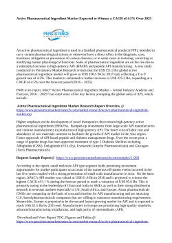
J. Phys. Chem. B 2007, 111, 4271-4279
4271
Peptide Bond Vibrational Coupling
Nataliya S. Myshakina and Sanford A. Asher*
Department of Chemistry, UniVersity of Pittsburgh, Pittsburgh, PennsylVania 15260
ReceiVed: August 14, 2006; In Final Form: January 3, 2007
Neutral trialanine (Ala3), which is geometrically constrained to have its peptide bond at Φ and Ψ angles of
R-helix and PPII-like conformers, are studied at the B3LYP/6-31+G(d,p) level of theory to examine vibrational
interactions between adjacent peptide units. Delocalization of the amide I, amide II, and amide III3 vibrations
are analyzed by calculating their potential energy distributions (PED). The vibrational coupling strengths are
estimated from the frequency shifts between the amide vibrations of Ala3 and the local amide bond vibrations
of isotopically substituted Ala3 derivatives. Our calculations show the absence of vibrational coupling of the
amide I and amide II bands in the PPII conformations. In contrast, the R-helical conformation shows strong
coupling between the amide I vibrations due to the favorable orientation of the CdO bonds and the strong
transitional dipole coupling. The amide III3 vibration shows weak coupling in both the R-helix and PPII
conformations; this band can be treated as a local independent vibration. Our calculated results in general
agree with our previous experimental UV Raman studies of a 21-residue mainly alanine-based peptide (AP).
Introduction
Vibrational spectroscopy is a powerful method for determining molecular structure and dynamics, as evident from the
numerous IR and Raman studies that have examined the
conformations and dynamics of peptides and proteins.1-15 These
studies interpret spectral changes in terms of alterations in
equilibrium conformations. Most often, the underlying assumption is that the systems are linear and that the measured spectra
derive from a weighted linear sum of the spectral contributions
of the individual conformations. In the case of peptide secondary
structure studies, it is assumed that the spectra are the linear
sum of the spectra of the individual peptide bonds and that the
individual peptide bond spectra are uniquely characteristic of
the conformation about each peptide bond. This presumption
assumes that there is negligible coupling between the atomic
motions of adjacent peptide bonds.
If significant coupling occurs between adjacent peptide bonds,
the individual peptide bond spectra would depend upon their
context. In this case, a simple linear interpretation of the resultant
observed spectra becomes impossible. It would, thus, become
necessary to understand the dependence of the peptide bond
spectra on coupling of atomic motions between adjacent peptide
bonds. This coupling can be studied theoretically by calculating
the normal modes of peptides.
Previous studies, which have examined vibrational coupling
between the same peptide bond vibrations between peptide bond
units, have mainly focused on the amide I vibration, which is
primarily a CdO stretching mode mixed with minor contributions from C-N stretching and CR-C-N deformation.2,4,5,16-27
It has been demonstrated that the amide I vibrations of
polypeptides show significant coupling and are delocalized
across many peptide bonds. The coupling between the amide I
vibrations is enabled by its large transition dipole moment as
shown by Krimm and co-workers.28-31
Very few studies have examined vibrational coupling between
the amide II (C-N stretching and N-H bending coupled out* Corresponding author. E-mail: [email protected]. Telephone: (412)-6248570. Fax: (412)-624-0588.
of-phase) vibrations of adjacent peptide units and the coupling
between amide III vibrations (C-N stretching and N-H bending
coupled in-phase) of adjacent peptide units.32 These couplings
should be smaller than that of the amide I vibrations because
the transition dipoles are smaller. Understanding the extent of
coupling of the amide III vibrations is important because the
amide III Raman bands are the most sensitive to peptide
conformation. In fact, the lowest frequency amide III band
component, which is denoted as the amide III3 vibration, is a
sensitive probe of the peptide bond Ψ angle.33-35 It is important
to understand whether the amide III3 vibration is a local mode
or if it involves collective peptide unit motion; is its frequency
determined solely by the conformation of the vibrating peptide
unit, or is its frequency also determined by the conformation
of adjacent peptide units?
To examine the extent of coupling of amide I, amide II, and
amide III3 vibrations between peptide units, we previously
experimentally studied this coupling by examining the UV
resonance Raman spectra (UVRS) of two linked amides in
mixed H2O/D2O solutions.36 We also examined the 21-residue
mainly poly Ala peptide (AP), which is ∼50% R-helix at 0 °C
and melts to a polyproline II (PPII) conformation at higher
temperatures.36,37 Our hypothesis was that if the peptide
vibrations were coupled, then partial deuteration of the amide
nitrogens would alter the amide bond vibrations dramatically,
because deuteration of the N-H decouples N-H bending from
C-N stretching. This large mass change would dramatically
change the specific peptide bond amide vibrations. Thus, the
partially deuterated spectrum would not simply be the sum of
the spectra of the fully deuterated and non-deuterated peptides.
These studies36,37 suggested essential vibrational independence
of the amide III3 and amide II bands for linked peptide bonds
in both the R-helix and PPII conformations of AP. In addition,
the amide I and amide I′ vibrations showed no evidence for
coupling in the case of the AP PPII conformation, and the amide
I band of the R-helix conformation showed significant interpeptide bond coupling.
10.1021/jp065247i CCC: $37.00 © 2007 American Chemical Society
Published on Web 03/30/2007
4272 J. Phys. Chem. B, Vol. 111, No. 16, 2007
The weakness of our experimental study was that we did not
know the magnitude expected for the band frequency perturbation due to coupling of adjacent peptide bonds. In the work
here, we further clarify the extent of interpeptide coupling using
vibrational analysis calculated by using electronic structure
methods. We examined trialanine (Ala3) as a model peptide to
study vibrational interactions between adjacent peptide units and
studied the normal mode compositions of the amide I, amide
II, and amide III3 vibrations for geometries constrained to occur
in the PPII and R-helical conformations.
We also calculated the normal modes for several Ala3
isotopomers where we reduce coupling between adjacent
substituents and peptide bonds by altering the atomic masses.
We used these calculated local mode frequencies to estimate
the magnitude of the amide band frequency shifts due to
vibrational coupling.
Computational Details
All calculations were performed at the density functional
theory38-40 (DFT) level by using the Gaussian 03 calculation
package.41 Geometry optimization and vibrational frequency
analysis were carried out by using B3LYP functional42-44 and
a 6-31+G(d,p) basis set. Partial geometry optimization was
performed for trialanine (Ala3) in vacuum with each specified
pair of fixed values of φ and ψ angles corresponding to R-helical
(φ ) -76°; ψ ) -44°) and PPII-like (φ ) -67°; ψ ) 132°)
secondary structures. The values of the φ- and ψ-angles were
taken from the molecular dynamics study performed by Mu et
al.45 The authors showed that conformations with these particular
values of ψ and φ angles are the most populated by short
peptides. To take into account frequency shifts that occur in
the PPII conformation due to peptide-water hydrogen-bonding,
we partially optimized the geometry and calculated frequencies
for Ala3 in a PPII conformation surrounded by water molecules.
We calculated the normal modes of Ala3 in both the R-helical
and PPII conformations for the natural abundance and the
following isotopomers: 13Cd18O substituted at the N-terminus
peptide unit, 13Cd18O substituted at the C-terminus peptide unit,
N-D substituted at the N-terminus peptide unit, N-D substituted at the C-terminus peptide unit, ND2 substituted at the
N-terminus amide group, simultaneous ND2 substitution and
N-D substitution at the N-terminus peptide unit, and simultaneous ND2 substitution and N-D substitution at the C-terminus
peptide unit. The harmonic vibrational frequencies were corrected by the previously suggested scaling factor of 0.97.46,47
We calculated the potential energy distribution (PED) by using
the GAR2PED Gaussian output processing program written by
J. M. L. Martin and C. Van Alsenoy.48
To distinguish contributions of the nuclear motions of
different peptide units to the calculated normal mode composi-
Myshakina et al.
Figure 1. Labeling of peptide atoms indicating definitions of peptide
unit and Ψ and φ angles.
tion, we labeled the internal vibrational coordinates (bonds,
valence and torsion angles corresponding to stretching, bending
and torsional vibrations, respectively, Figure 1).
Results and Discussion
Normal Mode Composition Analysis. Amide I. Our previous
experimental study of the UVRS of the 21-residue AP peptide
in pure water, pure D2O, and H2O/D2O mixtures concluded
that the amide I vibrations are coupled in R-helical AP and
uncoupled in the PPII conformation.37 This conclusion is
consistent with the results of Hochstrasser’s group.5,24
Our R-helical Ala3 vibrational analysis demonstrates two
amide I modes, a high frequency A symmetry vibration (1716
cm-1), and a lower frequency E1 symmetry vibration (1702
cm-1) (Table 1). Each mode contains CO stretching contributions from both peptide units.
In contrast, the two amide I modes calculated for the PPII
conformation of Ala3 are essentially local vibrations of the
individual CO stretches. These results agree with our experimental data, which indicated coupled amide I modes in the
R-helical conformation but local amide I modes in the PPII
conformation.
Amide II. In D2O, N-H deuteration causes the complex amide
II vibration to become an almost pure C-N stretch (amide II′)
due to decoupling of N-H bending from C-N stretching.49,50
The amide II′ appears as a doublet in N-methylacetamide (NMA)
and AP because of a Fermi resonance between the amide II′
vibration and a combination of the low frequency amide IV′
(632 cm-1) and a skeletal deformation vibration (∼873 cm-1).50
Our theoretical results for the amide II of R-helical Ala3
indicate more coupling than our experimental studies do.
Experimentally, we did not observe a clear frequency shift for
the amide II and amide II′ bands in the PPII and R-helix
conformations of AP upon adjacent peptide bond deuteration/
protonation. Unfortunately, we were unable to completely
exclude small frequency shifts for the amide II bands due to
TABLE 1: Amide I Frequencies and Normal Mode Compositions of Ala3 in r-Helix and PPII Conformationsa
conformer
ν, cm-1
PED, %
R-helix
1702
N-term PU: CO s (51), NCRC b (4), -CN s (4), -CRC s (2), NH inp b (1), -CNpCR b (1)
C-term PU: -CaOa s (27), CRaNaCa b (2), CaNa s (2), CRaCN b (1), CaCRa s (1), -CaCRbNa b (1)
C-term PU: CaOa s (52), CRaNaCa b (4), -CaNa s (3), -CaCRa s (2), -NaHa inp b (2), CaCRbNa b (1),
-CaNCRa b (1)
N-term PU: CO s (27), NCRC b (2), -CN s (2), -CRC s (1), -NH inp b (1), CRaCN b (1)
N-term PU: CO s (70), NCRC b (6), -CN s (5), -NH2 scs (4), -CRC s (3), -CaOa s (3), -NH inp b (2),
CNpCR b (2), CRaCN b (2), -CCβCR b (1)
C-term PU: CaOa s (75), -CaNa s (7), CRaNaCa b (6), CO s (3), -CaCRa s (3), -NaHa inp b (2),
CaCRbNa b (2), CaNCRa b (1)
1716
PPII
1695
1698
a
Frequencies are scaled by factor 0.97. Abbreviations: PU, peptide unit; s, stretch; b, bending; inp b, in-plane bending; scs, scissoring.
J. Phys. Chem. B, Vol. 111, No. 16, 2007 4273
Peptide Bond Vibrational Coupling
TABLE 2: Calculated Amide II Frequencies and Normal Mode Compositions (PED) for Ala3 in r-Helical and PPII
Conformationsa
conformer
ν, cm-1
PED, %
R-helix
1480
N-term PU: NH inp b (19), -CN s (10), -CHRb inp b (5), CH3b asym def′ (4), -CO inp b (2),
-CH3 asym def′ (2), CRC s (1), CO s (1)
C-term PU: NaHa inp b (26), -CaNa s (12), NaCRb s (4), -CaHRa inp b (2), NCRa s (3), -CaOa inp b (2),
CaCRa s (1), -CH3b asym def (1), CH3b rocking′ (1)
N-term PU: NH inp b (28), -CN s (15), CO inp b (3), CRC s (2), -CH3 asym def′ (1)
C-term PU: NaHa inp b (20), CaNa s (8), NCRa s (4), CHRa inp b (4), -NaCRb s (3), CH3a asym def′ (2),
-CHRa inp b (2), CaOa inp b (1), -CaCRa s (1), -CH3b asym def′ (1), -CaOa s (1)
N-term PU: NH inp b (39), -CN s (23), CRC s (4), -CO inp b (4), -CH3 asym def′ (2), CHR inp b (1),
CO s (1), -NH2 rocking (1)
C-term PU: NaHa inp b (5), NCRa s (5), -CHRa inp b (4), -CaNa s (3), CH3a asym def′ (2),
NCβaCRa b (1), NaCRb s(1)
C-term PU: NaHa inp b (44), -CaNa s (25), NaCRb s (6), -NaCRb s (4), CaCRa s (4), -CH3a asym def′ (2),
CRaHRa inp b (2), -CRbHRb inp b (1), -NCRa s (1), NaCβbCRb b (1), CH3b asym def′ (1),
CRbHRb outp b (1), -NaCpCRb b (1)
N-term PU: -NH inp b (3), CN s (2)
1495
PPII
1491
1495
a
Frequencies are scaled by factor 0.97. Abbreviations: PU, peptide unit; s, stretch; b, bending; inp b, in-plane bending; outp b, out-of-plane
bending; scs, scissoring.
TABLE 3: Calculated AmIII3 Frequencies and Normal Mode Compositions of Trialaninea
conformer
ν, cm-1
PED, %
R-helix
1222
C-term PU: CaNa s (18), NaHa inp b (17), -CaCRa s (16), CaOa inp b (7), -CHRb outp b (6), -NaCRb s (6),
CRbHRb inp b (4), NCRa s (3), CH3b rocking (2), -NCβaCRa b (2), CH3a rocking′ (2),
CaNCRa b (2), -CH3a rocking (1), CaCβaCRa b (1), -CH3a asym def′ (1), CH3b rocking′ (1),
CH3b asym def (1), NaCpCRb b (1), -CRaHRa outp b (1), NaCβbCRb b (1), CRaCβa s (1)
N-term PU: -NH2 rocking (2), CRHR outp b (1), -COp inp b (1)
N-term PU: NH inp b (17), CN s (10), -NH2 rocking (12), CHR outp b (10), -CRC s (8), CO inp b (4),
-CH3 rocking (2), -CβNpCR b (2), CO s (2), CH3a rocking (1), CNpCR b (1), CRCβ s (1),
CHR inp b (1), -CH3 asym def′ (1)
C-term PU: -CRaHRa outp b (12), CRaHRa inp b (6), CRbHRb outp b (2), -NCRa s (1), -NaHa inp b (1),
CH3a rocking′ (1), -CaNa s (1), -CHRb inp b (1), CH3a asym def (1)
C-term PU: CaNa s (21), NaHa inp b (16), -NaCRb s (14), NaCRb s (6), OHp* inp b (6), CRaHRa outp b (6),
CRaHRa inp b (5), CH3b rocking (4), -CRbHRb outp b (4), -CaCRa s (2), NaCβbCRb b (1),
-CH3a rocking′ (1), -CaNCRa b (1), -CNpCR b (1), NaCpCRb b (1), -COp inp b (1),
CN s (1), CaCRbNa b (1), CRbCp s (1)
N-term PU: -NH2 rocking (3), NH inp b (1), NpCR s (1)
N-term PU: NH2 rocking (18), -CN s (16), -NH inp b (13), -CO inp b (7), CNpCR b (6), -NpCR s (3),
-CRHR outp b (3), CRC s (2), -CCβCR b (2), -CO s (1), CH3 rocking′ (1), CH3 rocking (1),
CβNpCR b (1), CH3 asym def (1)
C-term PU: CRaHRa outp b (10), NRaCRa s (5), -CRaHRa inp b (2), -CH3a rocking (2),
-CH3a rocking′ (1), -CH3a asym def (1), NaCRb s (1), -CaNCRa b (1), NaHa inp b (1)
1247
PPII
1205
1227
a
Frequencies are scaled by factor 0.97. Abbreviations: PU, peptide unit; s, stretch; b, bending; inp b, in-plane bending; outp b, out-of-plane
bending; scs, scissoring. -NH2 rocking is a penultimate group vibration contribution.
overlap of HOD bending and arginine side-chain bands and the
molecular O2 stretching vibrations, which shows spectral
intensity variations.37
Table 2 indicates two different amide II modes for the
R-helical Ala3 conformation (1480 and 1495 cm-1), which
derive from the in- and out-of-phase coupled vibrations of both
peptide units. These amide II vibrations contain similar contributions of C-N stretching and N-H bending of both peptide
units. For example, the 1495 cm-1 amide II vibration contains
28% N-H bending of the N-terminal peptide units and 20%
N-H bending of the C-terminal peptide unit. In contrast, the
two amide II vibrations of Ala3 in the PPII conformation occur
much closer in frequency and are almost local modes (Table
2).
Amide III3. The amide III spectral region shows several bands
that originate from vibrations involving C-N, N-CR, CR-C,
and C-CR stretching and N-H and CR-Η bending motions.51
The “classic” amide III band (specifically the amide III3)
involves CN stretching with in-phase N-H bending, which
couples to CRH in-phase bending in the PPII conformation. This
conformation-dependent coupling makes this amide III3 band
extremely sensitive to peptide secondary structure.35 In the
R-helical AP, this band occurs at 1261 cm-1, and in the PPII
conformation, the amide III3 band occurs at 1245 cm-1. The
D2O decoupling of ND bending from CN stretching and CRH
bending upon deuteration results in the disappearance of the
amide III3 band, as well as the other amide III region
bands.49,51,52 Our UVRS comparison between the R-helical and
PPII bands of AP spectra in H2O/D2O mixtures indicated that
the spectra could be modeled as the sum of the deuterated and
protonated species in both the R-helical and PPII conformations.
We concluded that there was insignificant coupling between
the amide III3 vibrations of adjacent peptide units.37
The conclusions from our theoretical studies are confounded
by the fact that the two amide III3 vibrations in Ala3 differ
significantly because the N-terminal peptide unit amide III3
vibration selectively couples to the penultimate NH2 rocking
motion (Table 3). The amide III3 vibration is very complicated.
In addition to the atomic motions listed above, the amide III3
vibration also has contributions from CR-C stretching, CH3
rocking (from the Ala methyl side-chain), and CdO in-plane
bending. However, the main contributions are still from C-N
4274 J. Phys. Chem. B, Vol. 111, No. 16, 2007
Myshakina et al.
TABLE 4: Calculated Amide III3 Frequencies and Normal Mode Compositions of ND2-Substituted Ala3a
conformer
ν, cm-1
PED, %
R-helix
1218
C-term PU: CaNa s (14), NaHa inp b (12), -CaCRa s (11), CaOa inp b (6), -NaCRb s (4),
-CRbHRb outp b (4), -CRaHRa outp b (4), CRbHRb inp b (3), CH3a rock′ (3), CaNCRa b (2),
CH3b rock (1), CaCβaCRa b (1), -NCβaCRa b (1), -CH3a asym def′ (1), CH3b rock′ (1),
NCRa s (1), CH3b asym def (1), CRaHRa inp b (1)
N-term PU: NH inp b (8), CN s (5), -CRC s (5), -NH2 scs (3), CO inp b (2), CO s (1), CNpCR b (1)
N-term PU: NH inp b (15), CN s (13), -CRC s (10), CO inp b (5), CNpCR b (2), -NH2 scs (1),
CH3 rock′ (1), -NH2 rock (1), -CβNpCR b (1), CO s (1), -CH3 rock (1), CRCβ s (1),
CCβCR b (1), -CH3 asym def′ (1)
C-term PU: -NaHa inp b (6), -CaNa s (5), CaCRas (5), -CRaHRa outp b (5), -NCRa s (4), CRaHRa inp b (4),
CRbHRb outp b (4), CH3a rock (2), NaCRbs (2), -CRbHRb inp b (2), -CaOa inp b (2),
NCβaCRa b (1), -CH3b rock (1), -CH3b rock′ (1)
C-term PU: NaCR.b s (13), -CaNa s (13), -NaHa inp b (8), NCRa s (6), -OpHp* inp b (5), -CH3b rock (3),
-CHRb inp b (3), -CaOa inp b (2), -CH3a rock (2), CRbHRb outp b (2), -NaCβbCRb b (1),
-NCβaCRa b (1), -CRaNaCa b (1), -CRbCp s (1), -CaCRbΝa b (1), -NaCpCR.b b (1),
Cp.C βb CRb b (1)
N-terminal PU: -CN s (11), -NH inp b (8), -CRHR inp b (4), -CO inp b (4), -CRHR outp b (1),
CNpCR b (1), -NH2 scs (1), CRC s (1), NH2 rock (1), CH3 rock′ (1)
N-terminal PU: -CN s (12), -NH inp b (7), -CO inp b (4), -CRHR inp b (4), -CRHR outp b (2),
CNpCR b (1), -CH3a asym def (1), CH3 rock′ (1), CRC s (1), NH2 rock (1)
C-terminal PU: CaNa s (9), NaHa inp b (9), CHRa outp b (13), NCRa s (9), CaOa inp b (4), -CH3a rock (3),
-NaCRb s (3), -CRbHRb outp b (3), -CaNCRa b (2), -CH3a rock′ (2), -CaCRa s (2), OHp* inp b (2),
CH3b rock (1), -NCβaCRa b (1)
1232
PPII
1201
1212
a
Frequencies are scaled by factor 0.97. Abbreviations: PU, peptide unit; s, stretch; b, bending; inp b, in-plane; outp b, out-of-plane bending; scs,
scissoring.
stretching and N-H bending. The amide III3 normal modes in
both the R-helix and PPII conformations of Ala3 act as local
modes in terms of contributions from C-N stretching and N-H
bending from individual peptide units (Table 3).
The two amide III3 bands in the R-helical conformation were
calculated to occur at 1222 and 1247 cm-1, with the 25 cm-1
higher frequency band corresponding to the vibration of the
N-terminus peptide unit with contributions from the terminal
NH2 group rocking bending. A similar gap between two amide
III3 frequencies was calculated for the PPII conformation where
the N-terminal peptide unit vibration also has a contribution
from NH2 rocking.
Because these two amide III3 vibrations differ due to the
unique NH2 rocking contribution from the N-terminal group, it
is likely that this calculation may not be relevant to the amide
III3 bonds of the internal peptide unit of peptides, which cannot
include NH2 rocking. Thus, we attempted to examine amide
III3 normal modes in Ala3 isotopomers, which were uncoupled
to the terminal NH2 group motions; we examined the ND2deuterated derivative of Ala3.
ND2-deuteration decreases the frequency difference between
the two amide III3 bands to 14 cm-1 in the R-helical conformation and to 11 cm-1 in the PPII conformation (Table 4). In the
ND2 R-helical Ala3 derivative, the higher frequency amide III3
vibration is dominated by the N-terminus peptide unit but also
contains a smaller contribution of motion from the other peptide
unit. The low frequency amide III3 band corresponds mainly to
a C-terminus amide III3 vibration with significant but smaller
N-H bending and C-N stretching contributions from the other
peptide unit.
The amide III3 vibrations (1201 and 1212 cm-1) in the PPII
conformation of ND2 Ala3 are evidently coupled; both amide
III3 vibrations involve comparable contributions of CN stretching
and NH bending from each peptide unit (Table 4). Our
experimental results concluded little coupling evidenced by
insignificant spectral frequency shifts upon deuteration of
adjacent peptide units.
Frequency Shifts of the Amide Bands Due to Coupling.
The coupling between local motions is monitored by the value
of the coupling force constants, i.e., the off-diagonal elements
of the Hessian matrix. For a system with two peptide bonds,
the Hessian matrix is
F̃(φ,φ) )
[
K1(φ,φ) C12(φ,φ)
C12(φ,φ) K2(φ,φ)
]
where K1 and K2 are force constants of the local modes and
C12 is the coupling force constant between local modes.
Due to the nonzero coupling force constants, interaction
between two local modes results in formation of two coupled
delocalized modes with perturbed frequencies. In the case of
amide I local mode coupling, two coupled amide I modes of
symmetry A and E1 are formed. The frequencies of the A and
E1 modes will occur at higher and lower frequencies relative to
that of the unperturbed local CdO stretches. The A component
refers to the symmetrical, in-phase stretching of CdO bonds.
The E1 component originates from asymmetric or out-of-phase
stretching motions of CdO bonds. When the transition dipole
moments of the CdO stretch are perpendicular to the axis of
the peptide backbone (as in the PPII or β-strand-like conformations), the E1 component will show strong IR absorbance and
the A component will be active in Raman but vanish in IR
measurements. For these conformations, there is a difference
between the amide I frequency observable in IR and the amide
I frequency observable in Raman. This frequency difference
corresponds to the A-E1 splitting, which originates from
coupling of the local CdO stretches. By contrast, in the R-helix
conformation, where the transition dipole moment of each
peptide group is almost parallel to the helix axis, the A
component acquires a strong IR absorbance in addition to Raman
intensity. Therefore, R-helical peptides show an amide I band
of A symmetry, which is observable in both the IR and Raman
spectra; there will be no frequency difference between the IR
and Raman observed amide I bands in this case. There are few
published studies dedicated to the peptide secondary structure
determination from experimentally measured A-E1 splitting
between the amide I Raman and IR frequencies.25,53,54
The frequency difference between coupled vibrations depends
upon the magnitude of the vibrational coupling constant. The
larger the coupling constant, the stronger the coupling and the
J. Phys. Chem. B, Vol. 111, No. 16, 2007 4275
Peptide Bond Vibrational Coupling
TABLE 5: Calculated Amide I Frequencies (cm-1) of Normal Abundance Ala3 and Ala3 Isotopomers in r-Helical and PPII
Conformations
a
R-helix
PPII
C13O18-Ala3
1712a
l(CdO) ) 1.224 Å
1699
l(CdO) ) 1.229 Å
C13O18-Ala3
1708
l(CdO) ) 1.226 Å
1695
l(CdO) ) 1.231 Å
Ala3
1716 (A)
1702 (E1)
1698 (C term. PB)
1695 (N term. PB)
Frequencies and CdO bond lengths correspond to indicated PB.
TABLE 6: Normal Mode Compositions of Amide I Vibrations of D2N, ND-Isotopomers of Ala3a
R-helix
PU
deuterated
ν, cm-1
PED, %
N-terminus
1715
C-term PU: CaOa s (60), CRaNaCa b (5), -CaNa s (4), -CaCRa s (2), -NaHa inp b (2), CaCRbNb b (1),
-CaNCRa b (1)
N-term PU: CO s (19), -CN s (2), NCRC b (2), -CRC s (1)
N-term PU: CO s (60), -CN s (5), NCRC b (5), -CRC s (2), -CNpCR b (1)
C-term PU: -CaOa s (19), -CRaNaCa b (2), CaNa s (2), CRaCN b (1), CaCRa s (1)
C-term PU: CaOa s (43), CRaNaCa b (3), -CaNa s (3), -CaCR.a s (2)
N-term PU: CO s (37), NCRC b (3), -CN s (3), CRC s (2)
N-term PU: CO s (41), NCRC b (4), -CN s (3), -CRC s (2), -NH inp b (1), -CNpCR b (1)
C-term PU: -CaOa s (37), CaNa s (3), -CRaNaCa b (3), CaCRa s (1), -CaCRbNa b (1), CaNCR a b (1),
CRaCN b (1)
C-term PU: CaOa s (76), -CaNa s (8), CRaNaCa b (6), -CaCRa s (2), NaCaCRb b (2), CaNCRa b (1)
N-term PU: CO s (3)
N-term PU: CO s (75), NCRC b (7), -CN s (6), -CRC s (3), -ND inp b (2), CNpCR b (1)
C-term PU: -CaOa s (3), CRaCN b (2)
C-term PU: CaOa s (78), -CaNa s (7), CRaNaCa b (7), -CaCRa s (3), -NaDa inp b (2), CaCRbNa b (2),
CaNCRa b (1)
N-term PU: CO s (79), -CN s (7), NCRC b (7), -CRC s (3), CNpCR b (1), -NH inp b (1)
C-term PU: CRaCN b (2)
1699
C-terminus
1713
1701
PPII
N-terminus
1695
1690
C-terminus
1699
1685
a Frequencies are scaled by factor 0.97. Abbreviations: PU, peptide unit; s, stretch; b, bending; inp b, in-plane; outp b, out-of-plane bending; scs,
scissoring.
more the frequencies of coupled bands are perturbed compared
to the frequencies of the local uncoupled bands. Therefore, we
can estimate the strength of the amide band coupling by
monitoring the frequency differences between calculated amide
bands compared to the frequencies of the original uncoupled
local vibrations. To obtain these uncoupled frequencies, we
calculated the normal modes of various Ala3 isotopomers.
Amide I. The PED of the R-helical natural abundance Ala3
normal modes shows that the two calculated amide I bands
(1702 and 1716 cm-1) are coupled modes of A and E1
symmetry. We singly substituted C13-O18 at each of the two
Ala3 peptide bonds and calculated the frequency of the other
natural abundance peptide bond. In this case, the local amide I
vibrations at the carboxyl end occurred at 1712 cm-1 and at
the amine end occurred at 1708 cm-1. The frequencies differ
because their optimized structures have slightly different CdO
bond lengths (Table 5). If we assume a reference frequency for
the uncoupled local CdO stretching mode of Ala3 as a simple
average (1710 cm-1) of the local amide I frequencies calculated
for two Ala3 isotopomers, we conclude that the coupling
interactions in natural abundance Ala3 up-shift the high frequency A component by 6 cm-1 and down-shifts the E1
component by 8 cm-1 (Tables 5 and 10). In contrast, the PPII
Ala3 PED shows that two calculated amide I modes are
uncoupled and their frequencies are almost equal to the
uncoupled amide I frequencies of Ala3 isotopomers. These
results are fully consistent with our experimental data. Our
results are also consistent with Torii and Tasumi17 who
theoretically showed that the coupling constants between
adjacent amide I vibrations in the R-helix conformation are
larger than those in the β-region (close to that of PPII
conformation).
There are two main mechanisms of vibrational coupling:
through bond coupling (or mechanical coupling) and through
space coupling (or electrostatic coupling). Mechanical coupling
results from the impact of local atomic motion on the structure
of adjacent molecular bonds; electrostatic coupling results from
the motion of local dipolar bonds. The large dipole moment of
the C-O bond gives rise to significant electrostatic coupling
for the amide I modes in addition to significant mechanical
coupling. In contrast, mechanical coupling dominates the
coupling between the amide II and amide III3 vibrations due to
the smaller dipole moments of the vibrating bonds.
Amide I coupling in the R-helix conformation and the lack
of it in the PPII conformation result from the different relative
spatial orientations of the CdO bonds in the R-helix and PPII
conformations.
In the R-helix, the CdO bonds are almost co-parallel; in the
PPII conformation, they project out at angles of ∼120° from
one another. The more they project upon one another, the larger
4276 J. Phys. Chem. B, Vol. 111, No. 16, 2007
Myshakina et al.
TABLE 7: Calculated Amide I, Amide II, and Amide III3 Frequencies of Normal Abundance Ala3 and Ala3 Isotopomers in
PPII and r-Helical Conformations
amide I
deuteration
amide II
N-term.
none
R-helix
ND2
PPII
R-helix
1702 out-of-phase
1716 in-phase
1695
1698
1702 out-of-phase
1716 in-phase
1691
1699
1480 in-phase
1495 out-of-phase
1491
1495
1480 in-phase
1496 out-of-phase
1491
1496
1699 out-of-phase
1715 in-phase
1690
1699
1701 out-of-phase
1713 in phase
1694
1695
1699 out-of-phase
1715 in-phase
1685
1699
1701 out-of-phase
1713 in-phase
1690
1695
ND
1487
1201 in-phase
1212 out-of-phase
ND
1221
ND
1489
1495
ND
ND
1245
1206
ND
1492
ND
ND
1487
1226
ND
ND
1221
ND
1489
1495
ND
ND
1227
1206
ND
1491
ND
1207
ND
PPII
ND-N term
R-helix
ND-C term
PPII
R-helix
ND2, ND-N term
PPII
R-helix
ND2, ND-C term
PPII
R-helix
PPII
C-term.
N-term.
amide III3
conform
C-term.
N-term.
C-term.
1247
1222
1227
1232
1205
1218
TABLE 8: Normal Mode Compositions of Amide II Vibrations of D2N, ND-Isotopomers of Ala3a
R-helix
PPII
deuterated
PU
ν, cm-1 a
PED, %
N-terminus
1487
C-terminus
1489
N-terminus
1495
C-terminus
1491
C-term PU: NaHa inp b (47), -CaNa s (20), -CRbHRb inp b (9), NaCRb s (6), CH3b asym def′ (3),
-CaOa inp b (3), CaCR.a s (2), -CH3a asym def′ (2), -CH3b asym def (1), CH3b rocking′ (1),
CaOa s (1)
N-term PU: NH inp b (47), -CN s (25), -CO inp b (4), -CRC s (3), -CH3 asym def′ (2), CO s (1)
C-term PU: NCRa s (7), CRaHRa inp b (4), CH3a asym def′ (2), CRaHRa outp b (1), NCβaCRa b (1)
C-term PU: NaHa inp b (49), -CaNa s (28), NaCR.b s (7), -CaOa inp b (5), CaCRa s (4), -CRbHRb inp b (1),
NaCβbCRb b (1), -CH3a asym def′ (1), CH3b asym def′ (1), CRbHRb outp b (1), CRaHRa inp b (1),
-NaCpCRb b (1)
N-term PU: NH inp b (43), -CN s (25), -CO inp b (4), CRC s (4), -CH3 asym def′ (1), CO s (1),
CRHR inp b (1)
C-term PU: NCRa s (6), -CRaHRa inp b (6), CH3a asym def′ (3), NCβaCRa b (1), -CH3a asym def (1)
a Frequencies are scaled by factor 0.97. Abbreviations: PU, peptide unit; s, stretch, b, bending; inp b, in-plane; outp b, out-of-plane bending; scs,
scissoring.
TABLE 9: Normal Mode Compositions of Localized Amide III3 Vibrations of D2N, ND-Isotopomers of Ala3a
R-helix
PPII
deuterated
PU
ν, cm-1 a
PED, %
N-terminus
1221
C-terminus
1227
N-terminus
1206
C-terminus
1207
C-term PU: CaNa s (19), NaHa inp b (18), -CaCRa s (16), CaOa inp b (8), -CRbHRb outp b (7),
-NaCRb s (6), CRbHRb inp b (4), CH3b rock (2), -NCβaCRa b (2), CaNCRa b (2), NCRa s (2),
-CH3a rock (2), CH3a rock′ (2), -CH3a asym def′ (1), CH3a asym def′ (1), CH3b rock′ (1),
CH3b asym def (1), NaCpCRb b (1), CRaCβa s (1), NaCβbCRb b (1), -CpOp inp b (1),
CaCRbNa b (1)
N-term PU: NH inp b (24), CN s (19), -CRC s (16), -CO inp b (8), CNpCR b (3), -ND2 sciss (2),
-CO s (1), CH3 rock′ (1), -ND2 rock (1), CRCβ s (1), -CβNpCR b (1), -CH3 rock (1),
CCβCR b (1), -CH3 asym def′ (1)
C-term PU: CRaHRa outp b (10), NCRa s (2), CRaHRa inp b (4), CH3a rock′ (1), CH3a rock (1),
CH3a asym def (1)
C-term PU: CaNa s (23), NaHa inp b (17), -NaCRb s (13), CRaHRa outp b (10), CaOa inp b (7),
OHp* inp b (6), -CRbHRb outp b (4), CH3b rock (4), CRaHRa inp b (3), -CH3a rock′ (2),
-CaCRa s (2), -CaNCRa b (1), NaCβbCRb b (1), NaCpCRb b (1), -CpOp inp b (1)
N-term PU: NCRC b (1)
N-term PU: CN s (24), NH inp b (14), CO inp b (8), CRHR inp b (7), CRHR outp b (4), -CNpCR b (2),
-CRC s (1), -CH3 rock′ (1), -ND2 rock (1)
C-term PU: -NCRa s (16), CH3a rock (5), -CRaHRa outp b (4), NCβaCRa b (2), -NaCRa s (1),
CRaNaCa b (1), CRa CN b (1), CaNCRa b (1), CH3a asym def (1), CRaHRa inp b (1),
CaCRa s (1), CH3a rock′ (1)
a
Frequencies are scaled by factor 0.97. Abbreviations: PU, peptide unit; s, stretch; b, bending; inp b, in-plane; outp b, out-of-plane bending; scs,
scissoring.
will be the dipole-dipole interactions. In addition, the closer
the distance between CdO bonds, the stronger the dipoledipole interactions. An R-helix conformation has a smaller pitch
than the more extended PPII structure; the CdO and N-H
bonds of neighboring peptide bonds are closer to each other
compared to PPII or β-strand conformations.
Deuteration of the peptide bond NH and penultimate amino
group does not affect the coupled amide I frequencies and
J. Phys. Chem. B, Vol. 111, No. 16, 2007 4277
Peptide Bond Vibrational Coupling
TABLE 10: Vibrational Coupling Frequency Shifts of
Amide Bands
R-helix
PPII
Δνamide II,
cm-1
Δνamide I,
cm-1
conformation
Δνamide III3,
cm-1
-3
5
-5
6
in-phase
6
8
out-of-phase
-8
-8
in-phase
no coupling no coupling
out-of-phase
occurs
occurs
TABLE 11: Measured PPII AP Frequencies, Calculated
Frequencies of PPII Ala3 in Vacuum and in Ala3-Water
Complex, and Calculated Hydrogen-Bonding Frequency
Shifts
νamide I, cm-1
νamide II, cm-1
νamide III, cm-1
1699
1657
-42
1660
1496
1559
63
1558
1205
1249
44
1248
ala3 vacuum
ala3‚4H2O
ΔνHBa
PPII AP
a
ΔνHB ) νAla3×4W - νAla3
gas.
normal mode compositions in the R-helical conformation
compared to the amide I frequencies of normal abundance Ala3.
In the PPII conformation, the amide I frequencies slightly shift
upon deuteration (Tables 6 and 7). In the normal abundance
PPII Ala3 conformation, these modes are mainly CdO stretching
vibrations of each individual peptide unit containing minor
contributions (2%) from N-H bending. In addition to the N-H
bending, the local amide I vibration of the N-terminal peptide
unit has a contribution from NH2 scissoring (4%) (Table 1).
Deuteration of the NH2 group or N-H bond eliminates
contributions of N-H bending and NH2 scissoring vibrations
from the normal mode compositions, which results in slight
frequency shifts (4-10 cm-1) of the amide I frequencies (Table
7).
Amide II. No vibrational coupling occurs between amide II
bands in the PPII conformation of Ala3. These amide II
frequencies are almost equal to the local frequencies (1496 and
1491 cm-1) of isotopically substituted Ala3 (Tables 7 and 8).
The difference between two frequencies of regular Ala3 (4 cm-1)
and two local frequencies of the Ala3-isotopomers (5 cm-1)
originates from a slight difference between the C-N bond
lengths of the two peptide bonds and is unrelated to any
coupling.
We calculate that coupling between the amide II bands of
R-helical Ala3 results in formation of two delocalized vibrations
which are up- and down-shifted by 8 cm-1 compared to the
uncoupled localized amide II vibrations of the ND-substituted
Ala3 (Tables 7 and 10). However, for ND-deuterated R-helical
Ala3, we also calculate that the uncoupled amide II vibration
should occur around 1487-1489 cm-1 (excluding frequency
Figure 2. Water bridges in PPII structure.
shift due to hydrogen bonding). This differs from the coupled
amide II band frequencies calculated at 1480 and 1496 cm-1.
These results indicate that we should observe more often the
narrow amide II band in partially deuterated peptides than those
fully protonated bands in pure water. It is possible that this shift
was obscured by the overlap of the amide II band with an
arginine band and the molecular oxygen stretching band.
Amide III3. For R-helical NH2, NH-deuterated Ala3 isotopomers, our calculations show a 6 cm-1 frequency difference
between local amide III3 bands (1221 and 1227 cm-1) of the
N- and C-terminal peptide bonds.
In ND2-substituted R-helical Ala3, the amide III3 vibrations
are mainly localized. The higher frequency amide III3 band
(1232 cm-1) is dominated by the motion of the N-terminal
peptide unit with only a minor contribution from the C-terminal
peptide unit (Table 4); the low frequency amide III3 vibration
(1218 cm-1) contains a major contribution from the C-terminus
peptide unit. The local amide III3 vibrations of ND, ND2deuterated Ala3 isotopomers occur at 1227 cm-1 (N-terminus
vibration) and at 1221 cm-1 (C-terminus vibration) (Table 9).
Vibrational coupling with the other peptide unit up-shifts this
band by 5 cm-1. Consequently, amide III3 bands are 5 up- and
3 cm-1 down-shifted (Table 10). We conclude that R-helical
amide III3 band coupling is small because the frequencies shift
little from the frequencies of local vibrations. However, the
normal mode compositions of these two calculated amide III3
vibrations indicate some contributions from the adjacent peptide
unit.
The PPII conformation N- and C-terminus amide III3 vibrations of the ND2, ND-substituted Ala3 isotopomers have almost
identical frequencies (1206, 1207 cm-1). Therefore, in natural
abundance Ala3, the symmetrical component (1201 cm-1) of
amide III3 is down-shifted by 5 cm-1 and the asymmetrical
TABLE 12: Amide Frequencies of AP (Measured) and Ala3 (Calculated) in PPII Conformation in Gas-Phase and in
Peptide-Water Complex
amide I, cm-1
PPII-ala3, calc
PPII-AP in H2O,
experimental
PPII-AP in H2O/D2O
experimental
PPII-AP in H2O/D2O
modeled spectra
without coupling
a
amide II, cm-1
amide III3, cm-1
N-term.
C-term.
N-term.
C-term.
in-phase
1690
(1648)
1660
1699
(1657)
1491
(1554)
1558
1496
(1558)
1201
(1245)
1245
1655
1559
1248
1661
1560
1249
Values in parentheses include ΔυHB (see Table 11).
out-of-phase
1212
(1256)
4278 J. Phys. Chem. B, Vol. 111, No. 16, 2007
Myshakina et al.
TABLE 13: Measured Frequencies of Gas-Phase and Liquid
NMA, Calculated Hydrogen-Bonding Frequency Shifts
(ΔνHB), Calculated Frequencies of r-Helical Ala3 in Vacuum,
Frequencies of the r-Helical Ala3 Including ΔνHB, and
Measured Frequencies of r-Helical AP
νamide I, cm-1
νamide II, cm-1
νamide III3, cm-1
1731
1499
1255
1668
1558
1298
-63
1702
59
1480
43
1218
1639
1646
1539
1547
1261
1261
a
NMA, gas phase
measured
NMA, neat liquid
measured
ΔνHBb
R-Ala3, gas phase
calcd
R-Ala3 + Δν
R-AP, experimental
b
a
Kubelka, J.; Keiderling, T. J. Phys. Chem. A 2001, 10922-10928.
ΔνHB ) νNMA liquid - νNMA gas.
component (1212 cm-1) is up-shifted by 6 cm-1 due to
vibrational coupling (Tables 4 and 10). Our calculations show
weak coupling of the PPII amide III3 bands. This PPII amide
III3 coupling can be explained by its normal mode compositions.
Besides the C-N stretch and N-H bending, which are coupled
in-phase with CR-H bending, the PPII amide III3 modes contain
contributions from the N-CR stretching vibration (Table 4). The
N-CR bond “bridges” adjacent peptide units. This stretching
motion facilitates coupling with the CR-H bending motions.
The CR-H bending can efficiently couple with N-H bending
of the adjacent peptide unit. The contribution from the N-CR
stretch facilitates delocalization of the PPII amide III3 band.
The amide III3 vibration of R-helical Ala3 does not include the
N-CR stretching. Instead, this normal mode contains C-CR
stretching.
Frequency Shifts of the Amide Bands Due to Hydrogen
Bonding. Our calculated amide frequencies differ from the
experimentally measured frequencies because we have not taken
into account the effects of hydrogen bonding. The hydrogenbonding patterns differ in the R-helix and PPII conformations.
Hydrogen bonding in the PPII conformation involves hydrogen
bonding to water. Hydrogen bonding in the R-helix conformation
involves peptide bond-peptide bond hydrogen bonding.
Hydrogen-Bonding Shifts in PPII Conformation. To estimate
the effects of the hydrogen-bonding differences on the frequencies of the amide vibrations we compared amide I, amide II,
and amide III3 frequencies calculated for the PPII Ala3 in
vacuum to the Ala3-water complex.
Sreerama et al.55 studied the network of water molecules
involved in hydrogen bonding to the backbone of polyalanine
in β-strand and PPII conformations by molecular dynamics
simulations. They analyzed populations of different types of
hydrogen-bonding “bridges”. For the PPII conformation, they
found that a bridge of two water molecules connecting the i-th
CdO and (i + 2)-th N-H bonds was the most populated.
We calculated the frequencies of an Ala3-water complex in
the PPII conformation for this arrangement of water molecules
(see Figure 2). We included four water molecules, which made
two water bridges: one connects the first residue CdO bond
with the third residue N-H bond and the other connects the
remaining N-H and CdO groups in the second residue (Figure
2). This hydrogen-bonding network down-shifts the amide I
frequency of Ala3 by 42 cm-1 and up-shifts the amide II and
amide III3 bands by 63 and 44 cm-1, respectively (Table 11).
Overall, the amide frequencies calculated for the Ala3-water
complex agree well with the experimentally measured frequencies of PPII AP (Table 12).
Hydrogen-Bonding Shifts in R-Helical Conformation. Theoretical simulation of the intermolecular hydrogen bonding in
the R-helix conformation is more complicated. We estimated
the R-helix hydrogen-bonding frequency shifts from a difference
between UVRR amide band frequencies of the gas phase56 and
liquid state35 of N-methylacetamide (NMA) (Table 13). The
frequency of the amide I band decreases by 63 cm-1 in liquid
NMA, compared to the gas state, and the amide II and amide
III3 frequencies increase by ∼60 and 43 cm-1, respectively. We
applied these frequency shifts to correct the calculated frequencies of vacuum R-ala3. This correction brings calculated Ala3
frequencies into a good agreement with the measured frequencies of R-helical AP (Table 14).
Conclusion
Our calculations have demonstrated vibrational coupling
between adjacent peptide units of the amide I and amide II
modes in the R-helical conformation where these carbonyl bonds
are closest to each other and their projection angles are smallest.
This coupling decreases in the PPII conformation, because the
structure is more extended and the projection angle increases.
The favorable orientation of the CdO bonds in the R-helix
conformation facilitates transitional dipole coupling. Therefore,
coupling of the amide I bands is much larger than coupling
between the amide II bands.
Coupling interactions between amide III3 bands is less than
that of the amide I and amide II vibrations. The amide III3 bands
can be treated as a local vibrations of independently Raman
scattering peptide bonds. The calculated amide III3 frequencies
are not significantly perturbed compared to the frequencies of
local amide III3 modes even though our calculations indicate
that their normal mode compositions contain minor contributions
from the adjacent peptide units.
Acknowledgment. We gratefully acknowledge Alexander
Mikhonin for useful discussions. This work was supported by
NIH grant GM8RO1EB002053021.
TABLE 14: Measured and Calculated Frequencies of AP and Ala3 in r-Helical Conformationsa
amide I, cm-1
in-phase
R-Ala3, calcd
R-AP in H2O,
experimental
R-AP in H2O/D2O,
experimental
R-AP in H2O/D2O
modeled spectra
without coupling
a
1716
(1653)
1646
out-of-phase
1702
(1639)
amide II, cm-1
in-phase
1480
(1539)
1547
out-of-phase
1496
(1555)
amide III3, cm-1
in-phase
1218
(1261)
1261
1644
1535
1256
1643
1530
1262
Values in parenthesis include ΔνHB (see Table 11).
out-of-phase
1232
(1275)
Peptide Bond Vibrational Coupling
References and Notes
(1) Choi, J.-H.; Hahn, S.; Cho, M. Int. J. Quantum Chem. 2005, 104,
616.
(2) Edler, J.; Hamm, P. J. Chem. Phys. 2002, 117, 2415.
(3) Fang, C.; Wang, J.; Charnley, A. K.; Barber-Armstrong, W.; Smith,
A. B.; Decatur, S. M.; Hochstrasser, R. M. Chem. Phys. Lett. 2003, 2003,
586.
(4) Hamm, P.; Lim, M.; Hochstrasser, R. M. J. Phys. Chem. B 1998,
102, 6123.
(5) Fang, C.; Wang, J.; Kim, Y. S.; Charnley, A. K.; Barber-Armstrong,
W.; Smith, A. B. I.; Decatur, S. M.; Hochstrasser, R. M. J. Phys. Chem. B
2004, 108, 10415.
(6) Bowen, M.; Brunger, A. T. Proc. Natl. Acad. Sci. U.S.A. 2006,
103, 8378.
(7) Ahmed-Ouameur, A.; Diamantoglou, S.; Sedaghat-Herati, M. R.;
Nafisi, S.; Carpentier, R.; Tajmir-Riahi, H. A. Cell Biochem. Biophys. 2006,
45, 203.
(8) Perham, M.; Liao, J.; Wittung-Stafshede, P. Biochemistry 2006,
45, 7740.
(9) Xu, Y.; Wang, T.; Gai, F. Chem. Phys. 2006, 323, 21.
(10) Causgrove, T. P.; Dyer, R. B. Chem. Phys. 2006, 323, 2.
(11) Pozo Ramajo, A.; Petty, S. A.; Volk, M. Chem. Phys. 2006, 323,
11.
(12) Chin, W.; Piuzzi, F.; Dimicoli, I.; Mons, M. Phys. Chem. Chem.
Phys. 2006, 8, 1033.
(13) Du, D.; Tucker, M. J.; Gai, F. Biochemistry 2006, 45, 2668.
(14) Decatur, S. M. Acc. Chem. Res. 2006, 39, 169.
(15) Manning, M. C. Expert ReV. Proteomics 2005, 2, 731.
(16) Gorbunov, R. D.; Kosov, D. S.; Stock, G. J. Chem. Phys. 2005,
122, 224904/1.
(17) Torii, H.; Tasumi, M. J. Raman Spectrosc. 1998, 29, 81.
(18) Choi, J.-H.; Ham, S.; Cho, M. J. Chem. Phys. 2002, 117, 6821.
(19) Ham, S.; Cho, M. J. Chem. Phys. 2003, 118, 6915.
(20) Ham, S.; Cha, S.; Choi, J.-H.; Cho, M. J. Chem. Phys. 2003, 119,
1451.
(21) Choi, J.-H.; Ham, S.; Cho, M. J. Phys. Chem. B 2003, 107, 9132.
(22) Ham, S.; Kim, J.-H.; Lee, H.; Cho, M. J. Chem. Phys. 2003, 118,
3491.
(23) Bour, P.; Keiderling, T. A. J. Chem. Phys. 2003, 119, 11253.
(24) Fang, C.; Wang, J.; Charnley, A. K.; Barber-Armstrong, W.; Smith,
A. B.; Decatur, S. M.; Hochstrasser, R. M. Chem. Phys. Lett. 2003, 382,
586.
(25) Schweitzer-Stenner, R. J. Phys. Chem. B 2004, 108, 16965.
(26) Schweitzer-Stenner, R.; Sieler, G.; Mirkin, N. G.; Krimm, S. J.
Phys. Chem. A 1998, 102, 118.
(27) Huang, Q.; Schweitzer-Stenner, R. J. Raman Spectrosc. 2004, 35,
586.
(28) Krimm, S.; Abe, Y. Proc. Natl. Acad. Sci. U.S.A. 1972, 69, 2788.
(29) Moore, W. H.; Krimm, S. Proc. Natl. Acad. Sci. U.S.A. 1975, 72,
4933.
(30) Cheam, T. C.; Krimm, S. Chem. Phys. Lett. 1984, 107, 613.
(31) Krimm, S.; Bandekar, J. AdV. Protein Chem. 1986, 38, 181.
(32) Torii, H.; Tatsumi, T.; Tasumi, M. J. Raman Spectrosc. 1998, 29,
537.
(33) Asher, S. A.; Ianoul, A.; Mix, G.; Boyden, M. N.; Karnoup, A.;
Diem, M.; Schweitzer-Stenner, R. J. Am. Chem. Soc. 2001, 123, 11775.
J. Phys. Chem. B, Vol. 111, No. 16, 2007 4279
(34) Ianoul, A.; Boyden, M. N.; Asher, S. A. J. Am. Chem. Soc. 2001,
123, 7433.
(35) Mikhonin Aleksandr, V.; Bykov Sergei, V.; Myshakina, Nataliya,
S.; Asher, Sanford, A. J. Phys. Chem. B 2006, 110, 1928.
(36) Mix, G.; Schweitzer-Stenner, R.; Asher, S. A. J. Am. Chem. Soc.
2000, 122, 9028.
(37) Mikhonin, A. V.; Asher, S. A. J. Phys. Chem. B 2005, 109,
3047.
(38) Kohn, W.; Sham, L. J. Phys. ReV. 1965, 137, 1697.
(39) Parr, R. G.; W. Yang Density-functional theory of atoms and
molecules; Oxford University Press: Oxford, U.K., 1989.
(40) Hohenberg, P.; Kohn, W. Phys. ReV. 1964, 136, B864.
(41) Frisch, M. J. T., G. W.; Schlegel, H. B.; Scuseria, G. E.; Robb, M.
A.; Cheeseman, J. R.; Montgomery, Jr., J. A.; Vreven, T.; Kudin, K. N.;
Burant, J. C.; Millam, J. M.; Iyengar, S. S.; Tomasi, J.; Barone, V.;
Mennucci, B.; Cossi, M.; Scalmani, G.; Rega, N.; Petersson, G. A.;
Nakatsuji, H.; Hada, M.; Ehara, M.; Toyota, K.; Fukuda, R.; Hasegawa, J.;
Ishida, M.; Nakajima, T.; Honda, Y.; Kitao, O.; Nakai, H.; Klene, M.; Li,
X.; Knox, J. E.; Hratchian, H. P.; Cross, J. B.; Bakken, V.; Adamo, C.;
Jaramillo, J.; Gomperts, R.; Stratmann, R. E.; Yazyev, O.; Austin, A. J.;
Cammi, R.; Pomelli, C.; Ochterski, J. W.; Ayala, P. Y.; Morokuma, K.;
Voth, G. A.; Salvador, P.; Dannenberg, J. J.; Zakrzewski, V. G.; Dapprich,
S.; Daniels, A. D.; Strain, M. C.; Farkas, O.; Malick, D. K.; Rabuck, A.
D.; Raghavachari, K.; Foresman, J. B.; Ortiz, J. V.; Cui, Q.; Baboul, A.
G.; Clifford, S.; Cioslowski, J.; Stefanov, B. B.; Liu, G.; Liashenko, A.;
Piskorz, P.; Komaromi, I.; Martin, R. L.; Fox, D. J.; Keith, T.; Al-Laham,
M. A.; Peng, C. Y.; Nanayakkara, A.; Challacombe, M.; Gill, P. M. W.;
Johnson, B.; Chen, W.; Wong, M. W.; Gonzalez, C.; Pople, J. A. Gaussian
03; revision C.01; Gaussian, Inc.: Wallingford, CT, 2004.
(42) Becke, A. D. J. Chem. Phys. 1993, 98, 5648.
(43) Lee, C.; Yang, W.; Parr, R. G. Phys. ReV. B: Condens. Matter
Mater. Phys. 1988, 37, 785.
(44) Miehlich, B.; Savin, A.; Stoll, H.; Preuss, H. Chem. Phys. Lett.
1989, 157, 200.
(45) Mu, Y.; Stock, G. J. Phys. Chem. B 2002, 106, 5294.
(46) Irikura, K. K.; Johnson, R. D., III;, Kacker, R. N. J. Phys. Chem.
A 2005, 109, 8430.
(47) Halls, M. D.; Velkovski, J.; Schlegel, H. B. Theor. Chem. Acc.
2001, 105, 413.
(48) Martin, J. M. L.; Van Alsenoy, C. GAR2PED; University of
Antwerpen: Antwerpen, Belgium, 1995.
(49) Lee, S.-H. K., S. Biopolymers 1998, 46, 283.
(50) Chen, X. G.; Asher, S. A.; Schweitzer-Stenner, R.; Mirkin, N. G.;
Krimm, S. J. Am. Chem. Soc. 1995, 117, 2884.
(51) Mikhonin, A. V.; Ahmed, Z.; Ianoul, A.; Asher, S. A. J. Phys.
Chem. B 2004, 108, 19020.
(52) Lednev, I. K. K., A. S.; Sparrow, M. C.; Asher, S. A. J. Am. Chem.
Soc. 2001, 1231, 2388.
(53) Sugawara, Y.; Harada, I.; Matsuura, H.; Shimanouchi, T. Biopolymers 1978, 17, 1405.
(54) Painter, P. C.; Koenig, J. L. Biopolymers 1976, 15, 229.
(55) Sreerama, N.; Woody, R. W. Proteins: Struct., Funct., Gen. 1999,
36, 400.
(56) Kubelka, J.; Keiderling, T. A. J. Phys. Chem. A 2001, 105, 10922.
© Copyright 2026 Paperzz

![Solvent-free Synthesis of 5H-dibenzo[b,i]xanthene](http://s3.paperzz.com/store/data/007851911_1-f94b2583da3b31c5868ef4534268e319-250x500.png)