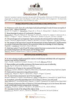
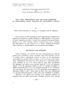
Molecular and Cellular Pathobiology
Cancer
Research
Oncogenic Role of miR-483-3p at the IGF2/483 Locus
Angelo Veronese1,3, Laura Lupini1, Jessica Consiglio3, Rosa Visone3, Manuela Ferracin1, Francesca Fornari2,
Nicola Zanesi3, Hansjuerg Alder3, Gemma D'Elia4, Laura Gramantieri2, Luigi Bolondi2, Giovanni Lanza1,
Patrizia Querzoli1, Adriano Angioni4, Carlo M. Croce1,3, and Massimo Negrini1
Abstract
hsa-mir-483 is located within intron 2 of the IGF2 locus. We found that the mature microRNA (miRNA)
miR-483-3p is overexpressed in 100% of Wilms' tumors. In addition, colon, breast, and liver cancers exhibit
high or even extremely high levels of miR-483-3p in ∼30% of the cases. A coregulation with IGF2 mRNA
was detected, although some tumors exhibited high expression of miR-483-3p without a concomitant increase
of IGF2. These findings suggested that miR-483-3p could cooperate with IGF2 or act as an autonomous oncogene. Indeed, here we prove that an anti-miRNA oligonucleotide against miR-483-3p could inhibit the miRNAs
without affecting IGF2 mRNA and it could suppress tumorigenicity of HepG2 cells, a cell line that overexpresses miR-483-3p and IGF2. Conversely, no antitumor effect was elicited by inhibition of IGF2. The oncogenic
mechanism of miR-483-3p was at least partially clarified by the finding that it could modulate the proapoptotic
protein BBC3/PUMA and miR-483-3p enforced expression could protect cells from apoptosis. Our results
indicate that miR-483-3p could function as an antiapoptotic oncogene in various human cancers and reveal
a new, potentially important target for anticancer therapy. Cancer Res; 70(8); 3140–9. ©2010 AACR.
Introduction
Genetic and epigenetic abnormalities at chromosomal
region 11p15.5 have been detected in various human neoplasms and in the cancer-predisposing Beckwith-Wiedemann
syndrome. In particular, loss of alleles and gene duplication
at 11p15 are typically detected in more than 80% of Wilms'
tumors (1–5) and, albeit less frequently, in more common
adult neoplasms (6–9).
Two close imprinted loci, which harbor at least eight
monoallelically expressed genes, are present at chromosome
region 11p15.5. The aberrant regulation of genomic imprinting at 11p15.5 was involved in human cancer (10–12). Aberrant methylation at the H19 maternal locus leads to the
reactivation of the silent IGF2 allele (10, 13–16) and the loss
of maternal allele methylation at the KvDMR1 locus was
linked to reduced expression of the cyclin-dependent kinase
inhibitor CDKN1C/p57 gene and other imprinted genes (11,
12, 17–19). These studies point to the existence of oncogenic,
Authors' Affiliations: 1 Dipartimento di Medicina Sperimentale e
Diagnostica, Università di Ferrara, Ferrara, Italy; 2 Dipartimento di
Medicina Interna e Gastroenterologia, Università di Bologna, Bologna,
Italy; 3 Department of Molecular Virology, Immunology, and Medical
Genetics, Ohio State University, Columbus, Ohio; and 4 Ospedale
Bambin Gesù, IRCCS, Struttura Semplice di Citogenetica e Genetica
Molecolare, Rome, Italy
Note: Supplementary data for this article are available at Cancer
Research Online (http://cancerres.aacrjournals.org/).
Corresponding Author: Massimo Negrini, Dipartimento di Medicina
Sperimentale e Diagnostica, Università di Ferrara, via Fossato di Mortara,
70, presso “Il Cubo”-Piano 2, 44121 Ferrara, Italy. Phone: 39-0532455399; Fax: 39-0532-455875; E-mail: [email protected].
doi: 10.1158/0008-5472.CAN-09-4456
©2010 American Association for Cancer Research.
3140
Cancer Res; 70(8) April 15, 2010
IGF2, as well as tumor-suppressive, CDKN1C/p57, functions
within the 11p15.5 chromosomal imprinted region.
IGF2 is a fetal growth factor, the abnormal expression of
which has been involved in the Beckwith-Wiedemann syndrome, which predisposes to the development of nephroblastoma, hepatoblastoma, and rhabdomyosarcoma. Biallelic
expression of IGF2 gene, consequent to the loss-of-imprinting
at the IGF2 locus, occurs in 40% to 50% of Wilms' tumors (5,
20), and it is thought to be an early event in carcinogenesis.
Therefore, it has been suggested that increased IGF2 could
lead to an enhanced cellular proliferation, differentiation failure, and tumor development. However, a transgenic mouse
model for IGF2 overexpression exhibited many of the features associated with the Beckwith-Wiedemann syndrome,
including prenatal overgrowth, polyhydramnios, fetal and
neonatal lethality, disproportionate organ overgrowth including tongue enlargement, and skeletal abnormalities, but this
mouse model did not develop tumors (21). These results suggest that additional cofactors should cooperate with IGF2 in
promoting human cancer. Recently, the IGF2 locus was
shown to harbor a microRNA (miRNA), the mir-483 locus,
within its second intron (22).
In the last 7 years, the involvement of miRNAs in human cancer has been proved. Aberrant expression of miRNAs has been detected in any human neoplasm and
miRNAs were found to play a central role in all molecular
pathways affecting cancer traits (23–25). Recently, the role
of the miR-17-92 miRNA cluster in Wilms' tumors has been
reported (26).
Here, we tested the potential oncogenic activity of miR-4833p, one of the mature products at chromosome 11p15.5. Our
results provide evidence for the role of this miRNA as an
antiapoptotic oncogene involved in human tumorigenesis.
miR-483-3p in Human Cancer
Materials and Methods
Primary tumors. Primary tumor RNAs were obtained
from 19 Wilms' tumors, 3 adjacent patient normal tissues,
2 adult kidneys; 27 hepatocarcinomas, 7 cirrhotic, and 2 normal liver tissues; 23 colorectal cancers and 5 normal colon
mucosa; 19 breast cancers and 4 normal breast tissues.
All tissue samples were collected at surgery, immediately
snap-frozen in liquid nitrogen, and stored at −80°C until RNA
extraction. Total RNA was isolated using Trizol (Invitrogen)
according to the instructions of the manufacturer.
Cell lines and transfection. HEK293, HepG2, and HCT116
cell lines (all from American Type Culture Collection) were
cultured with Iscove's modified Dulbecco's medium with
10% fetal bovine serum. hsa-miR-483-3p precursor (Sanger
accession no. MI0002467) and negative control 2 ribo-oligonucleotide were from Applied Biosystems/Ambion. Anti-miRNA
oligonucleotides (AMO) against miR-483-3p and against the
GFP gene (AMO Negative Control) were from Fidelity Systems.
RNA interfering for BBC3/PUMA and scramble control
were from Santa Cruz Biotechnology. Transfection of
miRNAs, AMOs, and expression vectors was carried out with
Lipofectamine 2000 (Invitrogen) in accordance with the
procedures of the manufacturer.
Luciferase assays and vectors. The human 3′-untranslated region (UTR) of BBC3/PUMA was amplified by PCR
using the primers indicated in Supplementary Table S1
and cloned downstream of the firefly luciferase gene in
the pGL3-Control vector (Promega). Substitutions into the
miR-483-3p binding sites of the BBC3/PUMA 3′UTR gene
was introduced by using Quick-Change site-directed mutagenesis kit following the instructions of the manufacturer
and using the primers indicated in Supplementary
Table S1. As a reference, the pRLTK vector (Promega),
which expresses the renilla luciferase, was used. Transfection was conducted in HEK293, HCT116, and HepG2 cells
cultured in 24-well plates, each well was cotransfected with
400 ng of pGL3-control vectors together with 40 ng of
pRLTK reference vector (Promega) and 30 pmol of miR483-3p or negative control 2 or AMOs, or methylated control oligonucleotide. Twenty-four hours after transfection,
firefly and renilla luciferase activities were measured using
the Dual-Luciferase Report Assay (Promega).
Western blot analyses. HEK293 and HepG2 cell lines
were transfected with 30 pmol of miR-483-3p, AMOs, and
control sequences in 24-well plates. After 48 h, cells were
collected, lysed in Laemmli 2× buffer, and analyzed by
Western blot to assess the expression of PUMA using
monoclonal antibodies (anti-PUMA antibody no. 4976; Cell
Signaling). Primary antibody was incubated for 2 h at room
temperature and then peroxidase-conjugated anti-mouse or
anti-rabbit antibodies were incubated for 30 min at room
temperature. Detection was conducted by chemiluminescent enhanced assay (WesternBreeze Chemiluminescent
Kit; Invitrogen). β-Actin antibody (β-actin antibody no.
4976; Cell Signaling) or Ponceau staining were used to
normalize protein loading. To quantify Western blot signals,
digital images of autoradiographies were acquired with
www.aacrjournals.org
Fluor-S MultiImager, and band signals were quantified in
the linear range of the scanner using specific densitometric
software (Quantity One).
Quantitative real-time reverse transcription PCR.
Mature miRNA expression was assayed by TaqMan MicroRNA assay (Applied Biosystems) specific for miR-483-3p
(P/N: 4378094) and normalized on RNU6B (P/N: 4373381).
Five nanograms of total RNA was reverse-transcribed using
the specific looped primer and quantitative real-time reverse transcription PCR (qRT-PCR) was conducted using
the standard TaqMan MicroRNA assay protocol on a BioRad-Chromo4 thermal cycler. The 20 μL PCR included
1.33 μL of reverse transcription product, 1× TaqMan Universal PCR Master Mix, No AmpErase UNG [P/N 4324018
(Applied Biosystems), 0.2 μmol/L TaqMan probe, 1.5 μmol/L
forward primer, and 0.7 μmol/L reverse primer]. The reaction
was carried out in a 96-well PCR plate at 95°C for 10 min followed by 40 cycles of 95°C for 15 s and 60°C for 1 min. Each
sample was analyzed in triplicate. qRT-PCR for mRNA was
performed using 500 ng of total RNA for each sample according to the instructions of the manufacturer (High-capacity
cDNA Reverse Transcription Kit; Applied Biosystems) and
the real-time reaction using SYBR green technologies (Power
SYBR green PCR Master Mix; Applied Biosystems) on the BioRad-Chromo4 instrument. The 20 μL PCR included 1 μL of
reverse transcription product, 1× Power SYBR green PCR
Master Mix (P/N 4368577; Applied Biosystems), 0.4 μmol/L
forward primer, and 0.4 μmol/L reverse primer. The reactions were incubated in a 96-well PCR plate at 95°C for
10 min followed by 40 cycles of 95°C for 15 s and 60°C for
1 min. Each sample was analyzed in triplicate. TaqMan
gene expression assays were performed for IGF2, BBC3/
PUMA, and CDKN1A/p21 genes using primers and probes
(Hs01005963_m1, Hs00248075_m1, and Hs99999142_m1) obtained from Applied Biosystems (Applied Biosystems). The
expression of 18S RNA was used as an endogenous reference
control. The levels of miRNA and mRNA were measured using
Ct (threshold cycle). The amount of target, normalized to an
endogenous reference and relative to a calibrator, is given
by 2−ΔΔCt (comparative Ct method; Applied Biosystems
User Bulletin no. 2).
Northern blot analysis. RNA samples (10 μg each) were
electrophoresed on 15% acrylamide and 7 mol/L of urea
Criterion precast gels (Bio-Rad) and transferred onto Hybond
N+ membrane (Amersham Biosciences). Membranes were hybridized as previously described (27) with oligonucleotide
probes corresponding to the complementary sequences of
the mature miRNAs: miR-483-3p 5′-UCACUCCUCUCCUCCCGUCUU-3′ and the reference U6 RNA 5′-GCAGGGGCCATGCTAATCTTCTCTGTATCG-3′.
Cell death and cell viability assays. MTT assays were
carried out on the HEK293 cell line. HEK293 was cultured
in 24-well plates the day before miR-483-3p or AMOs transfection. After 24 h from transfection, cells were treated
with doxorubicin (0.4 μg/mL) for an additional 24 h. The
assay was performed in accordance with the protocols of
the manufacturer (TOX-1; Sigma). Each experiment was
performed in triplicate. Nuclear fluorescein staining based
Cancer Res; 70(8) April 15, 2010
3141
Veronese et al.
Figure 1. miR-483-3p and IGF2
expression in Wilms' tumor. A,
miR-483-3p relative expression
analysis by quantitative real-time
PCR on 19 samples of primary
Wilms' tumor, 3 adjacent
non-tumoral tissues, fetal kidney,
cell line HEK293, and fetal liver
tissue. Each sample data was
normalized to the endogenous
reference RNU6B and related to
the fetal kidney (calibrator)
miR-483-3p expression (2−ΔΔCt
method). B, different expressions
from normal to tumoral tissue were
pointed out in samples (WT_38,
WT_39, and WT_40). C, Northern
blot analysis of miR-483-3p and
RNU6B in six Wilms' tumor
samples, two normal adult kidneys,
one normal adult liver, and a
HepG2 cell line. D, genomic
structure of IGF2 gene (dark gray
arrows) from the reference
sequence AF517226; coding
sequence (light gray arrows);
miR-483 is indicated in
the second IGF2 intron. Black bars
(e and f) show the cDNA-amplified
regions used to analyze IGF2
expression that was compared
with the miR-483-3p expression in
panels immediately below. IGF2
expression data were normalized
on RNA 18S and related to IGF2
fetal kidney expression (2−ΔΔCt).
R, correlation coefficients;
P, P values.
on labeling of DNA strand breaks by terminal deoxynucleotidyl transferase–mediated dUTP nick-end labeling reaction
was performed on the HepG2 cell line according to the
instructions of the manufacturer (In situ Cell Death
Detection Kit; Roche). The experiment was performed in
triplicate and analyzed on a Zeiss Axiovert 200 microscope
at 100× magnification, images were acquired with the CCD
Cascade Photometrics 512b. Quantification of positive
3142
Cancer Res; 70(8) April 15, 2010
nuclei was calculated by counting fluorescent spots in 10
images (10 × 10 magnifications) for each experiment. Caspase 3/7 activity assay was performed on HCT116 cell line.
HCT116 was cultured in 96-well plates the day before miR483-3p, AMO, and control transfection. After 24 h from
transfection, cells were treated with Nutlin-3 (5 μmol/L)
or 5-fluoruracil (from 0 to 50 μmol/L) for an additional
6 to 24 h, then the assay was performed in accordance with
Cancer Research
miR-483-3p in Human Cancer
the protocols of the manufacturer (Caspase-Glo 3/7 assay,
G8090; Promega).
Generation of stable cell lines overexpressing miR-483-3p
and anti–miR-483-3p. HCT116 cells were infected with the
PMIRH483 expression plasmid containing the full-length
miR-483-3p and the GFP gene under the control of two different promoters (PMIRH483PA-1; System Biosciences).
HCT116 and HepG2 cells were infected with the pSIH vector (System Biosciences) containing the antisense sequence for miR-483-3p (Supplementary Table S1) using the
Lentivector-based anti-miRNA technology (miRZIP,
System Biosciences). An empty vector was used as the control. Pre–miR-483-3p, anti–miR-483-3p expression, and control
constructs were packaged with pPACKH1 Lentivector Packaging Plasmid mix (System Biosciences) in 293-TN packaging
cell line. Viruses were concentrated using PEG-it Virus Precipi-
tation Solution and titers analyzed using UltraRapid Lentiviral
Titer Kit (System Biosciences). Infected cells were selected by
fluorescence-activated cell sorting analysis (FACSCalibur,
Becton Dickinson Immunocytometry Systems). An infection
efficiency of >90% was verified by fluorescent microscopy.
In vivo studies. Animal studies were approved by institutional ethical committees. HepG2 cells were transfected
in vitro with 2′-O-methyl RNA oligonucleotide complementary to miR-483-3p (anti-miR oligonucleotide: AMO-483-3p),
AMO-Negative controls from Fidelity Systems, and small interfering RNA (siRNA) against the IGF2 gene from ThermoScientific Dharmacon. At 24 h after the transfection, 107 viable cells per mouse were injected i.p. into 10 NOD-SCID
mice (Charles River Breeding Laboratories). After 35 d,
the mice were sacrificed, necropsies were performed, and
all tumors per mouse were weighed. For athymic “nude”
Figure 2. miR-483-3p and IGF2
expression in adult human
cancers. miR-483-3p relative
expression by quantitative
real-time RT-PCR. The expression
of miR-483-3p was normalized
to the endogenous gene RNU6B
(black bars) whereas the
expression of IGF2 was
normalized in 18S RNA (gray bars).
Expression data were related
to the average expression of
normal samples of each tissue
(2−ΔΔCt method). Expression of
the locus IGF2/miR-483-3p
was investigated in (A) 27
hepatocarcinomas and 7
non-tumoral cirrhotic livers (CE),
which were related to 2 normal liver
tissue samples, the positive
correlation between IGF2 and
miR-483-3p is indicated (R = 0.69,
P < 0.0001); B, 22 colorectal
cancer samples were related to
5 normal colon mucosa tissues,
the positive correlation between
IGF2 and miR-483-3p is indicated
(R = 0.86, P < 0.0001); C, 19
breast cancer and 4 normal breast
tissue samples were tested for
the expression of miR-483-3p.
www.aacrjournals.org
Cancer Res; 70(8) April 15, 2010
3143
Veronese et al.
mice experiments, HepG2 cells (10 × 106 cells/200 μL) were
injected s.c. into the flanks of nude mice 24 h after transfection with AMO-483-3p (four injections), AMO Negative
Control (four injections), or siRNA IGF2 (six injections).
Tumors were counted after 20 d.
Results
miR-483-3p is overexpressed in Wilms' tumor and
common adult human cancers in concert with IGF2.
Because of its location within the IGF2 locus, we evaluated
miR-483-3p expression in Wilms' tumor. Biopsies from
19 Wilms' tumors, 4 normal kidney tissues (three of which
matched with Wilms' tumor samples), 1 fetal kidney, and
1 fetal liver as positive control were analyzed. Upregulation
(from 4-fold to 40-fold) of miRNA expression was found in
73% of Wilms' tumors when compared with fetal kidney
and in 100% of the cases when compared with non-tumoral
kidney tissues (Fig. 1A and B). Northern blot analysis confirmed the qRT-PCR data (Fig. 1C).
Because mir-483-3p is located within the IGF2 gene, we
compared their levels of expression: a positive correlation
between IGF2 mRNA and miR-483-3p expression was found.
By using two sets of primers spanning the junctions between
exons 1 and 2 or exons 2 and 3 of the IGF2 cDNA sequence,
we found strong positive coefficients of correlation with
miR-483-3p in both cases (R = 0.94, P < 0.0001 for product
e, and R = 0.60, P = 0.004 for product f; Fig. 1D). These results
indicate that the joint overexpression of at least two functional elements from the IGF2 locus, the IGF2 protein
and miR-483-3p, may act in Wilms' tumors to promote
tumorigenesis.
We investigated the potential involvement of miR-483-3p
in common human neoplasms. We analyzed the expression
of miR-483-3p in breast, colon, and liver human cancers and
we found that overexpression of miR-483-3p was indeed present, suggesting a wider involvement of this miRNA in human tumorigenesis. In primary colon, breast, and liver
carcinomas (Fig. 2A–C), we compared the expression of
miR-483-3p of tumors with the average expression of nontumor histologic normal tissues. Considering the variability
of miR-483-3p expression in non-tumor counterparts, only
tumors exhibiting a fold-change of >10 in comparison with
the average expression of non-tumor normal tissues were
scored as overexpressed: 33% of hepatocarcinomas, 31% of
breast, and 27% of colorectal cancers exhibited significant
upregulation. Interestingly, cirrhotic liver tissues, a condition that predisposes to hepatocarcinoma, already exhibited
an increased miR-483-3p expression (from 3-fold to 14-fold)
when compared with normal liver tissues (P < 0.05; Fig. 2A).
We observed that, as in Wilms' tumors, the upregulation of
miR-483-3p was linked with IGF2 expression: a positive
Spearman coefficient of correlation of 0.69 for hepatocarcinomas (P < 0.0001) and 0.86 for colorectal cancers (P < 0.0001;
Fig. 2A and B). In spite of these significant positive correlations, some tumor samples (HCC_02, HCC_04, HCC_10,
HCC_13, HCC_16, HCC_21, HCC_26, HCC_29, CRC_188,
CRC_130, CRC_148) exhibited a divergent expression of
3144
Cancer Res; 70(8) April 15, 2010
Figure 3. AMO-483-3p induces cell death. A, cell viability measured by
the MTT assay in HEK293 cells. Each value represents the average
absorbance ± SD from three different experiments. The differences
between miR-483-3p versus negative control 2 (P = 0.002), and
AMO_miR-483-3p versus AMO negative control (P = 0.03) were both
statistically significant. B, cell viability of HepG2 cells at 48 h following
transfection of AMO-miR-483-3p or AMO negative control. Each value
represents the average ± SD from three different experiments (P = 0.02
on AMO_miR-483-3p versus AMO negative control) and related to
control data. C, terminal deoxynucleotidyl transferase–mediated
dUTP nick end labeling staining of HepG2 cells after transfection of
AMO-negative control or AMO miR-483-3p. Cells were stained 24 h
after transfection. Underneath the images, the average number of
fluorescent spots is shown: data in each column represents the average
± SD of fluorescent spots from three independent fields (P = 0.0162).
D, caspase 3/7 activity in HCT116 cells transfected with miR-483-3p,
anti-miR-483-3p AMO, or negative controls; after 24 h, cells were treated
with Nutlin-3 (5 μmol/L) for an additional 6 h.
Cancer Research
miR-483-3p in Human Cancer
IGF2 and miR-483-3p, suggesting the existence of multiple
mechanisms of miR-483-3 upregulation.
miR-483-3p protects cells from apoptosis. To start unraveling the molecular basis of the potential oncogenic role of
miR-483-3p, we focused on the apoptotic pathway because
important proapoptotic human genes are predicted targets
of miRanda and TargetScan algorithms (BBC3/PUMA, AMID,
BAX, BIK, SMAC/DIABLO, PDCD1, and PDCD7). Thus, we
evaluated apoptosis and cell viability in response to modulation of miR-483-3p in HEK293, HCT116, and HepG2 cells.
After treatment of HEK293 cells with doxorubicin to induce apoptotic cell death, transfection of miR-483-3p induced
a low (10%) but significant increase in cell viability (P = 0.05),
whereas transfection of anti–miR-483-3p AMOs induced a
further 15% decrease in cell viability (P = 0.02; Fig. 3A). These
results indicate that miR-483-3p could promote cell survival.
HepG2 cells express very high levels of miR-483-3p. Therefore, we evaluated only the effect of anti–miR-483-3p AMO in
this cell line. Cell growth was inhibited 30% to 40% by transfection of anti–miR-483-3p AMO (Fig. 3B). Simultaneously,
cells exhibited a 2.6-fold increase in the level of cell death
compared with control cells transfected with a negative
control AMO (P = 0.016; Fig. 3C), as detected by in situ cell
death using fluorescent labeling of DNA strand breaks with
terminal deoxynucleotidyl transferase.
To confirm that the mechanism was indeed apoptosis, caspase 3/7 activity was measured in HCT116 cells transfected
with miR-483-3p or anti–miR-483-3p and treated with the
apoptosis-inducing factor Nutlin-3A. Compared with the
controls, caspase activity exhibited an 80% increase in antimiR–treated cells and a 60% decrease in miR-483-3p–treated
cells (Fig. 3D); moreover, we detected an inverse correlation
between the amount of transfected miR-483-3p and caspase
3/7 activity, whereas a proportional increase was detected
between AMO-483-3p and caspase 3/7 activity (Supplementary
Fig. S2A).
PUMA is a target of miR-483-3p. In the list of potential
proapoptotic target genes, the BBC3/PUMA (BCL2 binding
component 3/p53 upregulated modulator of apoptosis) gene
was chosen for further analysis because of its known pivotal
role in induced cell death (28).
To test the direct interaction of miR-483-3p with 3′UTRs,
the predicted wild-type and mutant miR-483-3p target sites
of PUMA mRNA gene were cloned downstream of the luciferase reporter gene of pGL3-Control vector. The HEK293
cells were used as a biological system because they exhibited
a low expression of miR-483-3p, HCT116 cells had medium
expression, whereas the HepG2 cells exhibited a high expression of miR-483-3p (Supplementary Fig. S1A). Depending on
the miR-483-3p expression, the miR-483-3p responsive vector
Figure 4. BBC3/PUMA is target of
miR-483-3p. A, putative binding
site of miR-483-3p in PUMA
3′UTRs (miRBase Target
Database). Asterisks, nucleotides
substituted in miR-483-3p
predicted target site to perform
luciferase assay (GGAGT>AATTA).
B, PUMA 3′UTRs regulate
luciferase activity dependent on
miR-483-3p in HEK293, HCT116,
and HepG2 cell lines (WT,
wild-type; MUT, mutant;
P, P value). Firefly luciferase
activity was normalized on
Renilla luciferase activity of the
cotransfected pRL-TK vector.
C, Western blot analysis of PUMA
after miR-483-3p transfection in
HEK293 and HepG2 cell line; the
α (23 kDa) and β (18 kDa) isoforms
are indicated. Cells were collected
after 48 h of miRNA transfection.
D, Western blot analysis of PUMA
in Wilms' tumor samples (T) and
matched normal tissues (N).
www.aacrjournals.org
Cancer Res; 70(8) April 15, 2010
3145
Veronese et al.
was cotransfected with miR-483-3p into HEK293, either miR483-3p or anti–miR-483-3p oligonucleotide (anti–483-3p
AMO) into HCT116 cells and only anti–miR-483-3p into
HepG2 cells (Fig. 4A). In comparison with control vectors,
miR-483-3p induced a decrease in luciferase activity of
∼38% (HEK293) and 45% (HCT116) of the pGL3 vector carrying the PUMA-3′UTR, whereas in the mutated 3′UTR clone,
the luciferase activity was not significantly downregulated
by miR-483-3p. Conversely, the use of anti–miR-483-3p AMOs
induced an increase in luciferase activity of ∼70% and 39%
for HCT116 and HepG2 cells, respectively (Fig. 4B).
To further confirm PUMA as a target of miR-483-3p,
its protein level was assessed by Western blot analysis
on HEK293 and HepG2 cells transfected with miR-483-3p or
anti–miR-483-3p, respectively. Protein expression was reduced (70%) in HEK293 and induced (40%) in HepG2 cells
when compared with controls (Fig. 4C).
The analysis of two matched Wilms' tumors/normal kidney samples for the expression of PUMA protein revealed
that, as predicted by the molecular function of miR-483-3p,
the level of expression of these two proapoptotic proteins
was significantly lower in tumors, in which miR-483-3p is
expressed at higher levels (Fig. 4D).
To further support these data, we generated stable cell
lines overexpressing the miR-483-3p or the anti–miR-483-3p
to evaluate the expression of BBC3/PUMA after treatment
with 5-fluorouracil (5FU) as an apoptosis-inducing factor
that involves the p53 pathway. The HCT116 cells were infected with lentiviral vectors overexpressing miR-483-3p
(LV-483) or anti–miR-483-3p (LV-AS483), whereas the HepG2
cell line was infected with LV-AS483. An empty vector was
used as a control (LV-CTRL). The increased concentrations
of 5FU matched with the increased PUMA protein levels only in the HCT116 and HepG2 cells overexpressing the anti–
miR-483-3p (Fig. 5A), in spite of a homogeneous increment of
BBC3/PUMA mRNA levels (Supplementary Fig. S3A) in all
stable cell lines. The induction of both BBC3/PUMA and
CDKN1A/p21 mRNA levels by 5FU treatment suggests the
activation of the p53 pathway (Supplementary Fig. S3A
and S3B).
To assess the physiologic role of this finding, the caspase
3/7 activity of these stable cell lines was measured after 5FU
treatment. Compared with controls, the HCT116-LV-483 cells
showed a reduced caspase 3/7 activity (24–40%, P < 0.0001;
Fig. 5B), whereas the HCT116-LV-AS483 cells exhibited an
increased activity (4–28%, P = 0.03). As expected in HepG2LV-AS483 cells, the caspase activity was also increased (21–
70%, P < 0.0001; Fig. 5B). To further prove the connection
between miR-483-3p, PUMA, and apoptosis, we proved that
by knocking down PUMA using siRNA transfection, we could
prevent the AS483-3p effect in HepG2-LV-AS483 cells
(Fig. 5C). We used the HepG2 stable cell lines because the
efficacy of anti–miR-483-3p was more evident than the
HCT116 model (Fig. 5B).
AMO-miR-483-3p but not IGF2 siRNA can inhibit in vivo
tumorigenicity. The potential oncogenic role of miR-483-3p
was tested directly through tumorigenicity modulation of
the human HepG2 cells, which overexpresses miR-483-3p.
Twenty-four hours after the transfection of AMO-483-3p or
siRNA anti-IGF2 into HepG2 cells, qRT-PCR confirmed the
Figure 5. Stable cell lines
overexpressing anti–miR-483-3p are
more sensitive to apoptosis stimuli.
HCT116 cells overexpressing the
miR-483-3p (LV-483) or the
anti–miR-483-3p (LV-AS483) and
HepG2 cells overexpressing the
anti–miR-483-3p were treated with
different concentrations of 5FU.
LV-CTRL indicates stable cell line with
the empty vector. A, Western blot
analysis reveals an increment of PUMA
protein levels only in the LV-AS483
cells. B, caspase 3/7 activity was
decreased in LV-483 cells and
increased in LV-AS483 when
compared with LV-CTRL cells. Data
were normalized on LV-CTRL cells.
C, relative caspase 3/7 activity in
HepG2-LV-CTRL and LV-AS483 after
treatment with 5FU and siPUMA (+) or
siRNA scramble (−). Data were
normalized on the average caspase
activity of HepG2-LV-CTRL cells.
3146
Cancer Res; 70(8) April 15, 2010
Cancer Research
miR-483-3p in Human Cancer
Figure 6. AMO-483-3p reduces
tumors induced by HepG2 cells
in vivo. A, a qRT-PCR analysis to
verify the reduction of miR-483-3p
and IGF2 mRNA after transfection
of AMO-483-3p or siRNA anti-IGF2
in HepG2 cells. B, tumors from
each mouse were weighed: a
significant difference (P < 0.05) in
tumor weights was found between
AMO-483-3p and AMO negative
control transfected (CTRL) cells,
between siRNA IGF2 and
AMO-483-3p (P < 0.02), but not
between siRNA IGF2 and
AMO-CTRL–transfected HepG2
cells (P > 0.5). C, appearance of
intraperitoneal HepG2 induced
tumors in NOD-SCID mice (white
arrows, tumor formations).
specific reduction of miR-483-3p or IGF2 mRNA, respectively
(Fig. 6A). Then, 10 × 106 cells (cell viability greater than 97%)
were i.p. injected into NOD-SCID mice. Mice were sacrificed
on day 35 and all tumors for each mouse were weighed. The
AMO-483-3p mouse group showed a significant reduction of
number and weight of induced tumors compared with controls (P < 0.05). On the contrary, repression of the IGF2 gene
did not show any difference from controls (P > 0.5; Fig. 6B
and C). The three groups of data (CTRL, AMO-483-3p,
and siIGF2) were comparable because of the same nature
of the molecules used to silence the miR-483-3p and IGF2
gene (2′-O-methyl RNA oligonucleotide). Overlapping results
were obtained by subcutaneous injections of HepG2 cells
into athymic nude mice: AMO-483-3p induced a decrease
in the number of growing tumors (one of four) in comparison
with controls and IGF2 siRNA (AMO-CTRL, four of four;
siRNA anti-IGF2, four of six; Supplementary Fig. S4).
Discussion
We established a link between miR-483-3p, a miRNA located
within the IGF2 locus at chromosome 11p15.5, and human
tumorigenesis. Various evidences support this conclusion.
First, tumorigenicity of HepG2, a cell line that overexpresses
miR-483-3p, is suppressed by AMO anti–miR-483-3p; second,
miRNA is upregulated and overexpressed in Wilms' tumors
as well as in common human neoplasms; third, miR-483-3p
supports cell survival by protecting cells from apoptosis;
fourth, an important regulator of apoptosis, Puma, is inhibited
by the expression of miR-483-3p.
www.aacrjournals.org
To show the oncogenic properties of miR-483-3p, we
proved that silencing of miR-483-3p by AMO could reduce
the tumorigenicity of HepG2 cells, a cell line that exhibits
high expression of the IGF2/miR-483 locus and functional activity of IGF2 ligand (29, 30). Importantly, the reduction in
tumor formation by the specific silencing of miR-483-3p
was achieved without changing IGF2 gene expression. Moreover, the silencing of the IGF2 gene by siRNA technologies
did not cause a significant change in tumor formation by
HepG2 cells. These results clearly indicate that a crucial
oncogenic function was associated with the miR-483-3p
miRNA within the IGF2 locus. This conclusion might
explain why transgenic animal models for Igf2 overexpression
did not develop tumors (21). In fact, these animal models
were developed by using an Igf2 cDNA, which lacked the
mir-483 locus. It could be speculated that, by protecting cells
from apoptosis, miR-483-3p provides the additional element
required for supporting malignant transformation promoted
by the growth factor IGF2. Indeed, in the Wilms' tumors, hepatocarcinomas, and colorectal cancers studied, the expression of the IGF2/miR-483 locus is coregulated in almost
all of the samples analyzed. Interestingly, some cases present
divergent expressions of miR-483-3p and IGF2, suggesting
a possible IGF2-independent mechanism of miR-483-3p
regulation.
By proving that ectopic expression of miR-483-3p could
protect cells from apoptosis, whereas inhibition of endogenous miR-483-3p increases basal or induced apoptosis, we
show that the upregulation of miR-483-3p could protect
cells from apoptosis. This is in line with the finding that
Cancer Res; 70(8) April 15, 2010
3147
Veronese et al.
miR-483-3p can target and repress the expression of the proapoptotic protein PUMA, a BH3-only protein, the induction of
which is mediated by p53 (28, 31). Like other BH3-only proteins, it promotes apoptosis by interacting and inhibiting the
antiapoptotic factors BCL2 and BCLXL (31–33). Hence, an
overexpressed miR-483-3p might favor cell survival by reducing the level of Puma after p53 activation during apoptosis
stimuli, and therefore acting as an antiapoptotic oncogene.
Our results support this conclusion.
The involvement of miR-483-3p in cancer is supported by
the observation that it is overexpressed in ∼30% of common
human cancers and in 100% of Wilms' tumors. In addition,
Guled and colleagues reported the upregulation of miR-4833p in malignant mesothelioma (34). In human cancers, the
overexpression of miR-483-3p could be linked to its coregulation with IGF2 expression. Interestingly, through this mechanism, both growth (IGF2) and survival stimuli (miR-483-3p)
are simultaneously activated. However, some tumor samples
displayed a divergent expression between IGF2 and miR-4833p, thus revealing the existence of mechanisms in which IGF2
and hsa-mir-483 are not coregulated. This is an area that
needs further investigation.
The recent demonstration of the safe and effective use of
anti-miRNAs in animal models, including African green
monkeys, suggests the potential application of AMOs in anticancer therapy as well (35, 36). Our findings indicate miR483-3p as a potential target for antineoplastic intervention in
Wilms' tumors and possibly many other tumors. Thus, these
results not only improve our understanding on the molecular
mechanisms involved in tumor development, but they also
provide an indication for a novel potential therapeutic target.
Disclosure of Potential Conflicts of Interest
No potential conflicts of interest were disclosed.
Grant Support
Associazione Italiana per la Ricerca sul Cancro, Ministero dell'Università e
della Ricerca, Ministero della Salute, and Fondazione Cariplo (Progetto NOBEL; M. Negrini).
The costs of publication of this article were defrayed in part by the payment
of page charges. This article must therefore be hereby marked advertisement in
accordance with 18 U.S.C. Section 1734 solely to indicate this fact.
Received 12/08/2009; revised 01/15/2010; accepted 02/03/2010; published
OnlineFirst 04/06/2010.
References
1.
2.
3.
4.
5.
6.
7.
8.
9.
10.
11.
12.
13.
3148
Fearon ER, Vogelstein B, Feinberg AP. Somatic deletion and duplication of genes on chromosome 11 in Wilms' tumours. Nature 1984;
309:176–8.
Koufos A, Hansen MF, Lampkin BC, et al. Loss of alleles at loci on
human chromosome 11 during genesis of Wilms' tumour. Nature
1984;309:170–2.
Orkin SH, Goldman DS, Sallan SE. Development of homozygosity
for chromosome 11p markers in Wilms' tumour. Nature 1984;309:
172–4.
Reeve AE, Housiaux PJ, Gardner RJ, Chewings WE, Grindley RM,
Millow LJ. Loss of a Harvey ras allele in sporadic Wilms' tumour.
Nature 1984;309:174–6.
Satoh Y, Nakadate H, Nakagawachi T, et al. Genetic and epigenetic
alterations on the short arm of chromosome 11 are involved in a majority of sporadic Wilms' tumours. Br J Cancer 2006;95:541–7.
Ali IU, Lidereau R, Theillet C, Callahan R. Reduction to homozygosity
of genes on chromosome 11 in human breast neoplasia. Science
1987;238:185–8.
Fearon ER, Feinberg AP, Hamilton SH, Vogelstein B. Loss of genes
on the short arm of chromosome 11 in bladder cancer. Nature 1985;
318:377–80.
Weston A, Willey JC, Modali R, et al. Differential DNA sequence deletions from chromosomes 3, 11, 13, and 17 in squamous-cell carcinoma, large-cell carcinoma, and adenocarcinoma of the human
lung. Proc Natl Acad Sci U S A 1989;86:5099–103.
Lee JH, Kavanagh JJ, Wharton JT, Wildrick DM, Blick M. Allele loss
at the c-Ha-ras1 locus in human ovarian cancer. Cancer Res 1989;
49:1220–2.
Rainier S, Johnson LA, Dobry CJ, Ping AJ, Grundy PE, Feinberg AP.
Relaxation of imprinted genes in human cancer. Nature 1993;362:
747–9.
Schwienbacher C, Angioni A, Scelfo R, et al. Abnormal RNA expression of 11p15 imprinted genes and kidney developmental genes in
Wilms' tumor. Cancer Res 2000;60:1521–5.
Scelfo RA, Schwienbacher C, Veronese A, et al. Loss of methylation
at chromosome 11p15.5 is common in human adult tumors.
Oncogene 2002;21:2564–72.
Poirier K, Chalas C, Tissier F, et al. Loss of parental-specific
methylation at the IGF2 locus in human hepatocellular carcinoma.
J Pathol 2003;201:473–9.
Cancer Res; 70(8) April 15, 2010
14. Haruta M, Arai Y, Sugawara W, et al. Duplication of paternal IGF2 or
loss of maternal IGF2 imprinting occurs in half of Wilms tumors with
various structural WT1 abnormalities. Genes Chromosomes Cancer
2008;47:712–27.
15. Vu TH, Chuyen NV, Li T, Hoffman AR. Loss of imprinting of IGF2
sense and antisense transcripts in Wilms' tumor. Cancer Res 2003;
63:1900–5.
16. Sparago A, Russo S, Cerrato F, et al. Mechanisms causing imprinting
defects in familial Beckwith-Wiedemann syndrome with Wilms'
tumour. Hum Mol Genet 2007;16:254–64.
17. Diaz-Meyer N, Day CD, Khatod K, et al. Silencing of CDKN1C
(p57KIP2) is associated with hypomethylation at KvDMR1 in
Beckwith-Wiedemann syndrome. J Med Genet 2003;40:797–801.
18. Satoh A, Toyota M, Ikeda H, et al. Epigenetic inactivation of
class II transactivator (CIITA) is associated with the absence of
interferon-γ-induced HLA-DR expression in colorectal and gastric
cancer cells. Oncogene 2004;23:8876–86.
19. Gallagher E, Mc Goldrick A, Chung WY, et al. Gain of imprinting of
SLC22A18 sense and antisense transcripts in human breast cancer.
Genomics 2006;88:12–7.
20. Bjornsson HT, Brown LJ, Fallin MD, et al. Epigenetic specificity of
loss of imprinting of the IGF2 gene in Wilms tumors. J Natl Cancer
Inst 2007;99:1270–3.
21. Sun FL, Dean WL, Kelsey G, Allen ND, Reik W. Transactivation of Igf2
in a mouse model of Beckwith-Wiedemann syndrome. Nature 1997;
389:809–15.
22. Fu H, Tie Y, Xu C, et al. Identification of human fetal liver miRNAs by
a novel method. FEBS Lett 2005;579:3849–54.
23. Negrini M, Ferracin M, Sabbioni S, Croce CM. MicroRNAs in
human cancer: from research to therapy. J Cell Sci 2007;120:
1833–40.
24. Esquela-Kerscher A, Slack FJ. Oncomirs—microRNAs with a role in
cancer. Nat Rev Cancer 2006;6:259–69.
25. Calin GA, Croce CM. MicroRNA signatures in human cancers. Nat
Rev Cancer 2006;6:857–66.
26. Kort EJ, Farber L, Tretiakova M, et al. The E2F3-Oncomir-1 axis is
activated in Wilms' tumor. Cancer Res 2008;68:4034–8.
27. Fornari F, Gramantieri L, Ferracin M, et al. MiR-221 controls
CDKN1C/p57 and CDKN1B/p27 expression in human hepatocellular
carcinoma. Oncogene 2008;27:5651–61.
Cancer Research
miR-483-3p in Human Cancer
28. Jeffers JR, Parganas E, Lee Y, et al. Puma is an essential mediator of
p53-dependent and -independent apoptotic pathways. Cancer Cell
2003;4:321–8.
29. Shimizu M, Shirakami Y, Sakai H, et al. EGCG inhibits activation
of the insulin-like growth factor (IGF)/IGF-1 receptor axis in human
hepatocellular carcinoma cells. Cancer Lett 2008;262:10–8.
30. Lin SB, Hsieh SH, Hsu HL, Lai MY, Kan LS, Au LC. Antisense
oligodeoxynucleotides of IGF-II selectively inhibit growth of human hepatoma cells overproducing IGF-II. J Biochem 1997;122:
717–22.
31. Nakano K, Vousden KH. PUMA, a novel proapoptotic gene, is
induced by p53. Mol Cell 2001;7:683–94.
32. Yu J, Zhang L, Hwang PM, Kinzler KW, Vogelstein B. PUMA
www.aacrjournals.org
33.
34.
35.
36.
induces the rapid apoptosis of colorectal cancer cells. Mol Cell
2001;7:673–82.
Bouillet P, Strasser A. BH3-only proteins—evolutionarily conserved
proapoptotic Bcl-2 family members essential for initiating
programmed cell death. J Cell Sci 2002;115:1567–74.
Guled M, Lahti L, Lindholm PM, et al. CDKN2A, NF2, and JUN
are dysregulated among other genes by miRNAs in malignant
mesothelioma—a miRNA microarray analysis. Genes Chromosomes
Cancer 2009;48:615–23.
Elmen J, Lindow M, Schutz S, et al. LNA-mediated microRNA
silencing in non-human primates. Nature 2008;452:896–9.
Krutzfeldt J, Rajewsky N, Braich R, et al. Silencing of microRNAs
in vivo with ‘antagomirs’. Nature 2005;438:685–9.
Cancer Res; 70(8) April 15, 2010
3149
© Copyright 2026 Paperzz