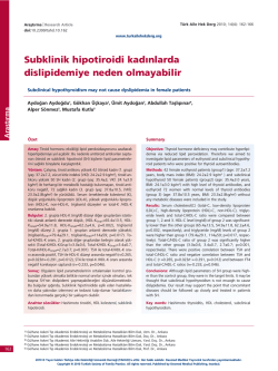
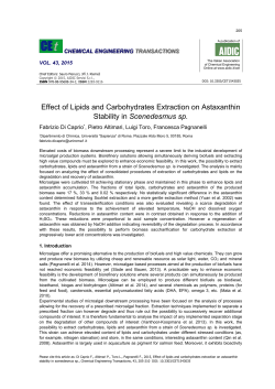
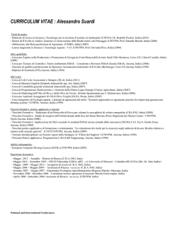
123
Extremophiles (2002) 6:437–444
Digital Object Identifier (DOI) 10.1007/s00792-002-0279-2
© Springer-Verlag 2002
ORIGINAL PAPER
Veronica M.T. Lattanzio • Angela Corcelli •
Giuseppe Mascolo • Aharon Oren
Presence of two novel cardiolipins in the halophilic archaeal community
in the crystallizer brines from the salterns of Margherita di Savoia (Italy)
and Eilat (Israel)
Received: January 17, 2002 / Accepted: May 10, 2002 / Published online: July 30, 2002
Communicated by W.D. Grant
Abstract Two novel cardiolipin derivatives were recently
detected in Halobacterium salinarum, namely an archaeal
analog of bisphosphatidylglycerol (BPG) and a glycocardiolipin (GlyC). GlyC was found to be tightly bound to
bacteriorhodopsin. To obtain information on the presence and distribution of these archaeal cardiolipins, we
have analyzed the lipids extracted from the crystallizer
ponds of the salterns of Margherita di Savoia (Italy) and
Eilat (Israel) and from cultures of representative species of
the Halobacteriaceae by electrospray ionization mass spectrometry. BPG was present as a minor lipid component in
the lipids extracted from the biomass of the Margherita di
Savoia and the Eilat salterns, while GlyC was detected only
in the extract of the biomass of Margherita di Savoia. Both
compounds were enriched in the membrane fraction
obtained by dialysis of the cells against distilled water. We
detected BPG in all members of the Halobacteriaceae
tested, but GlyC has so far been found only in the genera
Halobacterium and Haloarcula. A sulfated diglycosyl
diether was the major glycolipid detected in the biomass of
both salterns.
Key words Cardiolipins
Archaea • Salterns
•
Glycolipids
•
Halophilic
Communicated by W.D. Grant
V.M.T. Lattanzio • A. Corcelli (*)
Dipartimento di Fisiologia Generale ed Ambientale, Università di
Bari, via Amendola 165/a, 70126 Bari, Italy
Tel. +39-80-5443335; Fax +39-80-5443388
e-mail: [email protected]
G. Mascolo
IRSA-CNR, Bari, Italy
A. Oren
Division of Microbial and Molecular Ecology, The Institute of Life
Sciences, and The Moshe Shilo Minerva Center for Marine
Biogeochemistry, The Hebrew University of Jerusalem, Jerusalem,
Israel
Springer-VerlagTokyoExtremophilesExtremophiles007921431-06511433-4909s00792-002-0279-20279Springer-VerlagOriginal Paper
Introduction
Cardiolipin (bisphosphatidyglycerol) is well known as a
minor component of the lipids of the Eucarya. It is relatively abundant in the inner mitochondrial membrane,
where it is tightly bound to transmembrane enzymes associated with oxidative phosphorylation. The interaction of
eucaryal cardiolipin with the intrinsic membrane proteins of
mitochondria is relevant from a functional point of view.
Cardiolipin has been shown to be important to achieve
optimal activity of cytochrome c oxidase, NADH dehydrogenase, ATP synthase and other mitochondrial proteins
(Kagawa et al. 1973; Serrano et al. 1976; Dale and Robinson
1988). It mediates the cooperativity between cytochrome c
binding sites in the dimeric enzyme complex (Arnold and
Kadenbach 1997). It has been proposed that cardiolipin
ensures structural integrity of the environment of the proton-conducting protein and takes part directly in proton
uptake of the yeast citochrome bc1 complex (Lange et al.
2001).
Cardiolipin is a major phopholipid of various
Grampositive bacteria, mycobacteria and Gram-negative
bacteria (Ratledge and Wilkinson 1988; O’Leary and
Wilkinson 1988; Wilkinson 1988).
Recently, crystallographic evidence has been reported
for the interaction of cardiolipin with specific sites on the
protein surface of the photosynthetic reaction center in the
membrane of Rhodobacter sphaeroides (McAuley et al.
1999). However, our overall knowledge of the specific location and the functional role of bacterial cardiolipin is still
quite poor.
Bisphosphatidylglycerol is not the only tetraacylphospholipid in the prokaryotic world. Two tetraacyl glycophospholipids, having the basic structure of
phosphatidyl-α-kojibiosyl-diacylglycerol and a D-alanine
derivative of cardiolipin, have been isolated from streptococci and characterized (Fischer and Landgraf 1975; Fischer
and Arneth-Seifert 1998). Furthermore two novel glucosyl
and lysyl derivatives of cardiolipin have been found in
Gram-positive bacteria (Gutberlet et al. 2000).
438
Fig. 1. Structures of the novel
cardiolipin analogs in the purple
membrane of Halobacterium
salinarum: A glycocardiolipin,
B cardiolipin
1A,B
In the archaeal domain, cardiolipins have been detected
only recently. The first evidence that cardiolipins are present
in the Archaea came from our studies of the purple membrane of Halobacterium salinarum (Corcelli et al. 2000,
2002). Two structural analogs of eucaryal cardiolipin – a
phospholipid and a glycolipid – were detected in the purple
membrane of an engineered strain of Hbt. salinarum.
Their structures are, respectively, sn-2,3-di-O-phytanyl-1phosphoglycerol-3-phospho-sn-2,3-di-O-phytanylglycerol
(bisphosphatidylglycerol, BPG) and 3-HSO3-Galpβ16Manp α 1-2Glcp α -1-1-[sn -2,3-di-O -phytanylglycerol]-6[phospho-sn-2,3-di-O-phytanylglycerol] (glycocardiolipin,
GlyC). The molecular structures of the two archaeal cardiolipins are shown in Fig. 1. These novel cardiolipins were
found to be tightly bound to bacteriorhodopsin, the lightdriven proton pump of Halobacterium, and appear to have
a key role in its stability and function (Lopez et al. 1999;
Corcelli et al. 2000). GlyC and bacteriorhodopsin are
present in the purple membrane in equimolar amounts,
while BPG appears to be a minor component (Corcelli et al.
2002).
To obtain information on the presence and distribution
of cardiolipins among the halophilic Archaea, both in their
natural habitat and in culture, we used electrospray ionization mass spectrometry (ESI-MS) to analyze the lipids
extracted from the crystallizer ponds of the salterns of Margherita di Savoia (southern Italy) and Eilat (Israel) and
from cultures of representative species of the Halobacteriaceae. ESI-MS analysis is a highly sensitive method,
enabling the analysis of complex membrane lipid mixtures
and the selective detection of individual classes of phospholipids and other anionic membrane lipids in total lipid
extracts. We show here that the archaeal cardiolipin BPG is
present as a minor component in the lipids extracted from
the biomass of the Margherita di Savoia and the Eilat salterns, whereas GlyC was detected only in the lipid extract of
the biomass of Margherita di Savoia. We detected BPG in
all members of the Halobacteriaceae tested, but GlyC has
so far been found only in the genera Halobacterium and
Haloarcula.
Materials and methods
Sampling
Brine was collected from the crystallizer ponds of the salterns of Margherita di Savoia on 7 February, 2001 and of the
Israel Salt Company at Eilat on 3 October, 2001. Bacteria
were collected by centrifugation (30 min, 7,000 g), resuspended in 4 M NaCl or brine and kept at 4°C until use.
Archaeal strains and culture conditions
The following halophilic Archaea were used as reference
strains: Haloferax volcanii ATCC 29605T, Haloferax gibbonsii ATCC 33959T, Haloferax denitrificans ATCC 35960T,
Haloferax mediterranei ATCC 33500T, Halorubrum saccharovorum ATCC 29252T, Haloarcula vallismortis ATCC
29715T, Haloarcula marismortui ATCC 43049T, Halogeometricum borinquense ATCC 700274T, Halobacterium salinarum NRC 1 ATCC 700922, and Halobacterium salinarum
NRC 817.
The Archaea were grown in the light at 37°C in liquid
medium containing (all concentrations in g l–1): NaCl, 250;
439
MgSO4.7H2O, 20; trisodium citrate dihydrate, 3; KCl, 2;
CaCl2.2H2O, 0.2; neutralized peptone (L34, Oxoid), 10; prepared as previously described (Lanyi and McDonald 1979).
Cells were collected by centrifugation and resuspended in
4 M NaCl before lipid extraction.
Isolation of membranes from biomass by dialysis
Cell pellets were collected by centrifugation, resuspended
in 4 M NaCl and dialyzed against distilled water. The
disrupted cells were centrifuged at 40,000 g for 40 min
to collect membranes. Isolated membranes were resuspended in distilled water and immediately used for lipid
extraction.
Purple membrane isolation
Purple membrane was isolated and purified on a sucrose
density gradient, as previously described (Oesterhelt and
Stoeckenius 1974), from cultures of an engineered BR highproducing strain (L33) of Hbt. salinarum, the kind gift of
Richard Needleman (Ni et al. 1990).
Lipid extraction and analysis
All organic solvents used were commercially distilled
and of the highest available purity (Sigma-Aldrich). Plates
for thin-layer chromatography (TLC) (Silica gel 60A),
obtained from Merck, were washed twice with chloroform:methanol (1 : 1, v/v) and activated at 120°C before use.
The novel cardiolipin standards were isolated and purified
as previously described (Corcelli et al. 2000).
Lipids were extracted using the Bligh and Dyer method
as modified for extreme halophiles (Kates 1986).
Total lipid extracts were analyzed by thin-layer chromatography on silica gel 60A plates (Merck, 20 × 10 cm, layer
thickness 0.2 mm). Lipids were eluted with chloroform : methanol : 90% acetic acid (65 : 4 : 35, v/v), and
detected by spraying with 0.5% sulfuric acid in ethanol, followed by charring at 120°C.
For ESI-mass spectrometry analyses, dried samples of
lipid extracts were dissolved in chloroform:methanol (1 : 1,
v/v). Electrospray mass spectra (ESI-MS) were obtained
with an API 165 mass spectrometer (Applied Biosystems/
MSD Sciex, Concord, Ontario, Canada) equipped with a
turboion-spray interface. The samples were analyzed by
loop injection introducing, by means of a 7125 Rheodyne
valve, 5 µl of sample into a 25 µl/min flow of chloroform:methanol (1 : 1, v/v) delivered by a Harvard model 11
syringe pump (South Natick, MA, USA). The instrumental
conditions were as follows: nebulizer gas flow (air), 1.2 l/min;
curtain gas flow (nitrogen), 1.2 l/min; needle voltage,
5,600 V; interface temperature, ambient; orifice voltage,
–150 V; ring voltage, –200 V; mass range, 50–2,000 a.m.u.;
mass step, 0.1 a.m.u.; dwell time, 0.2 ms. With the orifice voltage used, CID-MS spectra were obtained showing [M-H]–
and [M-2H]2– parent ions as well as some fragmentation ions.
Results
Microscopic examination of the pellets obtained by centrifuging brines from the Margherita di Savoia and the Eilat
saltern crystallizer ponds showed a dominance of flat,
square-to-rectangular cells, similar to those reported earlier
from the Eilat salterns and similar systems worldwide
(Benlloch et al. 1995, 1996; Oren et al. 1996). No Dunaliella
cells were observed in the pellets upon microscopic examination. Dunaliella cells heavily loaded with β-carotene
granules, such as occur in the salterns, are lighter than the
brine and they float during centrifugation. We recovered
yields of 9 mg and 1 mg lipids per liter from the Margherita
di Savoia and Eilat brines, respectively.
ESI-MS analyses of the total unprocessed lipid extracts
of the biomass of Margherita di Savoia and Eilat are shown
in Fig. 2A, B. Figure 2C shows the TLC of the both lipid
extracts. The TLC lipid profiles of both extracts are quite
complex. Identification of the two novel archaeal cardiolipins, expected to be present as minor lipid components in
such halophilic microorganisms that may be present in natural brines, is therefore difficult on the basis of TLC data.
An additional problem in the identification of the
archaeal cardiolipins in the TLC lipid profile of Fig. 2C
arises from the fact that the Rf values of glycocardiolipin
and cardiolipin are very close to those of the major sulfoglycolipid S-DGD-1 and the abundant reddish pigments,
respectively.
The ESI-MS spectra, as presented in Fig. 2A, B are far
more informative on the biomass composition. The main
peaks present in Fig. 2A, B correspond to the molecular
ions of the different kinds of polar lipids (phospholipids and
glycolipids) present in the membranes of various halophilic
microorganisms of the biomass. As glycolipids of extremely
halophilic Archaea might serve as chemotaxonomic markers for classification of these organisms, the presence of certain glycolipid peaks in the total lipid extract of the biomass
can be considered indicative of the presence of various genera of the Halobacteriaceae family (Oren and Gurevich
1993; Oren 2001).
Table 1 lists the main peaks observed in the ESI-MS lipid
profiles of various halophilic microorganisms cultured in
our laboratory. It should be noted that unsulfated diglycosyl, triglycosyl, and tetraglycosyl lipids, when present,
appear as minor peaks in the ESI-MS spectra only, as they
are difficult to ionize. On the basis of these data it was possible to assign various ESI-MS peaks detected in the brine
extracts to known polar lipids of archaeal halophiles
(Table 2). The major peak at m/z 1,056 found in both salterns shows that monosulfated diglycosyl diether lipids, such
as occur in the genera Haloferax, Halobaculum, and
Halorubrum, are far more abundant in the saltern biomass
than, for example, in Halobacterium.
In a previous study we have shown that the two novel
phospholipids of the purple membrane, GlyC and BPG, can
easily be recognized within a mixture of phospholipids by a
set of specific peaks present in the ESI mass spectrum; in
particular we have shown that the two bicharged peaks at
m/z 760 and 966 are diagnostic for the presence of BPG and
440
GlyC, respectively. A small peak at m/z 760 is present in
both ESI-MS spectra of Fig. 2, whereas the peak at m/z
966 was detectable only in the lipids obtained from the
Margherita di Savoia biomass. It is interesting to point out
that the glycolipid S-TGA-1, which is part of the structure of
the glycocardiolipin and may be a precursor in its biosynthesis, is detectable among the lipids of Eilat biomass as well.
As the cardiolipins are lipid components specifically
associated with integral membrane proteins, they appear to
be enriched in isolated cellular membranes. We therefore
also analyzed the lipids of membranes isolated from the
saltern biomass in order to examine the presence of the glycocardiolipin in an enriched membrane preparation of the
biomass of Eilat. The lipid composition of membranes isolated by dialysis from the biomass of Margherita di Savoia
and Eilat is shown in Fig. 3A, B, respectively. In the case of
Margherita di Savoia, both the peaks at m/z 760 and m/z 966
appear to be enriched in the lipid profiles of the membranes
isolated from biomass compared to the lipid profile of nondisrupted biomass cells (Fig. 2A). The lipid extract of membranes isolated from Eilat cells shows the peak at m/z 760,
2A–C
Fig. 2. ESI-MS analyses (negative ion) of the total lipid extracts of the
biomass of Margherita di Savoia (A) and Eilat (B); C TLC profiles of
lipid extracts of Margherita di Savoia (M) and Eilat (E). Standard BPG
and GlyC, bisphosphatidylglycerol and glycocardiolipin, respectively,
were isolated and purified from the purple membrane of an engineered
strain of Hbt. salinarum (Ni et al. 1990)
Fig. 3. ESI-MS profiles (negative ion) of total lipid extracts of membranes isolated by dialysis of the biomass of Margherita di Savoia (A)
and Eilat (B)
3A,B
441
Table 1. Observed signals in the ESI-MS (negative ion) spectra of total lipid extracts from cultures of various halophilic archaeal microorganisms
Genus, Species
Hfx. gibbonsii ATCC 33959
Hfx. denitrificans ATCC 35960
Hfx. volcanii ATCC 29605
Hfx. mediterranei ATCC 33500
Hrr. saccharovorum ATCC29252
Har. vallismortis ATCC 29715
Har. marismortui ATCC 43049
Hbt. salinarum NRC 1
Hbt. salinarum NRC 817
a
b
Phospholipids
Glycolipids
806
886
900
976
1,056
x
x
x
x
x
x
x
x
x
x
x
x
x
x
x
x
x
x
x
x
x
x
x
x
x
x
x
x
x
x
x
x
x
1,138
x
a
1,218
1,381
760
966
x
x
x
x
x
x
x
x
x
x
x
xa
x
x
x
xb
xa
xa
xa
x
x
x
x
Present as a very small peak
Minor peaks
Table 2. Molecular ions and bicharged peaks (m/z values) of the
archaeal polar lipids present in halophilic microorganisms. All the lipids listed in the table are derivatives of the C20, C20isopranylglycerol
diether
Lipid
m/z, amu
Bisphosphatidylglycerol (BPG)
Phosphatidylglycerol (PG)
Phosphatidylglycerosulfate (PGS)
Phosphatidylglycerophosphate methyl ester (PGP-Me)
Glycocardiolipin (GlyC)
Sulfated diglycosyl diphytanylglycerol (S-DGD-1)
Diglycosyl diphytanylglycerol (DGD-1)
Sulfated triglycosyl diphytanylglycerol (S-TGD-1)
Triglycosyl diphytanylglycerol (TGD-1)
Sulfated tetraglycosyl diphytanylglycerol (S-TeGD-1)
760a
806
886
900
966a
1,056
976
1,218
1,138
1,381
a
Cardiolipins
Double-charged peaks
but not the peak at m/z 966. The peak at m/z 1,218, representing the molecular ion of the glycolipid S-TGA-1, is
clearly detectable as a minor lipid component in the mass
spectrum shown in Fig. 3B.
Discussion
The differences observed in the ESI-mass spectra of the lipids extracted from the biomass of the salterns of Margherita
di Savoia and Eilat show once more that in spite of the
superficial resemblance of the biota found in salterns worldwide, local variations do exist. Salterns may differ in nutrient levels, and the size of the biomass, both of algae and of
prokaryotes, can vary greatly (Javor 1983a, b). Qualitative
differences have also been documented in studies in which
the eutrophic salterns of San Francisco Bay have been compared with the oligotrophic ponds in Eilat (Litchfield et al.
2000; Litchfield and Oren 2001).
The large peak at m/z 1,056 shows that one or more
monosulfated diglycosyl diethers are the dominant glycolip-
ids in the crystallizer biomass, both at Margherita di Savoia
and at Eilat. Glycolipids are excellent chemotaxonomic
markers for different genera within the Halobacteriaceae,
and their classification is in part based on the presence of
specific glycolipids (Kamekura and Kates 1999; Oren 2001).
Earlier characterizations of the lipids present in the Eilat
crystallizer pond biomass by thin-layer chromatography
have shown a single glycolipid to be present, co-eluting with
S-DGD-1 of Haloferax (Oren 1994; Oren et al. 1996). In the
more nutrient-rich salterns in San Francisco Bay small
amounts of other glycolipids may be present, but the sulfated diglycosyl diether lipids are by far the most abundant
there (Litchfield et al. 2000; Litchfield and Oren 2001). Our
ESI-MS data for the two salterns examined thus confirm the
earlier findings.
In the present study we have shown for the first time
that both novel archaeal cardiolipins, BPG and GlyC, are
present among the lipids extracted from the microorganisms present in the halophilic community of the saltern of
Margherita di Savoia, whereas only BPG is detectable in the
total lipid extract of Eilat biomass. The first evidence that
these cardiolipins are present in Archaea came from our
studies of the purple membrane of an engineered strain of
Hbt. salinarum (Corcelli et al. 2000). The purple membrane
is enriched in cardiolipins, and these are tightly bound to
bacteriorhodopsin. They appear to play a key role in preserving the stability of solubilized bacteriorhodopsin and in
particular of the trimer aggregates (Lopez et al. 1999). The
molar ratio GlyC to bacteriorhodopsin in the purple membrane has been estimated to be 1 (Corcelli et al. 2000, 2002).
This molar ratio is therefore compatible with a role of GlyC
as a stabilizing agent of the trimer structure and a possible
location in the crevice between monomers. BPG is only a
minor lipid component of the purple membrane (Corcelli et
al. 2000, 2002). It has been suggested that it may be located
in the inter-trimer spaces, and that it could mediate specific
interactions between trimers by establishing bridging contacts between bacteriorhodopsin molecules in adjacent trimers in the purple membrane lattice (Corcelli et al. 2002).
The ubiquity of BPG in all examined members of the
Halobacteriaceae, including many species that do not pro-
442
duce purple membrane (Table 1), suggests that this lipid
may play additional roles in the extremely halophilic
Archaea. It may be worthwhile to investigate whether the
archaeal cardiolipins are present among the lipids tightly
bound to transmembrane enzymes performing oxidative
phosphorylation and in particular with archaeal cytochrome
c oxidase.
GlyC has been found thus far only in the genus Halobacterium and as a very minor component in Haloarcula
(Table 1). Bacteriorhodopsin proton pumps are also present
in Haloarcula, although the pigment has not been documented to be organized in purple membrane (Mukohata
1994; Tateno et al. 1994; Mukohata et al. 1999). We did not
detect GlyC in representatives of the genera Haloferax and
in Hrr. saccharovorum, all organisms that appear to lack
bacteriorhodopsin. Our results therefore suggest that GlyC
can be considered as a biomarker for the presence of bacteriorhodopsin and of the purple membrane in the biomass
of the salterns.
In general, little information is available on the presence
of the bacteriorhodopsin proton pump in halophile communities in salterns. In the archaeal community of an oligotrophic saltern crystallizer pond in Baja California
(Mexico), 2.2 nmol l–1 bacteriorhodopsin has been found,
being a very high concentration relative to the biomass
present (Javor 1983a). In a more eutrophic saltern in California the retinal pigment was not detected (Javor 1983b).
Experiments are in progress to assess the occurrence
of bacteriorhodopsin in the biomass of both salterns of
Margherita di Savoia and Eilat.
Microorganism(s) producing purple membranes could
easily be obtained in mixed cultures by inoculating the red
brine of Margherita di Savoia in a medium containing peptone. We have found that the production of the purple membrane was greatly stimulated in nutrient-rich media. The
novel cardiolipins have been found among lipids extracted
from the purple membranes isolated from these microorganisms cultured from the brine of Margherita di Savoia
(data not shown).
It would be interesting to ascertain the contribution
of BR-producing microorganisms of Halobacterium and
Haloarcula genera to the biomass of both salterns. It has
been shown that, using a selective enrichment based on its
ability to grow anaerobically on arginine, Halobacterium
could be isolated from salterns worldwide (Oren and
Litchfield 1999). Halobacterium has sulfated triglycosyl and
tetraglycosyl diethers: S-TGD-1 (1-O-[β-D-galactose-(3′SO3H)-(1′→6′)-α-D-mannose-(1′→2′)-α-D-glucose]-2,3-diO-phytanyl-sn-glycerol) (m/z = 1,218) and S-TeGD (1-O-[βD-galactose-(3′-SO 3H)-(1′→6′)-α- D-mannose-(3′←1′)-αD-galactofuranose-(1′→2′)-α-D-glucose]-2,3-di-O-phytanylsn-glycerol) (m/z = 1,381), as well as minor amounts of the
desulfated analogs TGD-1 (m/z = 1,138) and TeGD (m/z =
1,301) (Kamekura and Kates 1999; Kates and Deroo 1973;
Smallbone and Kates 1981).
S-TGD-1 was detected as a minor component in both the
Margherita di Savoia and the Eilat biomass, while the peak
at 1,138 m/z, which appears only as a small peak in the ESIMS lipid profiles of cultivated Halobacterium cells, could
not be detected. The glycolipid signature of Haloarcula was
not detected in the ESI-MS spectra of the Margherita di
Savoia and the Eilat crystallizer biomass. Haloarcula is
characterized by the presence of the triglycosyl diether
TGD-2 (1-O-[β-D-glucose-(1′→6′)-α-D-mannose-(1′→2′)α-D-glucose]-2,3-di-O-phytanyl-sn-glycerol) (Evans et al.
1980) (expected to give a peak at m/z = 1,138); evidence for
the presence of minor amounts of an unsulfated diglycosyl
diether has also been reported (Oren 2001). Both these glycolipids are difficult to ionize and appear only as small
peaks in the ESI-MS profile of the total cell lipid extract of
cultivated Haloarcula strains.
We have recently isolated bright-red, square-shaped representatives of the genus Haloarcula from the biomass of
Margherita di Savoia. The TLC and ESI-MS lipid profiles of
these strains were identical to those of Har. marismortui and
Har. vallismortis; furthermore, the 16S sequence of one of
these strains showed 99% similarity to Har. argentinensis
and Har. marismortui (F. Rodríguez-Valera, Universidad
Miguel Hernández, Alicante, Spain, unpublished results).
The possibility exists that the presence of cardiolipins in
the salterns may be attributed to an as yet uncultured type
of flat, square Archaea that is abundantly present in the
biomass. Microscopic examination shows that the most
frequently encountered type of cell has a flat, square
morphology, and often contains gas vesicles. Such square
gas-vacuolated cells were first discovered in a brine pool in
Sinai (Walsby 1980), and they have since been shown to be
ubiquitous in hypersaline environments at or approaching
halite saturation, including saltern crystallizer ponds
(Guixa-Boixareu et al. 1996; Oren et al. 1996). Lipid analysis of biomass collected from the Eilat saltern dominated
by these square cells showed the presence of S-DGD-1 as
sole glycolipid (Oren et al. 1996). Cells with such a morphology have not yet been obtained in culture. It is clear,
however, that this organism does not belong to any of the
cultured genera of the Halobacteriaceae. We do know its
phylogenetic position as based on 16S rRNA sequence comparisons. Amplification of archaeal 16S rRNA genes from
DNA extracted from the biomass of the saltern crystallizers
of Alicante (Spain) and Eilat consistently yielded a new
phylotype at the highest frequency, distantly related to the
genus Haloferax (Benlloch et al. 1995, 1996; RodríguezValera et al. 1999). This phylotype belongs to the square
Archaea, as shown by fluorescent oligonucleotide probes
designed to specifically interact with this new phylotype
(Antón et al. 1999). Electron microscopic examination of
the square Archaea collected from the Sinai brine pool did
not show any purple membrane to be present in thin
sections or freeze-fracture and freeze-etch preparations
(Stoeckenius 1981). However, flash spectroscopy of the
collected biomass showed evidence for the presence of both
bacteriorhodopsin and halorhodopsin activity (Stoeckenius
et al. 1985). Thus, it is possible that the cardiolipins detected
in the biomass of the salterns may be associated with retinal
pigments in the flat square Archaea that dominate the
community.
The present study shows that ESI-MS analysis of the
biomass lipids is a powerful experimental approach that
443
provides extensive information on the types of halophilic
microorganisms, that inhabit saltern brines. As ESI-MS can
also give reliable quantitative results by using appropriate
internal standards, we conclude suggesting that a combination of ESI-MS lipid analyses and 16S rRNA analyses may
allow a full description of the distribution of different halophiles along the salinity gradient of salterns.
Acknowledgments We thank Salvatore E. Carulli for microorganism
cultivation and PM isolation. We thank the owners of the Margherita di
Savoia salterns (Ente Tabacchi Italiani) and the Israel Salt Company in
Eilat for allowing us to sample the saltern ponds. Financial support was
provided by Provincia di Foggia IPCF-CNR and PRIN 2001–2002 of
Ministero Italiano dell’Università e della Ricerca (MIUR) (to A.C.)
and by the Moshe Shilo Minerva Center for Marine Biogeochemistry
(to A.O.).
References
Antón J, Llobet-Brossa E, Rodríguez-Valera F, Amann R (1999)
Fluorescence in situ hybridization analysis of the prokaryotic
community inhabiting crystallizer ponds. Environ Microbiol 1:517–
523
Arnold S, Kadenbach B (1997) Cell respiration is controlled by ATP, an
allosteric inhibitor of cytochrome c oxidase. Eur J Biochem 249:350–
354
Benlloch S, Martínez-Murcia AJ, Rodríguez-Valera F (1995)
Sequencing of bacterial and archaeal 16S rRNA genes directly
amplified from a hypersaline environment. Syst Appl Microbiol
18:574–581
Benlloch S, Acinas SG, Martínez-Murcia AJ, Rodríguez-Valera F
(1996) Description of prokaryotic biodiversity along the salinity gradient of a multipond saltern by direct PCR amplification of 16S
rDNA. Hydrobiologia 329:19–31
Corcelli A, Colella M, Mascolo G, Fanizzi FP, Kates M (2000) A novel
glycolipid and phospholipid in the purple membrane. Biochemistry
39:3318–3326
Corcelli A, Lattanzio VMT, Mascolo G, Papadia P, Fanizzi F (2002)
Lipid-protein stoichiometries in a crystalline biological membrane:
NMR quantitative analysis of the lipid extract of the purple membrane. J Lipid Res 43:132–140
Dale MP, Robinson NC (1988) Synthesis of cardiolipin derivatives with
protection of the free hydroxyl: its application to the study of cardiolipin stimulation of cytochrome c oxidase. Biochemistry 27:8270–
8275
Evans RW, Kushwaha SC, Kates M (1980) The lipids of Halobacterium
marismortui, an extremely halophilic bacterium in the Dead Sea.
Biochim Biophys Acta 619:533–544
Fischer W, Arneth-Seifert D (1998) D-Alanylcardiolipin, a major component of the unique lipid pattern of Vagococcus fluvialis. J Bacteriol
180:2950–2957
Fischer W, Landgraf HR (1975) Glycerophosphoryl phosphatidyl kojibiosyl diacylglycerol, a novel phosphoglucolipid from Streptococcus
faecalis. Biochim Biophys Acta 380:227–244
Guixa-Boixareu N, Caldéron-Paz JI, Heldal M, Bratbak G, PedrósAlió C (1996) Viral lysis and bacterivory as prokaryotic loss factors
along a salinity gradient. Aquat Microb Ecol 11:213–227
Gutberlet T, Dietrich U, Bradaczek H, Pohlentz G, Leopold K, Fischer
W (2000) Cardiolipin, α-D-glucopyranosyl, and L-lysylcardiolipin
from Gram-positive bacteria: FAB MS, monofilm and X-ray powder
diffraction studies. Biochim Biophys Acta 1463:307–322
Javor BJ (1983a) Planktonic standing crop and nutrients in a saltern
ecosystem. Limnol Oceanogr 28:153–159
Javor BJ (1983b) Nutrients and ecology of the Western Salt and
Exportadora de Sal saltern brines. In: 6th international symposium
on salt, vol 1. Salt Institute, Toronto, pp 195–205
Kagawa Y, Kandrach A, Racker E (1973) Partial resolution of the
enzymes catalyzing oxidative phosphorylation. XXVI. Specificity of
phospholipids required for energy transfer reactions. J Biol Chem
248:676–684
Kamekura M, Kates M (1999) Structural diversity of membrane lipids
in members of Halobacteriaceae. Biosci Biotechnol Biochem
63:969–972
Kates M (1986) Techniques of lipidology, 2nd ed. Elsevier, Amsterdam
Kates M, Deroo PW (1973) Structure determination of the glycolipid
sulphate from the extreme halophile Halobacterium cutirubrum. J
Lipid Res 14:438–445
Lange C, Nett JH, Trumpower BL, Hunte C (2001) Specific roles of
protein–phospholipid interactions in the yeast cytochrome bc1 complex structure. EMBO J 20:6591–6600
Lanyi JK, McDonald RE (1979) Light induced transport in
Halobacterium halobium. Methods Enzymol 88:5–10
Litchfield CD, Oren A (2001) Polar lipids and pigments as biomarkers
for the study of the microbial community structure of solar salterns.
Hydrobiologia 466:81–89
Litchfield CD, Irby A, Kis-Papo T, Oren A (2000) Comparisons of the
polar lipid and pigment profiles of two salterns located in Newark,
California, U.S.A., and Eilat, Israel. Extremophiles 4:259–265
Lopez F, Lobasso S, Colella M, Agostiano A, Corcelli A (1999) Lightdependent and biochemical properties of two different bands of bacteriorhodopsin isolated on phenyl-sepharose CL-4B. Photochem
Photobiol 69:599–604
McAuley KE, Fyfe PK, Ridge JP, Isaacs NW, Cogdell RJ, Jones MR
(1999) Structural details of an interaction between cardiolipin and an
integral membrane protein. Proc Natl Acad Sci USA 96:14706–14711
Mukohata Y (1994) Comparative studies on ion pumps of the bacterial
rhodopsin family. Biophys Chem 80:191–201
Mukohata Y, Ihara K, Tamura T, Sugiyama Y (1999) Halobacterial
rhodopsins. J Biochem 125:649–657
Ni BF, Chang M, Duschl A, Lanyi JK, Needleman R (1990) An efficient
system for the synthesis of bacteriorhodopsin in Halobacterium
halobium. Gene 90:169–172
Oesterhelt D, Stoeckenius W (1974) Isolation of cell membrane of
Halobacterium halobium and its fractionation into a red and purple
membrane. Methods Enzymol 31:667–678
O’Leary WM, Wilkinson SG (1988) Gram-positive bacteria. In:
Ratledge C, Wilkinson SG (eds) Microbial lipids. Academic Press,
London, pp 117–201
Oren A (1994) Characterization of the halophilic archaeal community
in saltern crystallizer ponds by means of polar lipid analysis. Int J Salt
Lake Res 3:15–29
Oren A (2001) The order Halobacteriales. In: Dworkin M, Falkow S,
Rosenberg E, Schleifer K-H, Stackebrandt E (eds) The Prokaryotes.
A handbook on the biology of bacteria: ecophysiology, isolation,
identification, applications, 3rd edn. Springer, Berlin Heidelberg
New York (electronic publication)
Oren A, Gurevich P (1993) Characterization of the dominant halophilic archaea in a bacterial bloom in the Dead Sea. FEMS Microbiol
Ecol 12:249–256
Oren A, Litchfield CD (1999) A procedure for the enrichment and isolation of Halobacterium species. FEMS Microbiol Lett 173:353–358
Oren A, Duker S, Ritter S (1996) The polar lipid composition of
Walsby’s square bacterium. FEMS Microbiol Lett 138:135–140
Ratledge C, Wilkinson SG (1988) Fatty acids, related and derived lipids. In: Ratledge C, Wilkinson SG (eds) Microbial lipids. Academic
Press, London, pp 23–53
Rodríguez-Valera F, Acinas SG, Antón J (1999) Contribution of molecular techniques to the study of microbial diversity in hypersaline
environments. In: Oren A (ed) Microbiology and biogeochemistry of
hypersaline environments. CRC, Boca Raton, pp 27–38
Serrano R, Kanner BJ, Racker E (1976) Purification and properties of
the proton-translocating adenosine triphosphatase complex of
bovine heart mitochondria. J Biol Chem 251:2453–2461
Smallbone BW, Kates M (1981) Structural identification of minor glycolipids in Halobacterium cutirubrum. Biochim Biophys Acta
665:551–558
Stoeckenius W (1981) Walsby's square bacterium: fine structure of an
orthogonal prokaryote. J Bacteriol 148:352–360
444
Stoeckenius W, Bivin D, McGinnis K (1985) Photoactive pigments in
halobacteria from the Gavish sabkha. In: Friedman GM, Krumbein
WK (eds) Hypersaline ecosystems: the Gavish sabkha. Springer,
Berlin Heidelberg New York, pp 288–295
Tateno M, Ihara K, Mukohata Y (1994) The novel ion pump rhodopsins from Haloarcula form a family independent from both the bac-
teriorhodopsin and archaerhodopsin families/tribes. Arch Biochem
Biophys 15:127–132
Walsby AE (1980) A square bacterium. Nature 293:69–71
Wilkinson SG (1988) Gram-negative bacteria. In: Ratledge C,
Wilkinson SG (eds) Microbial lipids. Academic Press, London, pp
299–488
© Copyright 2026 Paperzz