Acta Chim. Slov. 2015, 62, 261–271
DOI: 10.17344/acsi.2014.1041
Scientific paper
Supramolecular Potential of Vanadium
β-Diketonate and Picolinate Compounds and
The First One-dimensional Oxidovanadium(IV) Complex
with β-Diketonate Ligand
Tanja Kole{a-Dobravc, Anton Meden and Franc Perdih*
Faculty of Chemistry and Chemical Technology, University of Ljubljana, Ve~na pot 113, P. O. Box 537, SI-1000 Ljubljana,
Slovenia, and CO EN–FIST, Trg Osvobodilne fronte 13, SI-1000 Ljubljana, Slovenia
* Corresponding author: E-mail: [email protected]
Received: 22-09-2014
Dedicated to the memory of Prof. Dr. Jurij V. Bren~i~
Abstract
Three vanadium compounds with β-diketonato or picolinato ligands were prepared and structurally characterized. In
compounds [VO(tfpb)2]∞ (1) (tfpb = 4,4,4-trifluoro-1-phenylbutane-1,3-dionate) and [VO(acac)2(2-pyridone)] (2) the
coordination of vanadium atom is octahedral and in the compound Hpy[VO2(pic)Cl] (3) the central atom is pentacoordinated. X-Ray crystallographic studies reveal infinite chain formation with the V=O···V=O interactions in 1, while 2
and 3 are mononuclear compounds. Centrosymmetric hydrogen-bonded dimers are formed in 2 via N–H···O interactions due to the 2-pyridone ligand. In 3 the Hpy+ cation is hydrogen bonded to the complex anion and crystal structure
is further stabilized by π···π and C–H···O interactions.
Keywords: β-Diketonates, Picolinates, Vanadium, Crystal structure, π···π interaction
1. Introduction
The coordination chemistry of vanadium complexes
with various N-, O- and S-donor chelating ligands has received considerable attention due to their interesting properties and applications. Vanadium compounds, among
them also β-diketonates and picolinates, are intensively
studied for medical applications, they possess insulin enhancing activity,1 anticarcinogenic, antiproliferative, antiparasitic (Chagas disease) and other effects.2–4 The interactions of vanadium compounds with blood transport
proteins have also been studied.5 Additionally, vanadium
complexes are used as catalysts in oxidation reactions,6
ionic liquids7 and as precursors in metal–organic chemical
vapor deposition.8 Since the discovery of MIL-26 and
MIL-47 at the beginning of the last decade9 the field of vanadium metal-organic frameworks started to grow considerably.10 MOF materials can be constructed by covalent
and/or non-covalent bonds. The acetylacetonato (acac)
and picolinato (pic) ligands were selected in this research
as prototypical ligands still widely studied for medical ap-
plications and also enabling the study of weak interactions. We were interested in the formation of non-covalent
forces in these systems such as classical hydrogen bonds
as well as C–H···O/π and π···π interactions since they have
been demonstrated as important tools for assembling molecules in crystal lattices.11,12 Furthermore, the fluorinated
β-diketonato ligand 4,4,4-trifluoro-1-phenylbutane-1,3dionate (tfpb) was selected since it is not symmetric and
possesses the phenyl group enabling not only the formation of π···π and C–H···π interactions, but also C–H···F,
F···F and C–F···π interactions13 and also because no crystal structures of VO2+ compounds with tfpb were reported so far. We were also interested if the use of fluorinated
β-diketonates would facilitate the polymer formation via
V=O···V=O interactions in oxidovanadium(IV) compounds, since the introduction of fluorine atom to the ligand enhances the acid character of the central metal atom
and the increase of the coordination number can occur.
The molecular and crystal structures of [VO(tfpb)2]∞
(1), [VO(acac)(2-pyridone)] (2) and Hpy[VO2(pic)Cl] (3)
are presented and their geometries are discussed. This
Kole{a-Dobravc et al.: Supramolecular Potential of Vanadium ...
261
Acta Chim. Slov. 2015, 62, 261–271
262
study provides an insight into the influence of different
types of ligands on the crystal architecture, hydrogenbond formation and π···π interactions.
2. Experimental
2. 1. Materials and Characterization
Reagents and chemicals were obtained as reagent
grade from commercial sources and were used as purchased without any further purification. VO(acac)2 was prepared according to a literature procedure.14 Infrared (IR)
spectra (4000–600 cm–1) of the samples were recorded using a Perkin–Elmer Spectrum 100, equipped with a Specac Golden Gate Diamond ATR as a solid sample support.
Elemental (C, H, N) analyses were obtained using a Perkin–Elmer 2400 Series II CHNS/O Elemental Analyzer.
2. 2. Synthesis
Compound 1 (prepared by a modified procedure3a):
4,4,4-trifluoro-1-phenylbutane-1,3-dione (432 mg, 2.00
mmol) was dissolved in methanol (2 mL). After addition
of NaCH3COO·3H2O (272 mg, 2.00 mmol) in water (2
mL), the mixture was stirred for 5 min and then added
dropwise to the solution of VOSO4·5H2O (253 mg, 1.00
mmol) in water (3 mL). A light brown precipitate occur-
red. The mixture was stirred at 70 °C for additional 5 min.
The precipitate was filtered off and dried. Yield: 380 mg,
76%. Crystals suitable for X-ray analysis were obtained
either from ethanol/toluene solution of the product or
from organic layer obtained after extraction of reaction
mixture with chloroform (2 × 4 mL) and addition of toluene (2 mL). In both cases after a few days of slow evaporation of solvents yellow needles were grown from the solution. IR (ATR, cm–1): 1597s, 1572m, 1547w, 1538w,
1455m, 1321m, 1311w, 1293s, 1255m, 1181m, 1139s,
1095w, 1076w, 946w, 888s, 807m, 768m, 722m, 692m,
656m, 601m. CHN elemental analysis calculated for
C20F6H12O5V (%): C 48.31, H 2.43; found: C 48.24, H
2.32.
Compound 2: VO(acac)2 (66 mg, 0.25 mmol) was
dissolved in warm chloroform (5 mL) and 2-pyridone (24
mg, 0.25 mmol) was added. The reaction mixture was
stirred for 10 min and then allowed to stand at room temperature. Green crystals suitable for X-ray analysis were
obtained after slow evaporation of the solvent. Yield: 36
mg, 55%. IR (ATR, cm–1): 3088w, 2918w, 1647m,
1571m, 1520s, 1429m, 1383m, 1363s, 1279m, 1241m,
1168m, 1018m, 972s, 931m, 787s, 781m, 768m, 738m,
679m, 606m.
Compound 3: Ammonium metavanadate (59 mg,
0.50 mmol) was suspended in water (3 mL) and picolinic
acid (62 mg, 0.50 mmol) was added. The mixture was stir-
Table 1. Crystallographic and refinement data for 1a, 1b, 2 and 3.
Parameter
Mr
T (K)
Crystal system
Space group
a (Å)
b (Å)
c (Å)
α (°)
β (°)
γ (°)
Volume (Å3)
Z
Dcalc (Mg/m3)
μ (mm–1)
F(000)
Crystal size (mm)
Reflections collected
Reflections unique (Rint)
Parameters
R, wR2 [I>2σ(I)]a
R, wR2 (all data)b
GOF, Sc
Max/min Δρ (e/ Å3)
[VO(tfpb)2]
(1a)
497.24
150(2)
Orthorhombic
Ccc2
16.4708(7)
16.8106(8)
7.5120(4)
90.00
90.00
90.00
2079.95(17)
4
1.588
4.772
996
0.20 × 0.02 × 0.02
3131
1576 (0.0203)
241
0.0393, 0.1007
0.0468, 0.1087
1.059, 1.091
0.180/–0.199
[VO(tfpb)2]
(1b)
497.24
293(2)
Orthorhombic
Ccc2
16.7507(10)
16.8587(14)
7.5561(6)
90.00
90.00
90.00
2133.8 (3)
4
1.548
0.545
996
0.25 × 0.05 × 0.05
5510
1929 (0.0402)
234
0.0533, 0.1139
0.0808, 0.1295
1.104, 1.123
0.132/–0.187
[VO(acac)2(2-pyridone)]]
(2)
360.25
293(2)
Triclinic
P–1
7.5066(3)
10.6366(6)
10.7430(5)
88.063(3)
75.206(3)
85.574(3)
826.78(7)
2
1.447
0.629
374
0.20 × 0.10 × 0.08
6608
3673 (0.0355)
212
0.0472, 0.1200
0.0645, 0.1340
1.077
0.308/–0.366
Hpy[[VO2(pic)Cl]]
(3)
320.60
293(2)
Monoclinic
P21/c
10.5709(3)
9.9246(4)
13.4691(4)
90.00
115.553(2)
90.00
1274.85(7)
4
1.670
0.998
648
0.20 × 0.07 × 0.05
5461
2893 (0.0217)
174
0.0375, 0.0937
0.0552, 0.1047
1.042
0.230/–0.319
a
R = ∑||Fo| – |Fc||/∑|Fo|. b wR2 = {∑[w(Fo2 – Fc2)2]/∑[w(Fo2)2]}1/2. c S = {∑[(Fo2 – Fc2)2]/(n/p)}1/2 where n is the number of reflections and p is the
total number of parameters refined.
Kole{a-Dobravc et al.: Supramolecular Potential of Vanadium ...
Acta Chim. Slov. 2015, 62, 261–271
red for one minute and then pyridine (79 mg, 1.00 mmol),
concentrated hydrochloric acid (0.5 mL) and methanol (5
mL) were added. Yellow solution was stirred for an additional hour at room temperature and then filtered. After
few days of slow evaporation of solvents, green crystals
concomitantly with a few colorless crystals of NH4Cl were grown from the solution. They were filtered, washed
with water and methanol, and dried. Yield: 107 mg, 67%.
IR (ATR, cm–1): 3080w, 3062w, 2813br, 1685s, 1637w,
1606m, 1536w, 1485m, 1447w, 1374w, 1340s, 1289m,
1240w, 1202w, 1162m, 1049m, 946m, 924s, 908m, 857m,
754s, 740m, 709m, 676s, 650m, 607w.
2. 3. X-ray Crystallography
Single crystal X-ray diffraction data were collected
at room temperature with an Agilent Technologies SuperNova Dual diffractometer with an Atlas detector (1a, 1b)
and a Nonius Kappa CCD diffractometer (2, 3) using monochromated Mo-Kα radiation (λ = 0.71073 Å) (1a, 2, 3)
or Cu-Kα radiation (λ = 1.54184 Å) (1b). The data were
processed using CrysAlis Pro15 (1a, 1b) or DENZO16 (2,
3). The structures were solved by direct methods implemented in SHELXS17 (1b), SIR-9718 (2, 3) and SIR200419 (1a) and refined by a full-matrix least-squares procedure based on F2 with SHELXL-97.17 All the nonhydrogen atoms were refined anisotropically. All H atoms
were initially located in a difference Fourier maps and
then treated as riding atoms in geometrically idealized positions. In 1a C4–C10 atoms were refined with FLAT restrain. V atom, O atoms and C7–C10 atoms of phenyl
group are disordered over two positions in the ratio
0.57:0.43, and the CF3 group is disordered over two positions in the ratio 0.53:0.47. C–F and V=O bond distances
were restrained by SADI instruction, and ISOR restraint
was used in refinement of fluorine, O1A, and O1B atoms.
In 1b V atom, O atoms, CF3 group, and phenyl group are
disordered over two positions in the same ratios as in 1a.
The fluorine atoms were fitted into regular tetrahedron using SADI instruction for C–F and F–F distances. F1A and
F1B were additionally refined using ISOR restrain. The
crystallographic data are listed in Table 1. Due to severe
disorder the structure was also solved in the monoclinic
P21/c and in the orthorhombic Cccm space group, however, no improvement regarding the disordered part was achieved. For comparison, at the completion of the refinement the R-factor for 1a (150 K) was 0.0393 for Ccc2 and
0.0505 for Cccm.
3. Results and Discussion
3. 1. Literature Data and Possible Geometry
for Bischelated VO2+ Complexes
of β-Diketonates
The crystallographic evidence reveals that bis(diketonate) complexes of oxidovanadium(IV) [VO(diketonate)2] display a square-pyramidal geometry with two β-diketonates in the equatorial position and an oxido ligand in
the axial position (Scheme 1).20 In the case of nonsymmetric β-diketonates cis (A)21 and trans (B)3a,22 isomers are possible. However, the central metal atom is in
such cases coordinatively unsaturated. The completion of
Scheme 1
Kole{a-Dobravc et al.: Supramolecular Potential of Vanadium ...
263
264
Acta Chim. Slov. 2015, 62, 261–271
the coordination sphere can be achieved by adduct formation, in most cases by small neutral O- or N-donating ligands, leading to [VO(diketonate)2L] species with ligand
L in trans position to VO2+ moiety.23 In such octahedral
species the non-symmetric β-diketonates bound to the
central metal atom in equatorial position could still form
cis (C)3a and trans isomers (D). Additionally, the ligand L
may lie not only in the axial position (trans to the VO2+
moiety), but also in equatorial position (cis to the VO2+
moiety) (E).24 In none of these cases infinite structures are
obtained, instead only discrete molecules are formed. In
order to overcome this problem, we have to be aware that
the supramolecular frameworks in the case of diketonates
can be typically achieved using functionalized diketones
or by the introduction of additional neutral ligands linking
the metal centers thus enabling further connectivity.25 On
the other hand, in the case of oxidovanadium(IV) bis(diketonates) the third approach to form infinite covalently
bound structures is possible due to V=O···V=O interactions (F). The crystallographic evidence of such interaction is scarce, mostly polymeric oxidovanadium(IV)
Schiff base compounds have been reported.26,27 In CSD no
analogous polymeric crystal structures have been reported
on β-diketonates. In the last decades it has been established that the introduction of fluorine atom into organic framework of the ligand enhances the acid character of the
central metal atom and a stronger binding to the additional
ligands can therefore occur.28 The increase in the coordination number from five to six can also be achieved as, for
example, in the case of many copper β-diketonato complexes.28 It can be thus expected, that the enhanced acid
character of vanadium center due to fluorinated β-diketonates could facilitate the polymer formation via
V=O···V=O interactions.
3. 2. Synthesis and Characterization of
Bis(4,4,4-trifluoro-1-phenylbutane-1,3dionato)oxidovanadium(IV) (1a, 1b)
Compound 1 was prepared from VOSO4·5H2O and
4,4,4-trifluoro-1-phenylbutane-1,3-dione by a modified
procedure published by Sgarbossa et al.3a Yellow needles
of rather moderate quality but still suitable for X-ray
analyses were obtained from ethanol/toluene or chloroform/toluene solutions at room temperature after a few
days of standing on air. The IR spectrum of 1 fits well
with the previously published3a with a strong band at 889
cm–1, which is according to the literature characteristic
V=O stretching band for polymeric ···V=O···V=O··· linear
chain structures with distorted octahedral geometry.26
This observation is in accordance with results of the
single crystal analysis. Two different crystals were analyzed by single crystal X-ray diffraction at 150 K (1a) and at
room temperature (1b). Cell parameters at both temperatures are quite similar with the exception of axis a, that is
elongated at higher temperature (∼17%, 0.28 Å). Crystal
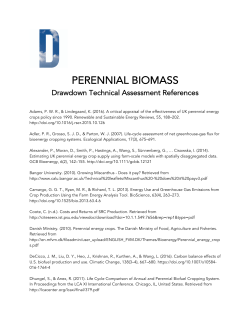
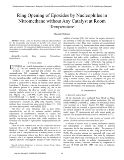
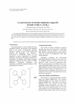
Figure 1. Structure of 1a with the atom-labeling scheme. Displacement ellipsoids are drawn at the 30% probability level. Disorder is
presented by dashed lines.
structures were refined in orthorhombic Ccc2 space group
and show the presence of one-dimensional polymeric
structure.
Structures 1a and 1b are very similar. Selected bond
distances and angles of 1a are summarized in Table 2, data of room temperature measurement 1b can be found in
Supplementary material. Asymmetric unit of 1a contains
one half of the bis(4,4,4-trifluoro-1-phenylbutane-1,3dionato)oxidovanadium(IV) complex with the VO2+ moiety on a twofold axis. Vanadium center has distorted octahedral geometry with carbonyl oxygen atoms of two bidentate tfpb ligands in the equatorial plane and with oxido
atoms in axial positions (Figure 1). The first oxido atom is
doubly bound to vanadium center, while the second is a
part of the adjacent VO2+ moiety. Each oxido atom is therefore also a bridging atom to the next vanadium center
enabling ···V=O···V=O··· interactions and causing the formation of one-dimensional coordination polymer.
The structure of 1a shows a great crystallographic
disorder over two positions. The VO2+ moiety, carbonyl
oxygen atoms and phenyl ring occupy two opposite positions with distinct refined occupation ratio of 0.57:0.43,
while an easily rotatable CF3 group is disordered with the
ratio 0.53:0.47. Both positions of the disordered complex
give the same, only inverted, polymeric structure. To simplify, only the major part will be discussed briefly. Bond
distance of V=O double bond is 1.601(11) Å and bond distance of V···O interaction is 2.155(11) Å. Vanadium is
shifted 0.31 Å out of the basal plane generated by the
equatorial oxygen atoms. Tfpb ligand is bound to vanadium in a bidentate manner with the chelate angle
88.3(2)° and V–O bond distances 1.985(5) and 1.982(5)
Å. Angles between O1 and equatorial oxygen atoms are
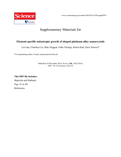
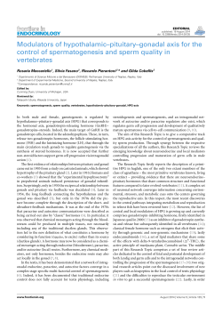
99.3(2) and 98.5(2)°. Polymeric chain is stabilized by
weak π···π stacking interactions with the centroid-tocentroid distance between phenyl rings of 4.215(8) Å (Figure 2).
Kole{a-Dobravc et al.: Supramolecular Potential of Vanadium ...
Acta Chim. Slov. 2015, 62, 261–271
265
Table 2. Selected bond distances and angles for 1a.
Distance
V1A–O1A
V1A–O2A
V1A–O3A
V1A–O1Aii
Angle
O1A–V1A–O1Aii
O1A–V1A–O2A
O1A–V1A–O3A
O2A–V1A–O2Ai
O2A–V1A–O3A
O2A–V1A–O3Ai
O3A–V1A–O3Ai
O1Aii–V1A–O2A
O1Aii–V1A–O3A
(Å)
1.601(11)
1.985(5)
1.982(5)
2.155(11)
(°)
180.000(4)
99.3(2)
98.5(2)
161.4(5)
88.3(2)
88.9(2)
162.9(4)
80.7(2)
81.5(2)
V1B–O1B
V1B–O2B
V1B–O3B
V1B–O1Biii
1.609(13)
1.989(7)
1.990(6)
2.147(13)
O1B–V1B–O1Biii
O1B–V1B–O2B
O1B–V1B–O3B
O2B–V1B–O2Bi
O2B–V1B–O3B
O2B–V1B–O3Bi
O3B–V1B–O3Bi
O1Biii–V1B–O2B
O1Biii –V1B–O3B
180.000(2)
97.7(3)
99.0(3)
164.6(6)
89.7(3)
87.9(3)
162.1(5)
82.3(3)
v81.0(3)
Symmetry codes: (i) –x, –y, z; (ii) –x, y, z – ½; (iii) –x, y, z + ½.
3. 3. Synthesis and Characterization
of Bis(acetylacetonato)(2-pyridone)
oxidovanadium(IV) (2)
Compound 2 was prepared by mixing VO(acac)2
with 2-pyridone in 1:1 ratio in warm chloroform and crystals suitable for X-ray analysis were obtained by slow
evaporation of the solvent at room temperature. In IR
spectrum of 2 a stretching vibration of the VO2+ moiety is
observed as a strong band at 972 cm–1. A band at 1635
cm–1 in the free 2-pyridone corresponding to C=O vibration is shifted to 1647 cm–1 upon coordination. This vibration confirms the presence of the lactam form of 2-pyridone in compound 2. Vibrations near 2918 cm–1 suggest the
involvement of the O–H group in the hydrogen bonding.
Compound 2 crystallizes as green prisms in the
triclinic P–1 space group. Selected bond distances and
Cg5i
Cg5
Cg5ii
Figure 2. Coordination polymer of 1a. For clarity only one orientation of the coordination polymer chain without a minor part of the
disorder is presented. Stabilization by π···π stacking interactions is
indicated by dashed lines (symmetry codes: (i) x, –y, z + ½; (ii) x,
–y, z – ½).
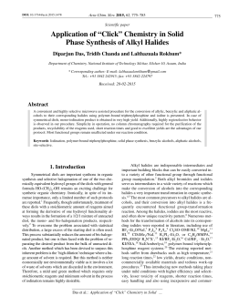
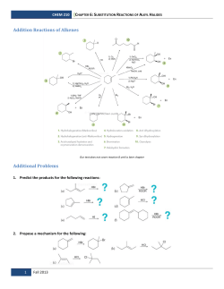
Figure 3. The asymmetric unit of 2 showing the atom-labeling
scheme. Displacement ellipsoids are drawn at the 30% probability
level.
Kole{a-Dobravc et al.: Supramolecular Potential of Vanadium ...
Acta Chim. Slov. 2015, 62, 261–271
266
Table 3. Selected bond distances and angles for 2.
Distance
V1–O1
V1–O2
V1–O3
Angle
O1–V1–O2
O1–V1–O3
O1–V1–O4
O1–V1–O5
O2–V1–O3
O2–V1–O4
(Å)
1.588(2)
1.9830(17)
1.9891(17)
(°)
100.14(10)
99.74(10)
100.16(10)
99.18(10)
88.86(7)
87.10(7)
V1–O4
V1–O5
V1–O6
1.9828(18)
1.9886(17)
2.515(2)
O2–V1–O5
O3–V1–O4
O3–V1–O5
O4–V1–O5
O1–V1–O6
160.66(8)
160.09(8)
88.56(7)
88.84(7)
174.65(9)
angles of 2 are summarized in Table 3. The vanadium
atom in compound 2 is octahedrally coordinated (Fig. 3)
with four oxygen atoms of two chelating acac ligands
with V–O distances 1.983–1.989 Å in a basal plane and
the vanadium atom lying 0.34 Å above this plane. Bite
angles O–V–O are ∼88.8°, while O–V–O in trans posi-
tions are ∼160°. The V=O double bond length is
1.588(2) Å and the V–O(pyridone) is 2.515(2) Å. This
distance is rather long; however, similar contacts in the
range from 2.46 even up to 2.58 Å were found in CSD.29
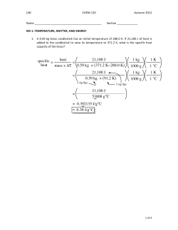
Even though the carbonyl oxygen of the 2-pyridone ligand is coordinated to the vanadium center, it can
still act as a hydrogen bond acceptor facilitating the formation of centrosymmetric hydrogen-bonded dimers via
N1–H1···O6(–x + 1, –y + 1, –z + 1) interactions (Fig. 4,
Table 4). Hydrogen-bonded dimers are further stabilized
by weak C15–H15···O5(–x + 1, –y + 1, –z + 1) interactions and 2D framework is achieved by weak C13–
H13···O2(–x + 1, –y + 1, –z) and C10–H10A···O3(–x + 1,
–y + 2, –z + 1) interactions (Fig. 4).
3. 4. Literature Data and Structures for VO2+
Complexes of Monochelated Picolinates
and Chlorido Compounds
The crystallographic evidence on bischelated picolinato complexes of VO2+ has been discussed earlier.5a,30
When examining the known monopicolinate complexes
of dioxidovanadium(V), VO2(picolinate), we found only
four structures and all display a trigonal bipyramidal geometry with a chelating picolinato or quinoline-2-carboxylato ligand and an additional monodentate ligand.31
Structures with chlorido ligand bonded to VO2+ are also
not numerous, as only five entries in CSD could be
found. All of these compounds were prepared under the
inert atmosphere since V–Cl bond can be easily hydrolyzed. Compounds [VO2Cl(py)2]32,33 and [VO2Cl(O-py)2]33
possess trigonal bipiramidal and PPh3Me[VO2Cl2]34 tetrahedral geometry.
3. 5. Synthesis and Characterization
of Pyridinium Chlorido(pyridin-2carboxylato-N,O)dioxidovanadate(V) (3)
Figure 4. The 2D network parallel to the bc plane in 2 formed by
weak C–H···O interactions (symmetry codes: (i) –x + 1, –y + 1, –z
+ 1; (ii) –x + 1, –y + 2, –z + 1; (iii) –x + 1, –y + 1, –z).
Compound 3 was prepared from ammonium metavanadate and picolinic acid with the addition of pyridine and
hydrochloric acid. Small cubic crystals of a side product
NH4Cl with similar solubility were present despite many
attempts of washing and recrystallization of the compound
3. Anyway, we managed to isolate some single crystals suitable for X-ray analysis. In IR spectrum of 3 bands for aromatic C–H vibrations are present at 3080 and 3062 cm–1,
Table 4. Hydrogen bonds for 2 [Å and °]
D–H···A
d(D–H)
d(H···A)
d(D···A)
<(DHA)
N1–H1···O6
0.86
2.02
2.884(2)
177
Symmetry transformation
for acceptors
–x + 1, –y + 1, –z + 1
C10– H10A···O3
C13–H13···O2
C15–H15···O5
0.96
0.93
0.93
2.57
2.55
2.46
3.367(4)
3.426(4)
3.249(4)
140
158
142
–x + 1, –y + 2, –z + 1
–x + 1, –y + 1, –z
–x + 1, –y + 1, –z + 1
Kole{a-Dobravc et al.: Supramolecular Potential of Vanadium ...
Acta Chim. Slov. 2015, 62, 261–271
as well as asymmetric and symmetric vibrations of COO–
group at 1685 and 1340 cm–1. The difference between
asymmetric and symmetric stretching vibrations (Δ = νas –
νs) of 345 cm–1 is in accordance with the monodentate
coordination of the COO– group to the VO2+ moiety.
Stretching vibrations of the VO2+ group are observed as a
medium band at 946 cm–1 and a strong band at 924 cm–1.
Compound 3 crystallizes as green prisms in the monoclinic P21/c space group. Selected bond distances and
angles of 3 are summarized in Table 5. Asymmetric unit of
3 consists of vanadium(V) complex anion and pyridinium
cation as its counter ion (Fig. 5). Vanadium(V) metal center is pentacoordinated. The distortion of a trigonal bipyramid can be best described by the structural parameter τ
(0 for an ideal square pyramid and 1 for an ideal trigonal
bipyramid),35 which in this case has the value of 0.44.
This value would imply the presence of distorted square
pyramid geometry; however, visual inspection would suggest distorted trigonal bipyramidal geometry. Although
the trigonality index τ is a well established structural parameter some of the reported examples show that τ is not always a reliable guidance.36 This seems to be the case for
compounds in which one metal–ligand bond is elongated,36 as is the case in 3. Reason for this is that the parame-
Figure 5. The asymmetric unit of 3 with atom labels. Ellipsoids are
drawn at the 30% probability level. Dashed lines indicate hydrogen
bonds.
267
Table 5. Selected bond distances and angles for 3.
Distance
V1–O1
V1–O2
V1–O3
Angle
O1–V1–O2
O1–V1–O3
O2–V1–O3
O1–V1–N1
O2–V1–N1
(Å)
1.6124(18)
1.6276(18)
2.0347(15)
(°)
108.41(10)
117.23(9)
132.76(9)
93.36(9)
91.19(9)
V1–N1
V1–Cl1
2.125(2)
2.3308(8)
O3–V1–N1
O1–V1–Cl1
O2–V1–Cl1
O3–V1–Cl1
N1–V1–Cl1
75.17(7)
100.32(7)
99.37(7)
84.42(5)
159.03(6)
ter τ is based solely on the two largest X–M–X angles,
and does not account for the distortion of the geometry
due to bond elongations.
Pyridin-2-carboxylato (picolinato) ligand is bound to
vanadium in a bidentate manner through nitrogen and carboxylato oxygen atoms with the chelate angle 75.17(7)°.
The angle between N1 and Cl1 atoms is 159.03(6)°, angles
between oxygen atoms are 108.41(10)–132.76(9)°, while
angles between N1 or Cl1 and oxygen atoms are
84.42(5)–100.32(7)°. The V–Cl distance is 2.3308(8) Å,
and the V=O double bond lengths are 1.6124(18) and
1.6276(18) Å. These lengths are in the same range as in the
other dioxidovanadates(V), but slightly shorter as in octahedral bis(picolinato)dioxovanadate(V) complex.37
Crystal structure of 3 is stabilized by hydrogen
bonds, π···π stacking interactions and weak C–H···O/Cl
interactions listed in Table 6. Hydrogen bonds are formed
between the pairs of a pyridinium cation and a complex
vanadate anion. N2–H2 group of the pyridinium cation is
hydrogen-bonded to the carboxylate O3, as well as to the
chlorido Cl1 atom in the same anion. Hydrogen-bonded
pairs are connected into infinite chains parallel to the b axis (Fig. 6) due to π···π stacking interactions between picolinato (ring centroid Cg2) and pyridinium rings or between two adjacent pyridinium rings (ring centroid Cg3)
with the centroid-to-centroid distances 3.5886(17) and
4.008(2) Å, respectively. Further stabilization is enabled
by weak hydrogen interactions between pyridinium C–H
and O1, O4 and Cl1 atoms spreading in all three dimensions (Fig. 7).
Table 6. Hydrogen bonds for 3 [Å and °]
D–H···A
d(D–H)
d(H···A)
d(D···A)
<(DHA)
N2–H2···O3
N2–H2···Cl1
0.86
0.86
2.07
2.72
2.872 (3)
3.357 (2)
154
132
Symmetry transformation
for acceptors
x, y, z
x, y, z
C7–H7···O4
C8–H8···Cl1
C9–H9···O1
C10–H10···O1
C11–H11···O4
0.93
0.93
0.93
0.93
0.93
2.55
2.70
2.60
2.42
2.40
3.347(3)
3.620(4)
3.357(4)
3.193(3)
3.222(4)
144
171
139
141
148
x, –y + ½ , z – ½
–x + 1, y – ½, –z + ½
–x + 1, –y, –z + 1
x + 1, –y + ½, z + ½
x, y, z
Kole{a-Dobravc et al.: Supramolecular Potential of Vanadium ...
Acta Chim. Slov. 2015, 62, 261–271
268
Cg2i
Cg3
Cg3i
Cg3ii
Cg2
Figure 6. Twisted infinite chains formed by the combination of hydrogen bonds and π–π stacking interactions in 3 forming infinite chain along b
axis. Dashed lines indicate hydrogen bonds, and dotted lines indicate centroid-to-centroid distances (symmetry codes: (i) –x + 1, –y + 1, –z + 1; (ii)
–x + 1, –y, –z + 1).
fluorinated β-diketonato ligand tfpb enhances the acid character of the vanadium atom and the additional interaction
with the adjacent oxidovanadium(IV) group occurs. Compound [VO(acac)(2-pyridone)] (2) has an octahedral structure with the 2-pyridone ligand in trans position to VO2+
group. Even though the carbonyl oxygen of the 2-pyridone
ligand is coordinated to the vanadium center, it can still act
as a hydrogen bond acceptor facilitating the formation of
centrosymmetric hydrogen-bonded dimers via N–H···O interactions with the adjacent molecule. In compound
Hpy[VO2(pic)Cl] (3) the metal center is pentacoordinated
with one picolinato and one chlorido ligand coordinated to
the dioxidovanadium(V) moiety, and the Hpy+ cation is
hydrogen bonded to the complex anion. The crystal structure is stabilized by π···π and C–H···O interactions. This
study provides an insight on the influence of different
types of ligands on the molecular structure and crystal architecture. Such cases are valuable examples that can help
to understand the covalent and non-covalent factors governing the supramolecular aggregation important for crystal
engineering and crystal structure prediction.
5. Supplementary Material
Figure 7. 3D network formed by weak hydrogen bonds in 3. Dashed lines indicate weak C–H···O interactions (symmetry codes: (i)
x, –y + ½, z – ½; (ii) –x + 1, y – ½, –z + ½; (iii) –x + 1, –y, –z + 1).
4. Conclusion
Three vanadium compounds with β-diketonato or picolinato ligands were prepared and structurally characterized. Compound [VO(tfpb)2]∞ (1) represents the first X-ray
structure of an oxidovanadium(IV) β-diketonate with infinite chain formed by the V=O···V=O interactions. The
CCDC 1024846–1024849 (1a–3) contain the supplementary crystallographic data for this paper. These data
can be obtained free of charge from The Cambridge Crystallographic Data Centre via www.ccdc.cam.ac.uk/data_request/cif.
6. Acknowledgment
The authors thank the Ministry of Education, Science and Sport of the Republic of Slovenia and the Slovenian Research Agency for financial support (P1–0230–
0175) as well as the EN–FIST Centre of Excellence, Trg
Osvobodilne fronte 13, 1000 Ljubljana, Slovenia for use
of the Supernova diffractometer.
Kole{a-Dobravc et al.: Supramolecular Potential of Vanadium ...
Acta Chim. Slov. 2015, 62, 261–271
7. References
1. (a) D. Rehder, Future Med. Chem. 2012, 4, 1823–1837;
http://dx.doi.org/10.4155/fmc.12.103
(b) H. Sakurai, Y. Yoshikawa, H. Yasui, Chem. Soc. Rev.
2008, 37, 2383–2392; http://dx.doi.org/10.1039/b710347f
(c) G. R. Willsky, L.-H. Chi, M. Godzala III, P. J. Kostyniak,
J. J. Smee, A. M. Trujillo, J. A. Alfano, W. Ding, Z. Hu, D. C.
Crans, Coord. Chem. Rev. 2011, 255, 2258–2269;
http://dx.doi.org/10.1016/j.ccr.2011.06.015
(d) D. C. Crans, J. J. Smee, E. Gaidamauskas, L. Yang,
Chem. Rev. 2004, 104, 849–902.
http://dx.doi.org/10.1021/cr020607t
2. (a) L. G. Naso, M. Valcarcel, M. Roura-Ferrer, D. Kortazar,
C. Salado, L. Lezama, T. Rojo, A. C. González-Baró, P. A.
M. Williams, E. G. Ferrer, J. Inorg. Biochem. 2014, 135,
86–99; http://dx.doi.org/10.1016/j.jinorgbio.2014.02.013
(b) I. E. Leon, V. Porro, A. L. Di Virgilio, L. G. Naso, P. A.
M. Williams, M. Bollati-Fogolín, S. B. Etcheverry, J. Biol.
Inorg. Chem. 2014, 19, 59–74;
http://dx.doi.org/10.1007/s00775-013-1061-x
(c) I. E. Leon, A. L. Di Virgilio, V. Porro, C. I. Muglia, L. G.
Naso, P. A. M. Williams, M. Bollati-Fogolin, S. B. Etcheverry, Dalton Trans. 2013, 42, 11868–11880.
http://dx.doi.org/10.1039/c3dt50524c
3. (a) S. Sgarbossa, E. Diana, D. Marabello, A. Deagostino, S.
Cadamuro, A. Barge, E. Laurenti, M. Gallicchio, V. Boscaro,
E. Ghibaudi, J. Inorg. Biochem. 2013, 128, 26–37;
http://dx.doi.org/10.1016/j.jinorgbio.2013.07.015
(b) Z. Gao, C. Zhang, S. Yu, X. Yang, K. Wang, J. Biol.
Inorg. Chem. 2011, 16, 789–798;
http://dx.doi.org/10.1007/s00775-011-0780-0
(c) J. Vinklárek, T. Dìdourková, J. Honzí~ek, A. Rù`i~ka, J.
Inorg. Biochem. 2010, 104, 936–943;
http://dx.doi.org/10.1016/j.jinorgbio.2010.04.012
(d) J.-Z. Lu, H.-W. Guo, X.-D. Zeng, Y.-L. Zhang, P. Zhao, J.
Jiang, L.-Q. Zang, J. Inorg. Biochem. 2012, 112, 39–48.
http://dx.doi.org/10.1016/j.jinorgbio.2012.02.034
4. D. Gambino, Coord. Chem. Rev. 2011, 255, 2193–2203.
http://dx.doi.org/10.1016/j.ccr.2010.12.028
5. (a) T. Kole{a-Dobravc, E. Lodyga-Chruscinska, M. Symonowicz, D. Sanna, A. Meden, F. Perdih, E. Garribba, Inorg.
Chem. 2014, 53, 7960–7976; (b) D. Sanna, M. Serra, G. Micera, E. Garribba, Inorg. Chem. 2014, 53, 1449–1464;
http://dx.doi.org/10.1021/ic402366x
(c) D. Sanna, G. Micera, E. Garribba, Inorg. Chem. 2013,
52, 11975–11985; http://dx.doi.org/10.1021/ic401716x
(d) S. Mehtab, G. Gonçalves, S. Roy, A. I.Tomaz, T. SantosSilva, M. F. A. Santos, M. J. Romão, T. Jakusch, T. Kiss, J.
Costa Pessoa, J. Inorg. Biochem. 2013, 121, 187–195.
http://dx.doi.org/10.1016/j.jinorgbio.2012.12.020
6. (a) A. M. Kirillov, G. B. Shul’pin, Coord. Chem. Rev. 2013,
257, 732–754; http://dx.doi.org/10.1016/j.ccr.2012.09.012
(b) J. A. L. da Silva, J. J. R. Fraústo da Silva, A. J. L. Pombeiro, Coord. Chem. Rev. 2011, 255, 2232– 2248;
http://dx.doi.org/10.1016/j.ccr.2011.05.009
(c) K.-H. Yang, Acta Chim. Slov. 2014, 61, 629–636.
7. A. Ohta, Y. Yamamoto, H. Kamihata, Y. H. Lee, F. Ichikawa,
K. Ohta, Y. Abe, N. Hoshino, M. Kojima, S. Hayami, Inorg.
Chem. Commun. 2012, 16, 89–91.
http://dx.doi.org/10.1016/j.inoche.2011.12.001
8. (a) D. Barreca, L. Armelao, F. Caccavale, V. Di Noto, A. Gregori, G. A. Rizzi, E. Tondello. Chem. Mater. 2000, 12,
98–103; http://dx.doi.org/10.1021/cm991095a
(b) D. Barreca, L. E. Depero, E. Franzato, G. A. Rizzi, L.
Sangaletti, E. Tondello, U. Vettori, J. Electrochem. Soc.
1999, 146, 551–558. http://dx.doi.org/10.1149/1.1391642
9. (a) M. Riou-Cavellec, M. Sanselme, G. Férey, J. Mater.
Chem. 2000, 10, 745–748;
http://dx.doi.org/10.1039/a909870d
(b) K. Barthelet, J. Marrot, D. Riou, G. Férey, Angew.
Chem., Int. Ed. 2002, 41, 281–284. http://dx.doi.org/10.
1002/1521-3773(20020118)41:2<281::AID-ANIE281>3.
0.CO;2-Y
10. (a) P. Kanoo, A. C. Ghosh, T. K. Maji, Inorg. Chem. 2011,
50, 5145–5152; http://dx.doi.org/10.1021/ic200463k
(b) X. Wang, J. Eckert, L. Liu, A. J. Jacobson, Inorg. Chem.
2011, 50, 2028–2036; http://dx.doi.org/10.1021/ic1025087
(c) A. Phan, A. U. Czaja, F. Gándara, C. B. Knobler, O. M.
Yaghi, Inorg. Chem. 2011, 50, 7388–7390;
http://dx.doi.org/10.1021/ic201396m
(d) I. Djerdj, J. Popovi}, J. Stare, G. Ambro`i~, S. D. [kapin,
B. Kozlev~ar, D. Paji}, Z. Jagli~i}, Z. C. Orel, J. Mater.
Chem. 2012, 22, 10255–10265; (e) I. Djerdj, S. D. [kapin,
M. ~eh, Z. Jagli~i}, D. Paji}, B. Kozlev~ar, B. Orel, Z. C.
Orel, Dalton Trans. 2012, 41, 581–589; (f) Y.-Y. Liu, S.
Couck, M. Vandichel, M. Grzywa, K. Leus, S. Biswas, D.
Volkmer, J. Gascon, F. Kapteijn, J. F. M. Denayer, M. Waroquier, V. Van Speybroeck, P. Van Der Voort, Inorg. Chem.
2013, 52, 113–120. http://dx.doi.org/10.1021/ic301338a
11. (a) G. R. Desiraju, Angew. Chem. Int. Ed. 2007, 46, 8342–
8356; http://dx.doi.org/10.1002/anie.200700534
(b) L. Brammer, Chem. Soc. Rev. 2004, 33, 476–489.
http://dx.doi.org/10.1039/b313412c
12. (a) F. Perdih, Monatsh. Chem. 2012, 143, 1011–1017;
http://dx.doi.org/10.1007/s00706-012-0760-2
(b) S. Shit, R. Sankolli, T. N. Guru Row, Acta Chim. Slov.
2014, 61, 59–66; (c) B. Ardan, Y. Slyvka, E. Goreshnik, M.
Mys’kiv, Acta Chim. Slov. 2013, 60, 484–490;
(d) F. Perdih, J. Coord. Chem. 2012, 65, 1580–1591;
http://dx.doi.org/10.1080/00958972.2012.676168
(e) M. A. Sharif, G. R. Najafi, Acta Chim. Slov. 2013, 60,
138–143; (f) S. Corti, W. T. Pennington, D. D. DesMarteau,
Acta Chim. Slov. 2013, 60, 556–560; (g) R. Vafazadeh, M.
Alinaghi, A. C. Willis, A. Benvidi, Acta Chim. Slov. 2014,
61, 121–125; (h) F. Perdih, Acta Cryst. C 2012, 68,
m64–m68; (i) L. Saghatforoush, H. A. Rudbari, F. Nicolò,
P. Asgari, F. Chalabian, M. Hasanzadeh, V. Panahiazar, Acta
Chim. Slov. 2013, 60, 300–309.
13. (a) F. Perdih, Struct. Chem. 2014, 25, 809–819;
http://dx.doi.org/10.1007/s11224-013-0344-8
(b) F. Marandi, S. Teimouri, H.-K. Fun, Acta Chim. Slov.
Kole{a-Dobravc et al.: Supramolecular Potential of Vanadium ...
269
270
Acta Chim. Slov. 2015, 62, 261–271
2013, 60, 328– 334.
14. C. Glidewell, in: J. D. Woollins (Ed.): Inorganic Experiments Wiley-VCH, Weinheim, Germany, 1994, pp. 149–151.
15. CrysAlisPRO, Agilent Technologies, Yarnton, England.
16. Z. Otwinowski, W. Minor, Methods Enzymol. 1997, 276,
307–326. http://dx.doi.org/10.1016/S0076-6879(97)76066-X
17. G. M. Sheldrick, Acta Cryst. 2008, A64, 112–122.
http://dx.doi.org/10.1107/S0108767307043930
18. A. Altomare, M. C. Burla, M. Camalli, G. Cascarano, C.
Giacovazzo, A. Guagliardi, A. G. G. Moliterni, G. Polidori,
R. Spagna, J. Appl. Cryst. 1999, 32, 115–119.
http://dx.doi.org/10.1107/S0021889898007717
19. M. C. Burla, R. Caliandro, M. Camalli, B. Carrozzini, G. L.
Cascarano, L. De Caro, C. Giacovazzo, G. Polidori, R. Spagna, J. Appl. Cryst. 2005, 38, 381–388.
http://dx.doi.org/10.1107/S002188980403225X
20. (a) S. Venkataraman, B. Varghese, B. K. Sadashiva, S. Subramanian, Mol. Cryst. Liq. Cryst. 2001, 357, 199–219;
http://dx.doi.org/10.1080/10587250108028254
(b) E. Shuter, S. J. Rettig, C. Orvig, Acta Cryst. C 1995, 51,
12–14; http://dx.doi.org/10.1107/S0108270194010462
(c) M. Hoshino, A. Sekine, H. Uekusa, Y. Ohashi, Chem.
Lett. 2005, 34, 1228–1229;
http://dx.doi.org/10.1246/cl.2005.1228
(d) K. S. Dichmann, G. Hamer, S. C. Nyburg, W. F. Reynolds, J. Chem. Soc. D 1970, 1295–1296;
http://dx.doi.org/10.1039/c29700001295
(e) U. K. Urs, K. C. Anitha, K. Lakshmi Raghunathan, S. A.
Shivashankar, W. T. Robinson, T. N. Guru Row, Acta Cryst.
E 2001, 57, m242–m243;
http://dx.doi.org/10.1107/S1600536801007760
(f) M. A. K. Ahmed, H. Fjellvag, A. Kjekshus, B. Klewe, Z.
Anorg. Allg. Chem. 2004, 630, 2311–2318;
http://dx.doi.org/10.1002/zaac.200400369
(g) S. S. Amin, K. Cryer, B. Zhang, S. K. Dutta, S. S. Eaton,
O. P. Anderson, S. M. Miller, B. A. Reul, S. M. Brichard, D.
C. Crans, Inorg. Chem. 2000, 39, 406–416;
http://dx.doi.org/10.1021/ic9905897
(h) U. Schilde, W. Bansse, E. Ludwig, E. Uhlemann, Z. Kristallogr. 1995, 210, 627–628.
http://dx.doi.org/10.1524/zkri.1995.210.8.627
21. (a) H. Adams, N. A. Bailey, D. E. Fenton, M. S. Leal Gonzalez, C. A. Phillips, J. Chem. Soc., Dalton Trans. 1983,
371–379; http://dx.doi.org/10.1039/dt9830000371
(b) P. Hon, R. L. Belford, C. E. Pfluger, J. Chem. Phys. 1965,
43, 3111–3115. http://dx.doi.org/10.1063/1.1697284
22. D. I. Arnold, F. A. Cotton, J. H. Matonic, C. A. Murillo,
Chem. Commun. 1996, 2113–2114.
http://dx.doi.org/10.1039/cc9960002113
23. (a) A. Sarkar, S, Pal, Eur. J. Inorg. Chem. 2009, 5391–5398;
http://dx.doi.org/10.1002/ejic.200900680
(b) S. Meicheng, W. Lifeng, Z. Zeying, Acta Chim. Sinica
1983, 41, 985–992; (c) S. Meicheng, W. Lifeng, T. Youqi,
Chin. Sci. Bull. 1984, 29, 759–764; (d) R. M. S. da Silva, C.
C. Spiazzi, R. Bortolotto, R. A. Burrow, Acta Cryst. E 2007,
63, m2422; http://dx.doi.org/10.1107/S1600536807039323
(e) L. Chen, F.-L. Jiang, W.-P. Su, C.-Y. Yue, D.-Q. Yuan,
M.-C. Hong, Inorg. Chim. Acta 2009, 362, 407–413;
http://dx.doi.org/10.1016/j.ica.2008.04.026
(f) H. Chen, Y. Si, C. Chen, Q. Liu, Acta Cryst. E 2008, 64,
m159; http://dx.doi.org/10.1107/S1600536807065439
(g) A. M. B. Bastos, J. G. da Silva, P. I. da S. Maia, V. M.
Deflon, A. Batista, A. V. M. Ferreira, L. M. Botion, E. Niquet, H. Beraldo, Polyhedron 2008, 27, 1787–1794;
http://dx.doi.org/10.1016/j.poly.2008.02.014
(h) C. Pretorius, J. A. Venter, A. Roodt, Acta Cryst. E 2012,
68, m1442. http://dx.doi.org/10.1107/S1600536812044686
24. (a) V. Stilinovi}, D.-K. Bu~ar, I. Halasz, E. Me{trovi}, New J.
Chem. 2013, 37, 619–623;
http://dx.doi.org/10.1039/C2NJ40780A
(b) T. Ishida, S. Mitsubori, T. Nogami, N. Takeda, M. Ishikawa, H. Iwamura, Inorg. Chem. 2001, 40, 7059–7064;
http://dx.doi.org/10.1021/ic010730n
(c) Z. C. Kadirova, D. S. Rahmonova, S. A. Talipov, J. M.
Ashurov, N. A. Parpiev, Acta Cryst. E 2009, 65, m819;
http://dx.doi.org/10.1107/S1600536809023113
(d) E. Mestrovi}, D.-K. Bu~ar, I. Halasz, V. Stilinovi}, Acta
Cryst. E 2004, 60, m1920–m1922.
25. I. Cvrtila, V. Stilinovi}, B. Kaitner, Struct. Chem. 2012, 23,
587–594. http://dx.doi.org/10.1007/s11224-011-9905-x
26. (a) M. Tsuchimoto, G. Hoshina, N. Yoshioka, H. Inoue, K.
Nakajima, M. Kamishima, M. Kojima, S. Ohba, J. Solid State Chem. 2000, 153, 9–15;
http://dx.doi.org/10.1006/jssc.2000.8710
(b) G. Grivani, S. Delkhosh, K. Fejfarová, M. Du{ek, A. D.
Khalaji, Inorg. Chem. Commun. 2013, 27, 82–87.
http://dx.doi.org/10.1016/j.inoche.2012.10.029
27. (a) N. Matsuoka, H. Kawamura, N. Yoshioka, Chem. Phys.
Lett. 2010, 488, 32–37;
http://dx.doi.org/10.1016/j.cplett.2010.01.057
(b) K. Nakajima, M. Kojima, S. Azuma, R. Kasahara, M.
Tsuchimoto, Y. Kubozono, H. Maeda, S. Kashino, S. Ohba,
Y. Yoshikawa, J. Fujita, Bull. Chem. Soc. Jpn. 1996, 69,
3207–3216; http://dx.doi.org/10.1246/bcsj.69.3207
(c) S. A. Fairhurst, D. L. Hughes, U. Kleinkes, G. J. Leigh, J.
R. Sanders, J. Weisner, J. Chem. Soc., Dalton Trans. 1995,
321–326. http://dx.doi.org/10.1039/dt9950000321
28. D. J. Bray, J. K. Clegg, L. F. Lindoy, D. Schilter, Adv. Inorg.
Chem. 2007, 59, 1–37.
29. For some examples see: (a) W. Plass, Angew. Chem., Int.
Ed. Engl. 1996, 35, 627–631;
http://dx.doi.org/10.1002/anie.199606271
(b) C. E. Schulz, H. Song, Y. J. Lee, J. U. Mondal, K. Mohanrao, C. A. Reed, F. A. Walker, W. R. Scheidt, J. Am.
Chem. Soc. 1994, 116, 7196–7203;
http://dx.doi.org/10.1021/ja00095a023
(c) D. Riou, G. Férey, J. Mater. Chem. 1998, 8, 2733–2735;
http://dx.doi.org/10.1039/a802711k
(d) W. Kläui, S. Schäfer, W. Peters, H. Wunderlich, Z.
Anorg. Allg. Chem. 2006, 632, 2447–2452;
http://dx.doi.org/10.1002/zaac.200600211
(e) S. K. Hanson, R. T. Baker, J. C. Gordon, B. L. Scott, D.
Kole{a-Dobravc et al.: Supramolecular Potential of Vanadium ...
Acta Chim. Slov. 2015, 62, 261–271
L. Thorn, Inorg. Chem, 2010, 49, 5611–5618;
http://dx.doi.org/10.1021/ic100528n
(f) C. Djordjevic, M. Lee, E. Sinn, Inorg. Chem. 1989, 28,
719–723; http://dx.doi.org/10.1021/ic00303a022
(g) V. Zerbib, F. Robert, P. Gouzerh, J. Chem. Soc., Chem.
Commun. 1994, 2179–2180.
http://dx.doi.org/10.1039/c39940002179
30. T. Kole{a-Dobravc, A. Meden, F. Perdih, Monatsh. Chem.
2014, 145, 1263–1275.
http://dx.doi.org/10.1007/s00706-014-1215-8
31. (a) H. Mimoun, L. Saussine, E. Daire, M. Postel, J. Fischer,
R. Weiss, J. Am. Chem. Soc. 1983, 105, 3101–3110;
http://dx.doi.org/10.1021/ja00348a025
(b) G. Süss-Fink, L. G. Cuervo, B. Therrien, H. StoeckliEvans, G. B. Shul’pin, Inorg. Chim. Acta 2004, 357,
475–484; http://dx.doi.org/10.1016/j.ica.2003.05.005
(c) L. E. Khoon, T. S. Guan, B. M. Yamin, Acta Cryst. E
2006, 62, m207–m209. (d) N. Smre~ki, B.-M. Kukovec, Z.
Popovi}, Monatsh. Chem. 2012, 143, 1471–1477.
32. M. Motevalli, D. Shah, S. A. A. Shah, A. C. Sullivan, Polyhedron 1996, 15, 2387–2395.
http://dx.doi.org/10.1016/0277-5387(95)00509-9
33. M. F. Davis, M. Jura, A. Leung, W. Levason, B. Littlefield,
G. Reid, M. Webster, Dalton Trans. 2008, 6265–6273.
http://dx.doi.org/10.1039/b811422f
34. D. Fenske, A.-F. Shihada, H. Schwab, K. Dehnicke, Z.
Anorg. Allg. Chem. 1980, 471, 140–146.
http://dx.doi.org/10.1002/zaac.19804710116
35. A. W. Addison, T. N. Rao, J. Reedijk, J. van Rijn, G. C.
Verschoor, J. Chem. Soc. Dalton Trans. 1984, 1349–1356.
http://dx.doi.org/10.1039/dt9840001349
36. S. Wöckel, J. Ga¦ezowska, S. Dechert, W. Meyer-Klaucke,
E. Nordlander, F. Meyer, Eur. J. Inorg. Chem. 2012, 4728–
4738. http://dx.doi.org/10.1002/ejic.201200269
37. M. Melchior, K. H. Thompson, J. M. Jong, S. J. Rettig, E.
Shuter, V. G. Yuen, Y. Zhou, J. H. McNeill, C. Orvig, Inorg.
Chem. 1999, 38, 2288–2293.
http://dx.doi.org/10.1021/ic981231y
Povzetek
Pripravili in strukturno okarakterizirali smo tri vanadijeve spojine z β-diketonato in picolinato ligandi. V spojinah
[VO(tfpb)2]∞ (1) (tfpb = 4,4,4-trifluoro-1-fenilbutan-1,3-dionat) in [VO(acac)2(2-piridon)] (2) je koordinacija vanadijevega atoma oktaedri~na, v spojini Hpy[VO2(pic)Cl] (3) je centralni atom pentakoordiniran. Rentgenska strukturna analiza razkriva tvorbo neskon~ne verige preko V=O···V=O vezi v 1, medtem ko sta 2 in 3 enojedrni spojini. V 2 se preko
N–H···O vezi s pomo~jo koordiniranega 2-piridona tvorijo centrosimetri~ni dimeri. V 3 je Hpy+ kation preko vodikovih
vezi vezan na kompleksni anion, kristalna struktura pa je dodatno stabilizirana s π···π in C–H···O interakcijami.
Kole{a-Dobravc et al.: Supramolecular Potential of Vanadium ...
271
© Copyright 2026 Paperzz