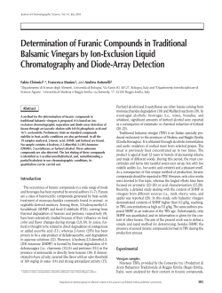
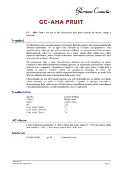
Standing Commission of Bee Pathology
PHARMACODYNAMICS OF OXALIC ACID IN THE HONEY BEE COLONY
3
1
2
2
2
A. NANETTI , P. BARTOLOMEI , Stefania BELLATO , Maria De SALVIO , E. GATTAVECCHIA , R. GHINI
2
1
E.N.E.A. – U.F.I.S. sede di Montecuccolino, Bologna, ITALY, E-mail: [email protected]
U.C.I. Scienze Chimiche Radiochimiche e Metallurgiche, Facoltà di Farmacia, Università di Bologna, ITALY
Tel.: +39 051 353103, E-mail: [email protected]
3
Istituto Nazionale di Apicoltura, Bologna, ITALY, E-mail: [email protected] and [email protected]
2
Abstract
The pharmacodynamics of oxalic acid (OA) administered by trickling was investigated by the means of radiochemical
methods. A sugar syrup containing 14C marked OA was trickled into a colony according to the dose and the technique commonly used
in the apicultural practice. In the first four days, the contamination of the adult bees reached 118 microg/g, but it decreased to less than
1/10 and 1/60 respectively one and two weeks after the treatment. During the following months a further decrease occurred.
Considerably lower levels were measured in 8-9 day old brood which, similarly, resulted only temporarily contaminated.
Autoradiographies put into evidence the presence of OA in the internal abdominal organs of the adult bees.
In freshly yielded honey the OA increase was as high as 0,6 mg/kg or less. This represents only a small fraction of the natural
OA content of honey. The radioactive marker was found also in wax collected from recently built combs, but it is not clear if it was due to
direct contamination involving chemical reactions between free OA and wax or to the presence of metabolites elaborated by the bees
from the absorbed OA.
In a second trial performed according to the same administration method, also other matrices were taken into account. The
radioactive marker was present either in the adult bee haemolymph or in the CO2 sampled within the colony. This seems to agree with
the hypothesis of an OA metabolization performed by the bees.
Keywords: varroa / residues / oxalic acid / distribution
Introduction
The trickling of sugar solutions added with oxalic acid into the honey bee colonies (NANETTI &
STRADI, 1997) is an increasingly spreading control method against varroosis in many European countries. A
huge mass of experiments have been made to verify efficacy and tolerability of this method in many
environmental and technical conditions, often leading to local set-ups (NANETTI, 2002).
Our knowledge about this active ingredient is far from complete, though. In particular, little is known
about the oxalic acid fate within colonies and single honey bees, the comprehension of which, aside from the
pure academic interest, is crucial to understand the treatment negative effects and to conceive
improvements in the applications. The few available data about the honey residues usually indicate a low
risk to jeopardise the honey authenticity after the oxalic acid treatments (MUTINELLI et al. 1997, DEL
NOZAL M.J. et al., 2000; BERNARDINI M & GARDI T., 2001; BOGDANOV et al., 2002; NANETTI A. et al.,
2002). However, the analytical techniques usually adopted cannot discriminate the contaminating oxalic acid
from the one which is naturally present in the honey, that greatly depends on its botanical origin (MUTINELLI
et al. 1997; NANETTI A. et al., 2002).
This paper represents a contribution to this knowledge. To overcome the problem represented by the
natural presence of the substance in the various honey bee colony matrices, the oxalic acid distribution was
studied through the administration of 14C labelled solutions and further radiochemical analyses.
Material and Methods
The experiment consisted of two replicates that were carried out during the summer seasons of the
years 1999 and 2002, respectively, close to the city of Bologna, Italy, during the honey yield. One colony
housed in DB hive per year was used. In both colonies the bees covered all the ten available combs, 6-7 of
which contained brood.
On July 19th, 1999 and on July 28th, 2002 the colonies were treated with oxalic acid according to the
above mentioned method of trickling. The treatments respectively consisted of 50 and 41 ml of aqueous
solution containing 4,2% of oxalic acid and 60 % of feed sugar (w/v) administered by the means of a syringe.
The solutions were beforehand added with 14C oxalic acid (Sigma N. 31,391-2), in amounts corresponding to
12,1 MBq and 7,4 MBq, respectively.
In both replicates, pre-treatment samples of the following matrices were collected from the colonies.
The post-treatment samples were initially collected once (1999) or twice a day (7 AM and 7 PM in 2002), but,
later on, they were drawn according to longer intervals.
In 1999, the following matrices were taken into account.
• adult honey bees from the side combs,
• eight-nine day old brood,
Standing Commission of Bee Pathology
• unsealed honey,
• wax from the walls of newly built cells (a foundation was used).
In 2002, samples were taken from:
• adult honey bees from the side combs,
• CO2 from the air of the colony,
• wax from natural new combs,
• fresh honey stored in new combs.
Samples of thoracic haemolymph were drawn from the honey bees collected in summer 2002 by the
means of capillary Pasteur pipettes (FLURI et al., 1981) into Eppendorf tubes. The fore wings of each honey
bee were cut and fixed on object-holder glasses with their external sides facing up, leaving the right ones as
they were and previously washing the left ones with abundant distilled water.
The intestines of other honey bees were extracted by gently pulling the sting off by the means of
tweezers. Then, they were put over object-holder glasses and stored until usage at about 40°C to allow mild
drying out. To decrease the preparation thickness, plenty honey sacs were pressed gently.
To give the queen further room to lay eggs, on the fifth day of the 2002 trial a side honey comb was
removed and replaced with an empty one. Earlier than the treatment only the upper part of the removed
comb was found to be filled with honey. Afterwards, it was progressively filled out towards the centre where
an area of unsealed honey cells was still present on the removal.
To collect the CO2 samples from the colony, a pipe was arranged within the hive, being one end
fixed to an external plug and the other one in the middle of the nest. Precautions were adopted to prevent
honey bees and solution droplets from entering the pipe. Four litres of air taken from the plug were allowed
to slowly bubble within a tube containing 5 ml of hydroxide of hyamine 10-X (1M in methanol; Packard
catalog no. 6003005).
All the samples, except wings, intestines and honey comb, were deep-freezed (about –25°C) until
analysis.
Autoradiographies were taken from washed and non-washed wings, from the dried intestines and
from the honey comb removed on the fifth day. Exposure lasted 33 and 23 days for the first three matrices
and the comb, respectively. To promote adhesion with film, the comb was lied on one of its sides. Honey
leakage was prevented by performing the autoradiography at about –25°C. The samples were coated with
polythene film (about 10 m) to avoid direct contact with the photosensitive emulsion. Kodak Biomax MR1
films were used; they were processed according to the manufacturer prescriptions.
Radioactivity measurement (instruments and sample preparation)
All radioactivity measurements were performed by liquid scintillation (LSC) in a Quantulus 1220
counter (LKB, Sweden). Twenty millilitre polyethylene vials coated with tephlon and Ultima Gold scintillating
cocktail (Packard Canberra, USA) were used.
Five lyophilised bees were exactly weighted, ground and suspended in a known volume of cold oxalic
acid. The suspension was sonicated for 10 min’, heated at 70-80 °C and centrifuged. One millilitre of the
supernatant was added to 18 ml of scintillating cocktail in a 20 ml vial.
An aliquot of honey (about 0,2 g) was exactly weighted directly in a scintillation vial, diluted with water
(about 1 mL) and added of 18 mL of scintillating cocktail.
About 0,1 g (exactly weighted) of wax were dissolved in 25 mL of cyclohexane and sonicated. One
millilitre of the solution was added to 18 ml of scintillating cocktail in a 20 ml vial.
An exactly weighed sample of 8-9 day old brood was suspended in 5 mL of cold oxalic acid,
sonicated for 10 min., heated at 60 °C for 10 min. and centrifuged. Three drops of trichloroacetic acid were
added to allow the solution clarification. After a second centrifugation, 1 ml of supernatant was added to 18
ml of scintillating cocktail in a 20 ml vial.
A weighted sample of pollen was suspended in 5 ml of cold oxalic acid, sonicated for 10 min., heated
at 80 °C for further 10 min’ and centrifuged. The supernatant was filtered through a 0,1 µm microfilter. One
millilitre of the resulting solution was added to 18 ml of scintillating cocktail in a 20 ml vial.
The amount of 1 ml of hydroxide of hyamine 10-X collected from the above described CO2 trap was
added to 18 ml of scintillating cocktail in a 20 mL vial.
The haemolymph was exactly weighted and completely transferred by consecutive amounts of
scintillating cocktail until a total volume of 3 mL to a 20 mL vial containing 16 ml of scintillating cocktail.
TLC chromatography of wax
A sample of wax, collected at the maximum of radioactivity, was dissolved in cyclohexane and
analyzed by TLC (silica gel, Merck). After elution with ethyl acetate, the distribution of radioactivity was
determined by a radioscanner (TLC linear scanner, Berthold, Germany).
Standing Commission of Bee Pathology
Results and Discussion
Adult honey bees and brood
Figure 1 shows that a noticeable contamination of the adult honey bees was detected 24 hours after
the 1999 treatment. One day later the peak reached a maximum of 118 µg/g, but further remarkable
decreases occurred on the seventh and on the eleventh days when contents of 10,8 and 2,0 µg/g were
found. If an average honey bee weight of about 100 mg is taken as a reference, the individual oxalic acid
contamination ranged around 12 µg, 1 µg and 0,2 µg, respectively. Further gradual decrease occurred
during the following months.
A lower contamination was found in old unsealed larvae, being the maximum value as high as 60
µg/g. Like in the honey bees, the peak was recorded 24 hours after the treatment. However, the decrease
started much earlier if compared to the bees and, subsequently, it settled to lower levels.
In the second replicate the presence of 14C-oxalic acid could be demonstrated in the honey bee
haemolymph. The highest value (10 ng/mg) was recorded 12 hours after the treatment, but a steep decrease
occurred further, leading to a concentration of 1,1 ng/mg on the 84th hour. Radioactivity almost completely
faded out about one month after the treatment (fig. 2).
Standing Commission of Bee Pathology
Little difference, if any, was shown by the autoradiographies of unwashed wings and of the wings
where oxalic acid was removed by thorough washing. Both left clear impression of their nervatures on the
film. This seems to indicate a low external contamination of the bees and is consistent with the detection of
contaminating oxalic acid in the haemolymph, which fills the wing nervatures.
The hypothesis of an oxalic acid metabolisation performed by the bees is supported by the detection
of radioactive CO2 in the air sampled within the colony (fig. 2), whose concentration peak was delayed in
comparison to the one recorded in the haemolymph. A sudden decrease followed by a daily cycle occurred,
with positive peaks in the air sampled in the evening and lows in the early morning. Although no
unambiguous explanation can be provided for this event, the different level of colony activity during the
daylight hours and in the night might have played an influence.
Figure 3 shows the distribution of radioactivity along the intestines. Twelve hours after the treatment,
all the tracts between honey sac and rectum showed the presence of the marker, but later on only
occasional presence of the radioactive marker was detected within the honey sacs. Generally the
contamination decreased with the time; the intestines collected on the 22nd and 31st days were not
contaminated with detectable amounts.
Figure 3 - Radioactivity in the honey bee intestines (coloured areas).
Standing Commission of Bee Pathology
Wax and combs
Figure 4 shows the µ-radioactivity detected in the 1999 wax samples. The peak is delayed of one
day if compared to the adult honey bees and brood. Notwithstanding the hydrophilic property of oxalic acid,
the presence of the radioactive marker (14C) was detected also at long term. From preliminary TLCs made
from some of these samples, it seems that the wax radioactive fraction might consist of more than one
compound. This would indicate that after the treatment not only oxalic acid crystals will end on the combs,
but also that the acid could react with the wax itself and/or that oxalic acid metabolites could follow the
pathway leading to the synthesis of some wax compound in the wax-secreting honey bees.
Consistently, in 2002 radioactivity was detected in fresh wax samples at long term too. In fig. 5 all the
recorded signals are converted into oxalic acid concentration but, also in this case, unknown amounts of
compounds coming from chemical reactions and/or metabolisation might be present, leading to a more or
less incorrect assessment of the contaminations. The hypothesis of an oxalic acid metabolisation performed
by the bees is consistent with the contemporaneity of the peaks recorded in wax and in CO2.
Standing Commission of Bee Pathology
The autoradiography performed on the honey comb (fig. 6) shows that the radioactive marker spread
over the comb surface (an uncontaminated comb did not leave any impression on the film). The darkest area
laid in the upper part, is close to the administration point. In unsealed honey cells, the contamination seemed
higher on cell border than in the honey itself.
Figure 6 - Autoradiography of an honey comb. The isolated dark spots are artefacts.
Residues in honey
After the 1999 treatment, the oxalic acid content of the unsealed honey collected from the supers
increased up to the value of 0,59 mg/kg that was reached on the fourth day. From the eighth day on, the
contamination was lower than 0,1 mg/kg. Honey stores sampled during the following autumn still contained
0,07-0,1 mg/kg. These values are small if compared to the natural content of oxalic acid in honey, which
ranges between 3 and 760 mg/kg according to the botanical type (NANETTI et al., 2002), and lay within its
natural range of variability. This indicates a low risk of important contamination in the honey intended for
extraction.
In 2002 the honey was collected from combs that were built by the bees in the period between two
subsequent samplings, reducing the possible influence of dilution with non-contaminated pre-existing honey.
This makes the relevant data much more representative of the actual transfer from bees to honey. In this
case, a maximum of 54,2 mg/kg was recorded 12 hours after the treatment (fig. 5) but a steep decrease
followed, leading to values ranging between 6 and 13,8 mg/kg in the period 1- 12 days after treatment.
Further recordings laid below 1 mg/kg.
Acknowledgments
The authors are grateful to Prof. Maria Adelaide VECCHI, University of Bologna, and to Prof. Adriano
PODESTÀ, University of Pisa, for the helpful discussions and for their valuable suggestions .
REFERENCES
Bernardini M., Gardi T. (2001), Impatto degli interventi sanitari per il controllo dell’acaro varroasulla qualità del miele e della cera.
Apitalia 28(7-8): 21-24
Bogdanov S., Charrière J.D., Imdorf A, Kilchenmann V., Fluri P. (2002), Determination of residues in honey after repeated field trials
with formic and oxalic acid. Apidologie 33(4): 399-409
Del Nozal M.J., Bernal J.L., Diego J.C., Gómez L.A., Ruiz J.M., Highes M. (2000), Determination of oxalate, sulphate and nitrate in
honey and honeydew by ion-chromatography. J. of Chromatography 881: 629-638
Fluri P., Sabatini A.G., Vecchi M.A., Wille H. (1981), Blood juvenile hormone, protein and vitellogenin titres in laying and non-laying
queen honeybees. J. Apic. Res. 20(4): 221-225
Mutinelli F., Baggio A., Capolongo F., Piro R., Biasion L. (1997), L’acido ossalico nella lotta alla varroasi. L’ape nostra amica (4): 4-7
Nanetti A. (2002), Oxalic acid treatments for varroa control (review). Symposium "Prevention of residues in honey", Celle (Germany),
October 10-11, 2002
Nanetti A., Ghini S., Gattavecchia E., Bartolomei P., Marcazzan G.L., Massi S. (2002), Pharmacodynamics of oxalic acid and treatment
residues in honey. Symposium "Prevention of residues in honey", Celle (Germany), October 10-11, 2002. Poster session
Nanetti A., Stradi G. (1997), Oxalsäure-Zuckerlösung zur Varroabekämfung. Allg. Dtsch. Imkerztg 31 (11): 9-11
© Copyright 2026 Paperzz