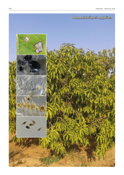
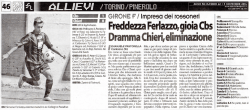
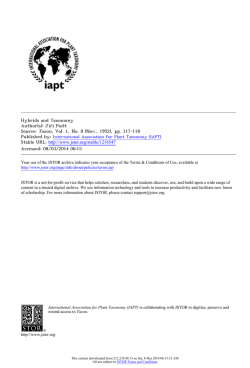
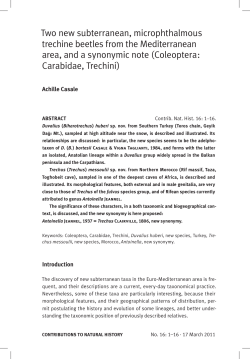
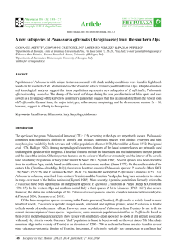
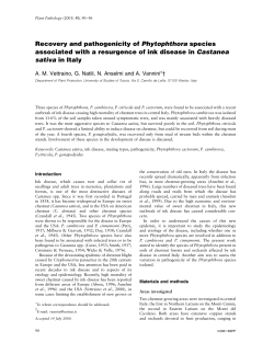
Mycosphere 7 (7): 962–977 (2016) www.mycosphere.org
ISSN 2077 7019
Article – special issue
Doi 10.5943/mycosphere/si/1b/8
Copyright © Guizhou Academy of Agricultural Sciences
Botryosphaeriaceae species associated with lentisk dieback in Italy and
description of Diplodia insularis sp. nov.
Linaldeddu BT1, Maddau L1, Franceschini A1, Alves A2 and Phillips AJL3
1
Dipartimento di Agraria, Sezione di Patologia vegetale ed Entomologia, Università degli Studi di Sassari, Viale Italia
39, 07100 Sassari, Italy
2
Departamento de Biologia, CESAM, Universidade de Aveiro, 3810-193 Aveiro, Portugal
3
Biosystems and Integrative Sciences Institute, Faculty of Science, University of Lisbon, Campo Grande, 1749-016
Lisbon, Portugal
Linaldeddu BT, Maddau L, Franceschini A, Alves A, Phillips AJL 2016 – Botryosphaeriaceae
species associated with lentisk dieback in Italy and description of Diplodia insularis sp. nov.
Mycosphere 7(7), 962–977, Doi 10.5943/mycosphere/si/1b/8
Abstract
Lentisk (Pistacia lentiscus L.) is an evergreen shrub that is widespread throughout the
Mediterranean region. Since spring 2012, a severe and unusual disease of unknown aetiology has
been observed on lentisk in six islands of the La Maddalena archipelago (Italy). The affected plants
showed leaf chlorosis, crown thinning, branch dieback and sunken cankers. When branches with
sunken cankers were cross-sectioned, internal wood symptoms included characteristic V-shaped
necrotic sectors. Frequently, the necrotic lesions girdled the branches resulting in death of the upper
crowns. Since there is no information about the aetiology of this disease and given the high
ecological importance of these natural ecosystems, from spring 2012 to summer 2014, 37 samples
of twigs and branches of lentisk showing sunken cankers were collected and processed.
Symptomatic woody samples yielded fungal isolates representing two distinct genera in the
Botryosphaeriaceae. On the basis of morphological features and DNA sequence data three distinct
species: Diplodia olivarum, Neofusicoccum cryptoaustrale and N. luteum were identified. In
addition, another Diplodia species morphologically distinct from all known species was isolated.
Phylogenetic analyses based on nucleotide sequences of ITS and tef1-α regions showed that this
new Diplodia species is most closely related to D. pseudoseriata and D. alatafructa. Pathogenicity
trials carried out in field conditions on asymptomatic branches of lentisk showed that all four
species are aggressive pathogens on this host and therefore directly involved in the severe dieback
that is currently threatening this typical shrub of the Mediterranean maquis.
Key words – Diplodia – Mediterranean maquis – Neofusicoccum – pathogenicity – phylogeny
Introduction
The La Maddalena archipelago (north-eastern Sardinia, Italy) is considered a hot spot of
biodiversity characterized by a large number of endemic species and multiple habitats typical of the
Mediterranean climate (Bocchieri 1992). The archipelago comprises 7 major islands and more than
50 small islets. The natural vegetation of the main islands is characterized by evergreen forest trees
such as Juniperus phoenicea, Olea europaea var. sylvestris, Quercus ilex, Salix atrocinerea and
Tamarix africana, mixed with species typical of the Mediterranean maquis such as Arbutus unedo,
Submitted 1 August 2016, Accepted 19 October 2016, Published online 29 October 2016
Corresponding author: Benedetto Linaldeddu – e-mail – [email protected]
962
Erica arborea, Myrtus communis, Phillyrea angustifolia, Pistacia lentiscus and Rhamnus alaternus
(Biondi & Bagella 2005).
Since 2008, a severe and widespread decline and mortality affecting several of these
Mediterranean species has been recognized on Caprera island. Results of etiological studies carried
out to establish the cause of these phenomena have revealed the primary role played by some
species belonging to the genera Diplodia and Neofusicoccum. In particular, Diplodia corticola and
Neofusicoccum parvum have been detected as the species most directly involved in the aetiology of
decline affecting Q. ilex trees (Linaldeddu et al. 2014), Diplodia africana, Neofusicoccum australe
and N. cryptoaustrale have been consistently associated with different disease symptoms on
branches of J. phoenicea (Linaldeddu et al. 2011, Andolfi et al. 2012), while Neofusicoccum luteum
has been recognized as the causal agent of a new disease of E. arborea (Linaldeddu et al. 2015b).
Furthermore, since spring 2012, a new disease of unknown aetiology has been observed on
P. lentiscus (lentisk) in six of the main islands belonging to the La Maddalena archipelago. Since
there is no information about the cause of this disease and given the high ecological importance of
these natural ecosystems, the aim of this study was to clarify the aetiology of this new disease and
determine the virulence of the major fungal species associated.
Materials & Methods
Field surveys and sampling procedure
Field surveys were carried out from spring 2012 to summer 2014 in six of the main islands
belonging to the La Maddalena archipelago (Budelli, Caprera, Mortorio, Santa Maria, Santo
Stefano and Spargi). A circular monitoring plot (MP, diameter 10 m) was established in each island
(two in Caprera), and the geographic coordinates of plots were recorded by a portable GPS (eTrex,
Garmin). The location of plots was based on the results of a preliminary survey about occurrence
and distribution of symptomatic plants. At each MP, the number of trees was detected and their
health status was assessed based on the occurrence of symptoms such as dieback, exudates, cankers
and epicormic shoots. From each symptomatic plant a branch showing an active canker was
collected. In total, 37 plants were sampled (Table 1).
Isolation and morphological characterization of canker-associated fungi
Symptomatic branch samples were taken to the laboratory and the outer bark surface tissue
was cut away with a scalpel. Longitudinal and transversal cuts from symptomatic branch samples
revealed internal symptoms. Fungi were isolated from chips of inner bark and xylem tissue (~5
mm2) removed from the margin of necrotic lesions with a sterile scalpel. All chips were cultured on
potato dextrose agar (PDA, Oxoid Ltd., Basingstoke, UK) in Petri dishes. After incubation at 25°C
for 5–7 days in the dark, fungal colonies were sub-cultured onto half-strength PDA supplemented
with autoclaved Q. ilex and P. lentiscus twigs and incubated at room temperature under natural
daylight until pycnidia developed.
For each fungal species, colony growth characteristics including surface and reverse colony
appearance were observed and recorded after 7 days of incubation at 25°C in the dark on PDA.
Identification of isolates to species level was based on colony and conidial morphology as
described by Phillips et al. (2013).
For novel species, cardinal temperatures for growth were determined on plates of PDA
incubated at 5, 10, 15, 20, 25, 30 and 35°C (±0.5°C) in the dark. Five replicate plates for each
isolate were made and colony diameters were measured after 4 days. For microscopy, the contents
of conidiomata were dissected and mounted in 100% lactic acid. Measurements of conidiogenous
cells and conidia were made with the Leica IM 500 measurement module from images recorded
with the ×100 objective on a Leica DFC 320 digital camera. Spore dimensions are presented as
mean values of 50 conidia with extreme values in parentheses. Dimensions of other structures are
given as means of at least 20 measurements.
963
Table 1 Characteristics of the seven monitoring plots and number of Pistacia lentiscus plants
sampled.
Island
Plot
Latitude
Longitude
Budelli
Caprera
1
2
3
4
5
6
7
41°16’48‖N
41°12’51‖N
41°12’36‖N
41°04’28‖N
41°17’47‖N
41°11’56‖N
41°14’04‖N
09°21’22‖E
09°27’01‖E
09°27’42‖E
09°36’10‖E
09°22’25‖E
09°24’38‖E
09°21’11‖E
Mortorio
Santa Maria
Santo Stefano
Spargi
Number of plants
Total
3
12
8
13
5
5
2
Sampled
3
10
8
6
3
5
2
Table 2 Diplodia isolates included in the phylogenetic analyses. The newly generated sequences
are indicated in italics and ex-type strains in bold face.
Isolate number
Species
Host
Country
CBS 120835
CBS 121104
CBS 132777
UCROK1429
CBS 124931
CBS 130408
CBS 130410
CBS 124254
CBS 124135
CBS 112549
BL7
BL10
BL11
MFLUCC 15-0648
CBS 168.87
BL102
CBS 140851
CBS 136010
CBS 136013
MFLUCC 15-0647
CBS 129750
CBS 129749
GUCC 0922-1
MFLUCC 14-1007
CBS 124462
CBS 112556
CBS 140350
BL99
BL183
BL132
BL133
BN-55
D. africana
D. africana
D. agrifolia
D. agrifolia
D. alatafructa
D. allocellula
D. allocellula
D. bulgarica
D. bulgarica
D. corticola
D. corticola
D. corticola
D. corticola
D. crataegicola
D. cupressi
D. cupressi
D. eriobotryicola
D. fraxini
D. fraxini
D. galiicola
D. guayanensis
D. guayanensis
D. huaxii
D. italica
D. intermedia
D. intermedia
D. insularis
D. insularis
D. insularis
D. insularis
D. insularis
D. insularis
Prunus persica
P. persica
Quercus agrifolia
Q. agrifolia
Pterocarpus angolensis
Acacia karroo
A. karroo
Malus sylvestris
M. sylvestris
Quercus suber
Quercus afares
Quercus ilex
Q. ilex
Crataegus sp.
Cupressus sempervirens
C. sempervirens
Eriobotrya japonica
Fraxinus angustifolia
F. angustifolia
Galium sp.
Acacia mangium
A. mangium
Platanus sp.
Crataegus sp
M. sylvestris
M. sylvestris
Pistacia lentiscus
P. lentiscus
P. lentiscus
F. angustifolia
F. angustifolia
E. japonica
South Africa
South Africa
USA
USA
South Africa
South Africa
South Africa
Bulgaria
Bulgaria
Portugal
Tunisia
Italy
Italy
Italy
Israel
Tunisia
Spain
Portugal
Italy
Italy
Venezuela
Venezuela
China
Italy
Portugal
Portugal
Italy
Italy
Italy
Italy
Italy
Spain
GenBank accession number
ITS
EF445343
EF445344
JN693507
JQ411412
FJ888460
JQ239397
JQ239399
GQ923853
GQ923852
AY259100
JX894190
JX894191
JX894192
KT290244
DQ458893
KF307722
KT240355
KF307700
KF307710
KT290245
JX545108
JX545106
KU848201
KU848202
GQ923858
AY259096
KX833072
KX833074
KX833076
KF307720
KF307721
KT240361
tef1-α
EF445382
EF445383
JQ517317
JQ512121
FJ888444
JQ239384
JQ239386
GQ923821
GQ923820
AY573227
JX894209
JX894210
JX894211
KT290248
DQ458878
KF318769
KT240193
KF318747
KF318757
KT290249
JX545126
JX545128
GQ923826
GQ923851
KX833073
KX833075
KX833077
KF318767
KF318768
KT240275
964
Isolate number
Species
Host
Country
CBS 124130
BL127
CBS 136014
CBS122553
CPC 22753
CPC 22754
CBS 121887
BL96
CBS 124906
GUCC G603-1
CBS 133852
CBS 133853
CBS 116470
CBS 116472
CBS 393.84
CBS 109725
CBS 109943
BL103
BL189
CBS 118110
CBS 109944
CBS 113423
CAP163
BL5
CBS 112555
CBS 119049
CAA502
BL130
CBS 124133
CBS 124131
CBS 418.64
D. malorum
D. malorum
D. mutila
D. mutila
D. neojuniperi
D. neojuniperi
D. olivarum
D. olivarum
D. pseudoseriata
D. pseudoplatani
D. quercivora
D. quercivora
D. rosulata
D. rosulata
D. sapinea
D. sapinea
D. sapinea
D. sapinea
D. sapinea
D. scrobiculata
D. scrobiculata
D. scrobiculata
D. scrobiculata
D. scrobiculata
D. seriata
D. seriata
D. seriata
D. seriata
D. subglobosa
D. subglobosa
D. tsugae
M. sylvestris
Populus alba
P. alba
Vitis vinifera
Juniperus chinensis
J. chinensis
Olea europaea
P. lentiscus
B. salicifolius
Platanus sp.
Quercus canariensis
Q. canariensis
Prunus africana
P. africana
Pinus nigra
Pinus patula
P. patula
Retama raetam
Corylus avellana
Pinus banksiana
Pinus greggii
P. greggii
O. europaea
Arbutus unedo
V. vinifera
V. vinifera
Fraxinus ornus
F. angustifolia
Lonicera nigra
F. ornus
Tsuga heterophylla
Portugal
Italy
Portugal
Portugal
Thailand
Thailand
Italy
Italy
Uruguay
China
Tunisia
Tunisia
Ethiopia
Ethiopia
Netherlands
Indonesia
Indonesia
Tunisia
Italy
USA
Mexico
Mexico
Italy
Italy
Portugal
Italy
Portugal
Italy
Spain
Italy
Canada
GenBank accession number
ITS
GQ923865
KF307717
KJ361837
AY259093
KM006431
KM006432
EU392302
KX833078
EU080927
KU848200
JX894205
JX894206
EU430265
EU430266
DQ458895
DQ458896
DQ458898
KX833080
KX833082
KF766160
DQ458899
DQ458900
EU392283
GU722102
AY259094
DQ458889
KJ361842
KF307723
GQ923856
GQ923855
DQ458888
tef1-α
GQ923833
KF318764
KJ361829
AY573219
KM006462
KM006463
EU392279
KX833079
EU863181
JX894229
JX894230
EU430267
EU430268
DQ458880
DQ458881
DQ458883
KX833081
KX833083
KF766399
DQ458884
DQ458885
EU392260
JX894231
AY573220
DQ458874
KJ361836
KF318770
GQ923824
GQ923823
DQ458873
Acronyms of culture collections: BL: B.T. Linaldeddu culture collection housed at Dipartimento di Agraria,
Università di Sassari, Italy; CAA: Collection of Artur Alves housed at Department of Biology, University of Aveiro,
Portugal; CAP, A.J.L. Phillips, Universidade Nova de Lisboa, Portugal; CBS: Centraalbureau voor Schimmelcultures,
The Netherlands; CPC: Collection of Pedro Crous housed at CBS; UCROK, Department of Plant Pathology and
Microbiology, University of California, Riverside; GUCC: Guizhou University Culture Collection (GUCC); MFLUCC:
Mae Fah Luang University Culture Collection.
Representative isolates of each species were stored on PDA slants under oil in the culture
collection of the Sez. di Patologia Vegetale ed Entomologia, Dipartimento di Agraria at the
University of Sassari. A representative culture of the new Diplodia species was also deposited at
the Centraalbureau voor Schimmelcultures (CBS), Utrecht, the Netherlands and nomenclatural data
in MycoBank (Crous et al. 2004) and Faces of Fungi (Jayasiri et al. 2015) databases. The holotype
was lodged with the herbarium of Instituto Nacional de Investigação Agrária e Veterinária I.P.,
Oeiras, Portugal (LISE).
DNA extraction, PCR amplification and sequencing
Genomic DNA was extracted from 5-day-old cultures grown on PDA at 25°C using
Instagene Matrix (BioRad Laboratories, Hercules, California, USA). The internal transcribed
965
spacer (ITS) region of the ribosomal DNA was amplified and sequenced using primers ITS1 and
ITS4 (White et al. 1990), while part of the translation elongation factor 1 alpha gene (tef1-α) was
amplified and sequenced with primers EF446f and EF1035r (Inderbitzin et al. 2010). PCR
amplification was carried out as described by Linaldeddu et al. (2013) and the products were
purified using the EUROGOLD gel extraction kit according to the manufacturer’s instructions
(EuroClone S.p.A., Pero, Italy). Both strands were sequenced by the BMR Genomics DNA
sequencing service. Sequences were edited with FinchTV v1.4.0 (Geospiza, Inc., Seattle,
Washington, USA) and compared with sequences deposited in GenBank using the BLAST
algorithm. New sequences were deposited in GenBank (Table 2 & Table 4) and alignments in
TreeBase (S19828).
Phylogenetic analyses
ITS and tef1-α sequences of the isolates obtained in this study were combined and aligned
with sequences retrieved from GenBank, representing all 28 Diplodia species known from culture.
Alignments were done with ClustalX v. 1.83 (Thompson et al. 1997) using the following
parameters: pairwise alignment parameters (gap opening = 10, gap extension = 0.1) and multiple
alignment parameters (gap opening = 10, gap extension = 0.2, transition weight = 0.5, delay
divergent sequences = 25%). Alignments were checked and manual adjustments made if necessary
using BioEdit v. 7.2.5 (Hall 1999). Maximum likelihood (ML) analyses were done using MEGA6
(Tamura et al. 2013) using the best fitting DNA evolution model determined by the program. ML
analyses were performed on a Neighbour-Joining starting tree automatically generated by the
software. Nearest-Neighbour-Interchange (NNI) was used as the heuristic method for tree inference
and 1000 bootstrap replicates were performed. The robustness of the trees was evaluated by 1000
bootstrap replications. Trees were visualized with TreeView v. 1.6.6 (Page 1996).
An initial ITS only phylogenetic analysis was carried out because for the species Diplodia
huaxii, D. italica and D. pseudoplatani there are no tef1-α sequences available. However, since ITS
alone cannot resolve all species in Diplodia an additional ITS plus tef1-α phylogenetic analysis was
performed with all Diplodia species excluding the previously mentioned D. huaxii, D. italica and
D. pseudoplatani.
Pathogenicity test
To verify the pathogenicity of each species investigated in this study, a field inoculation
trial was conducted in August 2015 on asymptomatic P. lentiscus plants located on Caprera island
(41°12’26‖N, 09°27’54‖E). During the experimental period, the daily mean air temperature was
20.3–31.5°C. Six branches (1–2.5 cm diameter) were inoculated with a representative isolate of
each fungal species, and six uninoculated branches were used as controls. The inoculated region of
the branch was surface-disinfected with 70% ethanol and a piece of outer and inner bark was
removed with a flamed scalpel and replaced with an agar-mycelium plug taken from the margin of
an actively growing colony on PDA. The inoculation site was covered with cotton wool soaked in
sterile water and wrapped in a piece of aluminium foil secured with masking tape. Controls were
inoculated with a sterile PDA plug applied as described above. After 1 month, the outer bark was
carefully removed with a scalpel and the length of necrotic lesion surrounding each inoculation site
was measured.
Re-isolation of inoculated species was attempted by transferring onto PDA 10 pieces of
inner bark and wood taken from around the margin of each lesion. Cultures were grown in daylight
and room temperature until fungal colonies developed.
Statistical analyses
Pathogenicity assay data were checked for normality, then subjected to analysis of variance
(ANOVA). Significant differences among mean values were determined using Fisher’s least
significant differences multiple range test (P = 0.05) after one-way ANOVA using XLSTAT
software (Addinsoft, Paris, France).
966
Fig. 1 – Disease symptoms observed in Pistacia lentiscus plants. a–c. Progressive dieback of twigs
and branches. d. epicormic shoots along branches. e. wood necrosis visible after bark removal at
level of a sunken cankers. f. cross section of a branch showing a characteristic wedge-shaped
necrotic sector in the wood.
Table 3 Number of samples examined for each plot and associated fungi isolated.
Island
Plot
Budelli
Caprera
1
2
3
4
5
6
7
Mortorio
Santa Maria
Santo Stefano
Spargi
Fungal species
D. insularis
10/10
5/8
3/3
-
D. olivarum
2/10
3/8
1/6
2/5
2/2
N. cryptoaustrale
2/3
2/6
2/5
-
N. luteum
1/3
1/6
-
Results
Field surveys
Out of 48 P. lentiscus plants investigated 37 displayed canopy disease symptoms, including
the progressive dieback of twigs and branches associated with the abnormal growth of epicormic
shoots (Fig. 1). Furthermore, symptomatic branches showed sunken cankers often associated with
reddish brown exudations. After removing the outer and inner bark from cankers, dark brown
necrotic lesions of variable size were visible on the xylem tissue. In cross-section, necrotic lesions
appeared with the characteristic wedge-shaped aspect typical of Botryosphaeriaceae infections
(Fig. 1).
Fungal isolation and identification
Isolation carried out from 37 cankered branch samples yielded a total of 36 fungal colonies
belonging to the Botryosphaeriaceae. On the basis of morphological features and DNA sequence
967
data (ITS and tef1-α), three distinct species were identified: Diplodia olivarum (10 isolates), N.
cryptoaustrale (6 isolates) and N. luteum (2 isolates) (Table 3). For each species BLAST searches
against GenBank showed 99–100% identity to reference sequences of representative strains
including those of ex-type isolates. In addition, eighteen Diplodia-like isolates on the basis of
morphological features and DNA sequence data could not be assigned to any formerly described
species. Although all processed samples showed the same symptoms the assemblages of pathogens
associated was very different among the islands (Table 3). Diplodia olivarum was the species more
widespread as it was isolated from plants in four of the six islands surveyed.
Phylogenetic analyses
The ITS ML analysis, as expected, did not resolve all species, especially within the clade
containing D. sapinea, D. scrobiculata and other closely related species (Fig. 2). Within this large
clade, the Diplodia-like isolates from lentisk (CBS 140350, BL99 and BL183) obtained in this
study formed a separated and well-supported (89% bootstrap) clade containing also isolates from
narrowed-leaved ash (BL132 and BL133) and loquat (BN-55) from previous studies. This clade
clustered with the clade containing D. alatafructa and D. pseudoseriata, which contained also the
ex-type strain of D. pseudoplatani GUCC G603-1 that could not be separated from D.
pseudoseriata CBS124906. Additionally, the ex-type strains of D. crataegicola MFLUCC 15-0648
and D. italica MFLUCC 14-1007 clustered together while the ex-type strains of D. huaxii
MFLUCC 15-0648 and D. galiicola MFLUCC 15-0647 clustered with isolates previously
identified as D. seriata including the ex-epitype strain CBS112555.
Combined ITS plus tef1-α ML analysis resolved all Diplodia species although not all
species clades received high (> 90%) bootstrap support (Fig. 3). The only exceptions were D.
scrobiculata/D. guayanensis and D. seriata/D. galiicola that did not form separate clades. The
Diplodia-like isolates from lentisk (CBS 140350, BL99 and BL183) formed a separate and highly
supported (98% bootstrap) clade clustering closely to the D. alatafructa and D. pseudoseriata clade
with 100% bootstrap support.
Taxonomy
Diplodia insularis Linaldeddu, A. Alves & A.J.L. Phillips, sp. nov.
Fig. 4
MycoBank: MB 818231
Facesoffungi number: FoF 02607
Etymology – the epithet refers to the insular environments, where the species was originally
found.
Sexual morph not seen. Conidiomata pycnidial, produced on Pistacia lentiscus twigs on ½
strength PDA within 2–4 weeks, solitary or aggregated, black, globose and uniloculate.
Conidiophores absent. Conidiogenous cells hyaline, smooth, cylindrical, sometimes slightly
swollen at the base, holoblastic, proliferating percurrently to form two or three distinct annellations
or proliferating internally giving rise to periclinal thickenings, average of 20 conidiogenous cells
10.1 × 3.9 µm. Conidia initially hyaline becoming pigmented even while still attached to the
conidiogenous cell, dark brown when mature, unicellular, rarely septate, ellipsoid to ovoid, wall
finely roughened on inner surface 18.2–(22.6)–25.9 × 9.1–(11.7)–14.4 µm, ± S.D. = 22.6 ± 1.4 ×
11.7 ± 0.6 µm, L/W 1.9 ± 0.2; n = 50).
Culture characteristics – Colonies on PDA attained 90 mm diameter before 7 days in the
dark at 25 °C, the mycelium was moderately aerial, surface white at first and later turned pale grey
to dark and dark in reverse.
Cardinal temperatures – Min. ≥ 5 °C, max. ≤ 35 °C, opt. 25 °C. All isolates failed to grow at
35 °C, but growth resumed when plates were moved to 25 °C.
Known distribution – Caprera, Santa Maria, Sardinia (Italy) and Castellón (Spain).
Habitat – On cankered branches of Eriobotrya japonica, Fraxinus angustifolia and Pistacia
lentiscus.
968
Material examined – Italy, Santa Maria island, isolated from a branch canker of Pistacia
lentiscus, 7 November 2013, Benedetto T. Linaldeddu, HOLOTYPE LISE 96309, a dried culture
sporulating on Pistacia lentiscus twigs, culture ex-holotype CBS 140350 = BL140. Other isolates
examined are listed in Table 2.
Notes – Diplodia insularis is phylogenetically closely related to D. alatafructa and D.
pseudoseriata. However, the latter two species can be distinguished from D. insularis by the size
and shape of conidia as expressed as the L/W ratio of about 1.9 for D. insularis and about 2.2 for
both D. alatafructa and D. pseudoseriata.
Diplodia scrobiculata J. de Wet, Slippers & M.J. Wingf., Mycol. Res. 107: 562. 2003. MycoBank
MB372427.
Synonym: Diplodia guayanensis F. Castro-Medina, J.R. Úrbez-Torres, S.R. Mohali & W.D.
Gubler, Plant Disease (http://dx.doi.org/10.1094/PDIS-05-16-0612-RE). MycoBank MB812480
Notes – According to Úrbez-Torres et al. (2016) D. guayanensis is closely related
phylogenetically to D. scrobiculata but can be distinguished by its larger conidia and the presence
of up to 4 septa in conidia while in D. scrobiculata conidia up to 3 septa may be present. Here we
show that both species are indistinguishable in phylogenetic analyses. Also, morphological
variability is common in these fungi making it unreliable for species differentiation (Phillips et al.
2013).
Diplodia seriata De Not., Micr. Ital. Dec. 4: 6. 1942. MycoBank MB180468.
Synonym: Diplodia galiicola Dissanayake, Camporesi & K.D. Hyde, Fungal Diversity 75: 54.
2015, Facesoffungi: FoF00884.
Notes – According to Ariyawansa et al. (2015) D. galiicola is phylogenetically most closely
related to D. seriata but can be distinguished by the shorter conidia. Here we show that both species
are indistinguishable in phylogenetic analyses. Also, as already mentioned above morphological
variability is common in these fungi making it unreliable for species differentiation (Phillips et al.
2013).
Pathogenicity test
All four Botryosphaeriaceae species proved to be pathogenic on P. lentiscus. At the end of
the experimental period, all branches inoculated with D. insularis, D. olivarum, N. cryptoaustrale
and N. luteum displayed dark brown bark lesions that spread up and down from the inoculation site
(Fig. 5). The average lesion length differed significantly between species (F3, 20 = 14.013, P <
0.001; Tab. 4), e.g. the lesions caused by N. cryptoaustrale (mean = 7 cm) were significantly larger
than those caused by D. insularis (mean = 3.8 cm), D. olivarum (mean = 2.6 cm) and N. luteum
(mean = 2.4 cm). In addition, the branches inoculated with D. insularis and N. cryptoaustrale
displayed wilting symptoms and a wedge-shaped necrotic sector in cross section congruent with
field observations.
Control branches inoculated with sterile PDA plugs remained symptomless. All four fungal
species were successfully re-isolated from symptomatic wood and inner bark tissues from all
inoculated plants, thus fulfilling Koch’s postulates (Tab. 4).
Discussion
This study represents the most comprehensive investigation of canker-causing agents of P.
lentiscus to date in the Mediterranean region. For the first time the direct involvement of
Botryosphaeriaceae species in the aetiology of canker and dieback symptoms of this important
component of Mediterranean maquis was ascertained in various natural ecosystems. In particular,
three fungal species belonging to two different genera were isolated and identified by means of
morphological characters and DNA sequence data. These species included D. olivarum, N.
cryptoaustrale and N. luteum. In addition, a novel species here described as Diplodia insularis was
isolated.
969
Fig.2 – Maximum Likelihood tree obtained from ITS sequence data and based on the Tamura 3parameter model. The rate variation model allowed for some sites to be evolutionarily invariable.
The tree is drawn to scale, with branch lengths measured in the number of substitutions per site.
Bootstrap support values in percentage (1000 replicates) are given at the nodes.
970
Fig. 3 – Maximum Likelihood tree obtained from combined ITS + tef1-α sequence data and based
on the Hasegawa-Kishino-Yano model. A discrete Gamma distribution was used to model
evolutionary rate differences among sites. The tree is drawn to scale, with branch lengths measured
in the number of substitutions per site. Bootstrap support values in percentage (1000 replicates) are
given at the nodes.
971
Fig. 4 – a. Colony morphology of Diplodia insularis after 7 days growth at 25 °C on PDA. b–d.
hyaline immature conidia developing on conidiogenous cells. e. brown conidia still attached to the
conidiogenous cell. f. mature conidia. g, h. two different focal planes to show the roughened inner
surface of the conidium wall. Bars = 10 µm.
All four species are reported here for the first time on P. lentiscus. Strains of the new
Diplodia species were first collected from declining Fraxinus angustifolia trees in Sardinia by
Alves et al. (2014), but they did not introduce a new species to accommodate these strains, although
the existence of important morphological and genetic differences between the fungal isolates from
F. angustifolia and those belonging to the complex Diplodia alatafructa/Diplodia pseudoseriata
were detected. These differences have been confirmed in the current study and the isolates obtained
from F. angustifolia and P. lentiscus in Italy as well as a strain recently isolated from E. japonica in
Spain by González-Domínguez et al. (2016), were considered representative of D. insularis.
Morphologically, it is interesting to remark that in some species belonging to this Diplodia
subclade, such as D. allocellula, D. eriobotryicola and D. insularis, conidia become pigmented
while still attached to the conidiogenous cell, similar to what is seen in species of Dothiorella and
Spencermartinsia. This has also been reported for D. seriata, D. pinea, D. intermedia and D.
scrobiculata (Phillips et al. 2013).
972
Fig. 5 – Symptoms observed on Pistacia lentiscus branches 30 days after inoculation with, a–c
Diplodia insularis. d–f. Diplodia olivarum. g–i. Neofusicoccum cryptoaustrale. j–l. Neofusicoccum
luteum. m, n. Control branches.
Table 4 Mean lesion length ± standard deviation caused by fungal species on branches of Pistacia
lentiscus.
Fungal species
Diplodia insularis
Diplodia olivarum
Neofusicoccum cryptoaustrale
Neofusicoccum luteum
Control
LSD critical value
Details of strains inoculated
Code
ITS
tef1-α
BL140
BL96
BL94
BL95
KX833072
KX833078
KX833084
KX833086
KX833073
KX833079
KX833085
KX833087
Mean
lesion
length (cm)
Re-isolation
frequency %
3.8±0.7b
2.6±0.6b
7.0±2.5a
2.4±0.7b
0.00
2.79
100
100
100
100
Phylogenetic analysis based on the ITS region raised some doubts about the status of the
recently described species D. huaxii, D. italica and D. pseudoplatani (Wijayawardene et al. 2016).
However, the ITS region is known to be insufficient to accurately discriminate species in Diplodia.
Thus, this issue can only be clarified when tef1-α sequences are available for these species. In ITS
plus tef1-α phylogenetic analysis the ex-type of D. galiicola clustered with the ex-epitype of D.
seriata and both species could not be separated. A comparison of the sequences from both species
showed that the differences reported between them are located at the start of the tef1-α sequence of
D. galiicola. This part of the tef1-α sequence, although located in a very conserved region, was
highly divergent compared to all other Diplodia species, which suggests sequencing errors and lack
of editing. For this reason, it was excluded from the alignments and coded as missing data.
973
Additionally, combined ITS and tef1-α phylogeny showed that the recently described D.
guayanensis and D. scrobiculata (Úrbez-Torres et al. 2016) cannot be differentiated. A careful
examination of sequence alignments showed that there are no differences in the ITS sequence and
no fixed polymorphisms in the tef1-α sequences of D. guayanensis and the ex-type isolate of D.
scrobiculata CBS118110 (= CMW189). Apparently these two species differ in a single nucleotide
in the sequence of the β-tubulin gene. The differences between our results and those reported by
Úrbez-Torres et al. (2016) arise from the fact that those authors used in their phylogenetic analyses
older sequences for D. scrobiculata (ITS: AY253292 and tef1-α: AY624253) while we used more
recent ones (ITS: KF766160 and EF: KF766399). Comparing the sequences it is evident that the
older ones are shorter and contain sequencing errors. Thus, for the reasons explained above, D.
guayanensis is considered as a synonym of D. scrobiculata and D. galiicola a synonym of D.
seriata.
The pathogenicity of all Botryosphaeriaceae species obtained in this study was confirmed
through an inoculation experiment in the field. All isolates tested were able to cause necrotic
lesions and the isolate of N. cryptoaustrale proved to be highly aggressive. Neofusicoccum
cryptoaustrale has previously been reported as an aggressive pathogen on other woody hosts in
Sardinia such as Vitis vinifera and Juniperus phoenicea (Linaldeddu et al. 2010, Andolfi et al.
2012). The high degree of aggressiveness observed in this study for D. insularis is in agreement
with the results obtained by González-Domínguez et al. (2016) on E. japonica in Spain.
Diplodia olivarum was first reported from rotting olive drupes and cankered branches of
Ceratonia siliqua in Italy (Lazzizera et al. 2008, Granata et al. 2011). Subsequently it was reported
as associated with declining Prunus dulcis trees in Spain (Gramaje et al. 2012) and cankered
branches of Quercus coccifera in Tunisia (Alves et al. 2014).
Neofusicoccum luteum is emerging as a common and cosmopolitan species on diverse host
plants, and it is now recognized as an aggressive pathogen of Crataegus mexicana, Eucalyptus
camaldulensis, Olea europaea, Persea americana, Quercus robur, Rhododendron spp., Syzygium
cordatum and V. vinifera (Pavlic et al. 2007, McDonald et al. 2009, Sergeeva et al. 2009, Pintos
Varela et al. 2011, Amponsah et al. 2012, Adesemoye et al. 2013, Barradas et al. 2013, Deidda et
al. 2016). Furthermore, N. luteum has recently been reported as a pathogen of E. arborea on
Caprera island (Linaldeddu et al. 2015b). Erica arborea represents a ―sporulation host‖ for N.
luteum, large numbers of conidiomata and ascomata develop on infected shoots throughout the
year, which serve as an important inoculum source (Linaldeddu unpublished data).
Members of Botryosphaeriaceae family represent a growing threat to agricultural crops,
urban and natural forest ecosystems in Sardinia (Linaldeddu et al. 2014, 2015a, 2016). In particular,
over the last few years there has been an exponential increase in the occurrence of diseases caused
by species of this family in natural areas on the east coast of Sardinia, in particular on the islands of
the La Maddalena archipelago, where in a limited geographic area of about 5,134 hectares, the
involvement of 16 different species in the aetiology of new or unusual diseases of species of
Mediterranean maquis have been detected (Linaldeddu unpublished data). This finding raises
questions about the origin, introduction and pathway of these fungi as well as underlining the need
to develop suitable actions to limit their further spread.
Acknowledgements
This research was supported by grants from the National Park of the La Maddalena
archipelago. The authors would like to thank Paola Brundu who provided help and logistic support
and Antonio Deidda for his invaluable assistance during field sampling and morphological analysis.
Artur Alves acknowledges financing from European Funds through COMPETE and National
Funds through the Portuguese Foundation for Science and Technology (FCT) to research unit
CESAM (UID/AMB/50017/2013 – POCI-01- 0145-FEDER-007638), himself (FCT Investigator
Programme – IF/00835/2013) and support by the Autonomous Region of Sardinia, Visiting
Professor Programme at the University of Sassari, Italy.
974
References
Adesemoye AO, Mayorquin JS, Eskalen A. 2013 – Neofusicoccum luteum as a pathogen on
Tejocote (Crataegus mexicana). Phytopathologia Mediterranea 52, 123−129.
Alves A, Linaldeddu BT, Deidda A, Scanu B, Phillips AJL. 2014 – The complex of Diplodia
species associated with Fraxinus and some other woody hosts in Italy and Portugal. Fungal
Diversity 67, 143–156.
Amponsah NT, Jones EE, Ridgway HJ, Jaspers MV. 2012 – Susceptibility of grapevine tissues to
Neofusicoccum luteum conidial infection. Plant Pathology 61, 719–729.
Andolfi A, Maddau L, Cimmino A, Linaldeddu BT, Franceschini A, Serra S, Basso S, Melck D,
Evidente A. 2012 – Cyclobotryoxide, a phytotoxic metabolite produced by the plurivorous
pathogen Neofusicoccum australe. Journal of Natural Products 75, 1785–1791.
Ariyawansa HA, Hyde KD, Jayasiri SC, Buyck B, Chethana KWT, Dai DQ, Dai YC, Daranagama
DA, Jayawardena RS, Lücking R, Ghobad-Nejhad M, Niskanen T, Thambugala KM, Voigt
K, Zhao RL, Li GJ, Doilom M, Boonmee S, Yang ZL, Cai Q, Cui YY, Bahkali AH, Chen J,
Cui BK, Chen JJ, Dayarathne MC, Dissanayake AJ, Ekanayaka AH, Hashimoto A,
Hongsanan S, Gareth Jones EB, Larsson E, Li WJ, Li QR, Liu JK, Luo ZL,
Maharachchikumbura SSN, Mapook A, McKenzie EHC, Norphanphoun C, Konta S, Pang
KL, Perera RH, Phookamsak R, Phukhamsakda C, Pinruan U, Randrianjohany E,
Singtripop C, Tanaka K, Tian CM, Tibpromma S, Abdel-Wahab MA, Wanasinghe DN,
Wijayawardene NN, Zhang JF, Zhang H, Abdel-Aziz FA, Wedin M, Westberg M,
Ammirati JF, Bulgakov TS, Lima DX, Callaghan TM, Callac P, Chang CH, Coca LF, DalForno M, Dollhofer V, Fliegerová K, Greiner K, Griffith GW, Ho HM, Hofstetter V,
Jeewon R, Kang JC, Wen TC, Kirk PM, Kytövuori I, Lawrey JD, Xing J, Li H, Liu ZY, Liu
XZ, Liimatainen K, Lumbsch HT, Matsumura M, Moncada B, Nuankaew S, Parnmen S, de
Azevedo Santiago ALCM, Sommai S, Song Y, de Souza CAF, de Souza-Motta CM, Su
HY, Suetrong S, Wang Y, Wei SF, Wen TC, Yuan HS, Zhou LW, Réblová M, Fournier J,
Camporesi E, Luangsa-ard JJ, Tasanathai K, Khonsanit A, Thanakitpipattana D,
Somrithipol S, Diederich P, Millanes AM, Common RS, Stadler M, Yan JY, Li XH, Lee
HW, Nguyen TTT, Lee HB, Battistin E, Marsico O, Vizzini A, Vila J, Ercole E, Eberhardt
U, Simonini G, Wen HA, Chen XH, Miettinen O, Spirin V, Hernawati. 2015 – Fungal
diversity notes 111–252—taxonomic and phylogenetic contributions to fungal taxa. Fungal
Diversity 75, 27–274.
Barradas C, Correia A, Alves A. 2013 – First report of Neofusicoccum australe and N. luteum
associated with canker and dieback of Quercus robur in Portugal. Plant Disease 97, 560.
Biondi E, Bagella S. 2005 – Vegetazione e paesaggio vegetale dell’arcipelago di La Maddalena
(Sardegna nord-orientale). Fitosociologia 42, 3–99.
Bocchieri E. 1992 – Flora of the small islands of the archipelago of Maddalena (north-eastern
Sardinia) and floristic contributions regarding some of the mains islands of the archipelago.
Flora Mediterranea 2, 33–64.
Crous PW, Gams W, Stalpers JA, Robert V, Stegehuis G. 2004 – MycoBank: an online initiative to
launch mycology into the 21st century. Studies in Mycology 50, 19–22.
Deidda A, Buffa F, Linaldeddu BT, Pinna C, Scanu B, Deiana V, Satta A, Franceschini A, Floris I.
2016 – Emerging pests and diseases threaten Eucalyptus camaldulensis plantations in
Sardinia, Italy. Iforest (doi: 10.3832/ifor1805-009).
González-Domínguez E, Alves A, León M, Armengola J. 2016 – Characterization of
Botryosphaeriaceae species associated with diseased loquat (Eriobotrya japonica) in Spain.
Plant Pathology (doi: 10.1111/ppa.12556).
Gramaje D, Agustí-Brisach C, Pérez-Sierra A, Moralejo E, Olmo D, Mostert L, Damm U,
Armengol J. 2012 – Fungal trunk pathogens associated with wood decay of almond trees on
Mallorca (Spain). Persoonia 28, 1–13.
975
Granata G, Faedda R, Sidoti A. 2011 – First report of canker disease caused by Diplodia olivarum
on carob tree in Italy. Plant Disease 95, 776.
Hall TA. 1999 – BioEdit: a user-friendly biological sequence alignment editor and analysis
program for Windows 95/98/NT. Nucleic Acids Symposium Series 41, 95–98.
Inderbitzin P, Bostock RM, Trouillas FP, Michailides TJ. 2010 – A six-locus phylogeny reveals
high species diversity in Botryosphaeriaceae from California almond. Mycologia 102,
1350–1368.
Jayasiri SC, Hyde KD, Ariyawansa HA, Bhat DJ, Buyck B, Cai L, Dai YC, Abd-Elsalam KA, Ertz
D, Gibertoni TB, Hidayat I, Jeewon R, Jones EBG, Karunarathna SC, Kirk P, Li WJ, Liu
JK, Luangsa-ard JJ, Maharachchikumbura SSN, Manamgoda DS, McKenzie EHC,
Moncalvo JM, Ghobad-Nejhad M, Nilsson H, Pang KL, Pereira OL, Raspé O, Rollins AW,
Romero AI, Salazar JAE, Stephenson S, Suetrong S, Taylor JE, Tsui CKM, Vizzini A,
Abdel-Wahab MA, Wen TC, Boonmee S, Dai DQ, Daranagama DA, Dissanayake AJ,
Ekanayaka AH, Hongsanan S, Jayawardena RS, Perera RH, Phookamsak R, De Silva NI,
Thambugala KM, Tian Q, Wanasinghe DN, Wijayawardene NN, Zhao RL, Kang JC,
Promputtha I. 2015 – The Faces of Fungi database - A unique perspective: Fungal names
linked with morphology, phylogeny and human impacts. Fungal Diversity 74, 3–18.
Lazzizera C, Frisullo S, Alves A, Lopes J, PhillipsAJL. 2008 – Phylogeny and morphology of
Diplodia species on olives in southern Italy and description of Diplodia olivarum. Fungal
Diversity 31, 63–71.
Linaldeddu BT, Alves A and Phillips AJL 2016 – Sardiniella urbana gen. et sp. nov., a new
member of the Botryosphaeriaceae isolated from declining Celtis australis trees in
Sardinian streetscapes. Mycosphere doi: 10.5943/mycosphere/si/1b/5
Linaldeddu BT, Deidda A, Scanu B, Franceschini A, Serra S, Berraf-Tebbal A, Zouaoui Boutiti M,
Ben Jamâa ML, Phillips AJL. 2015a – Diversity of Botryosphaeriaceae species associated
with grapevine and other woody hosts in Italy, Algeria and Tunisia, with descriptions of
Lasiodiplodia exigua and Lasiodiplodia mediterranea sp. nov. Fungal Diversity 71, 201–
214.
Linaldeddu BT, Franceschini A, Alves A, Phillips AJL. 2013 – Diplodia quercivora sp. nov.: a new
species of Diplodia found on declining Quercus canariensis trees in Tunisia. Mycologia
105, 1266–1274.
Linaldeddu BT, Scanu B, Maddau L, Franceschini A. 2011 – Diplodia africana causing dieback
disease on Juniperus phoenicea: a new host and first report in the northern hemisphere.
Phytopathologia Mediterranea 50, 473–477.
Linaldeddu BT, Scanu B, Maddau L, Franceschini A. 2014 – Diplodia corticola and Phytophthora
cinnamomi: the main pathogens involved in holm oak decline on Caprera island (Italy).
Forest Pathology 44, 191–200.
Linaldeddu BT, Scanu B, Schiaffino A, Serra S. 2010 – First report of Neofusicoccum australe
associated with grapevine cordon dieback in Italy. Phytopathologia Mediterranea 49, 417–
420.
Linaldeddu BT, Scanu B, Seddaiu S, Deidda A, Maddau L, Franceschini A. 2015b – A new disease
of Erica arborea in Italy caused by Neofusicoccum luteum. Phytopathologia Mediterranea
54, 124–127.
McDonald V, Lynch S, Eskalen A. 2009 – First report of Neofusicoccum australe, N. luteum, and
N. parvum associated with avocado branch canker in California. Plant Disease 93, 967.
Page RD. 1996 – TreeView: an application to display phylogenetic trees on personal computers.
Computer Applications in the Biosciences 12, 357–358.
Pavlic D, Slippers B, Coutinho TA, Wingfield MJ. 2007 – Botryosphaeriaceae occurring on native
Syzygium cordatum in South Africa and their potential threat to Eucalyptus. Plant Pathology
56, 624–636.
976
Phillips AJL, Alves A, Abdollahzadeh J, Slippers B, Wingfield MJ, Groenewald JZ, Crous PW
2013 – The Botryosphaeriaceae: genera and species known from culture. Studies in
Mycology 76, 51–167.
Pintos Varela C, Redondo Fernández V, Mansilla Vázquez JP, Aguín Casal O. 2011 – First report
of dieback on hybrid rhododendrons caused by Neofusicoccum luteum and N. parvum in
Spain. Plant Disease 95, 221.
Sergeeva V, Alves A, Phillips AJL. 2009 – Neofusicoccum luteum associated with leaf necrosis and
fruit rot of olives in New South Wales, Australia. Phytopathologia Mediterranea 48, 294–
298.
Tamura K, Stecher G, Peterson D, Filipski A, Kumar S. 2013 – MEGA6: molecular evolutionary
genetics analysis version 6.0. Molecular Biology and Evolution 30, 2725–2729.
Thompson JD, Gibson TJ, Plewniak F, Jeanmougin F, Higgins DG. 1997 – The CLUSTAL_X
windows interface: flexible strategies for multiple sequence alignment aided by quality
analysis tools. Nucleic Acids Research 25, 4876–4882.
Úrbez-Torres JR, Castro-Medina F, Mohali SR, Gubler WD. 2016 – Botryosphaeriaceae species
associated with cankers and dieback symptoms of Acacia mangium and Pinus caribaea var.
hondurensis in Venezuela. Plant Disease (in press) doi: 10.1094/PDIS-05-16-0612-RE
White TJ, Bruns T, Lee S, Taylor J. 1990 – Amplification and direct sequencing of fungal
ribosomal RNA genes for phylogenetics. In: PCR Protocols, a Guide to Methods and
Applications (eds. MA Innis, DH Gelfand, JJ Sninsky and J White) Academic Press. San
Diego, Ca, USA 315–322.
Wijayawardene NN, Hyde KD, Wanasinghe DN, Papizadeh M, Goonasekara ID, Camporesi E,
Jayarama Bhat D, McKenzie EHC, Phillips AJL, Diederich P, Tanaka K, Li WJ,
Tangthirasunun N, Phookamsak R, Dai DQ, Dissanayake AJ, Weerakoon G,
Maharachchikumbura SSN, Hashimoto A, Matsumura M, Bahkali AH, Wangshow Y. 2016
– Taxonomy and phylogeny of dematiaceous coelomycetes. Fungal Diversity 77, 1–316.
977
© Copyright 2026 Paperzz