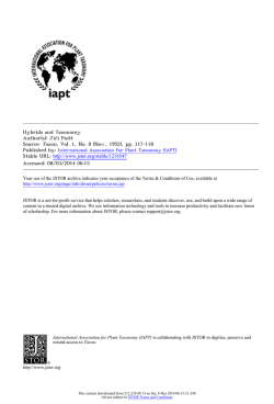
Plant Pathology (2001) 50, 90±96 Recovery and pathogenicity of Phytophthora species associated with a resurgence of ink disease in Castanea sativa in Italy A. M. Vettraino, G. Natili, N. Anselmi and A. Vannini*² Department of Plant Protection, University of Studies of Tuscia, Via S. Camillo de Lellis, 01100 Viterbo, Italy Three species of Phytophthora, P. cambivora, P. citricola and P. cactorum, were found to be associated with a recent outbreak of ink disease causing high mortality of chestnut trees in central Italy. Phytophthora cambivora was isolated from 11´6% of the soil samples taken around symptomatic trees, and was mainly associated with heavily diseased trees. It was the most aggressive species to Castanea sativa, but survived poorly in the soil. Phytophthora citricola and P. cactorum showed a limited ability to induce disease on chestnut, but could be recovered from soil during most of the year. A fourth species, P. gonapodyides, was recovered only from mud of stream beds within the chestnut stands. Involvement of these species in the development of disease is discussed. Keywords: Castanea sativa, ink disease, mating types, pathogenicity, Phytophthora cactorum, P. cambivora, P.citricola, P. gonapodyides Introduction Ink disease, which causes root and collar rot of seedlings and adult trees in nurseries, plantations and forests, is one of the most destructive diseases of Castanea spp. Since it was first recorded in Portugal in 1838, it has become widespread in Europe on sweet chestnut (Castanea sativa), and in the USA on American chestnut (C. dentata) and other chestnut species (Crandall et al., 1945). Two species of Phytophthora were shown to be responsible for the disease in Europe and the USA: P. cambivora and P. cinnamomi (Petri, 1917; Milburn & Gravatt, 1932; Day, 1938; Crandall et al., 1945). Other Phytophthora species have also been found to be associated with infected trees or to be pathogenic to Castanea spp. (Curzi, 1933; Smith, 1937; Cristinzio & Verneau, 1954; Wicks & Volle, 1976). Because of the devastating epidemic of chestnut blight caused by Cryphonectria parasitica in the 20th century in Europe and the USA, less attention has been paid in recent decades to ink disease and to aspects of its etiology and epidemiology. Recently, high mortality of sweet chestnut caused by ink disease has been reported from different areas of Europe (Abreu, 1996; Anselmi et al., 1996) and the USA (Vettraino et al., 2000), in some cases limiting the establishment of new groves or * To whom correspondence should be addressed. ² E-mail: [email protected] Accepted 14 July 2000. 90 the conservation of old ones. In Italy the disease has recently spread dramatically, apparently from infection foci, in most chestnut-growing areas (Anselmi et al., 1996). Large numbers of diseased trees have been found along roads and trails from which the disease has probably spread, carried by man and animals (Anselmi et al., 1999). Due to the high economic and environmental value of sweet chestnut in Italy, this new outbreak of ink disease has caused considerable concern. In order to understand the causes of this new epidemic, it is important to study the epidemiology and etiology of the disease, including whether one or more Phytophthora species are involved in addition to P. cambivora and P. cinnamomi. The present work aimed to identify the species of Phytophthora present in soils of chestnut forests and orchards affected by ink disease in central Italy. Another aim was to assess the variation in pathogenicity of the Phytophthora species isolated. Materials and methods Areas investigated Two chestnut-growing areas were investigated in central Italy: the first in Northern Latium on the Monti Cimini; the second in Eastern Latium on the Monti del Cicolano. Both areas have extensive coppice stands and orchards devoted to fruit production, ranging in Q2001 BSPP Ink disease in sweet chestnut in Italy 91 Table 1 Isolates of Phytophthora spp. used as standards for identification Species Strain Host Mating type Provided by P. gonapodyides P. gonapodyides AB4 P501 Quercus spp. Q. ilex Unknown A1 P. erythroseptica IMI 17028 Solanum tuberosum Homothallic P. cryptogea 13´4.9 Prunus avium Unknown P. cryptogea IMI 180615 Unknown Unknown P. drechsleri ± P. avium Unknown P. megasperma 27´1.5 Juglans nigra Homothallic P. cactorum PH31 Betula alba Homothallic P. cactorum PH6 Unknown Homothallic P. citricola P1013 Quercus spp. Homothallic P. P. P. P. P. 21/25-KV 20/95±21 21/95-KII 69094 8/88/92 Chamaecyparis lawsoniana Q. rubra C. lawsoniana C. lawsoniana C. lawsoniana Homothallic A2 A1 A1 A2 C. Delatour, INRA, France C.M. Brasier, Forest Research, Forestry Commission, UK G. Magnano di San Lio, University of Reggio Calabria, Italy G.C. Adams, Michigan State University, USA G. Magnano di San Lio, University of Reggio Calabria, Italy G.C. Adams, Michigan State University, USA G.C. Adams, Michigan State University, USA G. Magnano di San Lio, University of Reggio Calabria, Italy G. Tamietti, University of Turin, Italy C.M. Brasier, Forest Research, Forestry Commission, UK S. Werres, BBA, Germany S. Werres, BBA, Germany S. Werres, BBA, Germany S. Werres, BBA, Germany S. Werres, BBA, Germany citricola cambivora cambivora cinnamomi cinnamomi altitude from 400 to 1000 m a.s.l. Although ink disease was not uniformly present in these areas, an increasing number of infection foci were recorded each year, sometimes with a high incidence of the disease. Surveys were carried out between 1998 and 2000 by collecting soil samples from four different forest stands (three in the Monti Cimini and one in the Monti del Cicolano) where the disease had been recorded previously (Anselmi et al., 1996). Occasionally diseased trees recorded from additional stands were sampled. Samples were not taken from all stands at each sampling time. A total of 155 soil samples were collected: 119 soil samples were analysed from the chestnut area of the Monti Cimini; of these 93 were collected under symptomatic trees (both in coppice and orchards), and 26 under healthy-looking trees. Thirtysix soil samples were analysed from the chestnutgrowing area of the Monti del Cicolano: of these 19 were collected under symptomatic trees, seven under healthy-looking trees, and 10 from two stream beds crossing chestnut forest sites affected by the disease. Isolation of Phytophthora Each soil sample contained fine or coarse chestnut roots, and resulted from a mix of four monoliths of soil (25 £ 25 £ 25 cm) collected at four compass points around a tree at a distance of about 50 cm from the collar. After collection, soil samples were moistened with sterile distilled water and incubated at 208C for 3 days. About 200 mL soil was then flooded with Q 2001 BSPP Plant Pathology (2001) 50, 90±96 500 mL distilled water in plastic containers. Five freshly picked leaves of Rhododendron spp. (Themann & Werres, 1998) were placed directly on the water surface and incubated at 208C for 1 week until spots developed on the leaves or the leaves become discoloured. The leaves were then blotted on filter paper, cut into small pieces (0´5 £ 0´5 cm) and placed on PARBhy selective medium [10 mg pimaricin, 250 mg ampicillin (sodium salt), 10 mg rifampicin, 50 mg hymexazol, 15 mg benomyl, 15 g malt extract, 20 g agar in 1000 mL H2O] (Robin, 1991). Phytophthora isolates were maintained on carrot agar (CA) (Brasier, 1969) at 208C in darkness and subcultured at 4 week intervals. Identification of isolates Isolates were identified by comparing colony growth patterns and morphological features of sporangia, oogonia, antheridia, chlamydospores and hyphal swellings with known isolates (Table 1) and with species descriptions reported in literature (e.g. Stamps et al., 1990; Erwin & Ribeiro, 1996). Colony morphology was described on 10-day-old cultures grown on CA in 90 mm Petri dishes at 208C in darkness. Sporangia were produced by placing a disc of mycelium from a 7-day-old culture grown on CA in soil extract prepared according to Chee & Newhook (1965). Morphology was assessed by light microscopy, and the length and breadth of 100 sporangia were measured for each isolate. Identification of the isolates was confirmed by 92 A. M. Vettraino et al. comparing the RFLP patterns of their ribosomal DNA (rDNA) with those of known isolates listed in Table 1. Phytophthora spp. DNA suitable for PCR amplification was purified according to the methodology of Cenis (1992). ITS1 and ITS4 universal primers (White et al., 1990) were used to amplify Phytophthora rDNA. PCR amplification was performed using Ready-to-Go PCR Beads following the manufacturer's instructions (Amersham Pharmacia Biothec Inc., Uppsala, Sweden). Template DNA (10 ng), 1 m m of each primer and bi-distilled H2O were added to each reaction tube to make a final volume of 25 m L. The mixture was subjected to thermal cycling in a Techne Progene cycler (Techne, Cambridge, UK). An initial denaturation step at 958C for 2´5 min was followed by 35 cycles of annealing for 30 s at 558C, extension for 30 s at 728C, and denaturation for 30 s at 958C. Final extension was performed for 5 min at 728C. Amplification products were purified using a QIAquick PCR Purification kit (Qiagen Corp., Venlo, The Netherlands) following the manufacturer's directions. A 10 m L sample of the amplification product was digested with the restriction enzymes AluI, MspI and RsaI according to the manufacturer's instructions (Takara Shuzo Co. Ltd, Shiga, Japan). Digestion products were separated on a 1´5% Separide Gel Matrix (Life Technologies, Grand Island, NY, USA), stained with ethidium bromide, and visualized under UV light. Sexual behaviour For heterothallic species, the mating type of each isolate was determined directly on microscope slides by placing a CA plug of the `unknown' isolate in contact with a CA plug of an A1 or A2 tester strain. Slides were incubated at 208C in the dark at high relative humidity and scored with the aid of a light microscope for the presence of oospores after 10±15 days. Homothallic species produced oospores in single culture on CA. Morphology was studied using light microscopy, and the length and breadth of 100 oogonia, antheridia and oospores were measured for each colony after pairing. Pathogenicity tests Five field isolates of P. cambivora, P. citricola and P. cactorum and four isolates of P. gonapodyides were used for pathogenicity tests. Tests were performed on excised sprouts using a method described by Browne & Mircetich (1993, 1996), with some modifications, and also by soil infestation. One-year-old dormant sprouts (105), 2 cm in diameter and 1 m in length, were collected from a large stump, placed in test tubes with sterile H2O, and maintained at 208C with a 12 h photoperiod. Shoots were inoculated as the buds started to open. A cork borer was used to remove a 3 mm bark disc from the excised shoot. The bark disc was replaced by a 3 mm plug of a 10-day-old culture grown on PDA. Five replicates of each isolate were used. Controls (10) were represented by shoots inoculated with a PDA plug. After inoculation, shoots were incubated in a ventilated chamber for 1 week at 208C and 100% relative humidity. After incubation, the length of bark necrosis was measured on each shoot. For the soil infestation tests, C. sativa seedlings were obtained from surface-sterilized seeds planted in pots containing steam-pasteurized potting mix (50% peat, 25% sand, 25% ground pumice). Seedlings were grown in a greenhouse until their lower stems were well lignified and their average height was 20±30 cm. Subsequently they were transplanted into 1 L plastic containers filled with the same potting mixture that was either unamended or infested with Phytophthora spp. Phytophthora inoculum was prepared by growing isolates for 4±6 weeks at 208C on sterilized millet seeds thoroughly moistened with V8 broth (200 mL V8 juice, 3 g CaCO3, 800 mL distilled H2O). The inoculum was repeatedly rinsed with sterile water to remove unassimilated nutrients, then added to the potting mixture at the rate of 25 mL inoculum per 1000 mL potting mixture. Mixtures of inoculum and potting mixture were then flooded for 24 h to induce sporulation of Phytophthora isolates There were 50 replicate pots per isolate. Plants were watered to field capacity every other day. Two months after inoculation, 50 g samples of soil from each of six randomly chosen pots for each species of Phytophthora were flooded with water and baited separately with 15 Rhododendron leaf discs 1 cm in diameter. The health status of the plants was then scored on a scale of 0±4, where 0 healthy plant; 1 leaves turning pale green and necrosis on up to 10% of leaf surface; 2 yellowing of leaves on up to 50% of leaf surface and necrosis of up to 25% of leaf surface; 3 yellowing of leaves on up to 100% of leaf surface and necrosis of up to 50% of leaf surface; 4 dead plant. A disease index (DI) was calculated using the following formula: DI S(g £ n)/N, where g damage score, n number of samples belonging to each class, and N 50, the total number of inoculated plants. Statistical data analysis Statistical analysis of data was performed with the software instat 3 (GraphPad, San Diego, CA, USA). Results Recovery of Phytophthora and species identification Four Phytophthora species, P. cambivora, P. citricola, P. cactorum and P. gonapodyides, were identified on the basis of morphology. This was confirmed by the RFLP profiles characteristic of the rDNA of the four species (Fig. 1). Four different patterns were generated among the field isolates, corresponding to those of the standard isolates PH31 and PH6 (P. cactorum), P1013 and 21/25KV (P. citricola), 20/95-21 and 21/95-KII (P. cambivora), and AB4 and P501 (P. gonapodyides). Q 2001 BSPP Plant Pathology (2001) 50, 90±96 Ink disease in sweet chestnut in Italy 93 Figure 1 PCR amplification products of the ITS1/5´8S/ITS2 region of the RNA gene repeat of four species of Phytophthora isolated under chestnut trees (primers ITS1 and ITS4) digested with the DNA restriction enzymes RsaI (a), MspI (b) and AluI (c). Lane 1, 100 bp ladder; lane 2, P. gonapodyides; lane 3, P. cambivora; lane 4, P. citricola; lane 5, P. cactorum. Molecular weights (bp) of the 100 bp ladder are indicated on the left. Percentage recovery of Phytophthora spp. in the Monti Cimini and Monti del Cicolano chestnut forests is shown in Table 2. A total of 262 colonies of Phytophthora spp. were obtained by the Rhododendron leaf baiting technique. Up to three species were sometimes recovered from the same soil sample. The effect of sampling date on the isolation of four species of Phytophthora from diseased stands of C. sativa is shown in Table 3. Among the heterothallic species, P. gonapodyides was characteristically sterile. All the colonies (98) of P. cambivora isolated from the 13 soil samples belonged to the A2 sexual compatibility type. Pathogenicity tests The mean length of necrosis produced by each species is shown in Fig. 2. All four species caused necrosis, but P. cambivora was by far the most aggressive species. Similarly, in the soil infestation test, by far the highest DI was obtained with P. cambivora (Fig. 3). Phytophthora cambivora also caused the highest mortality rate among inoculated seedlings: 46% died, compared with 22% for P. citricola, 14% for P. cactorum, and 14% for P. gonapodyides. All four species of Phytophthora were re-isolated from the soils in the pots 2 months after adding inoculum. Statistical data analysis The contingency table analysis for presence/absence of Phytophthora spp. and health status of trees in the sampled stands revealed a 1´292-fold (P 0´015) higher risk that a chestnut with Phytophthora in its rooting zone would show above-ground symptoms. This value increased to 1´333 (P 0´040) for P. cambivora. Discussion In chestnut stands in central Italy affected by ink disease, Phytophthora spp. were recovered from soils around both diseased and healthy-looking trees. However, the isolation frequency was much higher around symptomatic trees than around healthy-looking ones (26´8 versus 6´0%). No ambiguous results were obtained when RFLP patterns were compared for the four species identified on the basis of morphological characters. As also demonstrated by Cooke & Duncan (1997), RFLPs of the rDNA region appear to be a relatively reliable method for species identification, especially if applied to genera such as Phytophthora, whose identification based on morphological characters is time-consuming and requires considerable experience. Table 2 Frequency of isolation of four Phytophthora species from soil from four diseased stands of Castanea sativa Source of the sample Total number of attempted isolations Number of successful isolations (all species) P. cactorum P. citricola P. cambivora P. gonapodyides a Figures in brackets are percentage. Q 2001 BSPP Plant Pathology (2001) 50, 90±96 Symptomatic trees Healthy-looking trees Stream bed Total 112 30 7 13 13 0 33 2 (6) 1 (3´0) 1 (3´0) 0 0 10 7 2 4 0 4 155 39 10 18 13 4 (26´8)a (6´3) (11´6) (11´6) (70) (20) (40) (40) (25´2) (6´4) (11´6) (8´4) (2´6) 94 A. M. Vettraino et al. Table 3 Effect of sampling date on isolation of four species of Phytophthora from four diseased stands of Castanea sativa Date of sampling Species Month Day±year P. citricola P. cactorum P. cambivora P. gonapodyides February 4±00 24±00 15±99 19±99 29±99 03±98 24±99 31±99 12±99 30±99 05±99 04±99 26±98 02±99 26±99 12±98 25±99 4±99 ± ± 1 1 1 1 1 1 1 1 1 1 ± ± 1 1 ± ± ± ± 1 1 1 ± 1 1 ± 1 ± ± ± ± 1 ± ± ± ± ± ± ± 1 1 ± ± ± ± ± ± 1 1 1 ± ± ± ± ± ± ± ± ± ± ± 1 ± 1 ± ± ± 1 ± ± ± March April May June July August September October November December Three species, P. cambivora, P. citricola and P. cactorum, were present in the rooting zone of chestnut trees affected by the new outbreak of the disease. Phytophthora cambivora was typically associated with heavily diseased trees, and was never recovered from soils around slightly or nonsymptomatic hosts. Furthermore, it was the only species isolated from V-shaped necroses at the base of the stems (A. V., unpublished data). These necroses are typical of the latest phase of ink disease development (Petri, 1917). However, an isolation rate of 11´6% of P. cambivora from soil around diseased trees and the relative risk to the host of 1´333 are low. One possible explanation could be that the Rhododendron leaf-baiting technique is not efficient enough to detect the presence of the pathogen in the soil. However, P. cambivora appears to survive poorly in the soil during most of the year, and in this study was recovered only in late April±May and late September± October during periods of rain and mild temperatures. The difficulty in isolating P. cambivora from soil could be associated with a failure to produce resting structures. Some oospores may be formed as result of selfing stimulated by ubiquitous soil fungi (e.g. Trichoderma; Brasier, 1971), but in general oospore formation is likely to be poor due to the apparent presence of only a single mating type (A2) in the populations analysed. Furthermore, chlamydospores are not known to be formed by this species. The above considerations would suggest that P. cambivora has the ability to spread from infected roots and establish new infections for only a limited period of the year, when climatic conditions are favourable to the formation, release and survival of zoospores. Phytophthora citricola, previously reported on chestnut by Biocca et al. (1993), was recovered between March and November, demonstrating its ability to survive under relatively adverse conditions, probably as oospores in the soil. This species is able to cause disease when inoculated on chestnut, although to a lesser extent than P. cambivora. Phytophthora cactorum has not Figure 2 Mean length of necrosis (mm) produced by the four species of Phytophthora on excised chestnut sprouts 1 week after inoculation. Vertical bars, SD. Figure 3 Index of disease (DI) (see Materials and methods for details) caused on 1-year-old chestnut seedlings growing in soil infested with four Phytophthora species. Vertical bars, SD. Q 2001 BSPP Plant Pathology (2001) 50, 90±96 Ink disease in sweet chestnut in Italy previously been reported to occur in the rooting zone of chestnut trees in areas affected by ink disease, although it is known to be pathogenic to Castanea spp. following artificial inoculation (Curzi, 1933; Smith, 1937; Cristinzio & Verneau, 1954). Whether P. citricola and P. cactorum are involved in disease development in the field remains a matter for speculation. Both species are serious pathogens of many other woody hosts (Erwin & Ribeiro, 1996), and both were able to cause necrosis in the pathogenicity tests reported here. The absence of these species in the typical V-shaped necroses (A.V., unpublished data) and the relatively low disease index (DI) suggest that P. cactorum and P. citricola are not strongly involved in ink disease of sweet chestnut. However, further isolations must be made to clarify their role in ink disease as fine root pathogens. Phytophthora gonapodyides was isolated only from stream beds. It is typically associated with aquatic habitats in Britain and North America (Brasier et al., 1993) and has also been recovered, together with P. citricola, by Jung et al. (1996) and Hansen & Delatour (1999) from streams running through oak stands. An involvement of this species in ink disease seems unlikely, since it was not recovered from soil around symptomatic trees. The present work has identified four species of Phytophthora present in chestnut stands affected by ink disease in central Italy. Phytophthora cinnamomi was not isolated in this study, but the results clearly show that, in addition to P. cambivora, at least two other species are associated with diseased trees and could contribute to disease development. As in other woody hosts (e.g. walnut, apple), root rot by Phytophthora on chestnut could be caused by a number of different species (Cristinzio & Verneau, 1954; Browne & Mircetich, 1993). Many Phytophthora species from other hosts have been shown to be pathogenic on Castanea spp. These include P. syringae (Day, 1938), P. citrophthora (Curzi, 1933; Smith, 1937; Cristinzio & Verneau, 1954); P. megasperma (Waterhouse, 1963); P. cryptogea (Wicks & Volle, 1976); and P. katsurae (Uchida, 1967). Further work is needed to identify the Phytophthora species present in chestnut stands in other areas of Italy and Europe, and to determine their pathogenicity and relevance in disease development. Acknowledgements We are very grateful to C.M. Brasier, C. Delatour, G. Magnano di San Lio, G. Tamietti and S. Werres for providing the strains of Phytophthora spp. used as standards in the present study. A special thanks to C.M. Brasier for the critical reading of the manuscript and for making many helpful suggestions that greatly improved this paper. This study was carried out with financial support from the project EC ± Latium Region DOCUP Objective 5b 1994±99 I.1.3.07.002 `Development of Q 2001 BSPP Plant Pathology (2001) 50, 90±96 95 control strategies against ``ink disease'' caused by Phytophthora spp. in Italy'. References Abreu CG, 1996. DoencËa da tinta: causas e consequeÃncias do declõÂneo do castanhal. Estudos Transmontanos 6, 269±89. Anselmi N, Giordano E, Vannini A, Troiani L, Napoli G, Crivelli L, 1996. Il mal dell'inchiostro del castagno in Italia: una vecchia malattia ritorna attuale. Linea Ecologica 28, 39±44. Anselmi N, Vettraino AM, Franco S, Chiarot E, Vannini A, 1999. Recrudescenze del Mal dell'Inchiostro del castagno in Italia: nuove aquisizioni e suggerimenti di lotta. Linea Ecologica 28, 53±8. Biocca M, Motta E, Cacciola SO, Magnano di San Lio G, 1993. Identification of Phytophthora spp. associated with ink disease of chestnut in Central Italy. Proceedings of the International Congress on Chestnut, Spoleto, Italy, 527± 32. Brasier CM, 1969. The effect of light and temperature on reproduction in vitro in two tropical species of Phytophthora. Transactions of the British Mycological Society 52, 105±13. Brasier CM, 1971. Induction of sexual reproduction in single A2 isolates of Phytophthora species by Trichoderma viride. Nature New Biology 231, 283. Brasier CM, Hamm PB, Hansen EM, 1993. Cultural characters, protein patterns and unusual mating behaviour of Phytophthora gonapodyides isolates from Britain and North America. Mycological Research 97, 1287±98. Browne GT, Mircetich SM, 1993. Relative resistance of thirteen apple rootstocks to three species of Phytophthora. Phytopathology 83, 744±9. Browne GT, Mircetich SM, 1996. Effects of month of inoculation on severity of disease caused by Phytophthora spp. in apple root crowns and excised shoots. Phytopathology 86, 290±4. Cenis JL, 1992. Rapid extraction of fungal DNA for PCR amplification. Nucleic Acids Research 20, 2380. Chee KH, Newhook FJ, 1965. Improved methods for use in studies on Phytophthora cinnamomi Rands and other Phytophthora species. New Zealand Journal of Agriculture Research 8, 88±95. Cooke DEL, Duncan JM, 1997. Phylogenetic analysis of Phytophthora species based on ITS1 and ITS2 sequences of the ribosomal RNA gene repeat. Mycological Research 101, 667±77. Crandall BS, Gravatt GF, Ryan MM, 1945. Root disease of Castanea species and some coniferous and broadleaf nursery stocks, caused by Phytophthora cinnamomi. Phytopathology 35, 162±80. Cristinzio M, Verneau R, 1954. The etiology of `black disease' of walnut in Campania. Review of Applied Mycology 35, 561. Curzi M, 1933. Phytophthora (Blepharospora) cambivora Petri on walnut. Review of Applied Mycology 13, 336. Day WR, 1938. Root-rot of sweet chestnut and beech caused by species of Phytophthora. Forestry 12, 101±16. Erwin DC, Ribeiro OK, 1996. Phytophthora Diseases Worldwide. St Paul, MN, USA: APS Press. 96 A. M. Vettraino et al. Hansen EM, Delatour C, 1999. Phytophthora species in oak forests of north-east France. Annales des Sciences ForestieÁre 56, 539±47. Jung T, Blaschke H, Neumann P, 1996. Isolation, identification and pathogenicity of Phytophthora species from declining oak stands. European Journal of Forest Pathology 26, 253± 72. Milburn M, Gravatt GF, 1932. Preliminary note on a Phytophthora root disease of chestnut. Phytopathology 22, 977±8. Petri L, 1917. Ricerche sulla morfologia e biologia della Blepharospora cambivora, parasitica del castagno (Research on the morphology and biology of Blepharospora cambivora, parasitica from chestnut). Atti Regia Accademia Dei Lincei 26, 297±9. Robin C, 1991. La Maladie de l'Encre du CheÃne Rouge (Quercus rubra L.) causeÂe par Phytophthora cinnamomi Rands. Perspectives pour l'AmeÂlioration GeÂneÂtique de la ReÂsistance. Bordeaux, France: Universite de Bordeaux, PhD thesis. Smith CO, 1937. Inoculation of some economic plants with Phytophthora cactorum and P. citrophthora. Phytopathology 27, 1106±9. Stamps DJ, Waterhouse GM, Newhook FJ, Hall GS, 1990. Revised Tabular Key to the Species of Phytophthora. Kew, UK: CMI. Mycological Papers no. 162. Uchida K, 1967. Phytophthora disease of chestnut. Plant Protection 21, 383±7. Themann VK, Werres S, 1998. Use of Rhododendron leaves to detect Phytophthora species in root and soil samples. Nachrichtenblatt des Deutschen Pflanzenschutzdienstes 50, 37±45. Vettraino AM, Vannini A, Anselmi N, Fulbright DW, 2000. Survey of Phytophthora species from soils surrounding diseased chestnut species in North America. In: Hansen E, Sutton W, eds. Phytophthora Diseases of Forest Trees. Corvallis, OR, USA: Oregon State University, 145±7. Waterhouse GM, 1963. Key to the species of Phytophthora de Bary. Kew, UK: CMI. Mycological Papers no. 92. White TJ, Bruns T, Lee SB, Taylor JW, 1990. Amplification and direct sequencing of fungal ribosomal RNA genes for phylogenetics. In: Innis MA, Gelfand DH, Sninsky JJ, White TJ, eds. PCR Protocols, A Guide to Methods and Applications. San Diego, USA: Academic Press, 315±22. Wicks TJ, Volle D, 1976. Phytophthora wilt of chestnuts in South Australia. Plant Disease Reporter 60, 700±2. Q 2001 BSPP Plant Pathology (2001) 50, 90±96
© Copyright 2026 Paperzz