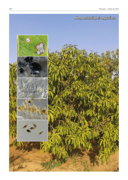
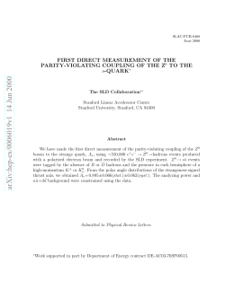
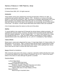
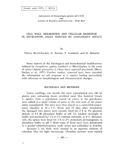
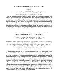
011_JPP197RP_487_COLORE
14-11-2008
17:48
Pagina 487
Journal of Plant Pathology (2008), 90 (3), 487-494
Edizioni ETS Pisa, 2008
487
PHIALOPHORA-LIKE FUNGI ASSOCIATED WITH KIWIFRUIT ELEPHANTIASIS
A. Prodi, S. Sandalo, S. Tonti, P. Nipoti and A. Pisi
Dipartimento di Scienze e Tecnologie Agroambientali, Alma Mater Studiorum, Università degli Studi,
Viale Fanin 40, 40127 Bologna, Italy
SUMMARY
An unusual disease, named elephantiasis for its typical
symptoms, has been seen in orchards of kiwifruit cv. Hayward in Emilia-Romagna (northern Italy) since 2001.
Phialophora-like isolates were obtained from the necrotic
wood and were studied in vitro for phenotype and tissue
colonization ability. We used primers amplifying the internal transcribed spacer (ITS) region of the ribosomal
DNA (rDNA) for molecular identification. Phaeoacremonium strains were further identified with b-tubulin-specific primers. Strains of Cadophora, Lecythophora and
Phaeoacremonium were classified and characterized. The
isolates differed in their ability to colonise tissue. P. aleophilum and C. melinii showed the highest colonization
index. To our knowledge, this is the first report of
Cadophora melinii isolated from kiwifruit plants.
Keywords: Actinidia deliciosa, fungal disease, Phaeoacremonium, Cadophora, molecular assays, ITS region.
INTRODUCTION
Kiwifruit (Actinidia deliciosa [(A. Chev.) C.F. Liang
et A.R. Ferguson] var. deliciosa) is an important crop of
temperate regions. Italy is the leading kiwifruit producer in the world with a total production of 422,335 tons
in 2006 (FAO, 2008).
In the last fifteen years, A. deliciosa plants with decline symptoms have been observed in several European
countries, Chile and New Zealand. In Chile, symptoms
of internal wood browning and small and silvery leaves
were attributed to Chondrostereum purpureum (Alvarez
et al., 1999).
Similar wood and leaf symptoms were also seen in
several Italian kiwifruit orchards. Symptoms included
deterioration of the wood structure and fruits which remained small and did not ripen properly. This syndrome was ascribed to a form of wood decay which can
Corresponding author: Paola Nipoti
Fax: +39 051 2096722
E-mail: [email protected]
affect 10-year-old plants (Calzarano et al., 1999). Longitudinal trunk sections showed brown areas of hard
necrotic tissue later followed by a spongy and friable
discolored zone, both symptoms developed from pruning cuts and progressed downwards. Analysis of deteriorated wood revealed the presence of fungi, typically responsible for complex diseases of woody tissue in different plants, e.g. Petri disease of grapevine. As reported
by Di Marco et al. (2000) the following fungi were isolated: Phaeoacremonium inflatipes W. Gams, P.W. Crous
& M.J. Wingf., P. aleophilum W. Gams, P.W. Crous,
M.J. Wingf. & L. Mugnai, P. rubrigenum W. Gams, P.W.
Crous & M.J. Wingf., Phaeomoniella chlamydospora (W.
Gams, P.W. Crous, M.J. Wingf. & L. Mugnai) P.W.
Crous & W. Gams, Phialophora spp., and agents of
white decay like Fomitiporia mediterranea M. Fisch.
Subsequent investigations, carried out in kiwifruit orchards of Emilia-Romagna (northern Italy), revealed the
presence of symptoms differing from normal “decay”.
The most obvious was an abnormal increase in trunk diameter (often over 60% at the collar) accompanied by
longitudinal bark cracks. In some orchards, the hypertrophy could be present at different heights of the
trunk. Because of the lack of decay, this was regarded as
a different disease called “elephantiasis” (Nipoti et al.,
2003). These symptoms generally occurred in plants
more than ten years old, occasionally in 7-year-old ones.
Affected plants were distributed at random in the orchard, and disease incidence was estimated to be less
than 10% (Nipoti et al., 2006). In cross section a
marked brown discoloration of the annual rings was visible, while in longitudinal sections the brown discoloration tended to diminish upwards, though it could extend to the branches. Diseased plants had reduced foliage and bore small unsalable fruits.
In Italy, the disease has also been recorded from
Piedmont (Mancini and Cotroneo, 1997), Latium (L.
Riccioni, personal communication), and Veneto (Nipoti
et al., 2007). It occurs also in France (Hennion et al.,
2001), northern Spain (Gonzalez Diaz, 2003) and New
Zealand (Manning et al., 2003).
Due to the importance of the Italian kiwifruit industry, the disease was studied further. In necrotic areas
Nipoti et al. (2006) found a complex mycoflora includ-
011_JPP197RP_487_COLORE
488
14-11-2008
17:48
Pagina 488
Phialophora-like fungi and kiwifruit elephantiasis
ing Fusarium Link ex Fr., Cylindrocarpon Wollenw.,
Phomopsis (Sacc.) Bubák and phialophora-like fungi
(Gams, 2000). Among phialophora-like endophytes,
Harrington and McNew (2003) have included some
morphologically little-differentiated anamorphs similar
to Phaeoacremonium W. Gams, P.W. Crous & M.J.
Wingf., Phialophora Medlar, Cadophora Lagerb. &
Melin, and Lecythophora Nannf.
From kiwifruit plants affected by elephantiasis Hennion et al. (2001) isolated mainly P. aleophilum and P.
viticola, whereas Manning et al. (2003) recovered
Phialophora alba F.H. Beyma. Recently, Riccioni et al.
(2007) have described a new kiwifruit disorder, called
“leader die-back”, from A. chinensis var. Hort16A (Zespri Gold) grown in the Latina area (central Italy), characterized by the death of a single cane or the entire
leader and by swelling and bark cracking similar to elephantiasis at the infection site. Fungi like Cryptosporiopsis actinidiae P.R. Johnst., M.A. Manning & X. Meier
and Cadophora spp were isolated from diseased plants
(Riccioni et al., 2007).
With our study we investigated the possible involvement of phialophora-like fungi in kiwifruit trunks showing elephantiasis.
MATERIALS AND METHODS
Fungal isolates and culture conditions. From 2001
onwards, in orchards of the Faenza area (Emilia-Romagna) we examined fifty 8- to 25-year-old kiwifruit
plants of cv. Hayward showing elephantiasis but no
Journal of Plant Pathology (2008), 90 (3), 487-494
macroscopic decay symptoms (Fig. 1A). Fragments of
discolored wood (Fig. 1B) were plated in Petri dishes
containing potato dextrose agar (PDA 39 g l-1, Difco,
USA), amended with streptomycin sulfate (300 mg l-1,
Sigma, USA), at 25°C in the dark. Five symptomless
plants were also tested as control. In this study, only
phialophora-like fungi, among the different fungi isolated, were considered. Seven phialophora-like isolates
kindly supplied by Dr. S. Di Marco (CNR, Bologna,
Italy) and Dr. J. Dupont (LCP, Laboratoire de Cryptogamie Paris, France), were used as reference isolates
(Table 1).
Morphological observations. A mycelial block (2–3
mm2), obtained from 14-day-old single conidial cultures
of each of the 34 phialophora-like strains, was transferred to Petri dishes and incubated on 2% malt agar
(MA, Sigma, USA) at 25, 30 and 35°C in the dark for 14
days. The capacity to grow at 35°C discriminates
phialophora-like taxa (Crous et al., 1996; De Hoog et
al., 1999; Dupont et al., 2000, 2002). Five plates for
each of the 34 strains were prepared for each temperature. This trial was repeated three times. The macroscopic features, i.e., colour of colonies, pigment diffusing into the agar, and microscopic observations, i.e.
conidiophore, phialides and conidial morphology, were
used to group the phenotypes (Crous et al., 1996;
Gams, 2000; Weber, 2002; Mostert et al., 2003, 2006).
DNA extraction, PCR amplification, sequencing and
phylogenetic analysis. Genomic DNA of phialophoralike strains was extracted from fresh mycelium grown on
Fig. 1. A. Kiwifruit plant affected by elephantiatis, B. Cross section of an infected trunk showing tissue discoloration.
011_JPP197RP_487_COLORE
14-11-2008
17:48
Pagina 489
Journal of Plant Pathology (2008), 90 (3), 487-494
Prodi et al.
MA for 14 days, using a protocol by Lohdi et al. (1994).
The universal oligonucleotide primers ITS4 and ITS5
(White et al., 1990) were used to amplify part of the nuclear ribosomal DNA (rDNA) by PCR carried out in a
final volume of 25 ml containing 5 ml of diluted sample,
0.75 units of GoTaq Flexi DNA Polymerase (Promega,
USA), 5x Green GoTaq Flexi Buffer, 0.2 mM dNTP, 3
mM MgCl2 and 2.5 pmol of each primer. Amplifications
were performed in a T3 Biometria Thermalcycler, using
an initial denaturation step of 94°C for 5 min, followed
by 30 cycles of denaturation at 94°C for 1 min, annealing
for 1 min at 56°C and elongation for 1 min at 72°C, with
a final extension for 10 min at 72°C. Products were analyzed by electrophoresis in 1% agarose gel in TBE
buffer, stained with ethidium bromide (0.4 mg ml-1) and
photographed under UV. The molecular weight of the
amplified DNA was estimated by comparison with a 100
bp DNA ladder (Promega, USA).
The amplified products were purified (Wizard SV
Gel and PCR Clean-Up System, Promega, USA) and
ligated into the pGEM-T easy vector (Promega, USA),
which was then used to transform competent cells of
JM109 Escherichia coli and recombinant plasmid DNA
from transformed cells was purified (Wizard Plus SV
Minipreps DNA Purification System, Promega, USA).
Colonies containing the insert were screened by PCR
with primers designed on the polylinker of the vector.
Sequencing was done by MWG (Germany).
Sequences were aligned using the multiple-sequence
alignment program Clustal V method (Higgins and
Sharp, 1989) from the Megalign package (DNAStar,
USA). Data were analyzed using TREECON software
(Van de Peer and De Wachter, 1994), with Jukes and
Cantor distance model to obtain a Neighbour-Joining
tree constructed using 1000 bootstrap replicates.
Bulleromyces albus CBS 6302 sequence (NCBI,
AF444663) was used as outgroup.
Since ITS data do not sufficiently discriminate all
species of Phaeoacremonium, primers amplifying btubulin were used in a touch-down PCR with the reverse primers Pbr6_1, Pbr12, Pbr11 and Pbr8 (Mostert
et al., 2006) in combination with the forward universal
primers T1 (O’Donnell and Cigelnik, 1997).
Colonization and pathogenicity assays. For better
and faster growth, each of the 34 phialophora-like isolates were streaked three times in each Petri dish containing MA and maintained at room temperature for 10
days. Three uninoculated MA Petri dishes were used as
control. Young sprouts of A. deliciosa cv Hayward, surface-disinfected in 2% vv-1 NaClO for 10 min, were cut
into 3 cm portions and laid horizontally over the fungal
streaks or placed in uninoculated dishes (Nipoti et al.,
1989; Riccioni et al., 2007). After 10 days incubation at
room temperature, the sprouts were cut longitudinally.
The experiment was replicated four times. The colonization index of each strain was evaluated by the
necrosis/length ratio of the sprout segment. Analysis of
variance (ANOVA) was performed using Statgraphic
Plus 2.1 software. Means were compared according to
Table 1. Reference strains used in this study.
Fungus
Strain number
Orign
Host
Phaeoacremonium aleophilum
teleomorph: Togninia minima (Tulasne
& C. Tulasne) Berlese.
CBS*101006(1)
Italy
A. deliciosa
P. aleophilum
CBS 101008 (1)
Italy
A. deliciosa
P. iranianum
CBS 101357 (1)
Italy
A. deliciosa
P. parasiticum teleomorph: T. parasitica
L. Mostert, W. Gams & P.W.Crous
LCP**883537 (2)
USA
Human
P. viticola teleomorph: T. viticola L.
Mostert, W. Gams & P.W. Crous
LCP 963886 (2)
France
Vitis vinifera
Cadophora malorum
CBS 101359 (1)
Italy
A. deliciosa
Sweden
Wood chips of
Betula verrucosa
and B. pubescens
teleomorph: T. minima
Phialophora verrucosa
1
489
CBS 839.69
(2)
Obtained from Dr. S. Di Marco; 2Obtained from Dr. J. Dupont
* CBS = Centraalbureau voor Schimmelcultures, Utrecht, The Netherlands
** LCP = Laboratoire de Cryptogamie Paris, France
011_JPP197RP_487_COLORE
490
14-11-2008
17:48
Pagina 490
Phialophora-like fungi and kiwifruit elephantiasis
Tukey’s test (P = 0.05).
Fifty 2-year-old plants of cv. Hayward from a commercial nursery, kept in plastic pots in a greenhouse,
were used for pathogenicity assays. A wound, made in
the stem 3 cm above soil level, was inoculated with a
mycelial agar plug from 2-week-old cultures of strains
showing a high colonization ability. To prevent drying,
the wounded area was covered with parafilm. Four sets
of 10 plants each were inoculated with a mix of
Cadophora melinii strains (2, 13, 75, 76), C. luteo-olivacea (20, 61), Lecythophora luteoviridis (26, 64) and P.
aleophilum (9, 10, 29, 30, 31). Controls were inoculated
with a plug of sterile agar.
After 18 months three plants from each set were examined. The stems were cut longitudinally to assess
wood discoloration. Fragments taken from 3 cm above
the inoculation site were surface-disinfected in 2% vv-1
NaClO for 5 min and plated onto Petri dishes containing MA. The remaining plants were transferred to a
field at Zattaglia, near Faenza, to allow development of
symptoms.
RESULTS
Fungal isolates and their morphology. Twenty-seven
phialophora-like fungi, were isolated and examined microscopically. Twenty-five were identified based on reference strains and the literature (Crous et al., 1996;
Gams, 2000; Weber, 2002; Mostert et al., 2003, 2006).
Two isolates, 435 and 29/1, were classified by the Centraalbureau voor Schimmelcultures, Utrecht (CBS) as
Cadophora melinii (CBS 111743) and Lecythophora luteoviridis, respectively; these were used, along with
those supplied by Drs S. Di Marco and J. Dupont, as
reference strains. No phialophora-like isolate was obtained from symptomless plants.
Observations of 14-day-old single conidial phialophoralike cultures allowed to recognize three phenotypes: A, B
and C (Table 2).
Phenotype A: Phaeoacremonium spp. (reference
strains: CBS 101006; CBS 101008; CBS 101357, LCP 88
3537, LCP 96 3886). The colonies, of varying colour,
usually smoke-grey or honey, reached a diameter of 2540 mm in 14 days at 25°C. Some strains produced a yellow pigment diffusible in the agar. Colonies were felty
and woolly. Mycelium had branched, septate, hyaline or
light brown hyphae occurring singly or in bundles.
Conidiophores were short and usually unbranched. The
phialides, arising singly from the conidiophores, were
subcylindrical or elongate-ampulliform, attenuated at
the base, with an inconspicuous funnel-shaped collarette. Conidia, aggregated into round slimy heads at
the phialide apices, were hyaline, aseptate, smoothwalled, typically allantoid and sometimes became twoguttulate with age. Eight strains belonged to this pheno-
Journal of Plant Pathology (2008), 90 (3), 487-494
type, 75% of them able to grow at 35°C (Table 2).
Phenotype B: Cadophora (reference strains: CBS
101359; CBS 111743). The colonies reached a diameter
of 38-55 mm in 14 days at 25°C, were predominantly
felt with a distinct grey-brown centre, with aggregated
hyphae in the centre and radiating hyaline runner hyphae at the edge. The submerged hyphae were
branched, hyaline or light brown. Conidiophores could
be branched or unbranched, phialides were flaskshaped with a distinct cup-shaped collarette. Conidia
were variously shaped ellipsoidal or obovate and heteropolar. Fourteen strains belonged to this phenotype,
and did not grow at 35°C (Table 2).
Phenotype C: Lecythophora (reference strain: 29/1).
The salmon-pink colonies turned brown ageing and
reached a diameter of 35-45 mm in 14 days at 25°C.
Phialides were single, flask-shaped, with inconspicuous
collarettes, often short necks arising as from intercalary
cells. Conidia, aggregated in slimy heads, were aseptate,
cylindrical to ellipsoidal and contained many small guttules. Five strains belonged to this phenotype and only
one strain was able to grow at 35°C (Table 2).
None of our isolates belonged to Phialophora sensu
stricto.
PCR amplifications, sequencing and phylogenetic
analysis. The DNA sequence alignment of phialophoralike strains (Fig. 2) supported the division into three distinct phenotypes: Phaeoacremonium spp. (A), Cadophora
spp. (B) and Lecythophora spp. (C).
The use of b-tubulin primers allowed the identification of six strains of P. aleophilum (548 bp) and two of
P. mortoniae (257 bp); none belonged to P. iranianum
(545 bp) or P. viticola (542 bp) (Fig. 3).
Within Cadophora spp. (phenotype B) seven strains
were identified as C. luteo-olivacea, seven as C. melinii
and none as C. malorum (Kidd & Beaum) W. Gams
(Fig. 2). In phenotype C, five strains of L. luteoviridis
were identified (Fig. 2).
The sequences of some of our strains are deposited in
GenBank (Table 2).
Colonization and pathogenicity assays. Blackening
in the kiwifruit sprouts was of variable length related to
the different phialophora-like strains. No necrosis was
visible in the control sprouts. The colonization capacity
of each strain was homogeneous in the four replications.
The differences were significant (F = 7.44**), and
Tukey’s test (P = 0.05) distinguished three groups differing in colonization index (Table 2). P. aleophilum and
C. melinii strains reached the highest values (letter c).
Eighteen months after inoculation, in the greenhouse
trials as well as in the plants transferred to the field, there
was no evidence of hypertrophy. In the longitudinally cut
stems we observed discolored tissues only in some plants
of two sets: P. aleophilum mix and C. melinii mix. Reiso-
011_JPP197RP_487_COLORE
14-11-2008
17:48
Pagina 491
Journal of Plant Pathology (2008), 90 (3), 487-494
Prodi et al.
491
Table 2. Phialophora-like isolates obtained from elephantiasis-affected kiwifruit plants, and some reference strains.
Strain and
collection number
NCBI
GenBank
Emilia-Romagna location Year
accession No.
9
DQ404356
Sarna - farm A(*)
2002
Abilty to
grow
at 35°C
Phenotype
Molecular
identification
Colonization
Index
Yes
A
P. aleophilum
1.00 c(1)
10
Sarna - farm A
2002
Yes
A
P. aleophilum
1.00 c
11
Sarna - farm A
2002
Yes
A
P. aleophilum
1.00 c
Sarna - farm B
2001
Yes
A
P. aleophilum
1.00 c
Sarna - farm B
2001
Yes
A
P. aleophilum
1.00 c
31
Marzeno
2002
Yes
A
P. aleophilum
0.75 abc
CBS 101006
Reference
Yes
A
P. aleophilum
1.00 c
29
30
69
DQ404355
Fossolo
2003
No
A
P. mortoniae
0.08 a
182
EU427312
Zattaglia
2001
No
A
P. mortoniae
0.12 ab
CBS 101008
Reference
No
A
P. mortoniae
0.07 a
CBS 101357
Reference
Yes
A
P. iranianum
1.00 c
LCP 88 3537
Reference
Yes
A
P. parasiticum
0.08 a
LCP 96 3886
Reference
No
A
P. viticola
0.51 abc
2
Sarna - farm A
2002
No
B
C. melinii
1.00 c
13
Sarna - farm A
2002
No
B
C. melinii
1.00 c
16
Sarna - farm B
2004
No
B
C. melinii
0.75 bc
Modigliana - farm B
2001
No
B
C. melinii
0.70 abc
75
Modigliana - farm A
2003
No
B
C. melinii
1.00 c
76
Modigliana - farm A
2003
No
B
C. melinii
0.98 c
19
DQ404352
435 CBS 111743
DQ404351
Sarna - farm B
2002
No
B
C. melinii
1.00 c
18
DQ404348
Zattaglia
2001
No
B
C. luteo-olivacea
0.07 a
20
DQ404349
Zattaglia
2001
No
B
C. luteo-olivacea
0.61 abc
21
Modigliana - farm B
2001
No
B
C. luteo-olivacea
0.07 a
23
Marzeno
2002
No
B
C. luteo-olivacea
0.46 abc
39
Zattaglia
2002
No
B
C. luteo-olivacea
0.16 ab
40
Modigliana - farm B
2002
No
B
C. luteo-olivacea
0.31 abc
Fossolo
2003
No
B
C. luteo-olivacea
0.42 abc
No
B
C. malorum
0.51 abc
61
CBS 101359
DQ404350
Reference
1
Sarna farm A
2002
Yes
C
L. luteoviridis
0.16 ab
26
Sarna farm A
2001
No
C
L. luteoviridis
0.48 abc
64
Sarna farm A
2002
No
C
L. luteoviridis
0.44 abc
65
Marzeno
2002
No
C
L. luteoviridis
0.15 ab
29/1
Sarna - farm B
2002
No
C
L. luteoviridis
0.20 ab
Yes
D
Phialophora verrucosa
0.19 ab
CBS 839 69
DQ404354
DQ404353
Reference
F = 7.44**
1
*Different farms in the same location. Means of colonization index followed by the same letter are not significantly different
(P=0.05) according Tukey’s test.
lations was possible from the discolored wood of all three
plants inoculated with P. aleophilum but only from one
plant inoculated with C. melinii.
DISCUSSION
Phialophora-like fungi isolated from kiwifruit plants
with trunk hypertrophy, comprised members of several
similar genera, which are difficult to distinguish phenotypically (Gams, 2000; Weber, 2002; Harrington and
McNew, 2003; Aroca and Raposo, 2007). These fungi
occur in many environments such as decaying wood,
soil, water and food, and can also cause human mycoses
(Schol-Schwarz, 1970; Mostert et al., 2005, 2006). As
mentioned, their identification is difficult, especially for
Phaeoacremonium, but the monograph of Mostert et al.
(2006) has made an important contribution to this aim.
011_JPP197RP_487_COLORE
492
14-11-2008
17:49
Pagina 492
Phialophora-like fungi and kiwifruit elephantiasis
Journal of Plant Pathology (2008), 90 (3), 487-494
Fig. 2. Phylogenetic relationships among phialophora-like isolates based on aligned internal transcribed spacer
(ITS) sequences and part of 18S rRNA, 5.8S rRNA and 28S sequences. The sequences of C. luteo-olivacea
AY249068 and B. albus AF 444663 are from the National Center for Biotechnology Information (NCBI)
GenBank. Bootstrap support values (1000 replicates) are shown at the nodes. Scale bar represents a genetic distance of 0.1. B. albus CBS 6302 selected as outgroup.
Phialophora-like fungi from elephantiasis-affected kiwifruit showed variation and comprised at least three
phenotypes and five molecular groups. The phenotype
A strains were identified as P. aleophilum and P. mortoniae, the former of which showed the highest ability to
colonize kiwifruit sprouts, confirming previous findings
by Di Marco et al. (2004). Other Phaeoacremonium
species have also been isolated from kiwifruit, i.e. P. parasiticum (Di Marco et al., 2004), P. iranianum (Mostert
et al., 2006), P. viticola (Hennion et al., 2001) and in our
study P. mortoniae, which showed poor colonization
ability. The latter fungus was reported by Gramaje et al.
(2007) to cause decline of young grapevines in Spain.
Phaeoacremonium species are associated with diseases of various woody hosts, such as grapevine, olive,
apricot, date palm, hop bush and oak, either as endophytes or as possible pathogens inducing wilting,
dieback or death (Mostert et al., 2005). In particular P.
aleophilum is thought to be a precursor of several other
pathogens and is considered to be a primary cause of
Petri disease and esca, two complex grapevine diseases
(Larignon and Dubos, 1997; Mugnai et al., 1999; Khan
et al., 2000; Mostert et al., 2006). This role is due to its
ability to colonise parenchyma and vascular tissues and
to break down polyphenolic compounds (Mugnai et al.,
1999). P. aleophilum can also infect kiwifruit propagat-
011_JPP197RP_487_COLORE
14-11-2008
17:49
Pagina 493
Journal of Plant Pathology (2008), 90 (3), 487-494
ing material (Nipoti et al., 2007) besides grapevine nursery plants (Zanzotto et al., 2007). Fourie and Halleen
(2004) emphasized the importance of infected propagation material as a major means of spread of these fungi.
The presence of C. melinii has been observed for the
first time in kiwifruit. In addition we isolated C. luteoolivacea but not C. malorum. Cadophlora species
showed variable ability to colonise kiwifruit sprouts. In
the literature some importance has been ascribed to
Cadophora species as tissue colonizers. For instance, C.
luteo-olivacea was reported as the causal agent of leader
die-back of kiwifruit, although this disease is not well
studied (Riccioni et al., 2007) and C. malorum, formerly
misidentified as Phaeomoniella chlamydospora (reference strain CBS 101359), is thought to be involved in
kiwifruit decay (Di Marco et al., 2004). In our study C.
melinii was able to colonise tissues better than C. luteoolivacea. Thus it is possible that C. melinii is involved in
the aetiology of elephantiasis.
All strains of phenotype C belonged to L.
luteoviridis. They showed intermediate ability to
colonise kiwifruit tissues. So far, Lecythophora spp. have
been reported as mainly associated with wood discoloration in Picea abies (Weber, 2002).
Fusarium spp., F. solani in particular, and phialophora-like organisms have been isolated with a high frequency in association from elephantiasis-affected plants
(Prodi et al., 2006). Since F. solani can cause wood discoloration and incite the development of small swellings
in Platanus x acerifolia (Pilotti et al., 2002), it may have
a role in the hypertrophy of kiwifruit (Nipoti et al.,
2004), thus supporting the suggestion that elephantiasis
may be caused by the interaction of different fungi. We
have found that P. aleophlium and C. melinii play a pioneering role in disease development due to their ability
to colonise kiwifruit tissue, but the subsequent possible
interaction with Fusarium spp. needs confirmation.
Pathogenicity trials using F. solani in association with
different phialophora-like phenotypes, artificially inoculated at different stages, are in progress.
The conclusion is that elephantiasis is a disease with
a complex etiology, for a better understanding of which
the identification of the different fungal species isolated
from affected plants represents a preliminary step.
ACKNOWLEDGEMENTS
We thank Dr J. Dupont and Dr S. Di Marco for
kindly supplying fungal strains, Dr. R. Credi for his research support, Dr. G. Spada and Dr. S. Graziani for
field technical help. This work was supported by Centro
Ricerche Produzioni Vegetali (CRPV) (Projects “Nuove
fitopatie dell’actinidia” and Fitopatie dell’actinidia:
”carie” ed elefantiasi – Regional Law 28/98).
Prodi et al.
493
Fig. 3. PCR products amplified from genomic DNA of
Phaeoacremonium strains using specific primers for b-tubulin
of: (i) P. aleophilum (Pbr6_1/T1 – expected product 548 bp):
lane 1, strain CBS 101006; lane 2, strain 9; lane 3, strain 10;
lane 4, strain 11, lane 5, strain 29; lane 6, strain 30; lane 7,
strain 31; (ii) P. iranianum (Pbr12/T1 - expected product 545
bp): lane 8, strain CBS 101357; (iii) P. viticola (Pbr 8/T1 – expected product 542 bp): lane 9, LCP 96 3836; (iv) P. mortoniae (Pbr 11 – expected product 257 bp): lane 10, strain CBS
101008; lane11, strain 69; lane 12, strain 182. Lane M, 100 bp
molecular marker.
REFERENCES
Alvarez M., Pinilla B., Elloriaga A., 1999. Chondrostereum
purpureum identified as the causal agent of silver leaf of kiwifruit in Chile. In: Proceedings of 4th International Symposium on kiwifruit, Santiago de Chile 1999: 33.
Aroca A., Raposo R., 2007. PCR-based strategy to detect and
identify species of Phaeoacremonium causing grapevine
disease. Applied and Envirormental Microbiology 93: 29112918.
Calzarano F., Spada G., Montuschi C., Di Marco S., 1999. Una
forma di deperimento lignicolo colpisce i frutteti italiani di
A. deliciosa. Informatore Fitopatologico 49 (11): 12-15.
Crous P.W., Gams W., Wingfield M.J., van Wyk P.S., 1996.
Phaeoacremonium gen. nov. associated with wilt decline
diseases of woody hosts and human infections. Mycologia
88: 786-796.
De Hoog G.S., Weenink X.O., Gerrits van den Ende A.H.G.,
1999. Taxonomy of the Phialophora verrucosa complex
with the description of two new species. Studies in Mycology 43: 107-121.
Di Marco S., Calzarano F., Gams W., Cesari A., 2000. A new
wood decay of kiwifruit in Italy. New Zealand Journal of
Crop and Horticultural Science 28: 69-73.
Di Marco S., Calzarano F., Osti F., Mazzullo A., 2004. Pathogenicity of fungi associated with a decay of kiwifruit. Australasian Plant Pathology 33: 337-342.
Dupont J., Magnin S., Cesari C., Gatica M., 2002. ITS and
[beta]-tubulin markers help delineate Phaeoacremonium
species, and the occurrence of P. parasiticum in grapevine
disease in Argentina. Mycological Research 106: 1143-1150.
Dupont J., Magnin S., Paronnaud J., Raquebert M.F., 2000.
The genus Phaeoacremonium from a molecular point of
view. Phytopathologia Mediterranea 39: 119-124.
FAO, 2008. FAOSTAT Statistical database. FAO Statistics Division. http://faostat.fao.org/default.aspx.
011_JPP197RP_487_COLORE
494
14-11-2008
17:49
Pagina 494
Phialophora-like fungi and kiwifruit elephantiasis
Fourie P.H., Halleen F., 2004. Occurrence of grapevine trunk
disease pathogens in rootstock mother plants in South
Africa. Australiasian Plant Pathology 33. 313-315.
Gams W., 2000. Phialophora and some similar morphologically little-differentiated anamorphs of divergent ascomycetes.
Studies in Mycology 45: 187-199.
Gonzalez Diaz I., 2003. Identificazione e caratterizzazione di
miceti associati ad una nuova malattia del legno di Actinidia deliciosa. Thesis Alma Mater Studiorum University of
Bologna, Faculty of Agriculture, Italy.
Gramaje D., Alaniz S., Pérez-Sierra A., Abad-Campos P.,
Garcia-Jiménez J., Armengol J., 2007. First report of
Phaeoacremonium mortoniae causing Petri disease of
grapevine in Spain. Plant Disease 91: 1206.
Harrington T.C., McNew D.L., 2003. Phylogenetic analysis
places the Phialophora-like genus Cadophora in the Helotiales. Mycotaxon 87: 141-151.
Hennion B., Baudry A., Lecomte P., Durpaire M. P., Mouyon
M., Tailleur J. L., Larignon P., 2001. Dépérissement du kiwi par maladie du bois. Infos-Ctifl 176: 25-27.
Higgins D.G., Sharp P.M., 1989. Fast and sensitive multiple
sequence alignments on a microcomputer. Computer Applications in the Biosciences 5: 151-153.
Khan A., Whiting C., Rooney S., Gubler W.D., 2000. Pathogenicity of three species of Phaeoacremonium spp. on
grapevine in California. Phytopathologia Mediterranea 39:
92-99.
Larignon P., Dubos B., 1997. Fungi associated with Esca disease in grapevine. European Journal of Plant Pathology 103:
147-157.
Lohdi M.A., Ye G., Weeden F., Reisch B.I., 1994. A simple
and efficient method for DNA extraction from grapevine
cultivars and Vitis species. Plant Molecular Biology Reporter 12: 6-13.
Mancini G., Cotroneo A., 1997. Bilancio fitosanitario, Piemonte
e Valle d’Aosta. Informatore Fitopatologico 47 (3): 4-6.
Manning M.A., Meier X., Olson T.L., 2003. Etiology of vine
decay in kiwifruit cv. Hayward in New Zealand. Proceedings of 8th International Congress of Plant Pathology,
Christchurch 2003: 263.
Mostert L., Crous P.W., Gronewald J.Z., Gams W., Summerbell R.C., 2003. Togninia (Calosphaeriales) is confirmed as
teleomorph of Phaeoacremonium by means of morphology,
sexual compatibility and DNA phylogeny. Mycologia 95:
646-659.
Mostert L., Groenewald J.Z., Summerbell R.C., Gams W.,
Crous P.W., 2006. Taxonomy and pathology of Togninia
(Diaporthales) and its Phaeoacremonium anamorphs. Studies in Mycology 54: 1-113.
Mostert L., Groenewald J.Z., Summerbell R.C., Robert V.,
Sutton D.A., Padhye A., Crous P.W., 2005. Species of
Phaeoacremonium associated with infections in humans
and environmental reservoirs in infected wood plants.
Journal of Clinical Microbiology 43:1752-1767.
Mugnai L., Graniti A., Surico G., 1999. Esca (black measles)
Received February 13, 2008
Accepted March 31, 2008
Journal of Plant Pathology (2008), 90 (3), 487-494
and brown wood streaking: two old and elusive disease of
grapevine. Plant Disease 83: 404-418.
Nipoti P., Finessi L., Manzali D., Gavina F., 1989. Methods
for estimating the resistance of strawberry plants to Verticillium dahliae Kleb., Rhizoctonia fragariae Husain and
McKeen, and Rhizoctonia solani. Acta Horticulturae 265:
609-613.
Nipoti P., Prodi A., Sandalo S., Credi R., 2004. Further studies on the main fungi associated with elephantiasis of kiwifruit. Journal of Plant Pathology 86: 327.
Nipoti P., Prodi A., Tonti S., Sandalo S., Credi R., 2007. Attuali
conoscenze sull’elefantiasi dell’actinidia. Atti del 8° Convegno Nazionale sull’Actinidia 2007, Cuneo-Torino 2007: 67.
Nipoti P., Riccioni L., Filippini G., Haegi A., Prodi A., Sandalo S., Sequino S., Tonti S., Valvassori M., 2006. Hypertrophic trunks in Italian kiwifruit orchards. Proceedings
12th Congress of the Mediterranean Phytopathological
Union, Rhodos 2006: 510-512.
Nipoti P., Sandalo S., Prodi A., Credi R., Spada G., Graziani
S., 2003. An unusual wood disease of kiwifuit in Italy. Acta
Horticulturae 610: 253-259.
O’Donnell K., Cigelnik E., 1997. Two divergent intragenomic
rDNA ITS2 types within a monophyletic lineage of the
fungus Fusarium are nonorthologous. Molecular Phylogenetics and Evolution 7: 103-116.
Pilotti M., Ponzio V., Motta E., 2002. Disorders of Platanus x
acerifolia in Italy associated with Fusarium solani. Forest
Pathology 32: 249-264.
Prodi A., Sandalo S., Tonti S., Credi R., Peressotti E., Nipoti P.,
Dal Prà M., 2006. Identification and characterization of
Fusarium spp. isolated from kiwifruit affected by elephantiasis. 9th European Fusarium Seminar Wageningen 2006: 55.
Riccioni L., Manning M., Valvassori M., Haegi A., Casanato
S., Spinelli R., 2007. A new disease: Leader die-back in Actinidia chinensis Hort16A in Italy. Acta Horticulturae 753:
669-676.
Schol-Schwarz M.B., 1970. Revision of the genus Phialophora
(Moniliales). Persoonia 6: 59-94.
Van de Peer Y., De Wachter R., 1994. TREECON for Windows: a software package for the construction and drawing
of evolutionary trees for the Microsoft Windows environment. Computer Application in the Biosciences 10: 569-570.
Weber E., 2002. The Lecythophora-Coniochaeta complex. I.
Morphological studies on Lecythophora species isolated
from Picea abies. Nova Hedwigia 74: 159-185.
White T.J., Burns T., Lee S., Taylor J., 1990. Amplification and
direct sequencing of fungal ribosomal RNA genes for phylogenetics. In: Innis M.A., Gelfand D.H., Sninsky J.J., White
T.J. (eds). PCR Protocols: A Guide to Methods and Applications, pp. 315-322. Academic Press, San Diego, CA, USA.
Zanzotto A., Autiero F., Bellotto D., Dal Cortivo G., Lucchetta G., Borgo M., 2007. Occurrence of Phaeoacremonium
spp. and Phaeomoniella chlamydospora in grape propagation materials and young grapevines. European Journal of
Plant Pathology 119: 183-192.
© Copyright 2026 Paperzz