The influence of the Accademia del Cimento in the development of medicine and the biological sciences
The legacy of the Accademia del Cimento continues to have a great influence over the perception of the
post-Galilean history of science. While it could be said that the main achievements of the group are
methodological, in that they promoted experimentation over acceptance of the facts based on Classical
authorities whose works had become part of established university learning, the group is also responsible
for the creation of a new philosophy entirely that would go on to have profound implications within the
fields of medicine, anatomy and the biological sciences. A new approach was developing throughout
Europe as a result of a convergence of factors, such as the mid-century push to debunk Galenic learning,
Paracelsan movements and the broader rebellion against elite institutions of traditional medicine, the
incremental unification of ‘physic’ (the work of a physician) and surgery undertaken by reformers in
London, and the greater production of books in the vernacular language allowing medical knowledge to
spread to previously excluded groups across the continent.1 This new philosophy emerging from the
members of the Accademia del Cimento (and other societies like it) would follow and set the ground for a
new way of thinking about physiology and the natural world as a whole.
In particular the experiments of Niels Stensen (or Nicolas Steno, 1638 – 1686) made important
contributions to the development of anatomy. In biographical accounts, Stensen began developing the skill
and dexterity that would serve him well in later life in the workshop of his father, Sten Pedersen. After
pursuing medical studies at the University of Copenhagen between 1656 and 1659, Stensen travelled
Europe and would generally remain on the move for the rest of his life. Upon arriving in Florence, the
Grand Duke appointed him as an anatomist at the Santa Maria Nuova hospital, which at that time had
facilities for around 1,500 patients, had several specialist departments and its own dedicated rooms for
1
Andrew Wear, Knowledge and practice in English medicine, 1550-1680 (Cambridge: Cambridge University Press,
2000), p. 41.
dissection. He soon learned Italian and developed close ties with the Grand Duke’s personal physician,
Francesco Redi (1626 – 98), as well as Vincenzio Viviani (1622 – 1703), the court mathematician, who had
been Galileo’s youngest pupil. Stensen was suited well to the spirit of inquiry that was very much in
evidence in the formation of the Accademia, and would eventually write that his years in Florence were
both the most important and the happiest of his life.2
One of Stensen’s major contributions had been his theories on the function of the heart. At this time
it was still argued that the heart had been the centre of warmth for the body, and had been written off as not
a ‘true muscle’ in the major anatomical work of Andreas Vesalius a century earlier. Stensen, however,
continued to defend his thesis that the heart ‘is simply a muscle’ based on his observations published in On
muscles and glands (1664),3 and went on to develop a model in which muscles were parallelepiped
integrations of fibers. This would serve as the methodological foundation on which figures such as
Giovanni Borelli (1608 – 97) would build a dynamic view of living beings, based on the muscles as objects
of physics.
Stensen is also credited with the discovery and descriptions of several glands and ducts in the body.
4
While the functions of some of the structures found and presented by Stensen remained conjectural until
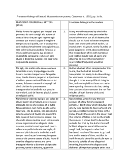
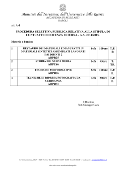
the eighteenth and nineteenth centuries, he set a new standard of accuracy in his descriptions of the parotid
duct (also known as the Stensen duct), and is known for his treatise on the tear glands, using illustrations of
the left eye of a calf to support his observations. (Figure 1)
Stensen described the ovarian follicle, though the name was granted to his contemporary Regnier
de Graaf (1641 – 73), and he played a lesser part in the fierce embryological controversies that followed,
such as the debate between ‘ovism’ and ‘animalcultism’ (in which followers of both schools argued
whether the spermatozoa or ovum was the true source of conception). Finally, in 1665, he delivered a
lecture in Paris on the anatomy of the brain, the Discours sur l'anatomie du cerveau ("A Dissertation on the
2
Karen Ascani and Gunver Skytte, Niccolo Stenone (1638-1686) Anatomista, Geologo, Vescovo. Conf Proceedings
Held 2000 Oct (Rome: L'Erma di Bretschneider, 2002), 31.
3
William F Bynum and Helen Bynum, Dictionary of medical biography (Westport, CT: Greenwood Press, 2007), 3,
p. 1188.
4
Niels Stensen, Observationes anatomicæ (Lugduni Batavorum: Apud Jacobum Chouët, 1662).
Anatomy of the Brain"), which is a seminal investigation on methods in neuroscience. 5
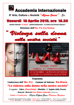
Among Stensen’s contacts and colleagues was Marcello Malpighi (1628 – 1694), who would take
advantage of the society’s advancements in optics and go on to make the first observations of plant and
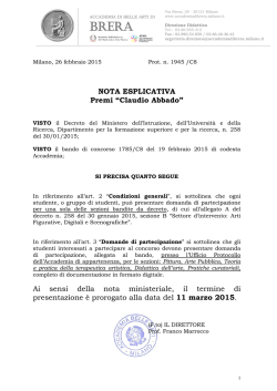
animal matter on the microscopic level.6 Malpighi’s discoveries would lead him to become a hugely
influential scientist in the fields of microscopical anatomy, histology, physiology and embryology.
(Figure 2)
5
Roy Porter, The greatest benefit to mankind : a medical history of humanity from antiquity to the present
(Hammersmith, London: HarperCollins, 1997), p. 226.
6
Ibid., p. 223.
Figure 1. Illustration of the tear glands, from Niels Stensen’s Observationes anatomicae. (I) shows the left
eye of a calf with the tear gland, labelled (A), with the parallel row of ducts (e) revealed. (II) depicts the
back of the upper eyelid turned upside down with the tear gland (b), its ducts (c) and openings (d). (III)
shows the tear gland and its two ducts in the corner of the eye. (IIII) shows the lachrymal duct (a) which
takes excess tears from the eye into the nasal cavity [Hans Kermit, Niels Stensen, 1638-1686: the scientist
who was beatified, (Leominster: Gracewing Publishing, 2003), p. 85].
Figure 2. Page from Malpighi’s thesis on the embryological development of chicks, as seen in the Opera
omnia, seu thesaurus locupletissimus botanico-medico-anatomicus, viginti quatuor tractatus complectens
et in duos tomos distributes (1686). The image labelled ‘Fig. 52’ on the right depicts Malpighi’s observation
of the development after twelve days [Marcello Malpighi and Luigi Belloni, Opere scelte. A cura di Luigi
Belloni (Torino: Unione tipografico-editrice torinese, 1967), p. 137].
Malpighi had taught theoretical medicine at Bologna, become a professor of medicine in Pisa and had been
close to the central work of the Accademia del Cimento, collaborating closely with Giovanni Borelli.
Malpighi had been heavily influenced by the experimental philosophy practiced under Medicean patronage,
and used the microscope to advance an essentially mechanical view of living beings. Taking influence from
Redi and Borelli, he was drawn to an Aristotlean and Democritean philosophy, believing that a model based
on tiny mechanisms (such as the ancient idea of the atom or corpuscle) was the most accurate way to
describe natural phenomena.
One of his most notable discoveries using the microscope included the differentiation of the fine
structures of the lungs. Despite the ambition of fifteenth and sixteenth century anatomists to challenge the
ancient sources, they had mostly still followed the writings of Galen and held that the lungs had been
formed from solidified bloody foam.7 Later theories had air and blood mingling freely in the fleshy
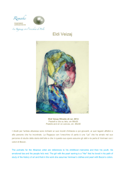
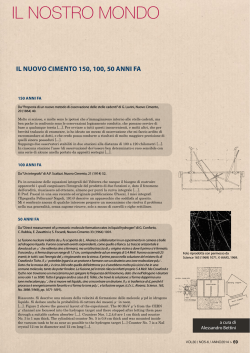
parenchyma of the lung. However it was Malpighi’s use of the microscope that revealed the membranous
alveoli at the ends of the traceho-bronchial ramifications,8 (Figure 3) leading Malpighi to correct one of the
largest missing links of William Harvey’s theory, and thus provide decisive confirmation of the blood’s
circulation: “I saw the blood, flowing in minute streams through the arteries, in the manner of a flood, and I
might have believed that the blood escaped into an empty space and was collected up again by a gaping
vessel, but an objection to the view was afforded by the movement of the blood being tortuous and scattered
in different directions and by its being united again in a definite path. My doubt was changed to certainty by
the dried lung of a frog which to a marked degree had preserved the redness of the blood in very tiny tracts,
which were afterwards found to be vessels, where by the help of a glass I saw not scattered points but
vessels drawn together in a ring-like fashion … Thus it was clear that the blood flowed along sinuous
vessels and did not empty into spaces, but was always contained within vessels, the paths of which
produced dispersion.”9
7
John B. West, 'Marcello Malpighi and the discovery of the pulmonary capillaries and alveoli', American Journal of
Physiology, 304.6 (2013), 383-90.
8
Porter, The greatest benefit to mankind … (cit. note 5), p. 223.
9
Ibid., p. 224.
Figure 3. Detail from Malpighi’s Opera Omnia. The left image depicts lungs with alveoli (E) and
capillaries (H). The right image depicts the pulmonary capillaries in a diagram of an alveolus that has been
opened up. These drawings were originally sent in letters to contemporary Giovanni Borelli [Marcello
Malpighi, Opera omnia, seu thesaurus locupletissimus botanico-medico-anatomicus, viginti quatuor
tractatus complectens et in duos tomos distributes (Lugduni Batavorum: apud Petrum vander Aa, 1686), p.
763].
Some of the most significant discoveries in Malpighi’s work were made possible by the invention of the
compound microscope, and during his association with the Accademia del Cimento. Though magnifying
spectacles using only one lens had been a long established invention, and many of Malpighi’s own
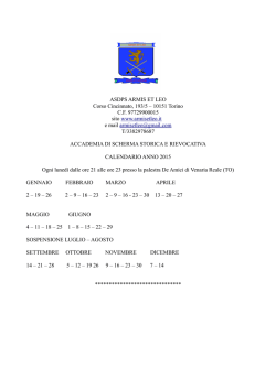
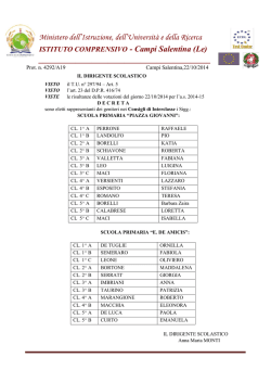
observations were apparently made using only a single lens, the illustrations made in De pulmonibus (1661)
are likely to have taken advantage of the recent invention in a similar way to Robert Hooke’s Micographia
(1665). The difference in the compound microscope is that it contained an objective lens and an eyepiece
lens, allowing for greater magnification. (Figure 4)
Figure 4. An example of a seventeenth century Galilean compound microscope, credited to optician
Giuseppe Gampani (1635 – 1715) [Museo Galileo, Florence, inv. no. 3429 – Photo J. Servello].
Malpighi’s observations of the alveoli and capillaries came only after many laborious and abortive efforts
in dissections of larger mammals. However, using what he called the ‘microscope of nature’, he could
visualise with a relatively small magnification the minute features of the lung in dissections of smaller
animals, in this instance frogs.10
Following on from Stensen’s work on the heart in De polypo cordis (1666), Malpighi offered the
first description of red blood corpuscles. In his De viscerum structura (1666), Malpighi showed that it was
the liver, not the gall bladder, that secreted bile and that the kidneys worked in a manner comparable to
sieves. In all, Malpighi was convinced that the mechanistic view was the best model when carrying out
investigations inside the body, and often compared bodily functions to machinery: “the mechanisms of our
bodies are composed of strings, thread, beams, levers, cloth, flowing fluids, cisterns, ducts, filters, sieves,
and other similar mechanisms. Through studying these parts with the help of Anatomy, Philosophy and
Mechanics, man has discovered their structure and function … With this and the help of discourse, he
apprehends the way nature acts and he lays the foundation of Physiology Pathology, and eventually the art
of Medicine.”11
Giovanni Borelli had also been convinced that the mechanical philosophy was the best way of
undertaking explorations of natural phenomena, and applied the views of Stensen and Malpighi to the
motion of the whole body. He studied muscular action, gland secretions, respiration, heart rhythm, and
neural response. In De motu animalium (1680), Borelli recorded incredible detail in his observations of
birds in flight, fish swimming, muscular contraction, respiration, and several other interrelated subjects, all
stemming from an attempt to analyse the body as it functions in terms of physics.12 (Figure 5)
10
John L Heilbron, The Oxford companion to the history of modern science (Oxford: Oxford University Press, 2003),
p. 485.
11
John M Forrester, 'Malpighi's De polypo cordis: An annotated translation', Medical history, 39.04 (1995), 477-92.
12
Giovanni Alfonso Borelli, De motu animalium (Rome: A. Bernabo, 1680).
Figure 5. Illustrations depicting the motions of animals, from Borelli’s De motu animalium (1680).
He argued that there was a ‘contractile element’ in the muscles, their operation being triggered by processes
similar to chemical fermentation. Respiration constituted a mechanical process where air was driven into
the lungs and into the bloodstream, possessing a life-sustaining function because it was a medium for
‘elastic particles’ entering the blood to impart internal motion to it. In this way he followed the study of
iatrophysics (medical physics) and iatrochemistry pushed for by previous groups such as Paracelsans.
Crucially, in this philosophy whatever could not be weighed or measured was mysticism.
Experimentation brought to bear the precision of his measurements, heavily influenced by the
central tenet of the Accademia del Cimento; ‘try and try again’. Borelli also followed closely in the
footsteps of the pioneer of iatromathematics; Santorio Santorio (or Sanctorius, 1561 – 1636), who studied at
Padua and would eventually set up practice in Venice where he collaborated with Galileo. His Methodi
vitandorum errorum omnium qui in arte medica contingent (1602), or Methods of Avoiding All Errors
Pertaining to the Art of Medicine, asserted that the highest priority should be given first to experience, then
second to reason, and only third to the study of the ancients. Santorio developed a thermometer specifically
to measure bodily temperature in physiological experiments, and his most influential book, De statica
medicina (1614), gave instructions on how to conduct experiments using other instruments as well, such as
a pendulum for measuring pulse-rate, an early hygrometer that would become a forerunner to Frencesco
Folli’s paper-ribbon type invention, a syringe to extract bladder stones, instruments for removing foreign
bodies from the ear, a trocar for tracheotomy, and the pulsilogium, a pulse watch.13 Between the later
experiments of Torricelli, Santorio and the members of the Accademia, new horizons opened in
thermometry, allowing for the development of graduated alcohol thermometers. (Figure 6)
13
Porter, The greatest benefit to mankind… (cit. note 5), p. 228.
Figure 6. An example of seventeenth century thermometers. [Museo Galileo, Florence, inv. no. 195 –
Photo J. Servello].
The idea of measurement and experimentation undertaken by the members of the Accademia del Cimento
would be continued in other fields by figures such as Giorgio Baglivi (1668 – 1707), who practised in
Bologna as Malpighi’s assistant. The mechanistic view of the body, not just understanding it in terms of
medical physics but also as an object where all functions could be quantified, would be a major driving
force in creating more a more sophisticated understanding of the treatment of disease. Baglivi illustrates
that the mechanistic view did not necessarily inhibit compassion or dehumanise its subjects, a sentiment
best expressed in his reiteration of Hippocratic ideals: “Length of life does not depend so much on a good
physical constitution as it does on the best use of six non-natural things, which if we rule alright, we shall
live long and healthy lives: to divide the day properly between sleep and waking; to adjust our air to the
needs of the body; to take more or less food and drink according to our age, our temperament, and whether
we live an active or inactive life; to take exercise or rest according to the quantity of our food and whether
we are lean or fat; to know ourselves, and be able to rule our emotions, and subject them to our reason.
Whoever handles these wisely will live long and seldom need a doctor.”14
The Accademia del Cimento, along with other scientific societies such as the English Royal Society
and the French Académie Royale des Sciences, were part of a movement which helped to establish a branch
of new philosophy that would go on to have direct implications for the treatment of sick individuals,
especially in the work of people like Thomas Sydenham. As Vesalius had once urged his students to ‘feel
with their own hands’ when studying anatomy, Sydenham embraced the new philosophy and implored
physicians to apply the empirical-experimental method to the treatment of disease, and to ‘earnestly study
with their own eyes’.15
14
15
Porter, The greatest benefit to mankind… (cit. note 5), p. 229.
Ibid., p. 230.
Museological notes on the Accademia del Cimento
The Museo Galileo - Istituto e Museo di Storia della Scienza holds an extensive collection of glassware
used in the experiments of the Accademia del Cimento.
Figure 7. Part of the collection of glassware used by the Accademia del Cimento, Room VIII of the Museo
Galileo, Florence [Photo J. Servello].
The tradition for such glassware dates back to the Venetian master Bortolo d’Alvise, who produced court
tableware for Cosimo I de’ Medici in 1569. Many of the pieces were produced by the “Gonfia”, the
glassblowers of Grand Duke Ferdinand II de’ Medici, took into account the nature of the research and
experiments carried out by the Accademia, familiarising themselves with the scientific ideas discussed by
their contemporaries. This means that while the objects come in beautiful and unusual shapes, produced in
Baroque style, most are tailored specifically to the needs of the Accademia and their intended experiments.
They were chiefly produced using experimental methods in the small glasshouse of the Boboli Gardens,
functioning between 1621 and 1670. Details and shapes of the items show similarities to those shown in
Giovanni Maggi’s Bichierografia (1604).
These artefacts of the Accademia were rediscovered near the original site of their creation by
Vincenzo Antinori in 1839, then director of the Museo di Fisica e Storia Naturale, when he came across the
discarded instruments in the storeroom of the Palazzo Pitti. The glassware is currently kept in Room VIII of
the Museo Galileo, where they offer a visual representation of the precise and experimental nature of the
Accademia del Cimento. (Figure 7) The large backlit case emphasises the shape of the objects, rather than
overhead lighting which would pick up their volume, a choice made for aesthetic value. The pieces, while
skilfully crafted, suffer from flaws that come from age and seventeenth century glass-blowing techniques
and make them exceptionally fragile. Humidity must be carefully controlled for all artefacts on display, but
this applies especially to older glass instruments where moisture can enter into surface imperfections. This
is a process that cannot be fully stopped, but can be slowed with careful display and storage.
Bibliography
Ascani, Karen and Gunver Skytte, Niccolo Stenone (1638-1686) Anatomista, Geologo, Vescovo. Conf
Proceedings Held 2000 Oct (Rome: L'Erma di Bretschneider, 2002), 31
Borelli, Giovanni Alfonso, De motu animalium (Rome: A. Bernabo, 1680)
Bynum, William F and Helen Bynum, Dictionary of medical biography (Westport, CT: Greenwood Press,
2007)
Forrester, John M, 'Malpighi's De polypo cordis: An annotated translation', Medical history, 39.04 (1995),
477-92
Heilbron, John L, The Oxford companion to the history of modern science (Oxford: Oxford University
Press, 2003)
Kermit, Hans, Niels Stensen, 1638-1686: the scientist who was beatified (Leominster: Gracewing
Publishing, 2003)
Malpighi, Marcello, Opera omnia, seu thesaurus locupletissimus botanico-medico-anatomicus, viginti
quatuor tractatus complectens et in duos tomos distributes (Lugduni Batavorum: apud Petrum vander Aa,
1686)
Malpighi, Marcello and Luigi Belloni, Opere scelte. A cura di Luigi Belloni (Torino: Unione
tipografico-editrice torinese, 1967)
Porter, Roy, The greatest benefit to mankind : a medical history of humanity from antiquity to the present
(Hammersmith, London: HarperCollins, 1997)
Stensen, Niels, Observationes anatomicæ, (Lugduni Batavorum: Apud Jacobum Chouët, 1662)
Wear, Andrew, Knowledge and practice in English medicine, 1550-1680 (Cambridge: Cambridge
University Press, 2000)
West, John B., 'Marcello Malpighi and the discovery of the pulmonary capillaries and alveoli', American
Journal of Physiology, 304.6 (2013), 383-90
© Copyright 2026 Paperzz