/ . Embryol. exp. Morph., Vol. 17, 1, pp. 213-227, February 1967
Printed in Great Britain
213
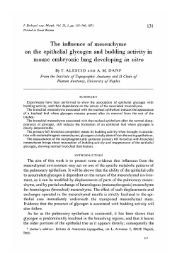
A quantitative assessment of
mesenchymal contribution to epithelial growth
rate in mouse embryonic lung developing
in vitro
ByTOMMASO ALESCIO 1 & EMILIA COLOMBO PIPERNO 1
From the Institute of Topographic Anatomy, University of Naples
INTRODUCTION
The experimental analysis of factors operating in lung morphogenesis has
shown that the normal pattern of the bronchial tree is related to a mesenchymal
budding-promoting activity, distributed non-uniformly throughout the mesenchymal mantle. No budding is present in the absence of mesenchyme (Dameron,
1961 a, b, 1962). The position of secondary epithelial buds may be changed by
substituting 'active' for 'inactive' mesenchyme (Alescio & Cassini, 1962a, b;
see also for a review and pertinent references Sorokin, 1965).
Recent in vitro experiments by Colombo Piperno (1966) dealt with the problem
of whether the inductive influence of pulmonary mesenchyme could also operate
as a stimulus for the expansion and global growth of the epithelial tree. In order
to answer this question a procedure was devised to modify, in a definite way, the
quantitative ratio of mesenchyme to epithelium of the 11-day pulmonary rudiment of mouse embryos, without disorganizing the natural topographic arrangement of the two tissues. The essential procedure was the removal of definite
parts of the epithelial tree while preserving the normal amount of mesenchyme.
In this way at the starting time of the experiments the normal 1:1 ratio of
epithelium to mesenchyme was changed to less than one.
The size of the epithelial tree of living lung rudiments was measured using a
planimetric procedure (Alescio, 1965), and growth curves were obtained showing
that, when the relative amount of mesenchyme was increased, an enhanced rate
of epithelial growth may be obtained.
The purpose of the present work is to give further evidence of the mesenchymal
growth-stimulating activity, therefore the experiments by Colombo Piperno
(1966) were repeated, with particular attention to the following points:
(1) In previous experiments a number of rudiments were eliminated from the
data because imperfect transparency due to precocious superimposition of buds
1
Authors' address: Istituto di Anatomia topografica, via L. Armanni 5, Napoli, Italy.
214
T. ALESCIO & E. COLOMBO PIPERNO
rendered them unfit for planimetric measurements. Since the rudiments showing
precocious superimposition of buds could represent the most rapidly growing
ones, whether their exclusion would affect the growth evaluation was studied.
(2) Conspicuous regeneration of part of the removed epithelial tree was
observed at the sites of the epithelial excision in several cases: hence, could the
regeneration process in some way modify the growth properties of the whole
epithelial tree ? When a pulmonary rudiment is experimentally deprived of part
of its epithelial tree and the removed part is being regenerated, one may expect
two different causes of error affecting growth assessment: (a) regeneration may
represent a special kind of growth, following a pattern very different from that
of the normal growth; (b) regeneration may also require part of the available
mesenchymal activity, which is therefore withdrawn from the normal growth
of the non-regenerating portion of the tree. The end result is that the inclusion
as well as the rejection of the regenerated part in the epithelial growth measurements may bias growth estimates.
(3) The increase of epithelial area was taken as an index of epithelial growth.
However, such an increase may also depend in part on spreading of the epithelial tree due to accumulation of fluid inside the tracheobronchial cavity.
Therefore, a second method of growth measurement, independent of the use of
area and based on computation of total number of terminal buds produced
during the cultivation period, was used as a collateral criterion.
The general features of the earlier results were confirmed, and further evidence of a mesenchymal participation in the determination of the epithelial
growth rate was obtained.
MATERIALS AND METHODS
Completely isolated lung rudiments from 11-day mouse embryos ($ C 57 BL x
S BALB/c) were cultured at the surface of a clot of chicken plasma and 9-day
chick embryo extract in hanging drop under the following experimental conditions (Fig. 1):
Exp. 1. Intact whole lung rudiment.
Exp. 2. Isolated right lung.
Exp. 3. Isolated left lung.
Exp. 4. Right primary bronchus of the epithelial tree with whole pulmonary
mesenchyme.
Exp. 5. Left primary bronchus of the epithelial tree with whole pulmonary
mesenchyme.
In Exps. 2 and 3 the left and the right parts, respectively, of the rudiment were
removed. Exps. 4 and 5 were done by externalizing the left or the right part of
the epithelial tree to be removed through a longitudinal cut in the mesenchymal
lining. The epithelium was then cut off as close as possible to the trachea
bifurcation, leaving the mesenchyme intact.
Epithelial growth rate in lung
215
Exp. 1 was used as a control procedure in order to obtain information about
the epithelial growth rate in a situation analogous to that of normal development in vivo. In Exps. 2 and 3 the total size of the rudiments was reduced to
about one-half, but no change was made in the epithelium/mesenchyme ratio.
They were devised to give information on the ability of the lung rudiment to
compensate for the loss of a conspicuous part of its tissue mass. In Exps. 4 and 5
the rudiment contained all the mesenchyme and only the right or left portion of
the epithelial tree (right or left primary bronchus), so that the epithelium/
Fig. 1. Plan of the research presented in form of schematic drawings from camera
lucida records. The epithelial surface is dotted in the figure. Exp. 1: intact whole lung.
Exp. 2: isolated right lung. Exp. 3: isolated left lung. Exp. 4: right primary bronchus
with whole mesenchyme. Exp. 5: left primary bronchus with whole mesenchyme.
mesenchyme ratio was changed to about one-half. It should be added that
because of the uneven size of the right and left lungs at explantation time the
left bronchus is smaller and less branched than the right one. Therefore Exps. 2
and 3, and respectively Exps. 4 and 5, are not fully parallel, since the left lung
epithelium represents about 34 % and the right lung epithelium about 60 % of
the total epithelial surface of the lung rudiment at the 11th day of gestation,
the remaining 6 % belonging to a small segment of trachea included in the whole
epithelial surface measurements made as described below.
Special care was taken to stretch the rudiments flat on the surface of the
plasma clot, in order to avoid any superimposition of bronchial buds. Indeed,
all the successfully cultured rudiments turned out to be well suited for an
accurate planimetric measurement.
216
T. ALESCIO & E. COLOMBO PIPERNO
The regeneration of the removed part of the epithelial tree was successfully
avoided by cutting the epithelium to be removed exactly at the level of the
tracheal bifurcation, so that no stump of the removed bronchus was left inside
the emptied mesenchymal mass.
Living cultures were drawn by camera lucida immediately after explantation
(zero time) and then again after 5, 20, 28, 44 and 52 h of growth at 37 °C.
0 hours
52 hours
A. = 19900
Left: nQ = 2
Right: n o = 4
Fig. 2. Schematic drawings from camera lucida records showing an example of
growth measurement by the two following criteria: (a) planimetric measurement of
the epithelial surface (dotted in the figure), given in arbitrary planimetric units,
and growth factor (G.F.) calculation; (b) computation of total number of terminal
buds developed from left and right primary bronchus.
The first criterion of growth measurement was the same as already used in
earlier experiments (Alescio, 1965; Colombo Piperno, 1966). The surface area
(Fig. 2 a) of the bronchial epithelial tree was measured on the camera lucida
drawings by planimetry; to obtain readily comparable data from all cases,
growth factors (AJAQ) were calculated as the ratio of the area at each observation time (At) to the area at zero time (.40). The mean growth factors were
plotted as a function of time and growth curves were obtained.
The use of growth factors (AtIA0) is necessary for two main reasons: (a) the
Epithelial growth rate in lung
217
size and branching stage of the epithelium on explantation differs to some extent
in the different rudiments; (b) all experimental treatments, implying the removal
either of half organ or half epithelium, reduce the total amount of epithelium.
The use of the ratios At\AQ, called 'growth factors' throughout this work,
makes the value of the epithelial tree always equal to 1 on explantation, in spite
of differences of absolute values of size. Therefore, the measures given here are
relative growth measures, allowing comparisons between epithelial rudiments of
differing size. For instance, a rudiment showing a growth factor equal to 2-0
grew to twice its initial size; its growth rate is therefore larger than that of
another rudiment having a growth factor of 1-5, but this does not necessarily
mean that the former is larger in absolute size.
The second criterion of growth evaluation (Fig. 2 b) is based on the assumption
that, during the developmental stage considered here, the main feature of lung
morphogenesis is the appearance of new epithelial buds. Accordingly, the total
Table 1. Number of cultured rudiments
Used for measurements
Degenerated
Total
Exp. 1
Exp. 2
Exp. 3
Exp. 4
Exp. 5
Total
20
12
11
17
11
71
4
9
13
6
6
38
24
21
24
23
17
109
number (nt) of terminal buds (mean value of all rudiments within each experimental group) was taken as an index of growth. This second criterion should be
considered as a collateral and less efficient one, for the following reasons: (a) it
does not take into account the considerable lengthening and shape modifications
of buds preliminary to their next dicotomous division, which participate in the
global pulmonary growth; (b) the computation of terminal buds may be considered a subjective and questionable matter. For the purpose of this work only
those terminal buds showing a longitudinal axis of at least 3 mm in the scale of
our camera lucida drawings were counted.
Table 1 shows that the percentage of degenerated rudiments was distributed
differently amongst the five different types of experiments. While the lowest
degeneration frequency was observed in the control group (Exp. 1) as might be
expected, the highest percentage of degenerated rudiments was present in
Exps. 2 and 3. This might be related to the considerable reduction of total mass
of the rudiments, perhaps making them too small to undergo good organotypic
development. Degeneration, when present, was very precocious, and already
clearly noticeable during the first 12-18 h of development. The degenerated
rudiments were not used for growth measurements.
218
T. ALESCIO & E. COLOMBO PIPERNO
RESULTS
Lung rudiments cultured as a whole (Exp. 1) showed a morphogenetic pattern
very similar to that already described in an earlier paper (Borghese, Alescio &
Cassini, 1963). New bronchial buds were produced in an orocaudal direction
from the lateral side of both main bronchi, and they grew and divided, giving
rise to smaller and smaller bronchial buds. No branches were produced during
this period from the medial side of the main left bronchus, while the infracardiac bronchus developed from the medial side of the main right one. This
pattern of branching is known to be perfectly comparable to what occurs in
normal conditions in utero, the main appreciable difference being that the rate of
branching in vitro is slower, so that the pulmonary tree produced in culture
conditions is simpler than it would be at the same total age in the in vivo
development.
When part of the rudiment was removed (Exps. 2 and 3), morphogenesis
went on in vitro without any qualitative divergency from the process outlined
above. An exception is that secondary buds are produced from the medial side
of the isolated primary left bronchus (Exp. 3) in about 60 % of the cases. As
earlier stated (Alescio & Cassini, 19626) this behaviour shows a shift from the
normal morphogenetic pattern, since budding from the medial aspect of the
primary left bronchus is never observed in normal conditions from the 11th to
the 13th day of development, in vivo or in vitro.
When the left or the right primary bronchus was removed (Exps. 4 and 5),
the mesenchymal mantle deprived of its own epithelium soon fused more or less
completely with the mesenchyme belonging to the contralateral lung, so that the
latter came in contact with a mesenchymal milieu about twice as large as normal.
In this case budding went on in an extremely active way and a very rich bronchial
arborization was produced. It may be relevant to the interpretation of our
results that the fusion of the emptied mesenchyme with the contralateral lung
was quantitatively different from case to case. In some instances spreading and
cellular migration from the mesenchyme reduced the amount of tissue gathered
round the rudiment.
The average growth factors (AJAQ) obtained from planimetric measurements
of the epithelial area are plotted as a function of time in Fig. 3. They become
larger from the intact lung rudiments (Exp. 1) to the isolated right (Exp. 2) and
left lung (Exp. 3), and then to the right (Exp. 4) and left (Exp. 5) primary
bronchus cultured in association with whole mesenchyme. Therefore, the hypothesis of an acceleration of growth rate is clearly suggested from the data.
Since the rate of growth with time appears linear in Fig. 3, the linear regression
equations were calculated from the average values of growth factors, to obtain
an easier comparison between growth rates of the different experiments. The
regression lines are shown in Fig. 4. The fit of the linear regression lines to the
data was tested by Fisher's F test, and highly significant values of F were
Epithelial growth rate in lung
219
obtained in all cases, thus showing that the hypothesis of a linear relationship
of growth factors to time may be accepted.
As one can expect from the average values of growth factors, the slope of the
regression lines increases sequentially (Fig. 4) from the whole lung rudiments
cultured in their normal anatomical conditions (Exp. 1), to isolated right lung
10 20 30 40 50 60 0 10 20 30 40 50 60
Fig. 3
Time (h)
Fig. 4
0 10 20 30 40 50 60
Fig. 5
Fig. 3. Growth curves of Exps. 1-5. The average values of growth factors (At/A0)
are plotted as a function of time. Standard errors (s/^) are indicated when larger than
the points as plotted.
Fig. 4. Regression lines of the average values of growth factors on time.
Fig. 5. Regression line of the average values of growth factors on time after Colombo
Piperno's (1966) experiments.
(Exp. 2), to isolated left lung (Exp. 3), to right epithelium cultured in association
with whole mesenchyme (Exp. 4), to left epithelium in the same conditions
(Exp. 5).
The statistical significance of the differences in slope of the various regression
lines was studied by comparing the regression coefficients two by two by means
of Student's t test. The results of these comparisons are given in Table 2, and
can be summarized as follows: (a) the rates of growth observed in the isolation
experiments (2 and 3) and mesenchyme doubling experiments (4 and 5) are first
compared with the rates of growth of the whole lung rudiments (Exp. 1). The
difference is not significant as far as the isolated right lung is concerned; it is
220
T. ALESCIO & E. COLOMBO PIPERNO
highly significant in all other cases, (b) Isolation experiments (2 and 3) and
mesenchyme doubling experiments (4 and 5) are then compared to each other;
the difference is not significant, (c) The rates of growth of right and left lung
associated with whole mesenchyme (Exps. 4 and 5) are compared with the rates
Table 2. Statistical comparisons of growth rates under
different experimental conditions
Experiments
1:2
1:3
1:4
1:5
2:3
4:5
2:4
3:5
Comparisons
D.F.
Whole lung: isolated right lung
0-02985 003526 2-2172
Whole lung: isolated left lung
0-02985 004080 3-8153
Whole lung:right epithelium
0-02985 0-04737 4-4130
with whole mesenchyme
Whole lung:left epithelium with
0-02985 0-05869 8-2165
whole mesenchyme
0-03526 0-04080 1-6390
Isolated right lung:isolated left
lung
0-04737 0-05869 2-2504
Right epithelium with whole
mesenchyme:left epithelium
with whole mesenchyme
0-03526 0-04737 2-7839
Isolated right lung:right epithelium with whole mesenchyme
Isolated left lung:left epi0-04080 0-05869 42494
thelium with whole mesenchyme
bx = Regression coefficient of the first variable on time.
b2 = Regression coefficient of the second variable on time.
n.s.
< 0010
< 0-005
< 0-001
n.s.
n.s.
< 0050
< 0-010
Table 3. Statistical comparisons of growth rates with those from
Colombo Pipemo's (1966) experiments
Experiments
Comparisons
D.F.
t
99 0-02990 0-02985 00295
Whole lung
100 0-03990 0-03526 1-6811
Isolated right lung
99 0-03726 0-04080 1-1606
Isolated left lung
Right epithelium with whole
100 0-04517 0-04737 0-5116
mesenchyme
5:5
Left epithelium with whole mesen- 101 005496 005869 10163
chyme
b, = Regression coefficient of growth on time from previous experiments.
b, = Regression coefficient of growth on time from the present experiments.
1:1
2:2
3:3
4:4
P
n.s.
n.s.
n.s.
n.s.
n.s.
of growth of the corresponding isolated lungs (Exps. 2 and 3). Both differences
are statistically significant, (d) The regression lines obtained in these experiments
were compared with the regression lines found in the same experiments by
Colombo Piperno (1966) and reported in Fig. 5. Table 3 presents the results of
statistical comparisons by Student's t test. In spite of slight changes in the slope
Epithelial growth rate in lung
221
of the regression lines, all the differences are not statistically significant. We may
conclude that the present results are in good agreement with those previously
reported.
Further evidence of a higher rate of growth and morphogenesis was obtained
from the data on the terminal buds. Fig. 6 is a plot as a function of time of the
average number of terminal buds observed in the isolated right lung (Exp. 2),
in the right primary bronchus with whole mesenchyme (Exp. 4) and, for comparison, in the right primary bronchus of whole rudiments (Exp. 1). Growth
10
20
30
40
50
60 .0
10
20
30
40
50
60
Time (h)
Fig. 6
Fig. 7
Fig. 6. Average number of terminal buds of the right lung as a function of time.
1, Right primary bronchus of whole lung rudiments (Exp. 1); 2, isolated right lung
(Exp. 2); 4, right lung epithelium with whole mesenchyme (Exp. 4). Standard errors
Cs/V'O are indicated, when larger than the points as plotted.
Fig. 7. Average number of terminal buds of the left lung plotted as a function of time.
1, Left primary bronchus of whole lung rudiments (Exp. 1); 3, isolated left lung
(Exp. 3); 5, left lung epithelium with whole mesenchyme (Exp. 5). Standard errors
(s/*Jri) are indicated when larger than the points as plotted.
curves are obtained showing the pattern of growth as measured in terms of
number of epithehal buds produced during the experimental period of time. It
appears that after both experimental treatments (Exps. 2 and 4) the right
primary bronchus produced an average number of buds clearly larger than
under normal conditions (Exp. 1). Budding activity in Exp. 4 appears only
slightly more intense than in Exp. 2.
The average number of terminal buds observed in cultures of isolated left
lung (Exp. 3) and left primary bronchus with whole mesenchyme (Exp. 5) in
comparison to that of the left lung of whole rudiments (Exp. 1) is plotted
222
T. ALESCIO & E. COLOMBO PIPERNO
against time in Fig. 7. Again, in both experiments (3 and 5) the average number
of terminal buds was well above that of the controls (Exp. 1). The relative
increase of mesenchyme (Exp. 5) also brought about a rate of budding clearly
higher than that of simple isolation experiments (Exp. 3), as the last point of the
Exp. 5 growth curve lies well above the corresponding point of Exp. 3.
Comparison of Fig. 6 with Fig. 7 shows that stimulation of budding activity
is present in right and left lung under both experimental treatments, irrespective
of their different size at starting time; the average number of buds at zero time
is about 4 for the right lung and about 2 for the left lung. The general pattern
of budding stimulation is the same. The main difference is that the mesenchyme
Table 4. Statistical comparisons of average number of buds
produced under different experimental conditions
Experiments
1:2
1:4
2:4
1:3
1:5
3:5
Comparisons
Right part of the whole lung:
isolated right lung
Right part of the whole lung:
right epithelium with whole
mesenchyme
Isolated right lung: right epithelium with whole mesenchyme
Left part of the whole lung :isolated left lung
Left part of the whole lung: left
epithelium with whole mesenchyme
Isolated left lung:left epithelium
with whole mesenchyme
D.F.
mx
m2
t
P
29
8-9473 92-0833
3-5139
< 0-005
33
8-9473
3-8218
< 0-001
26
120833
28
5-2105
7-6363
3-8936
< 0001
28
5-2105
10-3636
6-5782
< 0001
20
7-6363
10-3636
2-5574
< 0025
12-8750
12-8750 0-5520
n.s.
mx = Mean of first variable.
m2, = Mean of second variable.
doubling experiment seems to be more effective in the case of the left lung, as
a clear difference is present in the end results of Exps. 3 and 5. In the case of
the right lung, the results obtained with Exps. 2 and 4 are not significantly
different.
Table 4 reports a statistical analysis by Student's t test of average numbers of
buds present after 53 h cultivation. Budding activity of the isolated right lung
(Exp. 2) and of right primary bronchus with whole mesenchyme (Exp. 4) are
both significantly higher than that of the controls (Exp. 1). However, the difference of budding rate of Exps. 2 and 4 is not statistically significant. The
budding activity of isolated left lung (Exp. 3) and of left primary bronchus
associated with whole mesenchyme (Exp. 5) are significantly higher than the
controls (Exp. 1). Moreover, Exp. 5 shows a budding rate significantly faster
than that in Exp. 3.
Epithelial growth rate in lung
223
DISCUSSION
In the present research an attempt was made to estimate the growth rate of
the lung rudiment epithelial tree and its dependence upon mesenchymal factors.
A critical evaluation of these results must be primarily concerned with the use
of area as a measurement of growth. Mitotic activity is indeed thought to be the
basic mechanism of epithelial growth (Sorokin, 1965); therefore, budding and
epithelial expansion should be strictly correlated with cellular proliferation, but
the nature of their relationship still remains unknown. Furthermore, one should
remember that a number of mechanisms besides cellular proliferation do participate in the size determination of the epithelial tree; of these, the size of the
bronchial cavity, and changes in histological structure seem to be, at first sight,
of some importance. Also, research by Glucksmann (1964) on human embryonic
lungs developing in vitro points out the role of cell death in normal morphogenesis. However, all these factors take part in epithelial morphogenesis as
normal developmental processes. Therefore, our criterion, being a measure of
a quantity (size of the epithelial tree) which is the overall consequence of the
different actions participating in lung development, seems to be reliable in
giving an appraisal of epithelial growth, whatever elementary processes may be
involved in its determination. Even if not a measure of cellular proliferation
rate, it certainly may afford a useful approach for quantification of the morphogenetic process.
Causes of error may come from abnormal shape changes in the epithelial tree
of the cultured rudiments, such as spreading of the bronchial cavity due to the
accumulation of fluid in the ducts. However, such bronchial spreading was never
observed to any appreciable extent during the present research. Also, and more
important, the second criterion of growth measurement, namely computation
of the total number of terminal buds, may greatly help in a correct evaluation
of morphogenesis. Indeed, the appearance of new epithelial buds requires a
normal state of epithelial cavity distention; no or very little budding activity is
present where accumulation of fluid spreads out the bronchial epithelial wall.
Therefore, computation of total number of terminal buds, even if less accurate
as a full description of epithelial growth, may be a useful collateral criterion to
rule out some possible reasons of biased measurements. Its basic agreement with
the epithelial surface measurement data may improve the reliability of these
results.
The relation of growth to time remains linear in all experimental treatments
(extirpation of left or right lung; removal of the left or right part of the epithelial tree). However, the slope increases as one goes from the whole rudiment
to combining part of the epithelial tree with the whole amount of mesenchyme.
Growth of the right and left primary bronchus associated with whole mesenchyme (Exps. 4 and 5) is in fact remarkably faster than that of whole lung
rudiments (Exp. 1). The results are less clear as far as isolation experiments are
224
T. ALESCIO & E. COLOMBO PIPERNO
concerned. A rate of growth significantly higher than that of the controls is
demonstrated for the isolated left lung (Exp. 3) but not for the isolated right one
(Exp. 2), in contrast to the results of Colombo Piperno (1966). On the other
hand, the experiments in which the proportion epithelium/mesenchyme was
brought to about half normal (Exps. 4 and 5) gave clear evidence of a rate of
growth significantly faster than that of the corresponding isolation experiments
(2 and 3). This result is in complete agreement with the earlier findings.
The results of epithelial area measurements are confirmed by the data on the
rate of budding, the rudiments showing the same general trend towards an
acceleration of budding activity as a function of the experimental treatments.
The only difference concerns the effect of Exp. 2: right lung isolation. When
measured as rate of budding, the growth of isolated right lung is significantly
higher than that of the controls, closely approaching that obtained by the combination of right lung epithelium with whole mesenchyme (Exp. 4). .
The main purpose of these experiments was to confirm the earlier results by
clarifying some doubtful points, such as the effect of epithelial regeneration and
the possibility of distention and spreading of the epithelial tree due to accumulation of fluid within the bronchial cavity. Epithelial regeneration was successfully
avoided, and a new criterion of measurement independent of spreading gave
very similar general results. The present data are in good general agreement with
those previously reported, and we may also now conclude that a substantial
reproducibility of the experimental results is demonstrated.
As a general conclusion it appears that isolation experiments (2 and 3), in
which the epithelium/mesenchyme ratio is not changed but the total mass of
the rudiment is reduced, increase the growth rate of epithelium as compared
with that of the intact rudiments. The enhancement of growth under these
conditions might be tentatively interpreted as a kind of'compensatory growth',
as though the explant is attempting to reach the same order of magnitude, whatever its initial size. Of course, this expression is far from being an explanation
of how this phenomenon takes place; a reference could also be made to the
controversial point of growth autoregulation. Data in favour of such autoregulation were obtained with embryonic heart and metanephros cultures by
Weiss (1952, 1955), while other in vitro experiments by Nicoll (1965) provided
no evidence for autoregulation of duct growth in mammary glands.
A different situation occurs in Exps. 4 and 5, where a part of the epithelial
tree is combined with the whole mesenchyme. The epithelial growth rate is
markedly increased, not only in comparison with that of the whole rudiments,
but also when compared with that of the isolated lungs. Under these conditions
the ratio of mesenchyme to epithelium is increased to about twice normal at the
starting time of the experiment, and the result could support the idea of a
stimulating effect of the mesenchyme on the total growth of the epithelium.
Moreover, this interpretation becomes even more attractive if one considers
that the effect seems to be higher for the left epithelium (Exp. 5) than for the
Epithelial growth rate in lung
225
right (Exp. 4). In the former case, the epithelial tree being smaller at the beginning, this ratio was yet further increased.
A larger statistical fluctuation of the mean growth factors is indicated (Fig. 3)
in Exps. 4 and 5. Such a fluctuation may be explained in part by the variability of spreading and cellular migration from the emptied mesenchyme.
Where more migration is present, a smaller amount of mesenchyme fuses with
the contralateral lung. For this reason the rate of epithehal growth may be
inversely dependent on the rate of mesenchymal cell migration, but this point
was not established.
It may be concluded that under the reported conditions of growth in vitro,
lung mesenchyme exerts some inductive influence on the total growth of the
epithelium expressed as total amount of branching, and that this influence
appears to some extent proportional to the amount of mesenchyme relative to
epithelium. These results are in agreement with earlier observations by Grover
(1961), who found that an optimal ratio between mesenchymal and epithelial
cells was necessary to obtain development of tubuh during the reaggregation of
11-day embryonic chick lung cells in vitro, and by Dameron (1961a, b, 1962),
who observed that in chick embryo lungs cultured in vitro tubule formation is
in direct proportion to the amount of mesenchyme.
A cellular basis for this mesenchymal effect is indicated by the results by
Sobel (1958), who found that mitotic activity in the mouse embryonic pituitary
epithelium is present only when in association with mesenchyme. Rutter,
Wessells & Grobstein (1964) also reported results showing that the rate of
mitotic activity in pancreatic rudiments was significantly lower in the absence
of mesenchyme. Wessells (1963) showed that in chick epidermis the thymidine
incorporation rate is influenced by the quality or age of dermis present.
Other research (Cohen, 1964, 1965) deals with the problem of isolation and
chemical characterization of a substance stimulating epidermal growth found
in salivary glands. Evidence was given for its stimulating activity on epidermal
cell proliferation.
These results seem to strengthen the hypothesis that the duration of the
epithehal cell cycle may depend on mesenchymal factors. No deduction can be
drawn from our results on the action of mesenchyme on the cellular kinetics of
the growing bronchial epithelium, since the total epithelial growth, on our
criteria, may be only indirectly correlated with the rate of cellular proliferation.
If the model of growth of the epithehal tree as a whole reflects the kinetics of the
epithehal cells, namely if the cellular population growth follows a linear rate,
the fraction of dividing cells should drop with time, and the theoretical relation
should be of hyperbolic type. In this context it should be remembered that,
although the mitotic index alone cannot account for cellular kinetics, Sorokin
(1961) found a slowing down of the mitotic index with time in rat and guineapig lungs cultured in vitro.
15
J E E M 17
226
T. ALESCIO & E. COLOMBO PIPERNO
SUMMARY
1. The growth rate of the epithelial tree of mouse embryonic lung cultured
in vitro was studied after removal of the right or left part of the rudiment, or
after modification of the epithelium/mesenchyme ratio to about one-half of the
normal.
2. The epithelial growth rate is enhanced in both experimental conditions
well above that of the controls.
3. No satisfactory explanation can be put forward, so far, for the fact that
when the total mass of the rudiment is reduced without any change in the ratio
of the tissues, the growth rate of the epithelium is enhanced.
4. From the results of the experiments in which the ratio of the two tissues
was experimentally changed the conclusion may be suggested that, under the
reported conditions of growth in vitro, lung mesenchyme exerts some stimulating influence on the total growth of the epithelium, expressed as total amount
of branching, and that this influence is to some extent proportional to the amount
of mesenchyme as compared with epithelium.
RIASSUNTO
Valutazione quantitativa del contributo mesenchimale al tasso di
crescita epiteliale nel polmone di topo coltivato in vitro
1. £ stato studiato il tasso di crescita dell'abbozzo epiteliale del polmone
embrionale di topo coltivato in vitro dopo asportazione della porzione destra o
sinistra dell'abbozzo, e dopo modincazione del rapporto dell'epitelio col
mesenchima ad un valore di circa meta del normale.
2. II tasso di crescita dell'epitelio risulta aumentato in entrambe le condizioni
sperimentali a valori superiori a quelli dei controlli.
3. Non puo essere attualmente proposta alcuna spiegazione soddisfacente
dell'aumento del tasso di crescita epiteliale osservato dopo riduzione della
massa totale dell'abbozzo e senza modincazione delle quantita relative di epitelio
e mesenchima, che sembra esprimere una tendenza dell'abbozzo a ripristinare le
dimensioni normali.
4. I risultati delle esperienze in cui il rapporto fra i due tessuti veniva modificato aumentando la quantita relativa di mesenchima, suggeriscono che, in
condizioni di cultura in vitro, l'accrescimento globale dell'epitelio e sottoposto
ad una influenza mesenchimale direttamente proporzionale alia quantita
relativa di mesenchima.
The authors are indebted to Professor Italo Barrai for his helpful advice on the statistical
part of this work. This research was performed under contracts of U.S.A.E.C. (NYO-3355-5)
and Euratom (043-65-1BIOI) and with a contribution from the Italian C.N.R.
Epithelial growth rate in lung
227
REFERENCES
T. (1965). Indicazioni preliminari per lo studio quantitative dell'accrescimento di
organi embrionali coltivati in vitro. Ric. sci. 35 (IIB), 237-49.
ALESCIO, T. & CASSiNr, A. (1962a). Induction in vitro of tracheal buds by pulmonary mesenchyme grafted on tracheal epithelium. /. exp. Zool. 150, 83-94.
ALESCIO, T. & CASSINI, A. (19626). L'interazione epitelio-mesenchimale nell'organogenesi del
polmone embrionale di topo coltivato in vitro. Z. Anat. EntwGesch. 123, 369-96.
BORGHESE, E., ALESCIO, T. & CASSINI, A. (1963). Experiments on induction and effect of
gamma radiation in mouse lung developing in vitro. In Cinemicrography in Cell Biology,
ed. G. G. Rose. New York: Academic Press.
COHEN, S. (1964). Isolation and biological effects of an epidermal growth-stimulating protein.
Natn. Cancer Inst. Monogr. 13, 13-38.
COHEN, S. (1965). The stimulation of epidermal proliferation by a specific protein (EGF).
Devi Biol. 12, 394-407.
COLOMBO PIPERNO, E. (1966). Osservazioni sulla crescita dell'epitelio polmonare in vitro.
Z. Anat. EntwGesch. 125, 227-44.
DAMERON, F. (1961 a). Influence de divers mesenchymes sur la differenciation de l'epithelium
pulmonaire de l'embryon de poulet en culture in vitro. C. r. hebd. Seanc. Acad. Sci., Paris
252,3879-81.
DAMERON, F. (19616). L'influence de divers mesenchymes sur la differenciation de l'epithelium pulmonaire de l'embryon de poulet en culture in vitro. J. Embryol. exp. Morph. 9,
628-33.
DAMERON, F. (1962). Role du mesenchyme dans la differenciation de l'ebauche epitheliale du
poumon embryonnaire de poulet en culture in vitro. Path. Biol., Paris 10, 811-16.
GLUCKSMANN, A. (1964). Mitosis and degeneration in the morphogenesis of the human foetal
lung in vitro. Z. Zellforsch. 64, 101-10.
GROVER, J. W. (1961). The enzymatic dissociation and reproducible reaggregation in vitro of
11-day embryonic chick lung. Devi Biol. 3, 555-68.
NICOLL, C. S. (1965). Growth autoregulation and the mammary gland. /. natn. Cancer Inst.
34, 131-40.
RUTTER, W. J., WESSELLS, N. K. & GROBSTEIN, C. (1964). Control of specific synthesis in the
developing pancreas. Natn. Cancer Inst. Monogr. 13, 51-65.
SOBEL, H. (1958). The behaviour in vitro of dissociated embryonic pituitary tissue. /. Embryol.
exp. Morph. 6, 518-26.
SOROKIN, S. (1961). A study of development in organ culture of mammalian lungs. Devi Biol.
3, 60-83.
SOROKIN, S. (1965). Recent work on developing lungs. In Organogenesis, ed. R. L. DeHaan
and H. Ursprung. New York: Holt, Rinehart and Winston.
WEISS, P. (1952). Self-regulation of organ growth by its own products. Science, N. Y. 115,
487-8.
WEISS, P. (1955). Specificity in growth control. In Biological Specificity and Growth, ed. E. G.
Butler. Princeton University Press.
WESSELLS, N. K. (1963). Effects of extra-epithelial factors on the incorporation of thymidine
by embryonic epidermis. Expl Cell Res. 30, 36-55.
ALESCIO,
{Manuscript received 27 June 1966)
15-2
© Copyright 2026 Paperzz