From www.bloodjournal.org by guest on December 29, 2014. For personal use only.
Comparative
Density
Peripheral
Blood
of the
T Cells
By
A 65.000
dalton
strated
to
nant
be
T cells,
the
but
surface
tory.
not
(CLI).
cells
and
demon-
density
nonsecre-
to
that
on
cases.
normal
as well
the
CLI
immunoglobulins
flow
a
usually
bearing
with
of
light
ent on all normal
circulating
T lymphocytes,
as well
greater
than 95% of thymocytes.
By IF microscopy
finding
dual
size
analysis,
between
(�ells
and
(�dl
Heparinized
with
donors.
The
RPMI-l640
cell
cells.’
thymus
Blood.
Vol.
from
in this
fetal
the
samples
(a kind
obtained
abortion.
10%
have
presence
calf
gift
57.
No. 4 (April),
were
serum
characteristics
a fetus
thymus
CEM,67
study.
of human
Child
lines,
thymus
of
14-16
tissue
1981
was
wk
from
20
isotype
(RPC-5.
was
used
adult
MoIt-4,68
IIPB-
at 37#{176}Cin
(FCS)
without
antibiotics.
of
original
malignant
the
UCSD,
gestation
obtained
j.g/ml).
used
in this
La Jolla,
following
during
with
of cell
(8402)
to
implications
of
differentiation
(a kind
gift
La Jolla,
of Dr.
Calif.).
IgG
used
protein
of
the
same
Md.)
20 zg/mI).
a
the
(concen-
Kensington,
Fluores-
(FL-RAMIG,
as
IgG28
Miles
secondary
Labo-
antibody
for
staining.
Preparation
Fresh
heparinized
(Roger
bated
20
for
centrifuged
venous
Bellon
at
400
g for
at
Technicon
Biological
humid
atmosphere.
minced
into
the
incubated
layer
over
and
was collected
with
10%
fetal
N.Y.)
was
calf
serum
Veterans
FCS,
Administration
San
5% CO2
finely
mononuclear
cells
Medical
of Hematology/Oncology,
of (�alifornia,
in
(l0%
(Grand
in 10%
viable
and
overnight
at 37#{176}C
and
placed
and
5 ml
centri-
penicillin-streptomycin
tissue
Service,
layered
N.J.)
by incubation
l%
Reagent.
for 30 mm at 37#{176}C
shed
suspension.
Division
then
was then
Island.
Thymus
Research
the
Department
Diego,
School
of
of Medicine,
(�alif
Supported
in part
by
the
Veterans
NOI-(’B-84250-31.
(ontract
(;rant
and
Grand
cell
collected.
Piscataway,
lymphocyte
were
incu-
was
N.Y.)
mixture
Inc..
supplemented
University
La Jolla,
Tarrytown.
3 ml of
and
layer
Separator
proteins
a single
and
leukocyte
and
at 400 g. The
Co..
with
France)
( Lymphocyte
This
1-glutamine,
Island
mixed
resuspended,
cells.
media
was
Neuilly,
7 mm,
Corp..
Cytophilic
ml)
solution
(Pharmacia
for 40 mm
(�ancer
filing
phagocytic
Ficoll-I1)paque
(10
37#{176}C.The
Instruments
1%
blood
Laboratories,
mm
3 ml of an iron
Administration.
and
American
National
Cancer
Society
IM-207.
Submitted
Address
Calif.)
92093.
(i
myeloma
(concentration
was
the
to determine
[.aboratories,
anti-mouse
Ind.)
to
used
on the cell surface
mouse
control
rabbit
belonging
was
dalton)
Bionetics
immunofluorescent
Medicine,
saline
antibody
purified
as a negative
Elkhart.
study.
corrective
patient
Foundation,
T cells.”
(65.000
A
Plasmagel
with
mouse
human
[.itton
cein-conjugated
FCS).
10
7 healthy
propagated
were
of Dr. S. Sarkar.
obtained
other
staining
The
lymphocyte
on a 9-yr-old
antigen
tration
RPMI-1640
a relative
T lympho-
from
staining
Research
and
for
ofT65
From
were
and
the
performed
specific
washed.
METHODS
CLL
and
The
Clinic
a monoclonal
fuged
hematopoietic
cell
human
thymocytes
samples
T-cell
used
with
lines
o Two
Fetal
was
blood
diagnosed
leukemic
and 8402,69i0
surgery
Medicine,
clinically
to
surface
cells
(Molt-4).
regard
were
low
T cells.
heterogeneous
staining
with
R. Fox, Scripps
of
Lines
four
cardiac
(�enter,
peripheral
patients
ALL,6
AND
CLI
with
discussed.
to remove
detected
normal
For purposes
of comparison,
of T-cell
lineage
as well as
analyzed.
MATERIALS
The
also
and
the
intensity
Thymocytes
two
to normal
bright.
findings
(�elI
as
it
flow cytometry.
we
CLL
resembling
from
fluorescence
T cells.
populations.
low-density
indirect
analyses
of peripheral
blood
lymphofrom patients
with CLL
were performed
to those
of normal
donors.
Utilizing
parameter
difference
cytes.
lines
were
with
median
circulating
similar
varied
ratories.
the density
of the T65 surface
antigen
CLL
cells than
on normal
peripheral
In this study,
we have confirmed
and
this
a lower
three
of T65
TIOl.
.
Histogram
cytes
(PBL)
and compared
of
subclass,
(MoAb)
termed
TIOl
In contrast,
T65 was not found
on normal
B cells,
B cell lines,
or secretory
CLL
associated
with a circulating
M-protein,
but was pres-
extended
Cells
Antisera
to either
K or
A type.’
immunofluorescence
(IF)
and cytofluorographic
analysis4
to differ
from normal
B lymphocytes
by the presence
of significantly
lower
densities
of slg. Recently,
another
antigen,
T65,
was
demonstrated
on nonsecretory
CLL cells with the use
of a previously
described
monoclonal
antibody
appeared
that
was lower
on
blood
T cells.
are
Normal
Leukemia
normal
density
lines
on
more
is a
monothe
composed
these
lines
had
of
uniform.
peripheral
T65
I. Royston
and
that
higher
antigen
as cell
cells
(slg),
in each case restricted
cells were
shown
by
T65
and
density
lymphocytic
the
L. Collins,
homogeneous
on
and
of
Antigen
Lymphocytic
than
malig-
detected
with
lymphocytic
leukemia
of B lymphocytes
surface
chain
CLL
patients
thymocytes.
all
been
chronic
surface
human
In
HRONIC
proliferation
clonal
from
compared
lineage.
C
has
M.
and
of immunofluorescence
relative
was
T cells
T-ceII
B cells,
cells
B. Wormsley,
of normal
T-CeII
Chronic
previously
surface
normal
By means
the
blood
antigen
the
and
S.
immunoglobulin-positive
cytometry.
CLI
specific
on
of leukemic
surface
leukemia
on
T-cell
present
Human
September
reprint
V-Il
2, 1980;
requests
lE,
1981 by Grune
U(’SD
& Stratton.
accepted
November
to Ivor
Royston.
School
of
18, 1980.
M.D..
Medicine,
Department
La
Jolla.
of
Calif.
Inc.
()006--4971/8I/5704--0005$0I.00/0
657
From www.bloodjournal.org by guest on December 29, 2014. For personal use only.
WORMSLEY,
658
COLLINS,
AND
ROYSTON
500
NORMAL
250
I
0:
LU
500
-J
-J
LU
B
:
CLL
____
THYMUS
(-)
I
250
I
0
0
510
510
0
FLUORESCENCE
INTENSITY
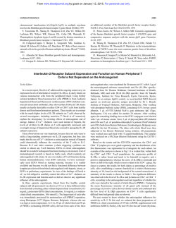
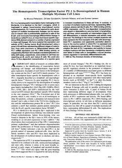
Fig. 1 .
Fluorescence
profiles
(control
versus positive)
of peripheral
(B). and normal
thymocytes
(C) stained
with TiOl . The arrows
designate
blood lymphocytes
from a normal
the median
intensity
fluorescence
donor (A).
values.
a patient
with
400
>-
(I)
z
uJ
I-
300
2nd
Peak
w
L)
w
L)
C,)
w
0:
200
0
:D
-J
1i
z
C
100
Is,
Peak
uJ
0
LU
Q->-x>
ck:(
ED
a
L)
tf
:i:
L)
c:
L)
J
Lu
NORMDAL
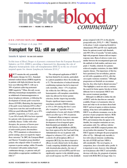
Fig. 2.
Summary
of median
:i:
:t
>
-,
-�
fluorescence
values
THYMUS
for
normal
�a’
OD
LiJ
Li
CLL
intensity
-
J
I-
PBL.
CLI
cells.
thymocytes.
CELL
and
cell
0
()
LINES
lines.
CLI
From www.bloodjournal.org by guest on December 29, 2014. For personal use only.
T65
ANTIGEN
ON T CELLS
separated
by
including
cell
cases
Ficoll-Hypaque
lines,
viability
Imni
washed
were
aliquots
each
resuspended
of
TIOl
FCS
and
and
resuspended
secondary
sample
(positive)
inactivated
and
through
and
FCS
Analysis
and
was
used
signal
for
as
analyzed.
sis,
with
were
fluorescence
determined.
the
the
the percent
positive
(y-axis).
dual
axis
each
cells
stained
nm)
of scatter
cells
were
and
analyscatter
were
on
centrally
presented
(x-axis)
as
versus
representing
sample
was
antibody
was
TIOl
cells
with
stained
with
broadly
revealed
with
median
blood
TiOl
are
tion of TIOl
shows
a more
cence
intensity
TIOI
shown
fluorescence
T cells (Fig.
distribution.
normal
PBL
cells
profiles
in Fig.
In this study,
was 60%-77%.
�
consisted
fluorescence
in patients
uniform
intensity
was of heterogeneous
stained
point
to the
Normal
peripheral
a broad
heterogeneous
range
ofTIOl
In contrast,
the
with
nonsecretory
show
IC). The
cells in
distribu-
two
first
peaks
peak
CLL
of
was
was
fluoresof low,
(median
channel
84), and the other
higher
intensity
(median
chan-
nd 332) staining.
Greater
were positive
for TIOI.
median
is shown
the
arrows
than
95%
of the thymocytes
fluorescence
intensity
of each
case
in Fig. 2. All CLL cases had a lower
value
than did normal
PBL,
while
the values
for
lines
varied
widely.
The
median
value
for the
intense
thymocyte
subpopulation
was comparable
that for CLL,
subpopulation
whereas
the
was comparable
more
intense
to the normal
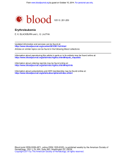
of light
(vertical)
are presented
scatter
sedimentation
at unit
had
PBL
thymocyte
T cells
does
versus
fluoin Fig. 3.
not
directly
gravity.’3
The
cytogram
low fluorescence
has
light
In all cases
a greater
degree
and
tended
to
of light
be more
for thymocytes
intensity
peak
of two distinct
size populations.
The
intensity
peak consisted
of a single
higher
popu-
case
( Fig. 4 A and B), considered
to be of non-T,
non-B
origin
(TIOl
slg
revealed
no TIOl
cells
above
background
control.
Analysis
of the second
case
,
(Fig. 4 C and
lating
M-protein
(4.8%)
ofTIOl
D),
),
which
was associated
with a circuof lgMK, revealed
a small
population
PBL with a fluorescence
distribution
for normal
T cells.
DISCUSSION
A 65,000
cells
thymocytes
lation
of cells midway
between
the other
two in relative size.
We also examined
two cases
of CLL
shown
to be
TIOl
(Fig.
4). Analysis
of the PBL
from
the first
TI 01.
cells in patients
with CLL
(Fig.
IB)
homogeneous
and decreased
fluoresthan
do normal
PBL.
The range
of
70%-90%.
Thymocytes
cence
intensity
(Fig.
The
studied
I (the
channels).
lA) show
of
TIOl
the
RESULTS
immunolluorescence
degree
distributed.
that
the
expected
channel
positive
with
positive
of
(488
located
channels
reactive
percent
50H
parameter
were
The
of
passed
in 2 ml
20,000
profiles
510
of
to each
as staining.
as generation
case
the
the CLL
cells
than
normal
then
source
Although
has
The
of the
coincide
with the cell size, a positive
correlation
been
found
between
relative
size measured
by
and
description
laser
signals
over
of cells
percent
peak.
tested,
scatter
FCS-AZ.
were
day
horizontal
intensity
percent
first
scatter
heat-
Cytotluorograf
by using
fluorescence
channel
in the
ml
added
A detailed
each
scatter
intensity
by subtracting
from
In
on
fall
a 30-mm
resuspended
Ortho
as well
size.
PBL
in each
The
determined
an
obtained
fluorescence
cells
50 il
microliters
was
finally
Mass.).
intensity
relative
of cells
median
control
cell
Normal
Fifty
1:30,
Two
I
1640-10T.
A 5 W argon
In addition,
of
and
activation
Cytograms
histograms
number
using
of
for convenience.
the
out
published.’2
After
on the same
rsis
fluorescence
axis.
with
The
azide
with
over
FCS-AZ.
Anal
a measure
the vertical
in all
sodium
stained
(control).
diluted
Wcstwood,
has been
and
washed
analyzed
carried
0.02%
layered
as above
were
Instrunients.
the system
and
x 10 cells/mI.
were
I640-10.
washed
Cells
was
RPC-5
at 0#{176}
for 30 mm.
Cviofluorographic
(Ortho
removed
FL-RAMIG
incubated
+
of 5.0
were
samples
in 25 l
of 1’7. formalin.
FCS
centrifuged.
antibody,
sample
PBL. Seventy-five
preparations.
Cytograms
displaying
scatter
rescence
intensity
(horizontal)
in l640-l0
0#{176}C,the
at
cell
to staining.
95..
to a concentration
of each
incubation
prior
Staining
FCS-AZ)
25-il
659
All
3 times
than
unofluorescent
Cells
CLL CELLS
centrifugation.
were
was greater
( 1640-l0T
AND
cell
less
to
in
been
dalton
T-cell-specific
shown by Royston
et al.
antigen
termed
T65
to be present
on slg
cells of patients
with nonsecretory
(absence
lating
M-protein)
CLL.
We have compared
fluorographic
analysis
using
a monoclonal
of circuby cytoantibody,
termed
TIOI,
the relative
density
of T65
on the
surface
of normal
peripheral
blood T lymphocytes
and
cells from patients
with CLL.
For further
comparison,
four cell lines of T-cell
lineage
and normal
human
thymocytes
were examined.
The CLL cells, in all cases
studied,
had lower surface
density
of T65 and were of
a more homogeneous
nature
than normal
circulating
T
lymphocytes.
The cell lines varied
from
high-density
heterogeneous
staining
of 8402
to the low-density
uniform
staining
of Molt-4.
Investigators’4’5
have
previously
suggested
that
CLL cells represent
immature
cells arrested
early
in
B-cell differentiation,
manifested
in part by the faint
immunofluorescent
compared
to normal
nonsecretory
CLL
staining
of slg on the
B cells. The presence
cells,
also
in decreased
provides
further
evidence
and may
indicate
arrest
lymphocyte
diflerentiation.
The absence
ofTiOl
from
other
B-cell
CLL
cells
of T65 on
density,
in support
of this hypothesis
at an even earlier
stage
reactivity
proliferative
with
diseases”
abnormal
of
cells
is consistent
From www.bloodjournal.org by guest on December 29, 2014. For personal use only.
WORMSLEY,
660
I9G2A
C0NTF0L
COLLINS,
AND
ROYSTON
1101
Normal
PBL
CLL
0
C-)
Cl)
LU
N-i
Cl)
-J
-J
LU
Child
Thymus
(-)
FLUORESCENCE
with the hypothesis
that these other
cies probably
represent
proliferations
tiated
B cells.15’6
Based
al.,5 postulate
that any
further
B-cell
with a serum
gen.
(Fig.
Consistent
4 C and
INTENSITY
B-cell
malignanof more differen-
on this reasoning,
CLL
that shows
differentiation,
M-protein,
will
such
as
not carry
with this hypothesis
D), who has “CLL”
Royston
evidence
cells.
(Leu
et
of
an association
the T65 antiwas patient
associated
LK
with
serum
monoclonal
1gM (Waldenstrom’s
macroglobulinemia).
His leukemic
cells stained
brightly
for 1gM
(not shown)
and had less than
5% cells reacting
with
TIOl,
cence
which
profile.
Other
monoclonal
resembled
investigators
anti-human
normal
have
T cells
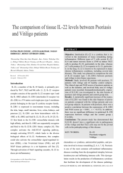
Fig. 3.
Dual parameter
analysis
of
narrow
forward
angle
scatter
(y-axis)
versus fluorescence
intensity
bc-axis) of
normal
PBL. CLI cells, and thymocytes
stained
with
lgG2 (control)
and TiOl
(positive).
in their
confirmed
the
T-cell
antibodies
fluores-
binding
of
to CLL
95%
with
Wang
et al.’7 have described
a p69,71
I
detected
by immunofluorescence,
),
of normal
human
sheep
erothrocytes
on
antigen
80%-
T cells,
which
form
rosettes
(E), and on slg
CLL
cells
from
I I of
14 patients.
Boumsell
et al.’5
have
described
a monoclonal
antibody,
termed
A50,
which
reacts
by complement-mediated
cytotoxicity
with
70%-90%
slg
CLL
ship
Leu
of E PBL and with greater
cells from
17 of 39 patients.
of monoclonal
1 await
further
Recently,
Reinherz
thymic
differentiation
escent
T-cell
staining
subset
antibodies
study.
TlOl,
than
70% of
The relationA50,
and
anti-
et al.’9 have postulated
an intrascheme
based
on immunofluor-
with a series of monoclonal
antibodies.
Their
studies
anti-human
suggest
that
From www.bloodjournal.org by guest on December 29, 2014. For personal use only.
T65
ANTIGEN
ON
T CELLS
AND
661
CLL CELLS
IQG2A
T 101
CONTROL
Null
cell
CLL
b.
gammopathy
0.
1gm
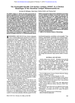
Fig.
4.
Dual
parameter
analysis
of
nar-
row forward
angle
scatter
(y-axis)
versus
fluorescence
intensity
(x-axis)
of cells from
patients
with
null cell CLI and monoclonal
gammopathy
stained
with lgG2 (control)
and
TiOl
(positive).
d.
C.
three
major
compartments
of thymic
differentiation
exist in humans.
By virtue
ofthe
fact that TlOl
reacts
with greater
than 95% of thymocytes,
we feel that T65
is present
on
regardless
based
all
thymus
of the stage
on
our
cells
in
varying
of differentiation.
own
hypothesis
fluorescence
analysis
with anti-Thy-l.2.
The majority
of mouse
thymocytes
are small
(90%)
and large
(5%)
cells
with
high
and intermediate
levels
of Thy-l.2,
respectively.
The third
population
of medium
sized
cells (5%) has low Thy- I .2 surface
density.
Weissman
amounts
Furthermore,
that
cells
with
less
surface
T65 are arrested
at an earlier
stage
of differentiation,
Molt-4
may be considered
to be less differentiated
than the other
T-cell
lines examined
(Fig. 2).
This notion
is consistent
with the findings
of Reinherz
et al.’9 that
Molt-4
expresses
tiation
antigens
than CEM.
Our studies
showed
three
cytes,
the majority
uniform,
low-density
medium
sized
cells
staining,
subgroups
strated
fewer
size
being
small
T65 and
with higher,
resembling
of mouse
by Fathman
monoclonal
stage
II differen-
populations
of thymo-
and large
cells with
a third
population
of
more
heterogeneous
circulating
T cells.
Similar
thymocytes
have
been
demonet al.’3 using
light
scatter
and
et al.2#{176}
have
cytes
high
surface
and
Thy-
further
characterized
the
mouse
thymo-
shown
that the small
and large
cells with
I .2 are derived
from the cortex
and have low
H-2,
while
the
medium
sized
cells
are
medul-
lary thymic
lymphocytes
and have the same phenotype
(low Thy-l.2,
high
H-2)
as peripheral
T cells.
We
propose
that
the weaker
staining
populations
in the
human
thymus
may also represent
cortical
thymocytes
and are in early
stages
more
mature
thymocytes,
cytes,
make
up
now underway
frozen
sections
studies.
the
of differentiation,
perhaps
medullary
brighter
population.
while
the
thymoStudies
are
to both localize
of thymus
and
these subpopulations
on
sort them for functional
marker
lymphoproliferative
REFERENCES
I
.
2.
tors.
3.
AC.
Aisenberg
neoplastic
Pernis
Ann
Bloch
lymphocytes.
B, Forni
NY
Acad
Preud’homme
KJ:
N EngI
L, Amante
Sci
190:420,
JL.
Seligmann
Immunoglobulins
J Med
287:272,
on the surface
L: Immunoglobulin
bulins
as cell
recep-
4. Slease
density
Surface
bound
as a cell
Blood40:777,
1971
M:
of
1972
immunoglo-
I 979
in
human
diseases.
1972
RB.
on human
Wistar
peripheral
R Jr.
Scher
blood
I: Surface
mononuclear
immunoglobulin
cells.
Blood 54:72,
From www.bloodjournal.org by guest on December 29, 2014. For personal use only.
WORMSLEY,
662
5.
Royston
Human
T
65.000
I, Majda
cell
dalton
cells
Meserve
BL,
monoclonal
(T65)
is also
bearing
GriflIths
JC:
antibodies.
surface
found
13.
on chronic
BIS,
Morikawa
5, Tatsumi
with
acute
G, Greaves
J NatI
leukemia
J
Cancer
Inst
GE.
Lazarus
McCarthy
RE:
Continuous
peripheral
blood
of
60:1269,
H,
with
associated
S.
of
Uzman
BG,
human
acute
Boone
BA,
lymphoblasts
from
Cancer
18:522,
leukemia.
8.
Minowada
of thymus
9.
of
8:434,
Ohnuma
cell
derived
Moore
ment
diseases.
lines.
T,
lymphocytes.
GE,
Woods
a leukemia
Moore
GE:
I. Establishment
in Clarkson
of
Cold
Normal
Springs
J NatI
LK,
Minowada
line
with
cell
Cancer
evidence
Inst
49:89
J, Mitchen
JR:
T cell
17.
for origin
Salmon
sharing
Establish-
J Exp
In
18.
Vitro
10.
I. Pius
against
cultured
Transplant
.
clonal
Peeters
I.
antibody
specific
Colloquium.
Super
Mullaney
York,
PF,
JA.
of the
The
Mendelsohn
Wiley
Graze
PR:
In vitro
T lymphoblastoid
immunicell
and
lines.
& Sons,
and
neoplastic
Biological
Fluids,
Pergamon.
ortho
ML
1979,
GY.
1980,
cytofluorograf.
(eds):
p 639
Flow
Baird
SM:
human
Mono-
T cells,
Wang
Proceedings
determinants
Med
Cytometry
and
AV,
JE (eds):
Cells.
DifferentiaNew
York,
p 8 11
B-cell
neoplasia
in man.
Lancet
B-type
Coppin
P. Dymbort
expressed
chronic
H.
A: An
chronic
Ammirati
complex
G,
on
Evans
human
lymphatic
RL:
T-cells
leukemic
cells.
1980
L.
J, Bernard
Pham
antigen
lymphocytic
Reinherz
EL,
of the
Proc
Acad
MR.
model
NatI
D,
shared
leukemic
Weissman
Dahlem
PC, Goldstein
of human
and
Sci USA
for selective
Neoplastic
Kung
stages
thymocytes
Sorting.
RA,
with
151:1539,
Boumsell
Discrete
20.
Melamed
PS, Till
M:
p 69,71
a
of normal
p 537
in
Good
CY.
of
B-cell
SF:
in
Seligmann
Hoffbrand
T-lymphoprolifera-
Raynal
B.
by a human
cells.
J Exp
Lemerle
T-cell
Med
J,
subset
I 52:229,
1980
19.
Yamamoto
for normal
Oxford,
BS:
RW.
B and
l):531, 1975
Majda
Protides
Smith
human
Proc7(Suppl
Royston
H (ed):
l2.
RB.
SE,
DAG.
Hematopoietic
I 978,
lyinpho-
1974
Dausset
Royston
Laboratory,
and
1977
Galton
B. Marks
IL:
“mature”
differentiation
deficiency
in B- and
Neoplastic
Harbor
Identification
1 , I 972
characteristics.
and
M,
Weissman
1975
6:355,
differentiation
ROYSTON
three
lymphocyte
Haematol
Cherchi
tive
Rosette-forming
and
D,
K: Cell
LA,
to immune
Clin
AND
of
15:109,
Human
application
Catovsky
16.
1973
zation
New
JT,
lymphoid
their
disorders,
Herzenberg
Differentiation
RA:
Good
Ganeshaguru
tion
M,
II.
Immunol
FP,
and
2:1230,
human
28th
Siegal
15.
leukemia-lymphoma
I965
I I
14.
proliferative
1978
Farber
culture
a child
T, Srivastava
of an antigen
in human
Small
maturation.
subclassesinvivo.Cell
immunoglobulin.
Tusubota
E: Expression
lymphoblastic
Foley
MD,
CG,
cell
markers
J, Janossy
lines.
Fathman
Thymus
The
125:725, 1980
Minowada
7.
SM,
by
of T cells
leukemia
lmmunol
cell
Baird
defined
antigen
lymphocytic
6.
JA,
antigens
COLLINS.
IL,
leukemic
77: I 588,
Baird
neoplastic
Transformation:
Konferenzen,
intrathymic
5:
Schlossman
of T-cell
Analysis
lineage.
I 980
Oncornavirus
leukemogenesis
transformation,
p 135
RH,
diflerentiation:
lymphoblasts
Mechanism
1977,
G, Levey
and
as a
in Koprowski
H (ed):
Consequences.
Berlin,
From www.bloodjournal.org by guest on December 29, 2014. For personal use only.
1981 57: 657-662
Comparative density of the human T-cell antigen T65 on normal peripheral
blood T cells and chronic lymphocytic leukemia cells
SB Wormsley, ML Collins and I Royston
Updated information and services can be found at:
http://www.bloodjournal.org/content/57/4/657.full.html
Articles on similar topics can be found in the following Blood collections
Information about reproducing this article in parts or in its entirety may be found online at:
http://www.bloodjournal.org/site/misc/rights.xhtml#repub_requests
Information about ordering reprints may be found online at:
http://www.bloodjournal.org/site/misc/rights.xhtml#reprints
Information about subscriptions and ASH membership may be found online at:
http://www.bloodjournal.org/site/subscriptions/index.xhtml
Blood (print ISSN 0006-4971, online ISSN 1528-0020), is published weekly by the American Society of
Hematology, 2021 L St, NW, Suite 900, Washington DC 20036.
Copyright 2011 by The American Society of Hematology; all rights reserved.
© Copyright 2026 Paperzz
![ドレッサー *トクラス*YEAB075AA[A/B]C 洗面化粧台[AFFETTO](http://s3.paperzz.com/store/data/006002757_1-aefa4c1ff6479d3b1e8686858be6b8bb-250x500.png)