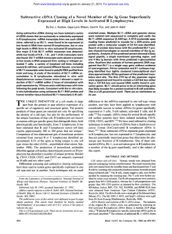
Molecular Microbiology (1998) 29(5), 1167–1177
The identification of Mycobacterium marinum genes
differentially expressed in macrophage phagosomes
using promoter fusions to green fluorescent protein
Lucia P. Barker,* Diane M. Brooks and P. L. C. Small
Microscopy Branch, Rocky Mountain Laboratories,
National Institute of Allergy and Infectious Diseases,
903 South 4th Street, Hamilton, MT 59840, USA.
Summary
Mycobacterium marinum , like Mycobacterium tuberculosis , is a slow-growing pathogenic mycobacteria
that is able to survive and replicate in macrophages.
Using the promoter-capture vector pFPV27, we have
constructed a library of 200–1000 bp fragments of M.
marinum genomic DNA inserted upstream of a promoterless green fluorescent protein (GFP) gene. Only
those plasmids that contain an active promoter will
express GFP. Macrophages were infected with this
fusion library, and phagosomes containing fluorescent bacteria were isolated. Promoter constructs that
were more active intracellularly were isolated with a
fluorescence-activated cell sorter, and inserts were
partially sequenced. The promoter fusions expressed
intracellularly exhibited homology to mycobacterial
genes encoding, among others, membrane proteins
and biosynthetic enzymes. Intracellular expression
of GFP was 2–20 times that of the same clones grown
in media. Several promoter constructs were transformed into Mycobacterium smegmatis , Mycobacterium
bovis BCG and Mycobacterium tuberculosis . These
constructs were positive for GFP expression in all
mycobacterial strains tested. Sorting fluorescent bacteria in phagosomes circumvents the problem of isolating a single clone from macrophages, which may
contain a mixed bacterial population. This method
has enabled us to isolate 12 M. marinum clones that
contain promoter constructs differentially expressed
in the macrophage.
Introduction
Mycobacterium marinum causes chronic skin lesions known
as ‘swimming pool granuloma’ on the extremities of human
hosts (Mollohan and Romer, 1961; Huminer et al ., 1986;
Received 23 March, 1998; revised 18 May, 1998; accepted 2 June,
1998. *For correspondence. E-mail [email protected]; Tel. (406)
363 9252; Fax (406) 363 9371.
Q 1998 Blackwell Science Ltd
Gluckman, 1995; Joe and Hall, 1995; Ramakrishnan,
1997). In immunocompromised human hosts, including
AIDS patients, infections can lead to systemic disease and
death (Tchornobay et al ., 1992; Hanau et al ., 1994; Parent
et al ., 1995). Consistent with the prevalence of human
disease in the extremities, the optimal growth temperature
of the organism is 25–358C, although some strains grow
well at 378C (Aronson, 1926; Clark and Shepard, 1963).
M. marinum is classified taxonomically as a slow-growing member of the genus Mycobacterium (Rogall et al .,
1990; Stahl and Urbance, 1990) and has been shown to
be closely related to M. tuberculosis by DNA–DNA homology and 16S RNA sequence studies (Tonjum et al ., 1998).
There have also been recent advances in the ability to
manipulate the organism genetically by performing gene
disruption via homologous recombination (Ramakrishnan
et al ., 1997a). Because of the relatively rapid growth of
M. marinum (4 h doubling time compared with 20 h for M.
tuberculosis ), the designation of the organism as a biosafety level two pathogen and the phenotypic similarity
of M. marinum and M. tuberculosis during a macrophage
infection (Barker et al ., 1997; Ramakrishnan et al .,
1997b), it is an ideal organism for the study of the virulence
mechanisms of pathogenic mycobacteria.
M. marinum and other pathogenic mycobacterial species
have been shown to survive and replicate in macrophages
and HeLa cells (Shepard, 1956; 1957; 1958; McDonough
et al ., 1993). In addition, several animal models are available for the study of M. marinum pathogenesis that have
shown a correlation of the ability of the organism to survive
in macrophages and its virulence in animal models (Ramakrishnan and Falkow, 1994; Ramakrishnan et al ., 1997b).
Many groups have sought to characterize the mechanisms
by which pathogenic mycobacteria survive the hostile
environment of the host macrophage (Sturgill-Koszycki et
al ., 1994; Xu et al ., 1994; Clemens and Horwitz, 1995).
In these studies, it has been shown that phagosomes containing pathogenic mycobacteria do not fuse with lysosomes, and the vesicles are only mildly acidified. Studies
in our laboratory have shown that the intracellular trafficking of M. marinum is analogous to that of M. tuberculosis
(Barker et al ., 1997). The molecular mechanisms by
which mycobacteria circumvent the host cell endosomal
network are, however, unclear.
Valdivia et al . (1996) have constructed a shuttle vector
1168 L. P. Barker, D. M. Brooks and P. L. C. Small
for the expression of a mutated green fluorescent protein
(mGFP) in M. marinum (Valdivia et al ., 1996). This vector
was developed as a tool for fluorescence-activated cell
sorter (FACS) isolation of individual or intracellular bacteria. The mGFP has been optimized for fluorescence
emission, solubility and kinetics of chromophore formation
in bacteria (Cormack et al ., 1996). M. marinum organisms
expressing mGFP were not adversely affected with respect
to replication or intracellular survival (Valdivia et al ., 1996).
This led to the development of a promoter-trap vector with
a multiple cloning site upstream of a promoterless mGFP
gene. Recently, a similar construct was used in differential
fluorescence induction (DFI) assays to sort Salmonella
typhimurium clones differentially expressing mGFP in low
pH environments (Valdivia and Falkow, 1996) and within
host cells (Valdivia and Falkow, 1997). In the DFI strategy,
a library of small fragments of bacterial DNA cloned
upstream of the promoterless mGFP gene is constructed,
and promoter activity is assayed under different environmental conditions by the level of fluorescence. Individual
or intracellular bacteria that are expressing the mGFP
are then isolated with the use of a FACS to separate and
isolate fluorescent bacteria or host cells.
It is our objective to elucidate the molecular mechanisms
by which pathogenic mycobacteria are able to survive in
the host cell environment. In this study, we have used the
DFI system to isolate M. marinum clones containing bacterial promoters, which are differentially expressed intracellularly, successfully. A unique aspect of this work is the
isolation of bacteria directly from phagosomes rather than
from intact macrophages. Mycobacteria are very hydrophobic and tend to clump in culture. Despite efforts to minimize clumping, in most experiments many bacteria are
taken up by macrophages in clusters (McDonough et al .,
1993). Even when individual bacteria are phagocytosed,
most macrophages will contain numerous organisms.
Therefore, the population of bacteria within a single macrophage is usually heterogeneous. However, within 2–3 days
of infection, the bacteria replicate intracellularly, and the
majority of mycobacteria are found distributed in vesicles
containing a single bacterium (Xu et al ., 1994). The isolation of vesicles from infected macrophages makes it possible to obtain a pure culture of an individual clone. In our
studies, vesicle preparations from infected macrophages
were sorted by FACS to isolate microorganisms that
expressed mGFP intracellularly. Using this method, we
identified 12 clones containing mGFP fusions differentially
expressed intracellularly. We further tested the ability of
representative clones to express mGFP in different mycobacterial species.
Results
Vesicle preparation was optimized for gentle lysis that
disrupted the cell membrane without compromising the
nuclear membrane. Nucleic acid contamination is viscous
and will tend to clump the preparation to the point at which
vesicles cannot be retrieved. We therefore chose to
Dounce homogenize vesicles and monitor cell and nuclear
membrane lysis by light microscopy. Vesicles were isolated
from RAW murine macrophages that were uninfected or
that were infected with 1 mm fluorescent latex beads. Figure
1A shows a transmission electron micrograph of a latex
bead enclosed in a macrophage vesicle. Very few nuclei
or whole cells were seen in electron micrographs of these
preparations. Preparations of latex beads in vesicles were
analysed by FACS to determine the best parameters by
which to sort fluorescent M. marinum of approximately
1–4 mm away from background vesicles and/or cell debris.
By calibrating the FACS instrument with latex beads of
known size (1–6 mm), we could gate the FACS to isolate
single organisms based on size and above-background
fluorescence.
After determining the optimal lysis and gradient spin
steps for vesicle isolation (see Experimental procedures ),
macrophages were infected with stationary phase or presorted library PCL2M. This library contains 200–1000 bp
fragments of M. marinum DNA cloned upstream of the promoterless mGFP gene. Approximately 6% of the PCL2M
library clones were fluorescent on solid media. Infections
of macrophages with PCL2M were allowed to proceed
for 3 days, after which macrophages were lysed and a
vesicle preparation performed to isolate individual M. marinum organisms (see Fig. 1B). As a majority of the organisms are in vesicles containing only one bacterium and the
phagosomal membrane surrounds the organism tightly,
the isolation of these phagosomes was relatively straightforward. Dilution plate counting of bacterial cfu from
the samples obtained with the FACS indicated that a
total of approximately 107 individual organisms in vesicles
could be isolated and analysed with the FACS in each
experiment.
Isolation of fluorescent clones by FACS
Two different preparations of the PCL2M promoter-capture
library were analysed by FACS. Parameters were set to
enrich for clones expressing mGFP intracellularly. In the
first experiment, a stationary-phase culture of PCL2M
was used to infect RAW murine macrophages. At 3 days
after infection, the point at which most vesicles contain
only one organism, the macrophages were lysed and the
vesicles isolated. This vesicle preparation was analysed
by FACS and was found to contain approximately 1.5%
fluorescent events that corresponded to the size of single
organisms in vesicles. Vesicles that were approximately
1–4 mm (i.e. that corresponded to a single bacterium
within a vesicle) and were fluorescing above background
Q 1998 Blackwell Science Ltd, Molecular Microbiology, 29, 1167–1177
M. marinum genes expressed in macrophages 1169
Fig. 1. Electron micrographs of vesicle preparations. Transmission electron micrographs of vesicle preparations from macrophages infected
with (A) 1 mm green fluorescent latex beads and (B) M. marinum . Bars represent 1 mm and large arrows point to (A) a 1 mm latex bead (lb)
within a vesicle and (B) M. marinum organisms in vesicles. A variety of sizes of vesicles can be seen in both preparations (small arrows).
As this is the post-nuclear fraction of the vesicle preparations, whole cells and nuclei have been removed by density gradient separation, and
a variety of vesicles of different sizes remain. A majority of the organisms are in vesicles containing only one bacterium, and the phagosomal
membrane surrounds the organism tightly.
levels were isolated away from the non-fluorescent population by gating the FACS to sort the desired population
away from other organisms and vesicles (see Fig. 2A). A
sample of the resulting suspension of organisms was
then analysed and found to contain 58.6% organisms of
high fluorescence and the proper size, an enrichment of
40-fold (Fig. 2B). This suspension was then diluted and
plated for single cfus. Clones obtained by this isolation procedure were designated F-series (FACS-isolated) clones
(e.g. F80).
In order to reduce the number of FACS-selected clones
that were constitutively expressing mGFP in tissue culture
media, we presorted fluorescent organisms away from the
PCL2M population. A culture of PCL2M was grown to stationary phase in DMEM media, and fluorescent clones
(approximately 6% of the population) were FACS isolated
away from the non-fluorescent population and discarded.
The remaining non-fluorescent clones were used to infect
RAW murine macrophages for 3 days, and vesicles were
isolated for FACS analysis. In this experiment, 1.1% of the
events corresponded to a single, fluorescent bacterium.
Q 1998 Blackwell Science Ltd, Molecular Microbiology, 29, 1167–1177
A sample of this suspension of organisms was then analysed and found to contain 16.2% organisms of high fluorescence and the proper size, an enrichment of 15-fold.
These organisms were also plated for single cfus. These
clones were designated P-series (presorted) clones.
A total of 400 individual F-series clones and 300 individual P-series clones were tested for fluorescence in tissue culture media and intracellularly by infecting tissue
culture wells with or without macrophages in parallel.
Each well was examined visually and scored for fluorescence in the macrophage that exceeded fluorescence in
media alone. In this way, four F-series clones (F80,
F154, F278 and F320) and 12 P-series clones (P40,
P99, P136, P140, P141, P144, P155, P163, P168, P170,
P232 and P238) were chosen for further characterization.
Although the enrichment of P-series clones was less successful than that for the F-series in the original FACS analyses, the percentage of P-series clones positively
screened for differential expression within macrophages
was higher [4/400 (1%) for F-series, 12/300 for P-series
(4%)].
1170 L. P. Barker, D. M. Brooks and P. L. C. Small
Fig. 2. FACS sorting of PCL2M.
A. Presort. Macrophages infected for 3 days with the PCL2M library were lysed and the vesicle fraction isolated. After calibration of the FACS
with 1 mm and 6 mm beads, the sorter was gated for events corresponding to 1–4 mm and above-background fluorescence (box). This sample
contained 1.5% of the total events. Approximately 106 events corresponding to the proper size and fluorescence were collected.
B. Post-sort. The sample gated as described above was reanalysed by FACS. Events of proper size and above-background fluorescence
(box) were determined to be 58.6% of the total sample collected.
Fluorescence of clones in media and intracellularly
by FACS analysis
In order to quantify promoter activity in the clones of interest, log-phase cultures of clones were seeded into 7H9
media, DMEM tissue culture media and RAW murine
macrophage cultures. After 3–4 days, the macrophages
were lysed with a mild detergent to release phagocytosed
bacteria. Bacteria from macrophage lysates and the same
bacteria grown in media were FACS analysed for fluorescence intensity. Strains GFP3R and G13 were included as
controls. Strain GFP3R is the wild-type strain 1218R transformed with construct pMV262gfpmut3. This construct has
the mGFP gene under the control of the mycobacterial
heat shock promoter HSP60. Clone G13 was picked
from solid media as a highly fluorescent constitutive promoter construct from the original PCL2M plating. Clone
G13 contains a promoter insert that fluoresces at levels
10–20 times higher than the mycobacterial HSP60 promoter in GFP3R.
Five of the 16 original clones (F80, P40, P136, P140
and P170) exhibited no induction of fluorescence (fold
induction # 1.0) and were characterized no further. Table
1 shows the fold fluorescence induction of the remaining
clones. Intracellular induction of M. marinum promoters
ranged from 1.4-fold for clones F320 and F154 to an
approximate 15-fold induction for clone P155. Most of
the promoters were induced approximately two- to threefold in the macrophage with low standard error values.
Construct P155, however, exhibits a great deal of variation
in the amount of fluorescence induction observed depending on the experiment. Fluorescence intensity of P155 is,
however, always higher in the macrophage than in tissue
culture media. Preliminary work suggests that mGFP
expression from the P155 construct may be dependent
upon growth phase as well as environmental signals (data
not shown), and kinetic studies are in progress.
Sequence analysis of selected clones
Partial sequences of the clones of interest were compared
with known sequences with BLAST (Altschul et al ., 1990)
and MycDB (Bergh and Cole, 1994) database programs.
Results of sequence comparisons are shown in Table 1.
Of the inserts, G13, F320, P99, P141/144, P163, P232
and P238 are completely sequenced. None of these
known sequences have coding regions that span the entire
insert. This indicates that intergenic regions that may contain mycobacterial promoters are probably present rather
than fortuitous promoters internal to open reading frames
(ORFs). Further, all clones obtained exhibited homology
to M. tuberculosis or M. leprae sequences, although, in
some cases, the percentage identity was low. Sequence
analysis identified clones P141 and P144 as sibling strains.
Inserts G13, P99, P141/144, P168 and P238 exhibited
homology to mycobacterial sequences or genes but did
not yield information concerning specific gene products.
Insert sequence from clone P163 showed high identity
Q 1998 Blackwell Science Ltd, Molecular Microbiology, 29, 1167–1177
M. marinum genes expressed in macrophages 1171
Table 1. Comparison of mGFP induction in
macrophages and associated nucleotide
sequences homologous to inserts.
Strain
Fold inductiona
1218R
GFP3R
G13
F154
F278
F320
P99
P141
P144
P155
P163
P168
P232
P238
0.7 6 0.2
3.4 6 1.2
1.9 6 0.4
1.4 6 0.4
2.9 6 0.2
1.4 6 0.3
3.0 6 0.5
2.4 6 1.1
1.8 6 0.1
14.5 6 12.9
2.8 6 0.1
2.8 6 0.8
2.8 6 0.5
1.9 6 0.1
Insert
size (bp)
–
–
462
> 1500
<2500
464
241
260
260
> 1500
844
1067
979
308
Sequence reading into mGFPb
–
HSP60 promoter
Homology to MTBc and M. leprae sequences
MTB arginyl-tRNA synthetase
Unknown MTB 46 kDa protein
Unknown MTB putative membrane protein
Homology to MTB and M. bovis sequences
Homology to MTB sequences
Homology to MTB sequences
MTB probable drug efflux pump
Pseudomonas pvdD-iron starvation box
M. leprae cosmid L308
MTB pdhC gene
MTB zinc metallopeptidase
a. Fold induction in macrophages is the fluorescence value in macrophage lysates divided by
the fluorescence values of the same clone grown in DMEM media supplemented with 10% fetal
bovine serum. Background wild-type fluorescence has been subtracted from mean fluorescence
values for each sample. Experiments were repeated at least twice, and values represent the
mean fluorescence induction 6 SEM.
b. The genetic nomenclature assigned to each promoter fusion was based on homology to either
known sequences or ORFs at the mGFP fusion junction. Those genes listed were determined
as the most likely sequences reading into mGFP based on the polarity of transcription/translation, as determined by sequence homology. Those genes listed in bold have demonstrated
homology with a P -value < 10¹20.
c. MTB, Mycobacterium tuberculosis .
(P < 10¹24 ) to the Pseudomonas aeruginosa gene pvd D,
a peptide synthetase-like gene that is located in an ironstarvation box in P. aeruginosa. A BLAST search of P163
ORFs yielded significant protein identity (P < 10¹17 )
between P163 and a peptide synthetase from Streptomyces pristinaespiralis.
The remaining clones, F154, F278, F320, P155 and
P232, showed significant identity to known M. tuberculosis
sequences. This is not surprising, as M. marinum has
been shown to be closely related to M. tuberculosis (Tonjum et al ., 1998). Clone P155 contained the insert sequence that exhibited the highest amount of induction in
macrophages. The P155 insert sequence most probably
reading into mGFP exhibits a high degree of identity
(P < 10¹81 ) with an M. tuberculosis gene encoding a probable drug efflux pump. Clones F154 and P232 contain
inserts with homology to M. tuberculosis housekeeping
genes. F278 and F320 contain inserts homologous to the
M. tuberculosis PPE protein family of unknown function
and a putative M. tuberculosis membrane protein of
unknown function respectively.
Transformation of other mycobacteria with control
strains and representative clones
The expression of most mycobacterial genes identified thus
far has not been restricted to a single species, e.g. many
genes isolated from M. tuberculosis can be expressed
in M. smegmatis (Jacobs et al ., 1991). Nonetheless, we
wanted to determine whether the clones isolated from
Q 1998 Blackwell Science Ltd, Molecular Microbiology, 29, 1167–1177
the M. marinum PCL2M library would be expressed in
other mycobacterial species. Strains M. bovis BCG, M.
smegmatis and M. tuberculosis H37Ra were transformed
with pMV262gfpmut3, G13, F154 and P155. Transformants were selected on kanamycin and colonies observed
on a fluorescent microscope. Figure 3 shows representative
transformant colonies compared with the same constructs
in M. marinum . In all of the strains, mGFP fluoresced
much more brightly with the G13 insert than with the
HSP60 promoter construct. Additionally, fluorescence
levels of F154 and P155 varied from strain to strain, but
all of the insert constructs exhibited colony fluorescence
to some degree, indicating that expression of mGFP from
the promoters isolated from M. marinum would be
expressed in other mycobacterial species as well.
Discussion
Using an mGFP promoter-capture strategy outlined in Fig.
4, we have successfully isolated a series of promoter constructs that are differentially expressed within macrophages.
By using a FACS to isolate macrophage vesicles rather
than intact macrophages, we were able to circumvent the
problems associated with isolating a mixed population of
organisms from intact macrophages. We have identified
12 clones containing fusions that exhibit more fluorescence
intensity when inside the macrophage than extracellularly.
DNA isolated from these mGFP fusion constructs exhibited
a high degree of identity with known M. tuberculosis DNA
sequences. As M. marinum and M. tuberculosis are both
1172 L. P. Barker, D. M. Brooks and P. L. C. Small
Fig. 3. Colonies of mycobacterial species transformed with promoter constructs. M. smegmatis , M. bovis BCG and M. tuberculosis H37Ra
were transformed with pMV262gfpmut3, G13 and representative constructs from clones F154 and P155. The resulting colonies were then
photographed on selective media and compared with M. marinum transformants (top row). Exposure times were identical for all strains (2 min),
except for M. tuberculosis , for which the exposure time was 1 min in order to avoid overexposure. However, M. tuberculosis transformed with
the P155 construct was exposed for 2 min.
able reside in similar phagocytic compartments (Barker et
al ., 1997) and are phylogenetically very closely related
(Tonjum et al ., 1998), it is probable that analogous molecular mechanisms are involved in the survival of these
organisms in a hostile host cell environment.
This is the first report of the use of GFP as a tool
for screening the promoter expression of a mycobacterial
library intracellularly. However, several other groups have
looked at the use of GFP as a reporter gene to quantify
the amount of gene expression by mycobacterial species
(Dhandayuthapani et al ., 1995; Kremer et al ., 1995; Parker and Bermudez, 1997). The addition of a procedure in
which we isolate vesicles containing individual mycobacteria has enabled us to separate these fluorescent organisms from other bacteria, even in the same host cell, that
express little or no mGFP. Further, by isolating vesicles
rather than intact macrophages, we would be less likely to
miss a weak promoter fusion that is differentially expressed
within the macrophage in a background of non-fluorescent
clones. The relatively low percentages of positive clones
(1% of F-series and 4% of P-series) can be explained by
the fact that this method is an enrichment rather than a
selection. The clones isolated by FACS were contaminated with non-fluorescent clones and clones constitutively
expressing mGFP. The sorting of organisms by FACS,
however, enabled us to screen many fewer individual
clones for differential expression in the macrophage.
Although the degree of induction of our promoter constructs appears to be relatively low in most cases, we
are confident that our screening methods have identified
several promoter constructs that are more active intracellularly. We believe that a reproducible two- or threefold
Q 1998 Blackwell Science Ltd, Molecular Microbiology, 29, 1167–1177
M. marinum genes expressed in macrophages 1173
Fig. 4. FACS isolation of fluorescent GFP fusions
expressed in macrophage phagosomes.
Macrophages are infected with the M. marinum
promoter-trap library PCL2M. After 3 days, the
majority of phagosomes contain only one
bacterium. Macrophages are lysed and vesicle
preparations are sorted by size and fluorescence
to enrich for clones specifically expressing GFP
intracellularly.
intracellular induction of a particular gene could indicate a
highly significant and physiologically relevant upregulation
of a particular gene product. We found it necessary to use
FACS analysis, rather than fluorometry, for quantification
of promoter activity. The high concentration of organisms
required for the measurement of population fluorescence
confounded our data, as light scatter from the sample
was too high to allow reproducible measurement of fluorescence intensity. The advantage of FACS analysis is that
the measurement of fluorescence intensity for each individual bacterium is highly reproducible.
Although the level of differential expression of these
M. marinum constructs is not as high as those found in
enteric bacteria (Valdivia and Falkow, 1997), it is consistent with previous work done with mycobacterial systems.
For example, Curcic et al . (1994) used a transcriptional
fusion system with the xylE gene from Pseudomonas as
a reporter gene and found a maximal eightfold reduction
of mtrA gene expression when M. smegmatis was grown
on various nitrogen sources. Dellagostin et al . (1995)
found a maximum 6.3-fold intracellular induction of an
Q 1998 Blackwell Science Ltd, Molecular Microbiology, 29, 1167–1177
M. leprae promoter sequence transformed into M. bovis
BCG as measured by b-galactosidase activity.
In contrast to the induction of specific promoters in different environmental backgrounds, several groups have
studied the range of promoter strength in mycobacterial
species. Using the chloramphenicol acetyltransferase
(CAT) reporter system and a LacZ reporter system, DasGupta et al . (1993) found that both M. tuberculosis and
M. smegmatis exhibited an approximate 20-fold range of
promoter activity, although M. tuberculosis promoters
were less active than those from M. smegmatis (Jain et
al ., 1997). In a related study by Timm et al . (1994) also
using a LacZ reporter system, an approximate 100-fold
range of promoter activity was described for M. bovis
BCG promoter constructs as well as M. smegmatis containing M. tuberculosis promoter constructs.
To summarize what is known about mycobacterial promoters thus far: (i) the induction of specific genes in different environmental backgrounds has not exceeded 10- to
20-fold using different reporter systems; (ii) the range of
values for promoter strength within mycobacterial species
1174 L. P. Barker, D. M. Brooks and P. L. C. Small
is similar; and (iii) slow-growing M. tuberculosis appears to
have many fewer strong promoters than does the fastgrowing M. smegmatis . It is reasonable to suggest that
the levels of induction that we have reported here represent biologically significant events.
Using the DFI strategy, we have identified 12 constructs
that are upregulated in the macrophage. It is important to
note that some of these fusions could contain chimeric
inserts (i.e. non-contiguous fragments of insert DNA),
and that this is most likely when the insert size is large.
We also cannot rule out the possibility of cryptic promoters
driving the expression of mGFP. In assigning gene identities to mGFP fusions, we have chosen the genes homologous to the insert sequences that are most likely to be
reading into and driving the expression of mGFP. Further
analysis is required before we know whether this preliminary assignment is correct.
All of the sequences we have characterized demonstrate homology to sequences from other mycobacterial
strains, and some can be assigned non-ambiguous designation as specific genes. Not surprisingly, some of these
sequences are homologous to housekeeping genes. We
also found that the mGFP construct on the HSP60 promoter (pMV262gfpmut3) was induced in the macrophage.
The upregulation of a heat shock/stress response promoter in the hostile environment of the macrophage was not
unexpected. Dellagostin et al . (1995), however, did not
measure any induction of the HSP60 promoter in macrophages when using a LacZ reporter system. This may be
because of differences in sensitivity between the two
assay systems. Another interesting result is the slight but
reproducible induction of the constitutive and highly active
G13 construct. The G13 promoter system expresses
mGFP highly in all mycobacterial strains tested and, in M.
marinum , is 10–20 times more active than the HSP60 promoter. As there are few strong mycobacterial promoters,
this promoter could be extremely useful for obtaining a
high yield of mycobacterial gene products. Work is in progress to characterize this promoter construct further.
The goal of this work is to identify mycobacterial genes
required for macrophage survival and/or virulence. Whether
the genes that we have identified are required for the survival of pathogenic mycobacteria in the macrophage can
only be addressed by deleting the gene of interest and
determining the effects of this deletion on the virulence of
the organism. We can then explore the effects of deleting
or interrupting these sequences in the wild-type strain in a
macrophage tissue culture system with regard to bacterial
survival and trafficking through the host cell.
Experimental procedures
Bacterial strains and growth conditions
M. marinum 1218R was originally obtained from the American
Type Culture Collection (ATCC 927), as were M. tuberculosis
H37Ra (ATCC 25177) and M. bovis BCG (ATCC 35743).
M. smegmatis mc2155 was provided by William R. Jacobs,
Albert Einstein College of Medicine, NY, USA. The M. marinum 1218R isolate has been shown to be virulent in the frog
model (Ramakrishnan et al ., 1997b) and in the guinea pig
and fish models (unpublished data). Individual isolates of M.
marinum 1218R were streaked for single colonies on 7H10
media (Difco) supplemented with 10% Middlebrook oleic acid,
albumin, dextrose and catalase enrichment (OADC; Difco)
and grown at 328C for 5–7 days. A single colony was inoculated into 7H9 media (Difco) supplemented with 10% OADC
and 0.2% Tween 80 and allowed to incubate at 328C for 10–
14 days. The cultures were then frozen in 0.5 ml aliquots at
¹708C in 7H9 media with 10% OADC. This culture was designated passage 2. Aliquots were inoculated as needed into
30 ml of liquid growth media and grown without shaking for
7–10 days. These cultures were designated passage 3. Passage 3 M. marinum used for experiments were in stationary
phase, having been in media for more than 10 but less than
30 days, unless otherwise specified. M. tuberculosis strain
H37Ra, M. smegmatis mc2155 and M. bovis BCG were cultured under similar conditions, but grown and maintained at
378C. Liquid and solid media were supplemented with
30 mg ml¹1 kanamycin as needed. Escherichia coli strain
DH5a was maintained in Luria broth or on LB agar with or
without kanamycin.
Macrophage cell line
The mouse macrophage cell line RAW 264.7 (ATCC TIB71)
was maintained at 378C in 5.0% CO2 in Dulbecco’s modified
Eagle medium (DMEM; Gibco) supplemented with 10% fetal
bovine serum (Gibco). RAW macrophages are fully capable
of phagocytosing bacteria and latex beads. Further, RAW
cells are able to kill avirulent mycobacteria in vitro (unpublished
data). RAW macrophages were not used beyond passage 10
from frozen stocks.
Library construction
Genomic DNA was isolated from M. marinum 1218R and
digested for various time intervals with Sau 3A. The digests
were resolved on a 1.0% agarose gel, and bands corresponding to DNA fragments between 200 and 1000 bp, were excised
from the gel. Size-fractionated DNA was phosphatase treated
and ligated into the Bam HI site of promoter-trap plasmid
pFPV27 (constructed and provided by Raphael Valdivia, Stanford University). This construct is derived from vector pFPV2
(Valdivia et al ., 1996) and contains the ori sequences of both
mycobacterial and E. coli strains, a kanamycin resistance
selectable marker and a promoterless mutant green fluorescent protein (mGFP) gene. Constructs were transformed into
E. coli DH5a. A total of 2.8 × 10⁵ individual kanamycin-resistant
clones were generated in E. coli . These colonies were suspended in Luria broth with 15% glycerol and frozen in 1 ml aliquots. This E. coli library was designated PCL2 (for promoter
capture library 2). Promoter capture library 1 was discarded
as not being representative of the M. marinum genome. An
aliquot of PCL2 was grown at 378C overnight, and the plasmid
library was isolated and transformed into M. marinum 1218R.
Q 1998 Blackwell Science Ltd, Molecular Microbiology, 29, 1167–1177
M. marinum genes expressed in macrophages 1175
A total of 3.5 × 10⁴ individual kanamycin-resistant clones was
generated in the 1218R transformation. Colonies were scraped
off plates into 7H9 media supplemented with 10% OADC and
frozen in 1 ml aliquots. This library was designated PCL2M
(for promoter capture library 2 in M. marinum ). Aliquots of
PCL2M were grown for 3–4 days in 7H9 media to ensure
that the bacteria were in log-phase growth. The log-phase
PCL2M bacteria were then diluted 1:10 in 7H9 media or
DMEM supplemented with kanamycin and grown for 10–14
days before use in experiments, unless otherwise specified.
Macrophage infections with bacterial strains
Macrophages were seeded into tissue culture dishes or microchambers at a concentration of 2 × 10⁵ cells ml¹1 total volume.
Macrophages were grown to semi-confluence before bacteria
were added at a multiplicity of infection (MOI) of 1–5:1. After
4 h of growth at 378C and 5% CO2 , the medium was removed,
and fresh DMEM supplemented with 100 mg ml¹1 amikacin
was added to kill extracellular organisms. Monolayers infected
with M. marinum were then transferred to 328C. At 24 h after
infection, the concentration of amikacin was reduced to
20 mg ml¹1.
Isolation and FACS sorting of vesicles
To isolate vesicles, 20 tissue culture dishes of infected or control macrophages were placed on ice and washed three times
with ice-cold phosphate-buffered saline (PBS), pH 7.2, and
monolayers were scraped into 2.5 ml of PBS per plate. Cells
were pooled into two tubes and spun down at 500 r.p.m.
(100 × g) in a Beckman J-6M centrifuge for 5 min at 48C. The
supernatant was removed, and the cell pellets were gently
resuspended in ice-cold homogenization buffer (HB). HB is
3 mM imidazole (Calbiochem) in 250 mM sucrose at pH 7.2,
to which was added one tablet of complete protease inhibitor
(Boehringer Mannheim) per 50 ml. This solution was immediately centrifuged at 1200 r.p.m. (200 × g ) for 10 min at 48C.
The supernatant was removed, and 0.5 ml of ice-cold HB
was added to each tube. Samples were combined and placed
in a 1.0 ml glass Dounce homogenizer (Wheaton Scientific)
and homogenized 35 strokes. A volume of 20 ml of this homogenized suspension was observed under light microscopy to
ascertain that at least 50% of the macrophages were lysed
without compromising the nuclear membranes. The homogenate was centrifuged at 2000 r.p.m. (500 × g ) for 2 min at
48C. The milky supernatant containing the vesicle suspension
was then gently separated from the whole-cell and nuclear
pellet for FACS analysis. Vesicle preparations of macrophages
infected with latex beads or M. marinum were fixed with
periodate–lysine–paraformaldehyde (Robertson et al ., 1963;
McLean and Nakane, 1974), subjected to 0.5% reduced
OsO4 (McDonald, 1984) and visualized on a Hitachi model
HU11-E1 transmission electron microscope at 75 kV. FACS
analysis was performed on a Facstar instrument modified
for five-parameter operation (Becton-Dickinson Immunocytometry Systems). Before each analysis, 1 mm and 6 mm green
latex beads (Polysciences) were used to calibrate and scale
the machine. For PCL2M library sorts, forward scatter
(size) was on the x -axis and the fluorescence intensity on
Q 1998 Blackwell Science Ltd, Molecular Microbiology, 29, 1167–1177
the y -axis. Fluorescence was measured using logarithmic
amplification.
Fluorescence screening of selected clones
Fluorescent bacterial clones that were sorted away from nonfluorescent vesicles and vesicles containing non-fluorescent
organisms were screened further by assaying fluorescence
visually on a Bio-Rad MRC 1000 scanning confocal microscope.
Control strains were GFP3R (pMV262gfpmut3 in 1218R) and
G13, a highly fluorescent constitutive promoter fusion isolated
from the original PCL2M library. Those clones that appeared
to fluoresce intracellularly but not on 7H10 agar or in media
were inoculated into DMEM media or into culture dishes containing RAW macrophages at a MOI of 1–5:1. Growth rates of
all clones measured by optical density did not vary from that
of the wild-type M. marinum 1218R (data not shown). After 3–
4 days of growth in either media or macrophages, bacteria
were harvested from macrophage culture by lysing macrophages with 0.1% Triton-X in PBS for 5 min, after which the
detergent was diluted with 9 volumes of PBS. A total of 5000
bacteria from media or macrophage lysates was assayed for
fluorescence by FACS. Bacteria were detected by side scatter
as described previously (Valdivia and Falkow, 1996), and fluorescence and side-scatter data were collected using logarithmic
amplification. Quantitative measurements and distribution of
fluorescence from the different fusions were determined with
the Consort 30 software program (Becton-Dickinson). To calculate the amount of fluorescence induction, the fluorescence
value of a given clone from the macrophage lysate was
divided by the value of fluorescence of the same clone
grown in DMEM.
Expression of mGFP in other mycobacterial species
Plasmid preparations from control and representative clones
were transformed into M. smegmatis , M. bovis and M. tuberculosis strains as described previously (Jacobs et al ., 1991).
Transformants were observed directly on selective media
for the expression of mGFP. A Leitz Orthoplan epifluorescent
microscope with a 2.5 × objective lens was used to observe
and photograph colonies.
Sequencing of selected clones
Plasmid mini-preps were performed on clones of interest as
described previously (Ramakrishnan et al ., 1997a). A total of
0.1 mg of DNA was transformed into library efficiency DH5a
competent cells (Gibco BRL) according to the manufacturer’s
instructions. Plasmid mini-preps were performed on at least
five single-colony transformants. Each plasmid preparation
was digested with Eco RI and resolved on an agarose gel to
ascertain that inserts were stable. Polymerase chain reaction
(PCR) amplification of insert sequences followed previously
described protocols (Maniatis et al ., 1989). Inserts were sequenced using PCR products with flanking vector sequences or
the native plasmid as templates. Primers that annealed 15 bp
away from the Bam HI insert site were chosen to flank the
insert DNA and read from both directions into the fragment.
All DNA sequencing was performed using a model 370A
1176 L. P. Barker, D. M. Brooks and P. L. C. Small
stretch automated DNA sequencer according to the manufacturer’s instructions (Applied Biosystems). Homologies to known
sequences were determined with MycDB (Bergh and Cole,
1994) and BLAST software (National Center for Biotechnology
Information at the National Library of Medicine) (Altschul et
al ., 1990).
Acknowledgements
We thank Olivia Steele-Mortimer for advice/expertise on
vesicle preparations, Raphael Valdivia for the provision of and
helpful discussion of vector pFPV27, Lalita Ramakrishnan
and Benjamin Chang for vector pMV262gfpmut3, Stanley F.
Hayes and Elizabeth Fischer for electron microscopy preparations, and Gary Hettrick and Bob Evans for help with graphics.
We also thank Patti Rosa, Jos van Putten, Katie George and
Jay Carroll for critical review of this manuscript. Thanks also
to K. and H. Fischer for helpful discussion. Our sincerest
thanks to Brian Stevenson, Steve Porcella and Stanley Falkow
for advice, expertise and encouragement.
References
Altschul, S.F., Gish, W., Miller, W., Meyers, E.W., and
Lipman, D.J. (1990) Basic local alignment search tool. J
Mol Biol 215: 403–410.
Aronson, J.D. (1926) Spontaneous tuberculosis in saltwater
fish. J Infect Dis 39: 315–320.
Barker, L.P., George, K.M., Falkow, S., and Small, P.L.C.
(1997) Differential trafficking of live and dead Mycobacterium marinum organisms in macrophages. Infect Immun
65: 1497–1504.
Bergh, S., and Cole, S.T. (1994) MycDB: an integrated mycobacterial database. Mol Microbiol 12: 517–534.
Clark, H.F., and Shepard, C.C. (1963) Effect of environmental temperatures on infection with Mycobacterium marinum
(balnei ) of mice and a number of poikilothermic species.
J Bacteriol 86: 1057–1069.
Clemens, D.L., and Horwitz, M.A. (1995) Characterization of
the Mycobacterium tuberculosis phagosome and evidence
that phagosomal maturation is inhibited. J Exp Med 181:
257–270.
Cormack, B.P., Valdivia, R.H., and Falkow, S. (1996) FACSoptimized mutants of the green fluorescent protein (GFP).
Gene 173: 33–38.
Curcic, R., Dhandayuthapani, S., and Deretic, V. (1994)
Gene expression in mycobacteria: transcriptional fusions
based on xylE and analysis of the promoter region of the
response regulator mtrA from Mycobacterium tuberculosis .
Mol Microbiol 13: 1057–1064.
DasGupta, S.K., Bashyam, M.D., and Tyagi, A.K. (1993)
Cloning and assessment of mycobacterial promoters by
using a plasmid shuttle vector. J Bacteriol 175: 5186–
5192.
Dellagostin, O.A., Esposito, G., Eales, L., Dale, J.W., and
McFadden, J. (1995) Activity of mycobacterial promoters
during intracellular and extracellular growth. Microbiology
141: 1785–1792.
Dhandayuthapani, S., Via, L.E., Thomas, C.A., Horowitz,
P.M., Deretic, D., and Deretic, V. (1995) Green fluorescent
protein as a marker for gene expression and cell biology of
mycobacterial interactions with macrophages. Mol Microbiol 17: 901–912.
Gluckman, S.J. (1995) Mycobacterium marinum . Clin Dermatol 13: 273–276.
Hanau, L.H., Leaf, A., Soeiro, R., Weiss, L.M., and Pollack,
S.S. (1994) Mycobacterium marinum infection in a patient
with the acquired immunodeficiency syndrome. Cutis 54:
103–105.
Huminer, D., Pitlik, S.D., Block, C., Kaufman, L., Amit, S.,
and Rosenfeld, J.B. (1986) Aquarium-borne Mycobacterium marinum skin infection. Dermatology 122: 698–703.
Jacobs, Jr, W.R., Kalpana, G.V., Cirillo, J.D., Pascopella, L.,
Udani, R.A., Jones, Jr, W.D., et al . (1991) Genetic systems
for mycobacteria. Methods Enzymol 240: 537–555.
Jain, S., Kaushal, D., DasGupta, S.K., and Tyagi, A.K. (1997)
Construction of shuttle vectors for genetic manipulation
and molecular analysis of Mycobacteria. Gene 190: 37–
44.
Joe, L., and Hall, E. (1995) Mycobacterium marinum disease
in Anne Arundel county: 1995 update. Md Med J 44: 1043–
1046.
Kremer, L., Baulard, A., Estaquier, J., Poulain-Godefroy, O.,
and Locht, C. (1995) Green fluorescent protein as a new
expression marker in mycobacteria. Mol Microbiol 17:
913–922.
McDonald, K. (1984) Osmium ferricyanide fixation improves
microfilament preservation and membrane visualization in
a variety of animal cell types. J Ultrastruct Res 86: 107–
118.
McDonough, K.A., Kress, Y., and Bloom, B.R. (1993) Pathogenesis of tuberculosis: Interaction of Mycobacterium
tuberculosis with macrophages. Infect Immun 61: 2763–
2773.
McLean, I.W., and Nakane, P.K. (1974) Periodate–lysine–
paraformaldehyde fixative: a new fixative for immunoelectron microscopy. J Histochem Cytochem 22: 1077–
1084.
Maniatis, T., Fritsch, E.F., and Sambrook, J. (1989) Molecular
Cloning: A Laboratory Manual . 2nd edn. Cold Spring
Harbor, NY: Cold Spring Harbor Laboratory Press.
Mollohan, C.S., and Romer, M.S. (1961) Public health significance of swimming pool granuloma. Am J Pub Health 51:
883–891.
Parent, L.J., Salan, M.M., Appelbaum, P.C., and Dossett,
J.H. (1995) Disseminated Mycobacterium marinum infection and bacteremia in a child with severe combined
immunodeficiency. Clin Infect Dis 21: 1325–1327.
Parker, A.E., and Bermudez, L.E. (1997) Expression of the
green fluorescent protein (GFP) in Mycobacterium avium
as a tool to study the interaction between Mycobacteria
and host cells. Microb Pathog 22: 193–198.
Ramakrishnan, L. (1997) Images in clinical medicine. Mycobacterium marinum infection of the hand. N Engl J Med
337: 612.
Ramakrishnan, L., and Falkow, S. (1994) Mycobacterium
marinum persists in cultured mammalian cells in a temperature-restricted fashion. Infect Immun 62: 3222–3229.
Ramakrishnan, L., Tran, H.T., Federspiel, N.A., and Falkow,
S. (1997a) A crtB homolog essential for photochromogenicity in Mycobacterium marinum: isolation, characterization,
Q 1998 Blackwell Science Ltd, Molecular Microbiology, 29, 1167–1177
M. marinum genes expressed in macrophages 1177
and gene disruption via homologous recombination. J Bacteriol 179: 5862–5868.
Ramakrishnan, L., Valdivia, R.H., McKerrow, J.H., and
Falkow, S. (1997b) Mycobacterium marinum causes both
long-term subclinical infection and acute disease in the
leopard frog (Rana pipiens ). Infect Immun 65: 767–773.
Robertson, J.D., Bodenheimer, T.S., and Stage, D.E. (1963)
The ultrastructure of Mauthner cell synapses and nodes in
goldfish brains. J Cell Biol 19: 159–199.
Rogall, T., Wolters, J., Flohr, T., and Bottger, E.C. (1990)
Towards a phylogeny and definition of species at the
molecular level within the genus Mycobacterium . Int J
Syst Bacteriol 40: 323–330.
Shepard, C.C. (1956) Growth characteristics of tubercle
bacilli and certain other mycobacteria in HeLa cells. J
Exp Med 105: 39–55.
Shepard, C.C. (1957) Growth characteristics in HeLa cells
of the rapidly growing acid fast bacteria, Mycobacterium
fortuitum , Mycobacterium phlei and Mycobacterium smegmatis . J Exp Med 106: 722–726.
Shepard, C.C. (1958) A comparison of the growth of selected
mycobacteria in HeLa, monkey kidney and human amnion
cells in tissue culture. J Exp Med 107: 237–250.
Stahl, D.A., and Urbance, J.W. (1990) The division between
fast- and slow-growing species corresponds to natural relationships among the mycobacteria. J Bacteriol 172: 116–
124.
Sturgill-Koszycki, S., Schlesinger, P.H., Chakraborty, P.,
Haddix, P.L., Collins, H.L., Fok, A.K., et al . (1994) Lack
of acidification in Mycobacterium phagosomes produced
Q 1998 Blackwell Science Ltd, Molecular Microbiology, 29, 1167–1177
by exclusion of the vesicular proton-ATPase. Science
263: 678–681.
Tchornobay, A.M., Claudy, A.L., Perrot, J.L., Levigne, V.,
and Denis, M. (1992) Fatal disseminated Mycobacterium
marinum infection. Int J Dermatol 31: 286–287.
Timm, J., Lim, E.M., and Gicquel, B. (1994) Escherichia
coli –Mycobacteria shuttle vectors for operon and gene
fusions to lacZ : the pJEM series. J Bacteriol 176: 6749–
6753.
Tonjum, T., Welty, D.B., Jantzen, E., and Small, P.L.C.
(1998) Differentiation of Mycobacterium ulcerans , M.
marinum and M. haemophilum : mapping of their
relationships to M. tuberculosis by fatty acid profile analysis, DNA–DNA hybridization and 16S rRNA gene
sequence analysis. J Clin Microbiol 36: 918–925.
Valdivia, R.H., and Falkow, S. (1996) Bacterial genetics by
flow cytometry: rapid isolation of Salmonella typhimurium
acid-inducible promoters by differential fluorescence induction. Mol Microbiol 22: 367–378.
Valdivia, R.H., and Falkow, S. (1997) Fluorescence-based
isolation of bacterial genes expressed within host cells.
Science 277: 2007–2011.
Valdivia, R.H., Hromockyj, A.E., Monack, D., Ramakrishnan,
L., and Falkow, S. (1996) Applications for green fluorescent protein (GFP) in the study of host–pathogen interactions. Gene 173: 47–52.
Xu, S., Cooper, A., Sturgill-Koszycki, S., van Heyningen, T.,
Chatterjee, D., Orme, I., et al . (1994) Intracellular trafficking in Mycobacterium tuberculosis and Mycobacterium
avium -infected macrophages. J Immunol 153: 2568–2578.
© Copyright 2026 Paperzz