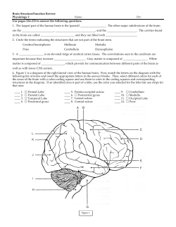
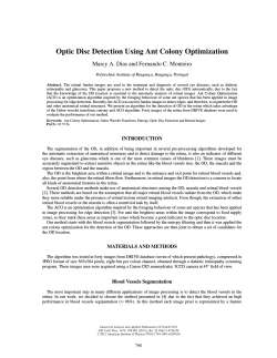
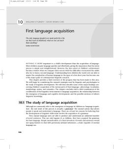
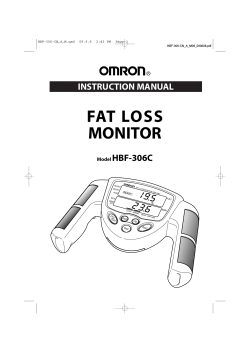
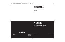
bj00078_ch19pg279_304.qxd 08/10/06 07:50 AM Page 279 E X E R C I S E 19 Gross Anatomy of the Brain and Cranial Nerves M A T E R I A L S O B J E C T I V E S ٗ Human brain model (dissectible) ٗ Preserved human brain (if available) ٗ Three-dimensional model of ventricles ٗ Coronally sectioned human brain slice (if available) ٗ Materials as needed for cranial nerve testing (see Table 19.1): aromatic oils (e.g., vanilla and cloves); eye chart; ophthalmoscope penlight; safety pin; Mall probe (hot and cold); cotton; solutions of sugar, salt, vinegar, and quinine; ammonia; tuning fork; and tongue depressor ٗ Preserved sheep brain (meninges and cranial nerves intact) ٗ Dissecting instruments and tray ٗ Disposable gloves ٗ The Human Nervous System: The Brain and Cranial Nerves videotape* See Appendix B, Exercise 19 for links to A.D.A.M.® Interactive Anatomy. 1. To identify the following brain structures on a dissected specimen (or slices), human brain model, or appropriate diagram, and to state their functions: *Available to qualified adopters from Benjamin Cummings. • Cerebral hemisphere structures: lobes, important fissures, lateral ventricles, basal ganglia, corpus callosum, fornix, septum pellucidum • Diencephalon structures: thalamus, intermediate mass, hypothalamus, optic chiasma, pituitary gland, mammillary bodies, pineal body, choroid plexus of the third ventricle, interventricular foramen • Brain stem structures: corpora quadrigemina, cerebral aqueduct, cerebral peduncles of the midbrain, pons, medulla, fourth ventricle • Cerebellum structures: cerebellar hemispheres, vermis, arbor vitae 2. To describe the composition of gray and white matter. 3. To locate the well-recognized functional areas of the human cerebral hemispheres. 4. To define gyri, fissures, and sulci. 5. To identify the three meningeal layers and state their function, and to locate the falx cerebri, falx cerebelli, and tentorium cerebelli. 6. To state the function of the arachnoid villi and dural sinuses. 7. To discuss the formation, circulation, and drainage of cerebrospinal fluid. 8. To identify at least four pertinent anatomical differences between the human brain and that of the sheep (or other mammal). 9. To identify the cranial nerves by number and name on an appropriate model or diagram, stating the origin and function of each. hen viewed alongside all nature’s animals, humans are indeed unique, and the key to their uniqueness is found in the brain. Each of us is a composite reflection of our brain’s experience. If all past sensory input could mysteriously and suddenly be “erased,” we would be unable to walk, talk, or communicate in any manner. Spontaneous movement would occur, as in a fetus, but no voluntary integrated function of any type would be possible. Clearly we would cease to be the same individuals. Because of the complexity of the nervous system, its anatomical structures are usually considered in terms of two principal divisions: the central nervous system and the peripheral nervous system. The central nervous system (CNS) consists of the brain and spinal cord, which primarily interpret incoming sensory information and issue instructions based on past experience. The peripheral nervous system (PNS) consists of the cranial and spinal nerves, ganglia, and sensory receptors. These structures serve as communication lines as they carry impulses—from the sensory receptors to the CNS and from the CNS to the appropriate glands or muscles. The PNS has two major subdivisions: the sensory portion, which consists of nerve fibers that conduct impulses toward the CNS, and the motor portion, which contains nerve fibers that conduct impulses away from the CNS. The motor portion, in turn, consists of the somatic division (sometimes called the voluntary system), which controls the skeletal muscles, and the autonomic nervous system (ANS), which controls smooth and cardiac muscles and glands. The W 279 bj00078_ch19pg279_304.qxd 08/10/06 07:50 AM Page 280 ANS is often referred to as the involuntary nervous system. Its sympathetic and parasympathetic branches play a major role in maintaining homeostasis. In this exercise both CNS (brain) and PNS (cranial nerves) structures will be studied because of their close anatomical relationship. The Human Brain During embryonic development of all vertebrates, the CNS first makes its appearance as a simple tubelike structure, the neural tube, that extends down the dorsal median plane. By the fourth week, the human brain begins to form as an expansion of the anterior or rostral end of the neural tube (the end toward the head). Shortly thereafter, constrictions appear, dividing the developing brain into three major regions— forebrain, midbrain, and hindbrain (Figure 19.1). The remainder of the neural tube becomes the spinal cord. During fetal development, two anterior outpocketings extend from the forebrain and grow rapidly to form the cerebral hemispheres. Because of space restrictions imposed by the skull, the cerebral hemispheres are forced to grow posteriorly and inferiorly, and finally end up enveloping and obscuring the rest of the forebrain and most midbrain structures. Somewhat later in development, the dorsal hindbrain also enlarges to produce the cerebellum. The central canal of the neural tube, which remains continuous throughout the brain and cord, enlarges in four regions of the brain, forming chambers called ventricles (see Figure 19.8a and b, page 288). A C T I V I T Y 1 Identifying External Brain Structures Identify external brain structures using the figures cited. Also use a model of the human brain and other learning aids as they are mentioned. (a) Neural tube Anterior (rostral) (b) Primary brain vesicles Generally, the brain is studied in terms of four major regions: the cerebral hemispheres, diencephalon, brain stem, and cerebellum. The relationship between these four anatomical regions and the structures of the forebrain, midbrain, and hindbrain is also outlined in Figure 19.1. Cerebral Hemispheres The cerebral hemispheres are the most superior portion of the brain (Figure 19.2). Their entire surface is thrown into elevated ridges of tissue called gyri that are separated by shallow grooves called sulci or deeper grooves called fissures. Many of the fissures and gyri are important anatomical landmarks. The cerebral hemispheres are divided by a single deep fissure, the longitudinal fissure. The central sulcus divides the frontal lobe from the parietal lobe, and the lateral sulcus separates the temporal lobe from the parietal lobe. The parieto-occipital sulcus on the medial surface of each hemisphere divides the occipital lobe from the parietal lobe. It is not visible externally. A fifth lobe of each cerebral hemisphere, the insula, is buried deep within the lateral sulcus, and is covered by portions of the temporal, parietal, and frontal lobes. Notice that most cerebral hemisphere lobes are named for the cranial bones that lie over them. Some important functional areas of the cerebral hemispheres have also been located (Figure 19.2d). The primary somatosensory cortex is located in the postcentral gyrus of the parietal lobe. Impulses traveling from the body’s sensory receptors (such as those for pressure, pain, and temperature) are localized in this area of the brain. (“This information is from my big toe.”) Immediately posterior to the primary somatosensory area is the somatosensory association area, in which the meaning of incoming stimuli is analyzed. (“Ouch! I have a pain there.”) Thus, the somatosensory association area allows you to become aware of pain, coldness, a light touch, and the like. Impulses from the special sense organs are interpreted in other specific areas also noted in Figure 19.2d. For example, (c) Secondary brain vesicles (d) Adult brain structures Cerebrum: cerebral hemispheres (cortex, white matter, basal ganglia) Lateral ventricles Telencephalon Prosencephalon (forebrain) Diencephalon (thalamus, hypothalamus, epithalamus), retina Third ventricle Diencephalon Mesencephalon (midbrain) Mesencephalon Brain stem: midbrain Cerebral aqueduct Metencephalon Brain stem: pons Rhombencephalon (hindbrain) Cerebellum Myelencephalon Posterior (caudal) (e) Adult neural canal regions Fourth ventricle Brain stem: medulla oblongata Spinal cord Central canal F I G U R E 1 9 . 1 Embryonic development of the human brain. (a) The neural tube becomes subdivided into (b) the primary brain vesicles, which subsequently form (c) the secondary brain vesicles, which differentiate into (d) the adult brain structures. (e) The adult structures derived from the neural canal. bj00078_ch19pg279_304.qxd 08/10/06 09:41 AM Page 281 Gross Anatomy of the Brain and Cranial Nerves F I G U R E 1 9 . 2 External structure (lobes and fissures) of the cerebral hemispheres. (a) Left lateral view of the brain. (b) Superior view. (c) Photograph of the superior aspect of the human brain. (d) Functional areas of the left cerebral cortex. The olfactory area, which is deep to the temporal lobe on the medial hemispheric surface, is not identified. Numbers indicate brain regions plotted by the Brodmann system. Central sulcus Postcentral gyrus Precentral gyrus Parietal lobe Frontal lobe Parieto-occipital sulcus (on medial surface of hemisphere) Lateral sulcus Occipital lobe Temporal lobe Transverse cerebral fissure Cerebellum Pons (a) Medulla oblongata Spinal cord Gyrus Cortex (gray matter) Frontal lobe Anterior Sulcus Longitudinal fissure White matter Precentral gyrus Fissure (a deep sulcus) Central sulcus Postcentral gyrus Parietal lobe Occipital lobe Posterior (b) (c) Motor Areas: Central sulcus Sensory Areas and Related Association Areas: Primary motor area 3 1 2 Premotor cortex 6 Frontal eye field 7 8 45 44 43 Taste General interpretation area (outlined by dots) 22 41 42 47 11 22 19 18 17 Primary visual cortex Visual association area Vision Auditory association area Primary auditory cortex (d) Somatic sensation Wernicke's area (outlined by dashes) Broca's area Solving complex, multitask problems Primary somatosensory cortex Somatosensory association area Gustatory cortex (in insula) Prefrontal Cortex: Working memory for spatial tasks Executive area for task management Working memory for object-recall tasks 5 4 281 Hearing bj00078_ch19pg279_304.qxd 08/10/06 07:50 AM Page 282 282 Exercise 19 the visual areas are in the posterior portion of the occipital lobe, and the auditory area is located in the temporal lobe in the gyrus bordering the lateral sulcus. The olfactory area is deep within the temporal lobe along its medial surface, in a region called the uncus (see Figure 19.4a, page 283). The primary motor area, which is responsible for conscious or voluntary movement of the skeletal muscles, is located in the precentral gyrus of the frontal lobe. A specialized motor speech area called Broca’s area is found at the base of the precentral gyrus just above the lateral sulcus. Damage to this area (which is located in only one cerebral hemisphere, usually the left) reduces or eliminates the ability to articulate words. Areas involved in intellect, complex reasoning, and personality lie in the anterior portions of the frontal lobes, in a region called the prefrontal cortex. A rather poorly defined region at the junction of the parietal and temporal lobes is Wernicke’s area, an area in which unfamiliar words are sounded out. Like Broca’s area, Wernicke’s area is located in one cerebral hemisphere only, typically the left. Although there are many similar functional areas in both cerebral hemispheres, such as motor and sensory areas, each hemisphere is also a “specialist” in certain ways. For example, the left hemisphere is the “language brain” in most of us, because it houses centers associated with language skills and speech. The right hemisphere is more specifically concerned Frontal lobe of cerebral hemisphere with abstract, conceptual, or spatial processes—skills associated with artistic or creative pursuits. The cell bodies of cerebral neurons involved in these functions are found only in the outermost gray matter of the cerebrum, the area called the cerebral cortex. Most of the balance of cerebral tissue—the deeper cerebral white matter—is composed of fiber tracts carrying impulses to or from the cortex. Using a model of the human brain (and a preserved human brain, if available), identify the areas and structures of the cerebral hemispheres described above. Then continue using the model and preserved brain along with the figures as you read about other structures. Diencephalon The diencephalon, sometimes considered the most superior portion of the brain stem, is embryologically part of the forebrain, along with the cerebral hemispheres. Turn the brain model so the ventral surface of the brain can be viewed. Using Figure 19.3 as a guide, start superiorly and identify the externally visible structures that mark the position of the floor of the diencephalon. These are the olfactory bulbs and tracts, optic nerves, optic chiasma (where the fibers of the optic nerves partially cross over), optic tracts, pituitary gland, and mammillary bodies. Frontal lobe Olfactory bulb (synapse point of cranial nerve I) Olfactory t r a c t Temporal lobe Pituitary gland Cerebral peduncle of midbrain Pyramid of medulla oblongata Decussation of pyramids Spinal cord Optic nerve (II) Optic chiasma Infundibulum of pituitary gland Temporal lobe Optic tract Mammillary body Pons Cerebellum Spinal cord F I G U R E 1 9 . 3 Ventral aspect of the human brain, showing the three regions of the brain stem. Only a small portion of the midbrain can be seen; the rest is surrounded by other brain regions. bj00078_ch19pg279_304.qxd 08/10/06 07:50 AM Page 283 Gross Anatomy of the Brain and Cranial Nerves Frontal lobe of cerebral hemisphere 283 Fornix Central sulcus Parietal lobe of cerebral hemisphere Lateral ventricle Corpus callosum Parieto-occipital sulcus Pineal body Anterior commissure Superior colliculi Inferior colliculi Corpora quadrigemina Hypothalamus Arbor vitae Optic nerve and chiasma Mammillary body Cerebellum Fourth ventricle Uncus Thalamus (a) Medulla oblongata Pons F I G U R E 1 9 . 4 Diencephalon and brain stem structures as seen in a midsagittal section of the brain. (a) Photograph. (Continues on page 284) Brain Stem Continue inferiorly to identify the brain stem structures— the cerebral peduncles (fiber tracts in the midbrain connecting the pons below with cerebrum above), the pons, and the medulla oblongata. Pons means “bridge,” and the pons consists primarily of motor and sensory fiber tracts connecting the brain with lower CNS centers. The lowest brain stem region, the medulla oblongata, is also composed primarily of fiber tracts. You can see the decussation of pyramids, a crossover point for the major motor tracts (pyramidal tracts) descending from the motor areas of the cerebrum to the cord, on the medulla’s anterior surface. The medulla also houses many vital autonomic centers involved in the control of heart rate, respiratory rhythm, and blood pressure as well as involuntary centers involved in vomiting, swallowing, and so on. Cerebellum 1. Turn the brain model so you can see the dorsal aspect. Identify the large cauliflower-like cerebellum, which projects dorsally from under the occipital lobes of the cerebrum. Notice that, like the cerebrum, the cerebellum has two major hemispheres and a convoluted surface (see Figure 19.6). It also has an outer cortex made up of gray matter with an inner region of white matter. 2. Remove the cerebellum to view the corpora quadrigemina (Figure 19.4), located on the posterior aspect of the midbrain, a brain stem structure. The two superior prominences are the superior colliculi (visual reflex centers); the two smaller inferior prominences are the inferior colliculi (auditory reflex centers). ■A C T I V I T Y 2 Identifying Internal Brain Structures The deeper structures of the brain have also been well mapped. Like the external structures, these can be studied in terms of the four major regions. As the internal brain areas are described, identify them on the figures cited. Also, use the brain model as indicated to help you in this study. Cerebral Hemispheres 1. Take the brain model apart so you can see a median sagittal view of the internal brain structures (see Figure 19.4). Observe the model closely to see the extent of the outer cortex (gray matter), which contains the cell bodies of cerebral neurons. The pyramidal cells of the cerebral motor cortex (studied in Exercise 17, page 257) are representative of the neurons seen in the precentral gyrus. 2. Observe the deeper area of white matter, which is composed of fiber tracts. The fiber tracts found in the cerebral hemisphere white matter are called association tracts if they connect two portions of the same hemisphere, projection tracts if they run between the cerebral cortex and the lower brain or spinal cord, and commissures if they run from one hemisphere to another. Observe the large corpus callosum, the major commissure connecting the cerebral hemispheres. The corpus callosum arches above the structures of the diencephalon and roofs over the lateral ventricles. Notice also the fornix, a bandlike fiber tract concerned with olfaction as well as limbic system functions, and the membranous septum pellucidum, which separates the lateral ventricles of the cerebral hemispheres. bj00078_ch19pg279_304.qxd 08/10/06 07:50 AM Page 284 Exercise 19 284 Parietal lobe of cerebral hemisphere Septum pellucidum Corpus callosum Interthalamic adhesion (intermediate mass of thalamus) Fornix Choroid plexus Occipital lobe of cerebral hemisphere Frontal lobe of cerebral hemisphere Thalamus (encloses third ventricle) Posterior commissure Interventricular foramen Pineal body/gland (part of epithalamus) Anterior commissure Corpora quadrigemina Hypothalamus Cerebral aqueduct Midbrain Optic chiasma Pituitary gland Arbor vitae Temporal lobe of cerebral hemisphere Fourth ventricle Mammillary body Pons Choroid plexus Cerebellum Medulla oblongata Spinal cord (b) F I G U R E 1 9 . 4 (continued) Diencephalon and brain stem structures as seen in a midsagittal section of the brain. (b) Diagrammatic view. 3. In addition to the gray matter of the cerebral cortex, there are several “islands” of gray matter (clusters of neuron cell bodies) called nuclei buried deep within the white matter of the cerebral hemispheres. One important group of cerebral nuclei, called the basal ganglia,* flank the lateral and third ventricles. You can see these nuclei if you have an appropriate dissectible model or a coronally or cross-sectioned human brain slice. Otherwise, Figure 19.5 will suffice. The basal ganglia, which are important subcortical motor nuclei (and part of the so-called extrapyronridal system), are involved in regulating voluntary motor activities. The most important of them are the arching, comma-shaped caudate nucleus, and the lentiform nucleus, which is composed of the putamen and globus pallidus nuclei. The closely associated amygdaloid nucleus (located at the tip of the caudate nucleus) is part of the limbic system. The corona radiata, a spray of projection fibers coursing down from the precentral (motor) gyrus, combines with sensory fibers traveling to the sensory cortex to form a broad band of fibrous material called the internal capsule. The internal capsule passes between the diencephalon and the basal ganglia, and gives these basal ganglia a striped appearance. This is why the caudate nucleus and the putamen portion of the lentiform nucleus are sometimes referred to collectively as the corpus striatum, or “striped body” (Figure 19.5a). *The historical term for these nuclei, basal ganglia, is a misleading term because ganglia are PNS structures. However, the name has been retained to differentiate these nuclei from the basal nuclei of the forebrain in the cerebrum. 4. Examine the relationship of the lateral ventricles and corpus callosum to the diencephalon structures; that is, thalamus and third ventricle—from the cross-sectional viewpoint (see Figure 19.5b). Diencephalon 1. The major internal structures of the diencephalon are the thalamus, hypothalamus, and epithalamus (see Figure 19.4). The thalamus consists of two large lobes of gray matter that laterally enclose the shallow third ventricle of the brain. A slender stalk of thalamic tissue, the interthalamic adhesion, or intermediate mass, connects the two thalamic lobes and bridges the ventricle. The thalamus is a major integrating and relay station for sensory impulses passing upward to the cortical sensory areas for localization and interpretation. Locate also the interventricular foramen (foramen of Monro), a tiny orifice connecting the third ventricle with the lateral ventricle on the same side. 2. The hypothalamus makes up the floor and the inferolateral walls of the third ventricle. It is an important autonomic center involved in regulation of body temperature, water balance, and fat and carbohydrate metabolism as well as in many other activities and drives (sex, hunger, thirst). Locate again the pituitary gland, which hangs from the anterior floor of the hypothalamus by a slender stalk, the infundibulum. (The pituitary gland is usually not present in preserved brain specimens.) In life, the pituitary rests in the hypophyseal fossa of the sella turcica of the sphenoid bone. Its function is discussed in Exercise 27. bj00078_ch19pg279_304.qxd 08/10/06 07:50 AM Page 285 Gross Anatomy of the Brain and Cranial Nerves Fibers of corona radiata Caudate nucleus Thalamus Lentiform nucleus Tail of caudate nucleus Internal capsule (projection fibers run deep to lentiform nucleus) (a) Anterior Cerebral cortex Cerebral white matter Corpus callosum Anterior horn of lateral ventricle Caudate nucleus Third ventricle Putamen Globus pallidus Lentiform nucleus Thalamus Inferior horn of lateral ventricle (b) Posterior F I G U R E 1 9 . 5 Basal ganglia. (a) Three-dimensional view of the basal ganglia showing their positions within the cerebrum. (b) A transverse section of the cerebrum and diencephalon showing the relationship of the basal ganglia to the thalamus and the lateral and third ventricles. 285 bj00078_ch19pg279_304.qxd 08/10/06 07:50 AM Page 286 286 Exercise 19 White matter of cerebellum Anterior lobe Brain stem (midbrain) Cerebellar cortex Primary fissure Posterior lobe Arbor vitae Horizontal fissure Vermis (a) Deep cerebellar nuclei Caudal (inferior) Vermis (cut) (b) F I G U R E 1 9 . 6 Cerebellum. (a) Posterior (dorsal) view. (b) The cerebellum, sectioned to reveal its cortex and medullary regions. (Note that the cerebellum is sectioned frontally and the brain stem is sectioned horizontally in this posterior view.) Anterior to the pituitary, identify the optic chiasma portion of the optic pathway to the brain. The mammillary bodies, relay stations for olfaction, bulge exteriorly from the floor of the hypothalamus just posterior to the pituitary gland. 3. The epithalamus forms the roof of the third ventricle and is the most dorsal portion of the diencephalon. Important structures in the epithalamus are the pineal body, or gland (a neuroendocrine structure), and the choroid plexus of the third ventricle. The choroid plexuses, knotlike collections of capillaries within each ventricle, form the cerebrospinal fluid. Brain Stem 1. Now trace the short midbrain from the mammillary bodies to the rounded pons below. Continue to refer to Figure 19.4. The cerebral aqueduct is a slender canal traveling through the midbrain; it connects the third ventricle to the fourth ventricle in the hindbrain below. The cerebral peduncles and the rounded corpora quadrigemina make up the midbrain tissue anterior and posterior (respectively) to the cerebral aqueduct. 2. Locate the hindbrain structures. Trace the rounded pons to the medulla oblongata below, and identify the fourth ventricle posterior to these structures. Attempt to identify the single median aperture and the two lateral apertures, three orifices found in the walls of the fourth ventricle. These apertures serve as conduits for cerebrospinal fluid to circulate into the subarachnoid space from the fourth ventricle. Cerebellum Examine the cerebellum. Notice that it is composed of two lateral hemispheres each with three lobes (anterior, posterior, and a deep flocculonodular) connected by a midline lobe called the vermis (Figure 19.6). As in the cerebral hemispheres, the cerebellum has an outer cortical area of gray matter and an inner area of white matter. The treelike branching of the cerebellar white matter is referred to as the arbor vitae, or “tree of life.” The cerebellum is concerned with unconscious coordination of skeletal muscle activity and control of balance and equilibrium. Fibers converge on the cerebellum from the equilibrium apparatus of the inner ear, visual pathways, proprioceptors of tendons and skeletal muscles, and from many other areas. Thus the cerebellum remains constantly aware of the position and state of tension of the various body parts. ■Meninges of the Brain The brain (and spinal cord) are covered and protected by three connective tissue membranes called meninges (Figure 19.7). The outermost meninx is the leathery dura mater, a double-layered membrane. One of its layers (the periosteal layer) is attached to the inner surface of the skull, forming the periosteum. The other (the meningeal layer) forms the outermost brain covering and is continuous with the dura mater of the spinal cord. The dural layers are fused together except in three places where the inner membrane extends inward to form a septum that secures the brain to structures inside the cranial cavity. One such extension, the falx cerebri, dips into the longitudinal fissure between the cerebral hemispheres to attach to the crista galli of the ethmoid bone of the skull (Figure 19.7a). The cavity created at this point is the large superior sagittal sinus, which collects blood draining from the brain tissue. The falx cerebelli, separating the two cerebellar hemispheres, and the tentorium cerebelli, separating the cerebrum from the cerebellum below, are two other important inward folds of the inner dural membrane. The middle meninx, the weblike arachnoid mater, underlies the dura mater and is partially separated from it by the subdural space. Threadlike projections bridge the subarachnoid space to attach the arachnoid to the innermost meninx, the pia mater. The delicate pia mater is highly vascular and clings tenaciously to the surface of the brain, following its convolutions. bj00078_ch19pg279_304.qxd 08/10/06 07:50 AM Page 287 Gross Anatomy of the Brain and Cranial Nerves 287 Skin of scalp Periosteum Bone of skull Periosteal layer Meningeal layer Superior sagittal sinus Dura mater Arachnoid mater Pia mater Subdural space Arachnoid villus Blood vessel Subarachnoid space Falx cerebri (in longitudinal fissure only) (a) Falx cerebri Superior sagittal sinus Straight sinus Tentorium cerebelli Crista galli of the ethmoid bone Cavernous sinus Internal carotid artery Skull Scalp Falx cerebelli Superior sagittal sinus (b) Occipital lobe Tentorium cerebelli Cerebellum Arachnoid mater over medulla oblongata (c) F I G U R E 1 9 . 7 Meninges of the brain. (a) Three-dimensional frontal section showing the relationship of the dura mater, arachnoid mater, and pia mater. The meningeal dura forms the falx cerebri fold, which extends into the longitudinal fissure and attaches the brain to the ethmoid bone of the skull. A dural sinus, the superior sagittal sinus, is enclosed by the dural membranes superiorly. Arachnoid villi, which return cerebrospinal fluid to the dural sinus, are also shown. (b) Position of the dural folds: the falx cerebri, tentorium cerebelli, and falx cerebelli. (c) Posterior view of the brain in place, surrounded by the dura mater. Dura mater Tranverse sinus Temporal bone bj00078_ch19pg279_304.qxd 08/10/06 07:50 AM Page 288 288 Exercise 19 Lateral ventricle Lateral ventricle Third ventricle Anterior horn Septum pellucidum Posterior horn Third ventricle Cerebral aqueduct Interventricular foramen Cerebral aqueduct Fourth ventricle Inferior horn Fourth ventricle Median aperture Lateral aperture Central canal Central canal (a) Anterior view (b) Left lateral view F I G U R E 1 9 . 8 Location and circulatory pattern of cerebrospinal fluid. (a) Anterior view. (b) Lateral view. Note that different regions of the large lateral ventricles are indicated by the terms anterior horn, posterior horn, and inferior horn. In life, the subarachnoid space is filled with cerebrospinal fluid. Specialized projections of the arachnoid tissue called arachnoid villi protrude through the dura mater to allow the cerebrospinal fluid to drain back into the venous circulation via the superior sagittal sinus and other dural sinuses. Meningitis, inflammation of the meninges, is a serious threat to the brain because of the intimate association between the brain and meninges. Should infection spread to the neural tissue of the brain itself, life-threatening encephalitis may occur. Meningitis is often diagnosed by taking a sample of cerebrospinal fluid from the subarachnoid space. ● Cerebrospinal Fluid The cerebrospinal fluid, much like plasma in composition, is continually formed by the choroid plexuses, small capillary knots hanging from the roof of the ventricles of the brain. The cerebrospinal fluid in and around the brain forms a watery cushion that protects the delicate brain tissue against blows to the head. Within the brain, the cerebrospinal fluid circulates from the two lateral ventricles (in the cerebral hemispheres) into the third ventricle via the interventricular foramina, and then through the cerebral aqueduct of the midbrain into the fourth ventricle in the hindbrain (Figure 19.8). Some of the fluid reaching the fourth ventricle continues down the central canal of the spinal cord, but the bulk of it circulates into the subarachnoid space, exiting through the three foramina in the walls of the fourth ventricle (the two lateral and the single median apertures). The fluid returns to the blood in the dural sinuses via the arachnoid villi. Ordinarily, cerebrospinal fluid forms and drains at a constant rate. However, under certain conditions—for example, obstructed drainage or circulation resulting from tumors or anatomical deviations—cerebrospinal fluid accumulates and exerts increasing pressure on the brain which, uncorrected, causes neurological damage in adults. In infants, hydrocephalus (literally, “water on the brain”) is indicated by a gradually enlarging head. The infant’s skull is still flexible and contains fontanels, so it can expand to accommodate the increasing size of the brain. ● bj00078_ch19pg279_304.qxd 08/10/06 07:50 AM Page 289 Gross Anatomy of the Brain and Cranial Nerves 289 Superior sagittal sinus Superior cerebral vein Arachnoid villus Choroid plexus Subarachnoid space Arachnoid mater Meningeal dura mater Cerebrum covered with pia mater Septum pellucidum Periosteal dura mater Corpus callosum Great cerebral vein Interventricular foramen Tentorium cerebelli Straight sinus Confluence of sinuses Third ventricle Pituitary gland Cerebellum Cerebral aqueduct Choroid plexus Lateral aperture Cerebellar vessels that supply choroid plexus Fourth ventricle Median aperture Central canal of spinal cord Spinal dura mater F I G U R E 1 9 . 8 (continued) (c) Cerebrospinal fluid flows from the lateral ventricles, through the interventricular foramina into the third ventricle, and then into the fourth ventricle via the cerebral aqueduct. (The relative position of the right lateral ventricle is indicated by the pale blue area deep to the corpus callosum and septum pellucidum.) Inferior end of spinal cord Filum terminale (inferior end of pia mater) (c) Cranial Nerves The cranial nerves are part of the peripheral nervous system and not part of the brain proper, but they are most appropriately identified while studying brain anatomy. The 12 pairs of cranial nerves primarily serve the head and neck. Only one pair, the vagus nerves, extends into the thoracic and abdominal cavities. All but the first two pairs (olfactory and optic nerves) arise from the brain stem and pass through foramina in the base of the skull to reach their destination. The cranial nerves are numbered consecutively, and in most cases their names reflect the major structures they control. The cranial nerves are described by name, number (Roman numeral), origin, course, and function in Table 19.1. This information should be committed to memory. A mnemonic device that might be helpful for remembering the cranial nerves in order is “On occasion, our trusty truck acts funny—very good vehicle anyhow.” The first letter of each word and the “a” and “h” of the final word “anyhow” will remind you of the first letter of the cranial nerve name. Most cranial nerves are mixed nerves (containing both motor and sensory fibers). But close scrutiny of Table 19.1 will reveal that three pairs of cranial nerves (optic, olfactory, and vestibulocochlear) are purely sensory in function. You may recall that the cell bodies of neurons are always located within the central nervous system (cortex or nuclei) or in specialized collections of cell bodies (ganglia) outside the CNS. Neuron cell bodies of the sensory cranial nerves are located in ganglia; those of the mixed cranial nerves are found both within the brain and in peripheral ganglia. Text continues on page 292 bj00130_ch19_279-304.qxd 11/20/07 9:07 AM Page 290 Team B 206:BEUS014:BJ00130A:mar9ch19: 290 Exercise 19 TA B L E 1 9 . 1 The Cranial Nerves (see Figure 19.9) Number and name Origin and course Function* Testing I. Olfactory Fibers arise from olfactory epithelium and run through cribriform plate of ethmoid bone to synapse in olfactory bulbs. Purely sensory—carries afferent impulses associated with sense of smell. Person is asked to sniff aromatic substances, such as oil of cloves and vanilla, and to identify each. II. Optic Fibers arise from retina of eye to form the optic nerve and pass through optic canal of orbit. Fibers partially cross over at the optic chiasma and continue on to the thalamus as the optic tracts. Final fibers of this pathway travel from the thalamus to the visual cortex as the optic radiation. Purely sensory—carries afferent impulses associated with vision. Vision and visual field are determined with eye chart and by testing the point at which the person first sees an object (finger) moving into the visual field. Fundus of eye viewed with ophthalmoscope to detect papilledema (swelling of optic disc, or point at which optic nerve leaves the eye) and to observe blood vessels. III. Oculomotor Fibers emerge from dorsal midbrain and course ventrally to enter the orbit. They exit from skull via superior orbital fissure. Primarily motor—somatic motor fibers to inferior oblique and superior, inferior, and medial rectus muscles, which direct eyeball, and to levator palpebrae muscles of the superior eyelid; parasympathetic fibers to iris and smooth muscle controlling lens shape (reflex responses to varying light intensity and focusing of eye for near vision). Pupils are examined for size, shape, and equality. Pupillary reflex is tested with penlight (pupils should constrict when illuminated). Convergence for near vision is tested, as is subject’s ability to follow objects with the eyes. IV. Trochlear Fibers emerge from midbrain and exit from skull via superior orbital fissure. Primarily motor—provides somatic motor fibers to superior oblique muscle (an extrinsic eye muscle). Tested in common with cranial nerve III. V. Trigeminal Fibers emerge from pons and form three divisions, which exit separately from skull: mandibular division through foramen ovale in sphenoid bone, maxillary division via foramen rotundum in sphenoid bone, and ophthalmic division through superior orbital fissure of eye socket. Mixed—major sensory nerve of face; conducts sensory impulses from skin of face and anterior scalp, from mucosae of mouth and nose, and from surface of eyes; mandibular division also contains motor fibers that innervate muscles of mastication and muscles of floor of mouth. Sensations of pain, touch, and temperature are tested with safety pin and hot and cold objects. Corneal reflex tested with wisp of cotton. Motor branch assessed by asking person to clench his teeth, open mouth against resistance, and move jaw side to side. VI. Abducens Fibers leave inferior pons and exit from skull via superior orbital fissure to run to eye. Carries motor fibers to lateral rectus muscle of eye. Tested in common with cranial nerve III. *Does not include sensory impulses from proprioceptors. PDF pass bj00078_ch19pg279_304.qxd 08/10/06 07:50 AM Page 291 Gross Anatomy of the Brain and Cranial Nerves TA B L E 1 9 . 1 291 (continued) Number and name Origin and course Function* Testing VII. Facial Fibers leave pons and travel through temporal bone via internal acoustic meatus, exiting via stylomastoid foramen to reach the face. Mixed—supplies somatic motor fibers to muscles of facial expression and parasympathetic motor fibers to lacrimal and salivary glands; carries sensory fibers from taste receptors of anterior portion of tongue. Anterior two-thirds of tongue is tested for ability to taste sweet (sugar), salty, sour (vinegar), and bitter (quinine) substances. Symmetry of face is checked. Subject is asked to close eyes, smile, whistle, and so on. Tearing is assessed with ammonia fumes. VIII. Vestibulocochlear Fibers run from inner-ear equilibrium and hearing apparatus, housed in temporal bone, through internal acoustic meatus to enter pons. Purely sensory—vestibular branch transmits impulses associated with sense of equilibrium from vestibular apparatus and semicircular canals; cochlear branch transmits impulses associated with hearing from cochlea. Hearing is checked by air and bone conduction using tuning fork. IX. Glossopharyngeal Fibers emerge from medulla and leave skull via jugular foramen to run to throat. Mixed—somatic motor fibers serve pharyngeal muscles, and parasympathetic motor fibers serve salivary glands; sensory fibers carry impulses from pharynx, tonsils, posterior tongue (taste buds), and from chemoreceptors and pressure receptors of carotid artery. A tongue depressor is used to check the position of the uvula. Gag and swallowing reflexes are checked. Subject is asked to speak and cough. Posterior third of tongue may be tested for taste. X. Vagus Fibers emerge from medulla and pass through jugular foramen and descend through neck region into thorax and abdomen. Mixed—fibers carry somatic motor impulses to pharynx and larynx and sensory fibers from same structures; very large portion is composed of parasympathetic motor fibers, which supply heart and smooth muscles of abdominal visceral organs; transmits sensory impulses from viscera. As for cranial nerve IX (IX and X are tested in common, since they both innervate muscles of throat and mouth). XI. Accessory Fibers arise from medulla and superior aspect of spinal cord and travel through jugular foramen to reach muscles of neck and back. Mixed (but primarily motor in function)—provides somatic motor fibers to sternocleidomastoid and trapezius muscles and to muscles of soft palate, pharynx, and larynx (spinal and medullary fibers respectively). Sternocleidomastoid and trapezius muscles are checked for strength by asking person to rotate head and shoulders against resistance. XII. Hypoglossal Fibers arise from medulla and exit from skull via hypoglossal canal to travel to tongue. Mixed (but primarily motor in function)—carries somatic motor fibers to muscles of tongue. Person is asked to protrude and retract tongue. Any deviations in position are noted. *Does not include sensory impulses from proprioceptors. bj00078_ch19pg279_304.qxd 08/10/06 07:50 AM Page 292 292 Exercise 19 Frontal lobe Filaments of olfactory nerve (I) Olfactory bulb Olfactory tract Optic nerve (II) Temporal lobe Optic chiasma Optic tract Infundibulum Oculomotor nerve (III) Facial nerve (VII) Trochlear nerve (IV) Vestibulocochlear nerve (VIII) Trigeminal nerve (V) Abducens nerve (VI) Glossopharyngeal nerve (IX) Vagus nerve (X) Cerebellum Accessory nerve (XI) Medulla Hypoglossal nerve (XII) F I G U R E 1 9 . 9 Ventral aspect of the human brain, showing the cranial nerves. (See also Figure 19.3.) A C T I V I T Y 3 Identifying and Testing the Cranial Nerves 1. Observe the ventral surface of the brain model to identify the cranial nerves. Figure 19.9 may also aid you in this study. Notice that the first (olfactory) cranial nerves are not visible on the model because they consist only of short axons that run from the nasal mucosa through the cribriform plate of the ethmoid bone. (However, the synapse points of the first cranial nerves, the olfactory bulbs, are visible on the model.) 2. The last column of Table 19.1 describes techniques for testing cranial nerves, which is an important part of any neurological examination. This information may help you understand cranial nerve function, especially as it pertains to some aspects of brain function. Conduct tests of cranial nerve function following directions given in the “testing” column of the table. 3. Several cranial nerve ganglia are named in the chart here. Using your textbook or an appropriate reference, fill in the chart by naming the cranial nerve the ganglion is associated with and stating its location. ■Cranial nerve ganglion Trigeminal Geniculate Inferior Superior Spiral Vestibular Cranial nerve Site of ganglion bj00078_ch19pg279_304.qxd 08/10/06 07:50 AM Page 293 Gross Anatomy of the Brain and Cranial Nerves 293 D I S S E C T I O N : Ventral The Sheep Brain The brain of any mammal is enough like the human brain to warrant comparison. Obtain a sheep brain, disposable gloves, dissecting tray, and instruments, and bring them to your laboratory bench. 1. Before beginning the dissection, turn your sheep brain so that you are viewing its left lateral aspect. Compare the various areas of the sheep brain (cerebrum, brain stem, cerebellum) to the photo of the human brain in Figure 19.10. Relatively speaking, which of these structures is obviously much larger in the human brain? Olfactory bulb Optic nerve (II) Mammillary body Cerebral peduncle 2. Place the intact sheep brain ventral surface down on the dissecting pan, and observe the dura mater. Feel its consistency and note its toughness. Cut through the dura mater along the line of the longitudinal fissure (which separates the cerebral hemispheres) to enter the superior sagittal sinus. Gently force the cerebral hemispheres apart laterally to expose the corpus callosum deep to the longitudinal fissure. 3. Carefully remove the dura mater and examine the superior surface of the brain. Notice that, like the human brain, its surface is thrown into convolutions (fissures and gyri). Locate the arachnoid mater, which appears on the brain surface as a delicate “cottony” material spanning the fissures. In contrast, the innermost meninx, the pia mater, closely follows the cerebral contours. Ventral Structures Figure 19.11a and b shows the important features of the ventral surface of the brain. Turn the brain so that its ventral surface is uppermost. Left cerebral hemisphere Pons Trigeminal nerve (V) Abducens nerve (VI) Medulla oblongata (a) F I G U R E 1 9 . 1 1 Intact sheep brain. (a) Photograph of ventral view. 1. Look for the clublike olfactory bulbs anteriorly, on the inferior surface of the frontal lobes of the cerebral hemispheres. Axons of olfactory neurons run from the nasal mucosa through the perforated cribriform plate of the ethmoid bone to synapse with the olfactory bulbs. How does the size of these olfactory bulbs compare with those of humans? Is the sense of smell more important as a protective and a food-getting sense in sheep or in humans? 2. The optic nerve (II) carries sensory impulses from the retina of the eye. Thus this cranial nerve is involved in the sense of vision. Identify the optic nerves, optic chiasma, and optic tracts. Brain stem Cerebellum F I G U R E 1 9 . 1 0 Photo of lateral aspect of the human brain. 3. Posterior to the optic chiasma, two structures protrude from the ventral aspect of the hypothalamus—the infundibulum (stalk of the pituitary gland) immediately posterior to the optic chiasma and the mammillary body. Notice that the sheep’s mammillary body is a single rounded eminence. In humans it is a double structure. 4. Identify the cerebral peduncles on the ventral aspect of the midbrain, just posterior to the mammillary body of the bj00078_ch19pg279_304.qxd 08/10/06 07:50 AM Page 294 294 Exercise 19 Olfactory bulb Olfactory tract Cerebrum Infundibulum (stalk of pituitary gland) Optic nerve (II) Optic chiasma Mammillary body Optic tract Cerebral peduncle Oculomotor nerve (III) Trochlear nerve (IV) Trigeminal nerve (V) Abducens nerve (VI) Pons Cerebellum Facial nerve (VII) Glossopharyngeal nerve (IX) Vestibulocochlear nerve (VIII) Vagus nerve (X) Hypoglossal nerve (XII) Spinal root of the accessory nerve (XI) Medulla oblongata (b) Dorsal Olfactory bulb Cerebrum Cerebrum Cerebellum Cerebellum Medulla oblongata Spinal cord (c) (d) F I G U R E 1 9 . 1 1 (continued) Intact sheep brain. (b) Diagrammatic ventral view. (c and d) Diagrammatic and photographic views of the dorsal aspect, respectively. bj00078_ch19pg279_304.qxd 08/10/06 07:50 AM Page 295 Gross Anatomy of the Brain and Cranial Nerves 295 F I G U R E 1 9 . 1 2 Means of exposing the dorsal midbrain structures of the sheep brain. Occipital lobe of cerebral hemisphere Pineal body Superior colliculi of corpora quadrigemina Inferior colliculi of corpora quadrigemina Cerebellum hypothalamus. The cerebral peduncles are fiber tracts connecting the cerebrum and medulla oblongata. Identify the large oculomotor nerves (III), which arise from the ventral midbrain surface, and the tiny trochlear nerves (IV), which can be seen at the junction of the midbrain and pons. Both of these cranial nerves provide motor fibers to extrinsic muscles of the eyeball. • 5. Move posteriorly from the midbrain to identify first the pons and then the medulla oblongata, both hindbrain structures composed primarily of ascending and descending fiber tracts. Dorsal Structures 1. Refer to Figure 19.11c and d as a guide in identifying the following structures. Reidentify the now exposed cerebral hemispheres. How does the depth of the fissures in the sheep’s cerebral hemispheres compare to that in the human brain? 6. Return to the junction of the pons and midbrain, and proceed posteriorly to identify the following cranial nerves, all arising from the pons: • • Trigeminal nerves (V), which are involved in chewing and sensations of the head and face. Abducens nerves (VI), which abduct the eye (and thus work in conjunction with cranial nerves III and IV). • Facial nerves (VII), large nerves involved in taste sensation, gland function (salivary and lacrimal glands), and facial expression. 7. Continue posteriorly to identify: Vestibulocochlear nerves (VIII), purely sensory nerves that are involved with hearing and equilibrium. Glossopharyngeal nerves (IX), which contain motor fibers innervating throat structures and sensory fibers transmitting taste stimuli (in conjunction with cranial nerve VII). Vagus nerves (X), often called “wanderers,” which serve many organs of the head, thorax, and abdominal cavity. Accessory nerves (XI), which serve muscles of the neck, larynx, and shoulder; notice that the accessory nerves arise from both the medulla and the spinal cord. • • • • Hypoglossal nerves (XII), which stimulate tongue and neck muscles. It is likely that some of the cranial nerves will have been broken off during brain removal. If so, observe sheep brains of other students to identify those missing from your specimen. 2. Examine the cerebellum. Notice that, in contrast to the human cerebellum, it is not divided longitudinally, and that its fissures are oriented differently. What dural falx (falx cerebri or falx cerebelli) is missing that is present in humans? 3. Locate the three pairs of cerebellar peduncles, fiber tracts that connect the cerebellum to other brain structures, by lifting the cerebellum dorsally away from the brain stem. The most posterior pair, the inferior cerebellar peduncles, connect the cerebellum to the medulla. The middle cerebellar peduncles attach the cerebellum to the pons, and the superior cerebellar peduncles run from the cerebellum to the midbrain. 4. To expose the dorsal surface of the midbrain, gently separate the cerebrum and cerebellum as shown in Figure 19.12. Identify the corpora quadrigemina, which appear as four rounded prominences on the dorsal midbrain surface. bj00078_ch19pg279_304.qxd 08/10/06 07:50 AM Page 296 296 Exercise 19 Corpus callosum Cerebral hemisphere Pineal body Fornix Corpora quadrigemina (midbrain) Septum pellucidum Cerebellum Intermediate mass of the thalamus Fourth ventricle Olfactory bulb Spinal cord Third ventricle Medulla oblongata Cerebral aqueduct Optic chiasma Pons Infundibulum (a) Pituitary gland Cerebral peduncle Mammillary body Cerebral hemisphere Parietal lobe Corpus callosum Cerebellum Frontal lobe of cerebrum Pineal body Fornix Arbor vitae Intermediate mass of thalamus Corpora quadrigemina Cerebral peduncle Fourth ventricle Medulla oblongata Optic chiasma Pons (b) F I G U R E 1 9 . 1 3 Sagittal section of the sheep brain showing internal structures. (a) Diagrammatic view. (b) Photograph. What is the function of the corpora quadrigemina? Also locate the pineal body, which appears as a small oval protrusion in the midline just anterior to the corpora quadrigemina. Internal Structures 1. The internal structure of the brain can only be examined after further dissection. Place the brain ventral side down on the dissecting tray and make a cut completely through it in a superior to inferior direction. Cut through the longitudinal fissure, corpus callosum, and midline of the cerebellum. Refer to Figure 19.13 as you work. 2. The thin nervous tissue membrane immediately ventral to the corpus callosum that separates the lateral ventricles is the septum pellucidum. Pierce this membrane and probe the lateral ventricle cavity. The fiber tract ventral to the septum pellucidum and anterior to the third ventricle is the fornix. How does the relative size of the fornix in this brain compare with the human fornix? _________________________________________________ bj00078_ch19pg279_304.qxd 08/10/06 07:50 AM Page 297 Gross Anatomy of the Brain and Cranial Nerves 297 Fornix Part of corpus striatum Caudate nucleus Lentiform nucleus Lateral ventricle Third ventricle Intermediate mass of thalamus Thalamic nuclei Third ventricle Hypothalamus F I G U R E 1 9 . 1 4 Frontal section of a sheep brain. Major structures revealed are the location of major basal ganglia in the interior, the thalamus, hypothalamus, and lateral and third ventricles. Why do you suppose this is so? (Hint: What is the function of this band of fibers?) 3. Identify the thalamus, which forms the walls of the third ventricle and is located posterior and ventral to the fornix. The intermediate mass spanning the ventricular cavity appears as an oval protrusion of the thalamic wall. Anterior to the intermediate mass, locate the interventricular foramen, a canal connecting the lateral ventricle on the same side with the third ventricle. 4. The hypothalamus forms the floor of the third ventricle. Identify the optic chiasma, infundibulum, and mammillary body on its exterior surface. You can see the pineal body at the superoposterior end of the third ventricle, just beneath the junction of the corpus callosum and fornix. 5. Locate the midbrain by identifying the corpora quadrigemina that form its dorsal roof. Follow the cerebral aqueduct (the narrow canal connecting the third and fourth ventricles) through the midbrain tissue to the fourth ventricle. Identify the cerebral peduncles, which form its anterior walls. 6. Identify the pons and medulla oblongata, which lie anterior to the fourth ventricle. The medulla continues into the spinal cord without any obvious anatomical change, but the point at which the fourth ventricle narrows to a small canal is generally accepted as the beginning of the spinal cord. 7. Identify the cerebellum posterior to the fourth ventricle. Notice its internal treelike arrangement of white matter, the arbor vitae. 8. If time allows, obtain another sheep brain and section it along the frontal plane so that the cut passes through the infundibulum. Compare your specimen to the photograph in Figure 19.14, and attempt to identify all the structures shown in the figure. 9. Check with your instructor to determine if cow spinal cord sections (preserved) are available for the spinal cord studies in Exercise 21. If not, save the small portion of the spinal cord from your brain specimen. Otherwise, dispose of all the organic debris in the appropriate laboratory containers and clean the laboratory bench, the dissection instruments, and the tray before leaving the laboratory. ■bj00078_ch19pg279_304.qxd 08/10/06 07:50 AM Page 298 bj00078_ch19pg279_304.qxd 08/10/06 07:50 AM Page 299 R E V I E W S H E E T NAME ____________________________________ EXERCISE LAB TIME/DATE ________________________ 19 Gross Anatomy of the Brain and Cranial Nerves The Human Brain 1. Match the letters on the diagram of the human brain (right lateral view) to the appropriate terms listed at the left. 1. frontal lobe 2. parietal lobe f a g b 3. temporal lobe 4. precentral gyrus c h d i 5. parieto-occipital sulcus 6. postcentral gyrus j e k l 7. lateral sulcus 10. medulla 8. central sulcus 11. occipital lobe 9. cerebellum 12. pons 2. In which of the cerebral lobes would the following functional areas be found? auditory area: olfactory area: primary motor area: visual area: primary sensory area: Broca’s area: 3. Which of the following structures are not part of the brain stem? (Circle the appropriate response or responses.) cerebral hemispheres pons midbrain cerebellum medulla diencephalon 4. Complete the following statements by writing the proper word or phrase on the corresponding blanks at the right. 1 _ is an elevated ridge of cerebral tissue. The convoA(n) _____ lutions seen in the cerebrum are important because they increase 2 _. Gray matter is composed of _____ 3 _. White matter is the _____ 4 _. A fiber tract that provides for communication composed of _____ between different parts of the same cerebral hemisphere is called 5 _, whereas one that carries impulses to the cerebrum a(n) _____ from, and from the cerebrum to, lower CNS areas is called a(n) 6 _ tract. The lentiform nucleus along with the caudate nuclei _____ 7 _. are collectively called the _____ 1. 2. 3. 4. 5. 6. 7. 299 bj00078_ch19pg279_304.qxd 08/10/06 07:50 AM Page 300 300 Review Sheet 19 5. Identify the structures on the following sagittal view of the human brain stem and diencephalon by matching the numbered areas to the proper terms in the list. a. cerebellum b. cerebral aqueduct c. (small part of) cerebral hemisphere d. cerebral peduncle e. choroid plexus f. corpora quadrigemina 10 1 2 12 3 13 4 14 5 15 6 16 7 17 8 18 9 19 g. corpus callosum h. fornix i. fourth ventricle j. hypothalamus k. intermediate mass n. optic chiasma q. pons l. o. pineal body r. septum pellucidum p. pituitary gland s. thalamus mammillary bodies m. medulla oblongata 11 6. Using the terms from question 5, match the appropriate structures with the descriptions given below. 1. site of regulation of body temperature and water balance; most important autonomic center 2. consciousness depends on the function of this part of the brain 3. located in the midbrain; contains reflex centers for vision and audition 4. responsible for regulation of posture and coordination of complex muscular movements 5. important synapse site for afferent fibers traveling to the sensory cortex 6. contains autonomic centers regulating blood pressure, heart rate, and respiratory rhythm, as well as coughing, sneezing, and swallowing centers 7. large commissure connecting the cerebral hemispheres 8. fiber tract involved with olfaction 9. connects the third and fourth ventricles 10. encloses the third ventricle bj00078_ch19pg279_304.qxd 08/10/06 07:50 AM Page 301 Review Sheet 19 301 7. Embryologically, the brain arises from the rostral end of a tubelike structure that quickly becomes divided into three major regions. Groups of structures that develop from the embryonic brain are listed below. Designate the embryonic origin of each group as the hindbrain, midbrain, or forebrain. 1. the diencephalon, including the thalamus, optic chiasma, and hypothalamus 2. the medulla, pons, and cerebellum 3. the cerebral hemispheres 8. What is the function of the basal ganglia? 9. What is the corpus striatum, and how is it related to the fibers of the internal capsule? 10. A brain hemorrhage within the region of the right internal capsule results in paralysis of the left side of the body. Explain why the left side (rather than the right side) is affected. 11. Explain why trauma to the base of the brain is often much more dangerous than trauma to the frontal lobes. (Hint: Think about the relative functioning of the cerebral hemispheres and the brain stem structures. Which contain centers more vital to life?) 12. In “split brain” experiments, the main commissure connecting the cerebral hemispheres is cut. First, name this commissure. Then, describe what results (in terms of behavior) can be anticipated in such experiments. (Use an appropriate reference if you need help with this one!) bj00078_ch19pg279_304.qxd 08/10/06 07:50 AM Page 302 302 Review Sheet 19 Meninges of the Brain 13. Identify the meningeal (or associated) structures described below: 1. outermost meninx covering the brain; composed of tough fibrous connective tissue 2. innermost meninx covering the brain; delicate and highly vascular 3. structures instrumental in returning cerebrospinal fluid to the venous blood in the dural sinuses 4. structure that forms the cerebrospinal fluid 5. middle meninx; like a cobweb in structure 6. its outer layer forms the periosteum of the skull 7. a dural fold that attaches the cerebrum to the crista galli of the skull 8. a dural fold separating the cerebrum from the cerebellum Cerebrospinal Fluid 14. Label the structures involved with circulation of cerebrospinal fluid on the accompanying diagram. Add arrows to the figure above to indicate the flow of cerebrospinal fluid from its formation in the lateral ventricles to the site of its exit from the fourth ventricle. Then fill in the blanks in the following paragraph. Cerebrospinal fluid flows from the fourth ventricle into the (1)__ space surrounding central canal of the spinal cord and the _____ the brain and spinal cord. From this space it drains through the (3)__. (2)__ into the _____ _____ 1. 2. 3. bj00078_ch19pg279_304.qxd 08/10/06 07:50 AM Page 303 Review Sheet 19 303 Cranial Nerves 15. Using the terms below, correctly identify all structures indicated by leader lines on the diagram. a. abducens nerve (VI) j. longitudinal fissure s. pituitary gland b. accessory nerve (XI) k. mammillary body t. pons c. cerebellum l. medulla oblongata u. spinal cord d. cerebral peduncle m. oculomotor nerve (III) v. temporal lobe of cerebral hemisphere e. decussation of the pyramids n. olfactory bulb f. facial nerve (VII) o. olfactory tract g. frontal lobe of cerebral hemisphere p. optic chiasma h. glossopharyngeal nerve (IX) q. optic nerve (II) i. hypoglossal nerve (XII) r. optic tract w. trigeminal nerve (V) x. trochlear nerve (IV) y. vagus nerve (X) z. vestibulocochlear nerve (VIII) bj00078_ch19pg279_304.qxd 08/10/06 07:50 AM Page 304 304 Review Sheet 19 16. Provide the name and number of the cranial nerves involved in each of the following activities, sensations, or disorders. 1. rotating the head 7. listening to music; seasickness 2. smelling a flower 8. secretion of saliva; tasting wellseasoned food 3. raising the eyelids; pupillary constriction 9. involved in “rolling’’ the eyes (three nerves—provide numbers only) 4. slows the heart; increases motility of the digestive tract 5. involved in Bell’s palsy (facial paralysis) 10. feeling a toothache 11. reading the newspaper 12. purely sensory in function (three nerves—provide numbers only) 6. chewing food Dissection of the Sheep Brain 17. In your own words, describe the firmness and texture of the sheep brain tissue as observed when cutting into it. Because formalin hardens all tissue, what conclusions might you draw about the firmness and texture of living brain tissue? 18. When comparing human and sheep brains, you observe some profound differences between them. Record your observations in the chart below. Structure Olfactory bulb Pons/medulla relationship Location of cranial nerve III Mammillary body Corpus callosum Intermediate mass of thalamus Relative size of superior and inferior colliculi Pineal gland Human Sheep
© Copyright 2026 Paperzz