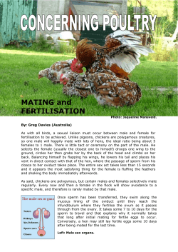
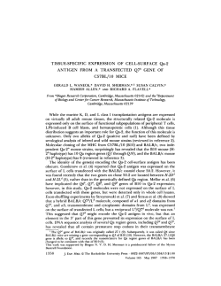
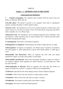
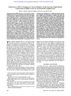
®¾ÅÊËÌÄÃËÆÃ Æ ÔÆÈÉ٠ξÅÀÆÐÆÝ ÀÌÂÌÎÌÏÉÃÇ UDC 582.261.1 + 581.162 Yu.A. PODUNAY, O.I. DAVIDOVICH, N.A. DAVIDOVICH Karadag Nature Reserve of the NAS of Ukraine, 98188 Feodosiya, Crimea, Ukraine e-mail: [email protected] MATING SYSTEM AND TWO TYPES OF GAMETOGENESIS IN THE FRESH WATER DIATOM ULNARIA ULNA (BACILLARIOPHYTA) Sexual reproduction and mating system of the freshwater diatom Ulnaria ulna (Nitzsch) Compère were studied by using clonal cultures. Mating system of the species involves homo- and heterothallic modes of reproduction; and both male and female clones were capable of homothallic reproduction. Two types of gametogenesis corresponding to two mating types were investigated. Analysis of the crossing table, gamete morphology, and sex distribution in the progeny resulted from intraclonal reproduction provided evidences that anisogamy and two mating types were determined in U. ulna genetically; male and female clones were hetero- and homogametic correspondently. Specific active movement of male gametes caused by the formation and retraction of pseudopodia-like structures on the gamete surface was described. The absence of reproductive isolation between clones gathered from geographically distant populations suggests continuity and broad distribution of the species. K e y w o r d s : Ulnaria ulna, mating system, sexual reproduction, gametogenesis, gametes movement, reproductive isolation. Introduction Last three decades brought us numerous publications, which indicated heterothallic character of mating system in many pennate diatoms (Roshchin, 1994; Davidovich et al., 1998, 2009, 2010; Chepurnov, Mann, 2001, 2004; Chepurnov et al., 2004; Mann, Chepurnov, 2005; Amato et al., 2007; Poulíþková et al., 2007; Trobajo et al., 2009; Mann, Poulíþková, 2010; Davidovich, Davidovich, 2011; and others). Biparental sex distribution suggests clear strategy of breeding experiments in pair combination of clones. Surprisingly, in the beginning experiments with Ulnaria ulna we received unexpected results. Signs of auxosporulation, i.e. gametes, empty gametangial frustules, zygotes, young and growing auxospores, etc. were found almost in all pair cross combinations. However, careful investigation of the pattern of sexual reproduction allowed us revealing two types of gametogenesis and corresponding modes of gametes behaviour that elucidated mating system and mysterious "panmixia" of clones in this species. The strategy of fertilization in araphid sessile diatoms is fairly diverse. In some araphid species parental cells need to be lain down © Yu.A. Podunay, O.I. Davidovich, N.A. Davidovich, 2014 ISSN 0868-8540. ÉÚÁÌÉÌÁÆÝ. 2014, 24(1) 3 Yu.A. Podunay, O.I. Davidovich, N.A. Davidovich close to each other; just on this occasion gametes can reach the place of syngamy by amoeboid movement, e.g. Licmophora C. Agardh (Chepurnov, Mann, 2004). Male gametes in Tabularia fasciculata (C. Agardh) D. Williams and Round and T. tabulata (C. Agardh) Snoeijs were found to be able to move for relatively long distance; mechanism of their movement turned to be highly unusual (Davidovich et al., 2012). Similar method of motion was reported in Pseudostaurosira trainorii E.A. Morales (Sato et al., 2011). In the present investigation we paid special attention to the strategy of fertilization, which is realized in U. ulna. Data on reproductive isolation are useful in the discovery of species, species validation, or for confirming that specimens taken from different localities belong or not belong to the same species (Mann, 2010). Some diatoms are known to be cosmopolitan (Casteleyn et al., 2008) while some others may represent a complex comprising cryptic or pseudo-cryptic species (Mann et al., 2008). Morphotype U. ulna is known from around the world that provokes questions about real distribution of this species, and how many species are hidden under this name. Direct breeding of clones gathered from geographically distant populations, which inhabit different river basins and separated one from another by thousand kilometres including marine straits may provide valuable information. The aim of the present article was in discussing breeding system, fertilization strategy, and means of gamete delivery to the place of syngamy in the araphid pennate diatom U. ulna. The subject of speculation was also worldwide distribution of the species that was examined in breeding experiments. Objects and methods Clonal cultures of Ulnaria ulna (Nitzsch) Compère in Jahn et al. 2001 (formerly, Synedra ulna (Nitzsch) Ehrenb. 1832) were derived from samples collected in 2008—2012 in geographically distant places, the Dnieper River, Kiev, Ukraine (3 clones), a spring in the south-eastern Crimea, Radostnoe, Ukraine (11 clones), the Moskva River, Zvenigorod, Russia (3 clones), and a small river flowing from the Roath Park Lake, Cardiff, the UK (6 clones). Single cells were isolated under the microscope MBS-9 (Russia) with the aid of glass micropipettes by repeated washing in 5—7 drops of culture medium placed on a slide. Clones were named as Y.MMDD-X, where Y is the last digit of the year of isolation, MM is a month, DD is a date, and X is a short name of the clone. Cultures were cultivated in glass Petri dishes (diam. 5—9 cm, medium volume from 8 to 45 mL). The composition of culture medium was close to the medium Dm (Mann, 2004). Cultures were maintained in the temperature stabilized room at 19—21 oC under diffuse daylight from northward-directed window. Every 4—6 days cultures were re-inoculated into fresh medium to maintain them in exponential growth phase. Cultures were inspected daily for signs of intraclonal sexual reproduction, general health and growth. Clones were periodically inoculated in pairs to stimulate interclonal reproduction. 4 ISSN 0868-8540. lgologia. 2014, 24(1) Mating system Observations were fulfilled by using light microscopes MBS-9 (LOMO, USSR) and Biolar PI (PZO, Poland). The last was equipped with Differential Interference Contrast (DIC) optics and adjusted to bright-field illumination following Keller (Fedin, Barsky, 1971). Microphotographs were taken using a Canon Power Shot A-640 digital camera, which was used also for gamete movement records. The cell length (apical size) was determined using an ocular ruler calibrated against object-micrometer. Mean values are presented as Mean ± Standard Error, N-number of measurements. Results Life cycle and critical cell sizes In the natural populations, Ulnaria ulna grew either as single cells or in clusters of cells being attached to the substratum by common mucilage pad. In cultures, if conditions were good, rapidly growing clones formed loose colonies on the bottom of Petri dishes. As a result of vegetative multiplication mean length of cells maintained in clonal cultures permanently decreased with the average rate of 5.5±0.4 ȝm/month (N = 32). The apical length of cultured cells on different stages of their life history and the length of cells derived from natural populations were measured; and their ranges are presented in Table 1. The apical length of vegetative cells in the Crimean population varied from 11.2 to 176.0 ȝm, with mean value 91.4± 2.4 ȝm (N = 658), initial cells had sizes 291—422 ȝm, mean 340.0±2.9 ȝm (N = 104). In Cardiff population, mean apical length was 148.0±3.2 ȝm (N = 104, range 62.3—235.3). In the Moskva River the cell length ranged from 90.0—290.6 ȝm, mean 188.0±4.6 ȝm (N = 144). Table 1 The apical size of cells of Ulnaria ulna in samples taken from geographically distant natural populations Location Sampling period Dnieper April, River 2010 Mean apical size, ȝm Size range, ȝm — — 332.9±5.5 Sept., 188.0±4.6 River 2011 N = 144 Cardiff — (304.5—359.8) N = 10 Moskva Crimea Mean and range of apical sizes of initial cells, ȝm in natural in culture population April, 91.4±2.4 2010 N = 658 April, 148.0±3.2 2012 N = 104 341.8±3.9 90.0—290.6 — (297.6—387.5) 340.0±2.9 353.9±1.25 (290.6—422.1) (276.8—415.2) N = 104 N = 182 328.2±2.1 (304.5—359.8) N = 45 N = 35 11.2—176.0 62.3—235.3 ISSN 0868-8540. ÉÚÁÌÉÌÁÆÝ. 2014, 24(1) — 5 Yu.A. Podunay, O.I. Davidovich, N.A. Davidovich The largest cell was found in the natural Crimean population, not in culture and its length was 422 ȝm. The smallest recorded cell had the length 11 ȝm. The length of cells entered sexual reproduction (gametangia) varied from 62 to 162 ȝm (in the Crimean population). The length of initial cells weakly depended on the size of gametangia (Fig. 1), correlation coefficient was equal to 0.27 (N = 23). There were no marked differences between the lengths of initial cells produced by different populations. Fig. 1. Relationship between gametangial and initial cell lengths. Gametangial cell length was calculated as the average of mean cell sizes in a pair of clones; initial cell length was calculated as the average length of initial cells arisen in the mixture of parental clones Male and female gametogenesis Each male and female gametangium yielded two gametes, which were morphologically identical (spherical) in the end of gametogenesis. However, early stages of male and female gametogenesis differed significantly. In the male gametangium, protoplast divided into two equal parts in transapical plan (Photo 1, a, b). Rearrangement of gametes was not observed. During early stages male gametes were rod-shaped with rounded ends, resembled "frankfurters", which moved to some extend independently along the apical axis in a reciprocal manner. Later the gametes rounded, and went out one after another from the gametangium frustule pushing its valves apart (Photo 1, d). After leaving the gametangium, male gametes became spherical. In contrast to male, female gametangia divided in the apical plane (Photo 1, e). In the beginning of their development female gametes were attached to the valves of the mother frustule, one against other (Photo 1, e, f). Development of the two gametes in the female gametangium was not completely synchronic, very often one of the gametes developed faster, but with time, both female gametes rounded and lost contact with valves (Photo 1, j). After detaching from the gametangium, female gametes also get spherical form. Usually mature female gametes lay between or near "mother's" gametangial frustules. Sometimes copulation may occur while female gamete was not yet detached from the valve. 6 ISSN 0868-8540. lgologia. 2014, 24(1) Mating system Photo 1. Male and female gametogenesis. a — the cell content of the male gametangium divides in the transapical plan (arrowhead) giving rise to two male gametes; b — an early stage of male gametes (arrows) partition, a gap (arrowhead) between two gametes (arrows); female gamete (double arrow) associated with mother frustules; c, d — both male and female gametes finally became round and detached from the gametangium valve; e—j — female gametangium content divides in the apical plan giving rise to two gametes attached to the gametangial thecae; with time the gametes narrow, became rounded and finally detach from the thecae. Scale 20 m ISSN 0868-8540. ÉÚÁÌÉÌÁÆÝ. 2014, 24(1) 7 Yu.A. Podunay, O.I. Davidovich, N.A. Davidovich Photo 2. Auxospore formation and paedogamous copulation. b—d — different stages of auxospore expansion; e — the initial cell (above) is prominently longer than one of the cells of the parental clone (below); f—h — paedogamous reproduction in the female clone. Scale: 20 m (a—e), and 10 m (f—h) 8 ISSN 0868-8540. lgologia. 2014, 24(1) Mating system Following physical contact male and female gametes fused giving rise to zygotes (Photo 2, a). Neither copulation tubes nor mucilage envelopes were ever observed. The zygotes were initially spherical (Photo 2, a, lower gamete). Approximately one to two hours later zygotes started bipolar expansion (Photo 2, a, upper gamete; b), and from this moment they may be regarded as auxospores. Growing auxospores had no tight contact with valves of mother frustule and could lie between or apart from them (Photo 2, c). Elongated auxospores were slightly curved. In contrast to some other diatoms (e.g. Mann, 1996; Trobajo et al., 2009), the remains of zygote envelopes in the form of "caps" at the ends of auxospores were not visible. Inside the fully expanded auxospore, two valves were deposited successively, giving rise to the frustule of the initial cell (Photo 2, e), which renews mitotic divisions. Gamete movement Gamete movement coincided with the formation of slender cytoplasmic projections on the gamete surface (Photo 3). Only male gametes were found to form these projections. They were not permanent structures and to a certain extent resembled pseudopodia. Pseudopodial activity could be revealed at the early stages of gamete formation, while they were laying between the gametangia valves, and periodically every several minutes after male gametes escaped from the gametangium. Initially the gametes were almost ideally spherical and immobile. After several minutes the gamete surface became active; a few relatively short and broad lamellipodium-like projections arose, which distorted spherical form of the gamete. With time one or two projections elongated into thin threads, while shorter and broader protrusions disappeared. Upon reaching the maximal length projections became flexible and started to retract. It appeared that projections winded around the gamete cell and the last was forced to rotate in the opposite direction, as "a ball of thread". Finally, projections vanished completely and the cell returned to spherical form. The cycle repeated in a few minutes. Breeding system For the first time sexual reproduction was detected in monoclonal culture 8.0626-D. All gametes which had been seen in this case were spherical and motile; in all aspects of their morphology and behaviour they were similar to male gametes which were founded later in heterothallic pairs. Intraclonal progeny was fertile; F-1 clones could mate with each other if they were sexually compatible and readily entered interclonal reproduction with clones originated from natural populations. Sex of 4 clones randomly derived from the first intraclonal generation of 8.0626-D was determined; and 2 of them turned to be male and 2 female (Table 2). Both male and female clones were able to enter intraclonal reproduction; in total a half of the clones examined (11 of 23) revealed such ability. Intraclonal reproduction was not abundant compared to the interclonal one and did not always result in the formation of viable initial cells, the process sometimes terminated at the stage of the gamete or zygote formation (Table 2). ISSN 0868-8540. ÉÚÁÌÉÌÁÆÝ. 2014, 24(1) 9 Yu.A. Podunay, O.I. Davidovich, N.A. Davidovich Photo 3. Time sequences of the two cytoplasmic projections formation cycles (a—g and h—l). The objects in the frames (h—l) are the same as in the Photo 1 (b—d); the male gamete (arrows) in contrast to the female one (double arrow) can produce slender cytoplasmic projection on the cell surface (arrowhead), the process is accompanied by change of the cell shape and rotation of the cell during projection retraction. Scale 10 m 10 ISSN 0868-8540. lgologia. 2014, 24(1) Mating system In male clones intraclonal reproduction was presumably allogamous; in the case of female clones we observed paedogamous reproduction, where two female gametes produced by the same gametangium fused while being atISSN 0868-8540. ÉÚÁÌÉÌÁÆÝ. 2014, 24(1) 11 Yu.A. Podunay, O.I. Davidovich, N.A. Davidovich tached to the valves of the mother frustule (Photo 2, f—h). Theoretically, auto-mixis in male clones as well as allogamy in female clones are also possible, but we did not acquire direct evidences. If clones were inoculated in pairs, signs of sexual reproduction could be observed in two thirds of crosses (139 of 221) that gave an impression of "panmixia". As a result, it was not possible to determine affiliation of clones to the certain mating type by using solely data of crossing table without taking into account other characteristics. It is worth noting that information on sex affiliation could not be elicited if late stages of gametogenesis were investigated; mature gametes were uniformly spherical. More careful investigation of the data obtained revealed pronounced difference in the frequency of auxosporulation and an ability to form viable auxospores and initial cells in different pairs of clones (Table 2). Relative abundance of sexual cells (gametes) and their products (zygotes, auxospores, and initial cells) were much higher in 84 pairs. To differentiate sexes, most valuable were morphological data obtained from early stages of gametes development. Clones isolated from geographically distant populations turned to be sexually compatible; they readily entered intercrossing sexual reproduction according to their mating types (Table 2). Discussion Sexual reproduction of the araphid freshwater diatom Ulnaria ulna (Photo 4) was thoroughly investigated by L. Geitler (1939a, b). He gave a description of allogamous reproduction as most usual in the species and showed that during early stages of gametogenesis cell protoplast divided in the longitudinal plane being attached to the valves; later gametes rounded, separated from mother's frustules, and fused with the gametes, which in other cells had not yet detached from the valves. As an exception to the normal cross-copulation, Geitler described automictic (paedogamous) fertilization, when a single mother cell formed only one zygote attached to both valves of the frustule. He pointed out, not entering into details, an ability of Synedra ulna's gametes to move and suggested that gamete movement correlated more with the timing of gamete formation rather than genetic sexual constitution. Of course, Geitler could not investigate breeding system, distribution of sexes in the population, as well as mechanism of sex determination and differentiation because he worked mainly on material derived from natural populations; breeding experiments with clonal cultures were of little used. Comparison of gamete morphology and mating type distribution (Table 2) suggests, however genetic determination of sexes. Genotypic type of sex determination is also confirmed by appearance of “male” and “female” clones in the intraclonal progeny that demonstrates heterogametic nature of male sex (Table 2, intraclonal descendants of the clone 8.0626-D). Accordingly, we were able to distinguish two types of gametogenesis corresponding to two mating types, male and female. 12 ISSN 0868-8540. lgologia. 2014, 24(1) Mating system Photo 4. Valve (a, b, d) and girdle (c) view of cleaned frustules and the life form (colonies on the filamentous substrate, e; and on the bottom of a Petri dish, f). Light microscopy, differential interference contrast optics. Scale: 20 m (a), and 10 m (b—d) Results obtained in the present study confirmed that Ulnaria ulna belongs to the category IA2b in Geitler's (1973) system of patterns of sexual reproduction and auxospore formation. This category is characterized by formation of two auxospores in the pair of gametangia, each of which produces two gametes. Gametes behaviour is anisogamous, and both active gametes are produced by the same gametangium and move to fuse with the stationary gametes. While differing in the beginning, male and female gametes of U. ulna are finally equal by size and have similar shape. In this connection mature female gametes may be misinterpreted as male. Rearrangement of gamete was not ISSN 0868-8540. ÉÚÁÌÉÌÁÆÝ. 2014, 24(1) 13 Yu.A. Podunay, O.I. Davidovich, N.A. Davidovich observed both in male and female parental gametangia. The mode of protoplast division, either in apical (females) or transapical (males) plane, does not fully agree with Geitler's system. As more data are gathered, it becomes clear that Geitler's classification needs in further modification (Stickle, 1986; Mizuno, 1994; Chepurnov, Mann, 2004). Thus, following recommendations by M. Mizuno (1994) we have to place U. ulna to the subgroup IA2(d). In many principal features mode of sexual reproduction in U. ulna is very similar to that of Tabularia fasciculata and T. tabulata (Roschin, 1994; Davidovich et al., 2010; Davidovich, Davidovich, 2011). In contrast to U. ulna, in T. fasciculata and T. tabulata female gametes were more tightly associated with gametangial thecae. Rearrangement of male gametes was supposed in T. fasciculata (Davidovich et al., 2010), however this question must be elucidated in view of the data for T. tabulata (Davidovich, Davidovich, 2011) and new data obtained for U. ulna where rearrangement of male and female gamete was not observed. Interestingly, in the araphid pennate Licmophora ehrenbergii (Kütz.) Grunow female gametes never rearranged, but gametes in male gametangia rearranged or not, depending on the relative position of mating gametangia (Chepurnov, Mann, 2004). In the life cycle, the upper border of the size range suitable for sexual reproduction may be from 30 to 75 % of the maximal cell size, depending on the species, but in most diatoms it corresponds to 45—55 % (Davidovich, 2001). U. ulna has the upper critical size close to 40 % of the maximal size. At the same time, there is no correlation between the cell size of parental and descendent cells (Fig. 1), that may result in the situation when the smallest gametangia produce the biggest initial cells, as well as the biggest gametangia may produce the smallest initial cells. Consequently, the size restoration coefficient may change from 1.7 to 6.8 (Fig. 2). Following L. Geitler (1939a, b) automictic auxosporulation was quite usual in U. ulna, and was reported to be the only mode of sexual reproduction in Synedra vaucheriae (Kütz.) Kütz. (Geitler, 1958). Geitler's description of homothallic allogamy in U. ulna (Geitler, 1939a, b) not brought into correlation with any certain sex of clones. Moreover, he suggested phenotypic rather than genotypic sex expression in U. ulna. Theoretically, monoparental but allogamous reproduction is possible if gametes have a chance to be delivered to the place of syngamy that may be achieved because of pronounced male gamete motility caused for example by pseudopodia-like structures (Davidovich et al., 2012). The mystery would be a mode of allogamous copulation, if exists, of female gametes, which in contrast to male ones can not produce pseudopodia and are immobile. Unfortunately, we found only paedogamous (Photo 2, f—h) but not allogamous copulation in the female clones. At the same time, homothallic allogamous reproduction of U. ulna seems to be normal for male clones judging from a number of empty gametangia that were observed near growing auxospores, and the fact that usually a couple of auxospore arose in the space between opened thecae of the gametangial frustule. 14 ISSN 0868-8540. lgologia. 2014, 24(1) Mating system Fig. 2. Cardinal points. Diagram represents stages in the life cycle of Ulnaria ulna. The numbers indicate minimal and maximal sizes of cells at the appropriate stages of the life cycle. The coefficients of the size restoration are shown near the arrows As more data were gathered it became clear that vast majority of pennate diatoms revealed heterothallic mode of sexual reproduction, and this mode is principal, while homothally is accidental (Chepurnov et al., 2004). In some diatoms, e.g. raphid Eunotia species (Mann et al., 2003) or araphid Licmophora communis (Heib.) Grun. (Chepurnov, Mann, 2004), the only known way of reproduction was heterothallic. At the same time, there are much more examples when homo- and heterothallic ways of reproduction are combined in the mating system; that was applicable to Tabularia tabulata and T. fasciculata (Roshchin, 1989, 1994; Davidovich et al., 2010), Nitzschia longissima (Brèb. ex Kütz.) Grunow (Davidovich, 2002), Haslea species (Davidovich et al., 2009, 2012), and now has been revealed in U. ulna too. In the last case, sexually compatible clones being mated in pairs yielded great number of gametes and initial cells, while homothallic reproduction was sporadic and unpredictable. Different types of the sexual reproduction processes may be presented at homo- and heterothallic ways of reproduction. For example, cis-type of anisogamous heterothallic reproduction coexisted with apparent isogamy in the case of homothallic reproduction in Fragilaria delicatissima Proshk.-Lavr. (Roshchin, 1994); anisogamous heterothallic reproduction was combined with haploid parthenogenesis in Licmophora ehrenbergii (Kütz.) Grunow (Roshchin, Chepurnov, 1994) and L. abbreviata Ag. (Chepurnov in: Roshchin, 1994), etc. The male gametes of U. ulna produce structures, which we refer to as pseudopodia. For now we can not designate the exact type of pseudopodia we deal with. In fact they share morphological and growth characteristics of several types of pseudopodia. At the beginning projections emerging on the cell surface are similar to lamellipodia. When fully formed, pseudopodia are long, slender, equally thin throughout their entire length; that relates them to either axopodia or filopodia, which are both long and slender. Filopodia contain actin filaments, sometimes in bundles and fibrils (Mattila, Lappalainen, 2008), while axopodia have an internal rod composed of numerous tubulin microtubules (Weber et al., 1977). Projections (threads) produced by the gametes of the diatom Pseudostaurosira trainorii were shown to contain microISSN 0868-8540. ÉÚÁÌÉÌÁÆÝ. 2014, 24(1) 15 Yu.A. Podunay, O.I. Davidovich, N.A. Davidovich tubules (Sato et al., 2011). During retraction the threads wounded up tightly around the gamete cell body, rather than depolymerized from the base. Our observations on male gametes of U. ulna revealed similar projection behaviour (Photo 3). Cytoplasmic projections arose and retracted on the surface of gametes in accordance with their nondirectional movement. Projections observed in U. ulna were very similar to those of Tabularia tabulata and T. fasciculata (Davidovich et al., 2011). Projections seem to be sticky and might serve as "lasso" to catch and draw gametes to each other. We observed this episode once. The motility of male gametes due to cytoplasmic projections is probably crucial for sexual reproduction of U. ulna, because this diatom, as a member of the araphid pennate group, is non-motile in its vegetative stage. Female gametes produced by U. ulna normally left the gametangial thecae, but we didn't see that they formed pseudopodia and moved in the same manner as male gametes. This may explain the fact that not every time gametogenesis observed in intraclonal crosses was succeeded by the formation of auxospores and viable initial cells; in all cases of the female crosses it terminated at the stage of gamete formation (Table 2). Gametes were found in a number of mixtures of two male clones, but in contrast to female, auxospores and initial cells were also possible in some of them. Viable progeny resulted from crosses between the clones gathered in the Crimea, the Dnieper River, the Moskva River, and a small river in Cardiff (Wales) suggests the absence of reproductive barriers. How powerful is the gene flow between these populations remains unknown, but in principal because of such a possibility we have to consider these populations belonging to the same biological species. Freshwater U. ulna was cited to be found in Europe, South-west and South-east Asia, North and South America, Australia and New Zealand (Morales et al., 2007; Guiry, Guiry, 2012). Our data support an idea of world-wide distribution of the species. Nonetheless, assertion about cosmopolitan distribution requires more thorough examination; and we would not be at all surprised if several species were discovered in the complex hidden under the name U. ulna. REFERENCES Amato A., Kooistra W.H.C.F., Levialdi Ghiron J.H. et al. Reproductive isolation among sympatric cryptic species in marine diatoms // Protistologica. — 2007. — 158. — P. 193—207. Casteleyn G., Chepurnov V.A., Leliaert F. et al. Pseudo-nitzschia pungens (Bacillariophyceae): A cosmopolitan diatom species? // Harm. Algae. — 2008. — 7(2). — P. 241—257. Chepurnov V.A., Mann D.D. Izuchenie zhiznennyh tsiklov i reproduktivnogo povedeniya diatomovyh vodoroslej v Karadagskom prirodnom zapovednike: istoriya voprosa, rezul'taty i perspektivy // Karadag. Istoriya, biologiya, arheologiya: Sb. nauch. tr. — Simferopol': Sonat, 2001. — P. 159—175. [Rus.] Chepurnov V.A., Mann D.G. Auxosporulation of Licmophora communis (Bacillariophyta) and a review of mating systems and sexual reproduction in araphid pennate diatoms // Phycol. Res. — 2004. — 52(1). — P. 1—12. 16 ISSN 0868-8540. lgologia. 2014, 24(1) Mating system Chepurnov V.A., Mann D.G., Sabbe K., Vyverman W. Experimental studies on sexual reproduction in diatoms // Int. Rev. Cytol. — 2004. — 237. — P. 91—154. Compère P. Ulnaria (Kütz.) Compère, a new genus name for Fragilaria subgen. Alterasynedra Lange-Bertalot with comments on the typification of Synedra Ehrenb. // LangeBertalot Festschrift. Studies on diatoms dedicated to Prof. Dr. h.c. Horst LangeBertalot on the occasion of his 65th birthday. — Ruggell: A.R.G. Gantner Verlag K.-G., 2001. — P. 97—101. Davidovich N.A. Sexual heterogeneity of the clones of Nitzschia longissima (Brèb.) Ralfs (Bacillariophyta) // Int. J. Algae. — 2002. — 3(4). — P. 104—116. Davidovich N.A. Species specific sizes and size range of sexual reproduction in diatoms // Proc. 16th Int. Diatom Symp. (Athens & Aegean Islands, 25 Aug. – 1 Sept. 2000). — Athens, 2001. — P. 191—196. Davidovich N.A., Bates S.S. Sexual reproduction in the pennate diatoms Pseudo-nitzschia multiseries and P. pseudodelicatissima (Bacillariophyceae) // J. Phycol. — 1988. — 34. — P. 126—137. Davidovich N.A., Davidovich O.I. Sexual reproduction and mating system of the diatom Tabularia tabulata (C. Agardh) Snoeijs (Bacillariophyta) // Int. J. Algae. — 2011. — 13(1). — P. 18—36. Davidovich N.A., Gastineau R., Gaudin P. et al. Sexual reproduction in the newly-described blue diatom, Haslea karadagensis // Fottea, Olomouc. — 2012. — 12(2). — P. 219— 229. Davidovich N.A., Kaczmarska I., Ehrman J.M. Heterothallic and homothallic sexual reproduction in Tabularia fasciculata (Bacillariophyta) // Fottea. — 2010. — 10(2). — P. 251—266. Davidovich N.A., Kaczmarska I., Karpov S.A. et al. Mechanism of male gamete motility in araphid pennate diatoms from the genus Tabularia (Bacillariophyta) // Protistologica. — 2012. — 163(3). — P. 480—494. Davidovich N.A., Mouget J.-L., Gaudin P. Heterothallism in the pennate diatom Haslea ostrearia (Bacillariophyta) // Eur. J. Phycol. — 2009. — 44. — P. 251—261. Ehrenberg C.G. Über die Entwickelung und Lebensdauer der Infusionsthiere; nebst ferneren Beiträgen zu einer Vergleichung ihrer organischen Systeme // Abhandl. der Königlichen Akad. Wiss. zu Berlin, Physik. Klasse. — 1831 (1832). — S. 1—154. Fedin L.A., Barskij I.Ya. Mikrofotografiya. — Leningrad: Nauka, 1971. — 220 p. [Rus.] Geitler L. Die Auxosporenbildung von Synedra ulna // Ber. Dtsch. Bot. Ges. — 1939a. — P. 432—436. Geitler L. Gameten- und Auxosporenbildung von Synedra ulna im Verleich mit anderen pennaten Diatomeen // Planta, Archiv für Wiss. Bot. — 1939b. — 30(3). — P. 551—566. Guiry M.D., Guiry G.M. 2012. AlgaeBase. World-wide electronic publication, Nat. Univ. Ireland, Galway. http://www.algaebase.org; searched on 31 July 2012. Mann D.G. Discovering diatom species: is a long history of disagreements about specieslevel taxonomy now at an end? // Plant Ecol. Evol. — 2010. — 143(3). — P. 251—264. Mann D.G. The systematics of Stauroneis (Bacillariophyta): sexual reproduction and auxospore development in S. anceps var. Siberica // Nova Hedw., Beih. — 1996. — 112. — P. 307—319. ISSN 0868-8540. ÉÚÁÌÉÌÁÆÝ. 2014, 24(1) 17 Yu.A. Podunay, O.I. Davidovich, N.A. Davidovich Mann D.G., Chepurnov V.A. Auxosporulation, mating system and reproductive isolation in Neidium (Bacillariophyta) // Phycologia. — 2005. — 44. — P. 335—350. Mann D.G., Chepurnov V.A., Idei M. Mating system, sexual reproduction, and auxosporulation in the anomalous raphid diatom Eunotia (Bacillariophyta) // J. Phycol. — 2003. — 39(6). — P. 1067—1084. Mann D.G., Poulíþková A. Mating system, auxosporulation, species taxonomy and evidence for homoploid evolution in Amphora (Bacillariophyta) // Phycologia. — 2010. — 49(2). — P. 183—201. Mann D.G., Thomas S.J., Evans K.M. Revision of the diatom genus Sellaphora: a first account of the larger species in the British Isles // Fottea, Olomouc. — 2008. — 8(1). — P. 15—78. Mattila P.K., Lappalainen P. Filopodia: molecular architecture and cellular functions // Nat. Rev. Mol. Cell Biol. — 2008. — 9(6). — P. 446—454. Mizuno M. Sexual reproduction and auxospore formation in Achnanthes javanica f. subconstricta // Diatom Res. — 1994. — 9(1). — P. 133—141. Morales E.A., Hamsher S.E., Manoylov K.M. et al. Synedra ulna (Nitzsch) Ehrenb. and allies from rivers in the United States // Thirteenth NAWQA Workshop on Harmonization of Algal Taxonomy (December 9—12, 2004). — 2007 Rep. N 07-03. — P. 6—29. Poulíþková A., Mayama S., Chepurnov V.A., Mann D.G. Heterothallic auxosporulation, incunabula and perizonium in Pinnularia (Bacillariophyceae) // Eur. J. Phycol. — 2007. — 42(4). — P. 367—390. Roshchin A.M, Chepurnov V.A. Allogamnyj polovoj protsess i gaploidnyj partenogenez u dvudomnoy vodorosli Licmophora ehrenbergii (Kütz.) Grun. (Bacillariophyta) // Algologia. — 1994. — 4. — P. 3—10. Roshchin A.M. Proyavlenie intsukhta u diatomovoj vodorosli Synedra tabulata // Zhurn. Obsh. Biol. — 1989. — 50. — P. 412—416. [Rus.] Roshchin A.M. Zhiznennye Tsikly Diatomovikh Vodoroslej. — Kiev: Nauk. Dumka, 1994. — 170 p. [Rus.] Sato S., Beakes G., Idei M., Nagumo T., Mann D.G. Novel sex cells and evidence for sex pheromones in diatoms // PLoS ONE. — 2011. — 6(10). — e26923. Stickle A. J. Mastogloia smithii has a method of sexual reproduction hitherto unknown in raphid diatoms // Diatom Res. — 1986. — 1. — P. 271—82. Trobajo R., Clavero E., Chepurnov V.A. et al. Morphological, genetic and mating diversity within the widespread bioindicator Nitzschia palea (Bacillariophyceae) // Phycologia.— 2009. — 48(6). — P. 443—459. Weber K., Osborn M., Herth W. Identification of microtubular structures in diverse plant and animal cells by immunological cross-reaction revealed in immunofluorescence microscopy using antibody against tubulin from porcine brain // Eur. J. Cell Biol. — 1977. — 15. — P. 285—302. ÌÏÐÑÍÆɾ 18 ÒÃÀξÉÝ 2013 Á. ÌÂÍÆÏ¾É À ÍÃÕ¾ÐÚ ¦.¼. ¨ÌÏÐÆÈÌÀ 18 ISSN 0868-8540. lgologia. 2014, 24(1) Mating system ¼.. ÌÂÑ˾Ç, ¬.¦. ¢¾ÀÆÂÌÀÆÕ, «.. ¢¾ÀÆÂÌÀÆÕ ¨¾Î¾Â¾ÁÏÈÆÇ ÍÎÆÎÌÂËÙÇ Å¾ÍÌÀÃÂËÆÈ «« ±ÈξÆËÙ, ÑÉ. «¾ÑÈÆ 24, Í. ¨ÑÎÌÎÐËÌÃ, 98188 ²ÃÌÂÌÏÆÝ, ® ¨ÎÙÊ, ±ÈξÆ˾ ¯¦¯°£ª ¯¨®£·¦ «¦½ ¦ ¢ °¦ ¡ª£°¬¡£«£¥ ± ®£¯«¬ ¬¢«¬§ ¬¢¬®¬¯©¦ ULNARIA ULNA (BACILLARIOPHYTA) ¦ÅÑÕÃËÙ ÍÌÉÌÀÌà ξÅÊËÌÄÃËÆÃ Æ ÏÆÏÐÃʾ ÏÈÎÃ×ÆÀ¾ËÆÝ ÍÎÃÏËÌÀÌÂËÌÇ ÂƾÐÌÊÃÆ Ulnaria ulna (Nitzsch) Compère Ï ÆÏÍÌÉÚÅÌÀ¾ËÆÃÊ ÈÉÌËÌÀÙÓ ÈÑÉÚÐÑÎ. ¯ÆÏÐÃʾ ÏÈÎÃ- ×ÆÀ¾ËÆÝ Àƾ ÏÌÏÐÌÆÐ ÆÅ ÁÌÊÌ- Æ ÁÃÐÃÎÌоÉÉÆÕÃÏÈÆÓ ÏÍÌÏÌ¿ÌÀ ÀÌÏÍÎÌÆÅÀÃÂÃËÆÝ, ÍÎÆÕÃÊ È¾È ÊÑÄÏÈÆÃ, Ð¾È Æ ÄÃËÏÈÆà ÈÉÌËÙ ÏÍÌÏÌ¿ËÙ ÀÌÏÍÎÌÆÅÀÌÂÆÐÚÏÝ ÁÌÊÌоÉÉÆ- ÕÃÏÈÆ. ÙÉÆ ÆÅÑÕÃËÙ ÂÀ¾ ÐÆ; Á¾ÊÃÐÌÁÃËÞ, ÈÌÐÌÎÙà ÏÌÌÐÀÃÐÏÐÀÌÀ¾ÉÆ Î¾ÅÂÃÉÃËÆÜ ÈÉÌËÌÀ ÍÌ ÍÌÉÌÀÌÊÑ ÍÎÆÅ˾ÈÑ. ˾ÉÆŠо¿ÉÆÔÙ ÏÈÎÃ×ÆÀ¾ËÆÝ, ÊÌÎÒÌÉÌÁÆÆ Á¾ÊÃÐ Æ Î¾ÏÍÎÃÂÃÉÃËÆÝ ÍÌɾ À ÍÌÐÌÊÏÐÀÃ, ÍÌÉÑÕÃËËÌÊ ÍÎÆ ÀËÑÐÎÆÈÉÌËÌÀÌÊ ÀÌÏÍÎÌÆÅÀÃÂÃËÆÆ, ÏÉÑľРÂÌȾžÐÃÉÚÏÐÀÌÊ ÐÌÁÌ, ÕÐÌ ¾ËÆÅÌÁ¾ÊÆÝ Æ Î¾ÅÂÃÉÃËÆà ˾ ÂÀ¾ ÍÌɾ ÍÎÃÂÌÍÎÃÂÃ- ÉÃËÙ Ñ U. ulna ÁÃËÃÐÆÕÃÏÈÆ, ÊÑÄÏÈÌÇ Æ ÄÃËÏÈÆÇ ÈÉÌËÙ ÝÀÉÝÜÐÏÝ ÁÃÐÃÎÌ- Æ ÁÌÊÌÁ¾ÊÃÐËÙÊÆ ÏÌÌÐÀÃÐÏÐÀÃËËÌ. ¬ÍÆϾËÌ Ð¾ÈÄà ¾ÈÐÆÀËÌà ÂÀÆÄÃËÆà ÊÑÄÏÈÆÓ ÍÌÉÌÀÙÓ ÈÉÃ- ÐÌÈ, ÀÙÅÀ¾ËËÌà ̿ξÅÌÀ¾ËÆÃÊ Æ ÀÐÝÁÆÀ¾ËÆÃÊ Ë¾ ÍÌÀÃÎÓËÌÏÐÆ Á¾ÊÃÐ ÏÐÎÑÈÐÑÎ, ÍÌÂÌ¿- ËÙÓ ÍÏÃÀÂÌÍÌÂÆÝÊ. ¬ÐÏÑÐÏÐÀÆà ÎÃÍÎÌÂÑÈÐÆÀËÌÇ ÆÅÌÉÝÔÆÆ ÊÃÄÂÑ ÈÉÌ˾ÊÆ, Ï̿ξË- ËÙÊÆ ÆÅ ÁÃÌÁξÒÆÕÃÏÈÆ Ñ¾ÉÃËËÙÓ ÍÌÍÑÉÝÔÆÇ, ÍÎÃÂÍÌɾÁ¾ÃÐ ÔÃÉÌÏÐËÌÏÐÚ Æ ÖÆÎÌÈÌà ξÏÍÎÌÏÐξËÃËÆà Àƾ. ¨ É Ü Õ Ã À Ù Ã Ï É Ì À ¾ : Ulnaria ulna, ÏÆÏÐÃʾ ÏÈÎÃ×ÆÀ¾ËÆÝ, ÍÌÉÌÀÌe ÀÌÏÍÎÌÆÅÀÃÂÃËÆÃ, Á¾ÊÃÐÌÁÃËÃÅ, ÂÀÆÄÃËÆà Á¾ÊÃÐ, ÎÃÍÎÌÂÑÈÐÆÀË¾Ý ÆÅÌÉÝÔÆÝ. ISSN 0868-8540. ÉÚÁÌÉÌÁÆÝ. 2014, 24(1) 19
© Copyright 2026 Paperzz