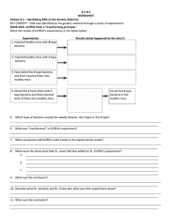
1 Journal of Genetic Toxicology Sarmishtha Chanda et al., Vol. 1. No.1 (Jun 2008) A comparative study of p53 promoter hypermethylation in Arsenic induced cancer in two different tissue compartment of the body: Cancer biopsy tissue and blood. 1 1 2 Sarmishtha Chanda , Uma Dasgupta , D.N.Guha Mazumder , U Chaudhuri 1 1 Department of Biophysics, Molecular biology & Genetics, University of Calcutta, Kolkata 700092, West Bengal ,India. 2 Department of Gastroenterology, Institute of post graduate medical education & research, Kolkata, West Bengal, India. Correspondence : [email protected] Abstract: Arsenic is a known carcinogen. Numerous studies link arsenic exposure to human cancer in a variety of tissues, including skin and lung. It is known to cause aberrant methylation in tissue culture system. Epigenetic changes therefore explored as a plausible route of arsenic induced cancer. We have reported in peripheral blood DNA that the degree of methylation in p53 and p16 gene promoter region is altered significantly in a dose dependent manner in persons chronically exposed to arsenic. Significant hypermethylation also demonstrated in peripheral blood leukocyte DNA in arsenic induced cancer patients (Chanda et al.2006). But till to date there is no report about the degree of methylation in p53 promoter region of arsenic induced cancer tissue. In this text we are therefore going to demonstrate the degree of methylation in persons with arsenic induced cancer where the DNA was isolated from the cancer biopsy tissue samples. DNA was also isolated from non arsenic related skin cancer biopsy sample and the p53 promoter methylation was studied to compare with the arsenic induced cancer biopsy sample. The degree of methylation in p53 promoter region in cancer biopsy sample shows a significant hypermethylation in comparison to p53 promoter region of DNA of peripheral blood leukocytes. Further, the degree of methylation in both blood and tissue samples in arsenic induced cancer and non arsenic related cancer ware significantly higher than the normal counterpart of the population where DNA was isolated from peripheral blood lymphocyte. Key words: Hypermethylation, carcinogenesis, arsenic, cancer biopsy tissue, p53 promoter . Introduction : Arsenic is a metalloid found in earth crust. It produces a number of clinical problems involving different organ systems of the body. Chronic exposure to inorganic arsenic Running title: DNA hypermethylation in arsenic cancer tissue produces cancer of skin, bladder and lung. The carcinogenic role of inorganic arsenic was chronicled over a hundred of years ago, when a patients treated with arsenicals were noted to develop an unusual numbers of tumors. Epidemiological studies indicate that chronic arsenic exposure has an association with several internal cancers along with skin cancer. Arsenic has been conclusively identified as a human carcinogen for skin, lung and bladder cancer (IARC 2004). Due to its carcinogenic potential, study of effect of arsenic on DNA is of particular interest. Arsenic is a poor mutagen (Rossman et al., 1980; Jacobson et al., 1985; Lee et al., 1985). Although it produces effects in sister chromatid exchange, micronuclei development and anueploidy, it does not induce significant point mutation. It is reported that arsenic causes deletional mutation in human chromosome 11 in a human hamster hybrid cell (NRC 1999). A recent report showed that sodium arsenite induces mutations in L5178Y/TK+/- cells (NRC 1999). Therefore epigenetic mechanism is explored as a probable route of arsenic induced carcinogenicity. Biotransformation of arsenic, on the other hand, involves methylation of inorganic arsenic to organic monomethyl arsonic acid (MMA) and dimethyl arsinic acid (DMA), using the same methyl donor S-Adenosyl methionine (SAM) which is also involved in DNA methylation (Vahter. 1999). The interference of the DNA methylation pathway with the arsenic detoxification pathway can lead to aberrant DNA methylation, precipitating aberrant expression and/or silencing of genes and has been mooted as the possible mechanism of arsenic induced carcinogenesis. Cytosine methylation has immense importance in animal development. Cytosine-5 methylation at the CpG islands in the regulatory sequence of http:/www.genetox.net 2 Journal of Genetic Toxicology Sarmishtha Chanda et al., Vol. 1. No.1 (Jun 2008) a gene is one of the key mechanism of gene inactivation. DNA methylation/demethylation constitutes a major consequence in all biological processes involving transcription, differentiation, development, DNA repair, recombination, and chromosome organization. Perturbation of DNA methylation has been correlated with many cases of cancer incidence and progression (Jones & Baylin 2002). The hypothesis that arsenic perturbs DNA methylation has been tested successfully on tissue culture system (Mass et al 1997). They demonstrated that CpG island of the p53 promoter region is hypermethylated in arsenic exposed murine cell line. We have tested the hypothesis on arsenic exposed population of West Bengal,India and reported significant hypermethylation of the promoter region of p53 and p16 genes in DNA extracted from peripheral blood leucocytes of persons exposed to different doses of arsenic (Chanda et al. 2006). lesion and chronic arsenicosis are the primary feature of arsenic induced skin cancer [fig 1]). Criteria of diagnosis of arsenicosis are based on the parameters described earlier (Guha Mazumder et al 1998), briefly, a) history of exposure to arsenic contaminated water (>50µg/L) as a source of drinking water for more than 5years. (The arsenic cancer patients had a degree of arsenic exposure of 15 years on average.) b) presence of definitive skin manifestation of chronic arsenic toxicity are divided into three main category, which are hyperpigmentation, hypopigmentation and keratosis. In our present study we are going to demonstrate that in arsenic induced cancer patients p53 promoter region is significantly hypermethylated in DNA extracted from the biopsy tissues from the site of lesion and the degree of methylation is significantly higher in cancer biopsy tissue than the DNA isolated from peripheral blood leukocytes. We are first to report arsenic induced hypermethylation in p53gene of human DNA isolated from tissue samples taken from site of lesion of arsenic induced skin cancer patients. Hypopigmentation :- Spotty hypopigmentation or leucodermia or leukomelanosis are spotty depigmentation which may also present all over the body, distributed in a bilaterally symmetrical fashion. Materials & Methods: Subjects and Selection criteria: Skin cancer tissue and blood samples were collected from arsenic and non arsenic associated skin cancer patients admitted in the plastic surgery department ( Samples of this study were taken from the surgery department) of Seth Sukhlal Karnani Memorial (S.S.K.M.) Hospital, Kolkata, India. Blood samples of control subjects were collected from non arsenic exposed people living in the area from where the cancer patients came. The study subjects were all residents of South & North 24 Parganas, West Bengal, India, the region severely affected with arsenic problem. All cancer patients had characteristic nodular or ulcerated skin lesion and confirmation of diagnosis was made by histological examination of biopsy tissue taken from the lesions. Arsenic induced skin cancer patients had associated features of arsenicosis ( Running title: DNA hypermethylation in arsenic cancer tissue Hyperpigmentation:-The hyperpigmentation of chronic arsenic poisoning commonly appears in a finely freckled �raindrop’ pattern that is particularly pronounced on the trunk and extremities distributed in a bilaterally symmetrical pattern. Keratosis :- Hyperkeratosis is characterised by diffuse thickening and / or nodularity of palm and sole or dorsum of feet and are distributed in a bilaterally symmetrical fashion. Arsenic induced skin cancer develops many years after the onset of (is the end stage clinical) skin manifestation of chronic arsenic toxicity. Skin Cancer :- Skin cancer of chronic arsenicosis is quite distinctive. The lesions of skin cancers are nodular or ulcerated of varying size. Arseincal skin cancers are frequently multiple, usually involve covered areas of the body and are associated with arsenical skin lesion. In contrast, non arsenical skin cancers are mostly single and occur in exposed parts of the body. Cancer diagnosis was based on the histological evidences of malignancy of biopsy tissues obtained from affected areas of the body. All cases studied were found to be suffering from squamous cell carcinoma. (On the basis of such definitive features study subjects were selected and cancer biopsy sample was collected to analyse the arsenic induced skin cancer as well as non arsenic related skin cancer.) Blood samples from cancer http:/www.genetox.net 3 Journal of Genetic Toxicology Sarmishtha Chanda et al., and control subjects and biopsy tissues from arsenic and non arsenic associated skin cancer patients were collected for methylation study of promoter region of tumour suppressor gene p53. All male participants were small traders or were involved in office jobs in small concerns. Women were housewives. Men were smokers and women were all non-smokers. The demographic data is presented in table1. Drinking water samples were also collected from each participant for analysis of arsenic. All arsenical cancer patients took arsenic contaminated subsoil water (300-1000µg/l) for drinking and cooking purpose. The non arsenic related cancer patients used surface water or well water containing very low arsenic level (<10 µg/l) of arsenic. Control subjects were selected from people of the same area from where arsenic cancer patients came. They were drinking water from tube wells having arsenic level less than 10 µg/l, and they did not have any skin lesion. Written informed consent was obtained from all participants before drawing their blood. The name of the institutes where human studies were carried out are Institute of Post Graduate Medical Education and Research, Kolkata (IPGME&R), and University of Calcutta both of which are run by Govt. of West Bengal, a state government within the framework of Republic of India. Ethical principles followed by the institute are guided by rules as formulated by Indian Council of Medical Research and these are in agreement with Helsinki rules. Determination of Arsenic concentration in water: Level of arsenic in water was determined by atomic absorption spectrophotometer with hydride generation system (AAS-HG). Collection of samples: Blood and biopsy tissue samples were collected from arsenic exposed/unexposed cancer patients during surgery. All the samples were transported in icebox from IPGME & R to the Department of Biophysics, Molecular Biology & Genetics, University of Calcutta for further storage at -70°C and subsequent analysis after DNA extraction. Isolation of DNA from cancer biopsy sample (Tissue) and from blood: The Tissue from which the DNA has to be extracted was fixed in liquid nitrogen. Running title: DNA hypermethylation in arsenic cancer tissue Vol. 1. No.1 (Jun 2008) It was then grind to fine powder and suspended in 1.2 ml digestion buffer for every 100 mg of tissue (100 mM NaCl, 10 mM Tris-Cl (pH 8.0), 25 mM EDTA (pH 8.0), 0.5% w/v SDS, 0.1 mg/ml PK added freshly from a 20mg/ml stock) and then incubated at 50° C for 16 to 18 hrs in a water bath. The material was then subjected to organic extraction twice by 1:1 Plhenol – Chloroform mixture. Finally 600 µl of 5 M NaCl and 2.5 volume of ethanol were added to each tube and the content was mixed well and DNA was spooled out and washed in 200 µl 70% ethanol, to remove the residual salt. The DNA was then air-dried. From blood samples the DNA was isolated by usual phenol –chloroform method (Miller 1988). Dried DNA was dissolved in 100 µl freshly autoclaved triple distilled water. Concentration and purity of DNA was then checked by spectrophotometry. Analysis of p53 promoter methylation DNA obtained from the biopsy sample of arsenic induced cancer and non arsenic related cancer were then subjected to PCR amplification for the study of methylation status in the promoter region of p53 tumour suppressor gene. The promoter region of p53 gene contains two CCGG sites beginning at bases 703 and 883 (Gen Bank accession no: X 54156). We choose two restriction enzyme to analyse the methylation status of p53 promoter methylation as a function of arsenic exposure, one is HpaII and the other is MspI. The Hpa II restriction enzyme will cleave CCGG sequences, which are not methylated at the internal or external cytosine. Msp I is the isocshizomer of Hpa II, which will cleave the CCGG sequence irrespective of its methylation status. Therefore Msp I is used as a control for Hpa II digestion. 300 ng of genomic DNA for each sample was subjected to restriction digestion separately by HpaII and MspI. For each sample, along with the Hpa II and MspI treatment, one aliquot was prepared for control containing no enzyme. Each of these three aliquot was subjected to overnight incubation at 37° C in a water bath. PCR amplification of p53 promoter region: The PCR was performed to amplify a 340 bp (638-978 bp) long region in the promoter of p53 http:/www.genetox.net 4 Journal of Genetic Toxicology Sarmishtha Chanda et al., gene. 150 ng of the digested product was then used as a template in PCR reaction mixture containing 200 mM dNTP, 25 pmol primers, i.e. 32 638 UN and 938 LN, 5mci of a P dCTP [4000 Ci/m mol) and 1 unit of Taq.Polymerase (Bangalore Genei Pvt. Ltd. India) in total volume of 25 ml according to the method described earlier (Chanda et al 2006). The PCR product was then precipitated using 7.5 M ammonium acetate and ethanol to wash out the unbound radioactivity. Electrophoresis was done in 2% agarose gel to resolve the product. Taking the control band as a guide the analogous bands were cut melted in 0.1N HCl and then the 32 percent incorporation of ά- P was measured by scintillation counting. Count in MspI was taken as a background. The fractional increase of count in the Hpa II digested DNA band over the background was considered as a measure of the level of template DNA present in the HpaII digested sample and is indicative of methylation level in the DNA in arbitrary units. Statistical Analysis: For finding the significance of the differences in DNA methylation status between different arsenic exposure groups we assumed no particular pattern of distribution of data and performed two tail Median test and MannWhitney U Test (Das & Das,1998) . Result: Blood samples were obtained from 24 normal unexposed people, 22 arsenic induced cancer patients and 17 non arsenic related cancer patients. Among these study subjects, 11 biopsy tissue samples were obtained from arsenic induced cancer patients and 10 from non arsenic related cancer patients. p53 gene methylation was observed in 11 arsenic induced skin cancer patients and 10 non arsenic related cancer patients where the DNA was isolated from biopsy tissue sample and compared with the methylation value obtained from peripheral blood leukocyte DNA. Statistical analysis using non-parametric median test indicate that there is a significant hypermethylation of promoter region of p53 gene in cancer biopsy tissue sample of arsenic induced cancer patients compared to normal counterpart of the population where DNA was isolated from peripheral blood leukocyte(p<0.001). The median value of p53 Running title: DNA hypermethylation in arsenic cancer tissue Vol. 1. No.1 (Jun 2008) promoter methylation is significantly higher in cancer biopsy tissue sample of arsenic induced cancer in comparison to peripheral blood DNA of the same study subjects (p<0.05). Similarly, in peripheral blood DNA the degree of methylation in arsenic induced cancer was also significantly higher than the normal counterpart of the population (p<0.01). Hypermethylation also observed in p53 promoter region of DNA isolated from cancer tissue samples of non arsenic related cancer in comparison to peripheral blood leukocyte DNA collected from the control(p<0.05). Median test and Mann-Whitney U test also indicate that the median value of methylation status in cancer biopsy tissue samples collected from patients of arsenic induced cancer is significantly higher (p<0.05) than the cancer patients unrelated to arsenic. Similarly it is demonstrated in peripheral blood DNA that the p53 methyaltion value is also higher in non arsenic related cancer the than control at 10% (p<0.1) level of significance. In non arsenic related cancer the methylation value in biopsy tissue sample is not significantly higher than the peripheral blood leukocyte DNA. The methylation value in p53 promoter region is significantly higher in peripheral blood DNA of arsenic cancer than non arsenic related cancer. However, there was no difference in methylation value regarding age, sex and smoking status. Discussion: Cytosine methylation has immense importance in animal development. CpG island methylation in the promoter region of any structural gene is the known route of transcriptional silencing. Knockouts of Cytosine methyltransferase produce embryonic lethality in mice. In human also it is presumed to cause abnormal cell growth and neoplastic development. DNA methylation is known as one of the epigenetic factors that promotes altered level of transcription of genes in vertebrates. Silencing of functional gene due to altered level of methylation is now well established. Hypermethylation and consequent silencing of tumour suppressors and repair genes as an early event in certain types of malignancy like lung, colorectal, breast and many other types of neoplasia (Heish et al. 1997, Herman et al. 1994, Graff et al. 1995, Merlo et al. 1995, Baylin & Herman 2000). Hypomethylation, on the other http:/www.genetox.net 5 Journal of Genetic Toxicology Sarmishtha Chanda et al., hand offers growth advantage and is an important step in genesis of many cancers rd (Strachan & Read , 1999, 3 Edition). Specific investigations on p53 tumour suppressor gene demonstrated hypermethylation after exposure of cells to sodium arsenite. It has also been shown that inorganic arsenic exposure reduces p53 expression and up regulates mdm2 (Hamadeh et al. 1999). Further, post UV p53 dependent p21 expression is greatly suppressed after arsenic exposure, thus overriding the post UV block in cell cycle progression (Vogt et al. 2001.). Studies from endemic areas of Taiwan have reported mutations of p53 in arsenic induced skin cancer and these mutations are apparently of different types than those found in UV induced skin cancer (Hsu et al. 1999). Arsenic induces mutation in human-hamster hybrid cell but all the mutations are of deletion mutation and not the point mutation. Due to the poor in vitro mutagenicity of arsenic, epigenetic mechanism seemed one of the probable routes of arsenic induced malignancy. Mass and Wang hypothesised that arsenic alter the DNA methylation level. They tested the hypothesis on human lung and kidney cell line and reported that arsenic produces hypermethylation in p53 gene promoter region (Mass &Wang 1997). Hypermethylation in the CpG island of promoter region blocks the transcription and when it occurs in any tumour suppressor gene may produce cancer. Hypomethylation of genes after arsenic exposure has also been reported in studies done on cell lines (Zhong & Mass 2001). Mass in a arbitrarily primed methyl sensitive PCR assay, found both hypermethylation and hypomethylation of different DNA sequences, their ratio being 3 : 1. The sequences corresponded mostly to promoter regions (Zhong & Mass 2001). In another significant work it has been shown that chronic low level of arsenic exposure on rat liver epithelial cells produced highly aggressive malignant cells, which showed DNA hypomethylation detected using a satellite DNA (Zhao et al. 1997). Woodson et al (Woodson et al.2001), in a study with 100 lung cancer patients and controls showed that genome wide hypomethylation level was related to disease risk and hypomethylation in p53, particularly within the hypermutable 5-8 exons related significantly with lung cancer risk. It was also reported that genomic hyomethylation could induces tumours in knockout mice (Gaudet et al 2003). Inorganic arsenic-induced transformation in human Running title: DNA hypermethylation in arsenic cancer tissue Vol. 1. No.1 (Jun 2008) prostate cells is associated with genomic DNA hypomethylation and K-ras overexpression (Tallaa et al. 2005). Human DNA methyltransferase mRNA pool is increased in arsenic exposure and also in different types of human cancer. Arsenic deplete the cellular pool of SAM, the methyl donor and represses the expression of DNMT1 and DNMT 3A and as a consequences of these two complimentary mechanisms produces DNA hypomethylation on human HaCaT keratinocytes (Reichard et al. 2007). It has been reported that genomic DNA methylation status is altered in arsenictransformed cells (Zhao et al. 1997). Long-term exposure of mice to arsenic in the drinking water can induce aberrant gene expression, global DNA hypomethylation (Chen et al. 2004). Cytosine methylation profiling in cancer cell shows a wide variation in promoter region (hypermethylation in promoter region) and over all genomic DNA (Global hypomethylation). A large proportion of genes (78 of 400 genes) are epigenetically altered in cancer. Although most genes show methylation changes in only one tumor type (Ehrlich et al 2008). Arsenicassociated increase in overall genomic hypomethylation and increase in genomic instability have been demonstrated in arsenicexposed animal models, which is a characteristic of aberrant methylation pattern, seen in human solid tumors also (Ehrlich et al. 2002). Thus, aberrant methylation has been documented as a probable route of arsenic induced carcinogenesis and various other types of neoplasm. We reported in our previous work that hypermethylation of cytosines in the promoter region of p53 and p16 gene (DNA isolated from peripheral blood leukocyte) shows a dose response pattern in people chronically exposed to arsenic. However, a small fraction of persons in high arsenic exposure showed hypomethylation though not at significant level (Chanda et al 2006). We are first to report that perturbation of DNA methylation occur in human population suffering from chronic arsenic exposure. In this text we are now reporting that alteration of DNA methylation occur significantly in the arsenic induced cancer tissue where the degree of methylation is higher than the peripheral blood leukocyte, another compartment of tissue http:/www.genetox.net 6 Journal of Genetic Toxicology Sarmishtha Chanda et al., in our body. We are trying to show the degree of methylation in cancer tissue where actually the transformation (normal cell to cancer cell) begins. This is the first report of arsenic induced hypermethylation carried out in cancer biopsy tissue samples obtained from the arsenic induced cancer patients. Arsenic may have multiple roles in altering the epigenetic environment of the cell. It may modify the steady-state levels of SAM, contributing to genomic hypomethylation already tested in animal models. Further, it may generate an environment for selection of cells with epigenetic silencing of specific genes such as RASSF1A and PRSS3 through promoter hypermethylation. It has been demonstrated that in lung cancer peteints also there is prevalence of p16INK4A promoter hypermethylation (Marsit et al., 2006). Sporadic tumorigenesis is driven by clonal somatic alterations of specific cellular pathways. These alterations take the form of activation or functional disregulation of oncogenes and concomitant inactivation of tumor suppressor genes. Numerous somatic alterations have been described in bladder cancer, most notably mutation or functional inactivation of the p53 tumor suppressor (Sidransky.et al. 1991, Kelsey.et al. 2004). Other tumor suppressors are subjected to epigenetic silencing, through promoter hypermethylation and in bladder cancer, this silencing has been described for genes such as p16INK4A and RASSF1A (Chan. et al. 2003). Significant (42%) p16 gene promoter hypermethylation has been shown to occur in chronic arseniasis patient in comparison to normal (2%) individual (Zhang et al. 2007). p16 promoter hypermethylation was also demonstrated in chronic arsenic exposed people as well as arsenic induced cancer patients of West Bengal (Chanda et al 2006). Urothelial carcinoma patients with high arsenic exposure shows DAPK promoter hypermethylation. This promoter hypermethyltion is closely associated with low level of expression of the DAPK gene (Chen et al 2007). But little is known about the molecular event of arsenic induced carcinogenesis. The mechanism of DNA hypermethylation after arsenic exposure is not clear. Pentavalent inorganic arsenic (AsV) after entering the body is methylated to MMA and DMA, which are significantly less toxic and are excreted through urine. The arsenic methyltransferase uses the same methyl donor SAM as DNA methyltransferase and other methyltransferases. It has been reported that Running title: DNA hypermethylation in arsenic cancer tissue Vol. 1. No.1 (Jun 2008) arsenite inhibits selenocysteine methyltransferase (Goering et al. 1999). Inhibition of other cellular methyltransferases resulting in increase of SAM pool might be postulated (Mass & Wang 1997). This increased substrate concentration would induce some unaffected methyltransferase like cytosine methyltransferase to overmethylate the CCGG sequences in DNA. Alternately, arsenite exposure causes induction of cytosine methyltransferase, as increase in mRNA level of this enzyme after arsenite exposure has been reported (Goering et al 1999). Induction of DNA methyltransferase has also been reported after arsenic exposure as well as in different types of neoplasia. Higher arsenic concentration, on the other hand is presumed to cause depletion of SAM pool and concomitant hypomethylation (Mass & Wang 1997). Measurement of intracellular SAM pool and activities of related enzymes are necessary to test the hypothesis, which, to our knowledge are not available yet. In conclusion, this study confirming the results obtained with tissue culture cells and result obtained from peripheral blood leukocyte DNA and cancer biopsy tissue sample, that chronic arsenic exposure in a population produces hypermethylation of DNA which is associated with arsenic induced malignancy. Acknowgement: Research funding was obtained from D.N.G.M research foundation. Cancer biopsy samples were obtained from plastic surgery department S.S.K.M hospital, Kolkata. Authors are also grateful to Dr. Tushar Chakraborty for his valuable guidance and a few supplementary works were also carried out in his laboratory, IICB, Kolkata. Arsenic testing of the water samples was carried out in trace element laboratory developed by funding from USEPA grant No: R 826137–01–0 and NIEHS grant No: P 42 E 504705 through Center for Occupational and Environmental Health, University of California, Berkeley, CA. Conflict of interest: none declared http:/www.genetox.net 7 Journal of Genetic Toxicology Sarmishtha Chanda et al., Vol. 1. No.1 (Jun 2008) Table 1 Demographic distribution of the study population Age group Normal/unexposed (blood) Arsenic cancer (DNA isolated from tissue) Non-arsenic cancer (DNA isolated from tissue) Arsenic cancer (DNA isolated from blood) 20-40 Male 8,Female 3 Male3,female2 Male 5, female 3 41-60 > 61 Male 6,Female 3 Male 4 Male3 Male2,female1 Smoking status Smoker 18 Non-smoker 6 Smoker 11 Non-smoker 5 Average duration of exposure Total No of samples 15 years 19 years Male2, female2 Male3, Male 2, female1 Smoker 9, non-smoker 4 ------- 19 years Nonarsenic cancer (DNA isolated from blood) Male2, female2 Male5, Male 2, female2 Smoker 9, nonsmoker 4 ------ 24 11 10 22 17 Male6,Female3 male4,female1 Smoker 15 Non-smoker 7 Table2. Comparison between degree of DNA methylation in arsenic cancer and non arsenic cancer. (in both cases DNA taken from blood and cancer biopsy tissue) Degree of exposure <50µg/l (a) 300-1000 µg/l (b) <50µg/l (c) 300-1000 µg/l (d) <50µg/l (e) Sample name normal Arsenic cancer Non-arsenic cancer Arsenic cancer Non arsenic cancer Sample type Blood Tissue Median value of methylation and level of significance 0.35 4.17, p<0.001 a Vs b Tissue 1.21, p<0.05 a Vs c, p<0.05 b Vs c Blood 3.01, p<0.01 a Vs d, p<0.05 b Vs d Blood 0.91, p<0.1 a Vs e, p <0.05 d Vs e. Running title: DNA hypermethylation in arsenic cancer tissue http:/www.genetox.net 8 Journal of Genetic Toxicology Sarmishtha Chanda et al., Fig 1a: lesions in the planter aspect of feet the fingers of the foot was amputed due to gangreene Vol. 1. No.1 (Jun 2008) Fig 1b: First stage of skin cancer (Bowen’s disease) Fig 1c: nodule in palm and cancer in middle finger(keratosis) Running title: DNA hypermethylation in arsenic cancer tissue http:/www.genetox.net 9 Journal of Genetic Toxicology Sarmishtha Chanda et al., References: 1. IARC. (2004). Some Drinking Water Disinfectants and Contaminants, Including 2. Arsenic. IARC monograph on the evaluation of carcinogenic risks to 3. humans, Vol. 84, pp. 209–214. International Agency for Research on Cancer, Lyon, France. 4. Absence of arsenite mutagenicity in E. coli and Chinese hamster cells. Rossman, TG., Stone, D, Molina, M., and Troll, W. Environ.Mutagen. volume 2, 1980, pages: 371–379. 5. The Reproductive Effects 6. Assessment Group’s report on the mutagenicity of inorganic arsenic, Jacobson, KD., and Moltalbano, D. Environ. Mutagen. 7, 1985, pages 787–804. 7. Comparison of arsenic induced cell transformation, cytotoxicity, mutation, and cytogenetic effects in Syrian hamster embryo cells in culture. Lee, T.C., Oshimura, M., and Barrett, J. C. Carcinogenesis 6, 1985, pages1421–1426. 8. NRC (National Research Council): Arsenic in Drinking Water, (1999), National Academy Press, Washington DC, U.S.A. 9. Vahter, M. (1999 ). Variation in human metabolism of arsenic. Arsenic Exposure and Health Effects III. (Edited by Chappel W.R., Abernathy C.O., Calderon R.L.), pages 267-275. Elseveir Science Ltd. Oxford. 10. The Fundamental Role of Epigenetic Events in Cancer. Jones, PA., and Baylin SB. Nature Rev. Genet. Volume 3, 2002, pages 415-428 . 11. Arsenic alters cytocine methylation patterns of the promoter of the tumor suppressor gene p53 in human lung cells : a model for a mechanism of carcinogenecis, Mass MJ, Wang L. Mutat. Res. Volume 386, 1997, pages 263-277. 12. DNA Hypermethylation of Promoter of Gene p53 and p16 in Arsenic-Exposed People with and without Malignancy. Chanda, S., Dasgupta, U.B., GuhaMazumder, D., Gupta, M., Chaudhuri, U., Lahiri. S., Das, S., Ghosh, N., Chatterjee, D. Toxicol.Sci. Volume 89, 2006. pages 431-7. 13. Arsenic levels in drinking water and the prevalence of skin lesions in WestBengal, India. GuhaMazumder DN, Haque R, Ghosh N, De BK, Santra A, Chakrabarty D, Smith Running title: DNA hypermethylation in arsenic cancer tissue 14. 15. 16. 17. 18. 19. 20. 21. 22. 23. 24. 25. Vol. 1. No.1 (Jun 2008) AH. Int.J. Epidemiol. Volume 27, 1998, pages 871-877. A simple salting out procedure for extracting DNA from human nucleated cells. Miller SS, Dykes DD, Polesky HF. Nucleic Acid Res. volume16, 1988 , page 1215. Das D, Das A. Statistics in Biology and Psychology. Pub: Academic publishers rd (1998), Eds 3 Calcutta 700073. Biosynthesis of dimethylselenide from sodium selenide in rat liver and kidney cellfree systems. Heish HS, Ganther HE. Biochim. Biophys. Acta .volume 497, 1997, pages 205-217. Silencing of the VHL tumour suppressor gene by DNA methylation in renal carcinoma. Herman JG, Latif F, Weng Y, Proc. Natl. Acad. Sci USA. Volume 91, 1994, pages 9700-9704. Cadrehin expression is silenced by DNA hypermethylation in human breast and prostate cancer. J.R. Graff, J.G. Herman, R.G. Lapidus et al. Cancer. Res. Volume 55, 1995, 5195-5199. 5’ CpG island methylation is associated with transcriptional silencing of the tumour p16/CDK N2/ MTS1 in human cancers. Merlo A, Herman JG, Mao L., Nat. Med, volume 1, 1995; pages 686-692. Promoter-region hypermethylation and gene silencing in human cancer. Herman J G and Baylin S B. Curr Top Microbiol Immunol. Volume 249, 2000, pages 35-54. Strachan T, Read AP, Human Molecular rd Genetics,3 Edition, Garland Science Arsenic disrupts cellular levels of p53 and mdm2 : A potential mechanism of carcinogesis. Hamadeh HK, Vargas M, Lee E, Menzel DB. Biochem. Biophys. Res. Commun. Volume 263: 1999, pages 446449. Effects of arsenite on p53, p21 and cyclin D expression in normal human fibroblasts- a possible mechanism for arsenite’s comutagenicity. Vogt BL & Taylor, Rossman TG. Mutat.Res. Volume 478, 2001, pages159-168. Mutational spectrum of p53 gene in arsenic related skin cancers from the blackfoot disease endemic area of Taiwan. Hsu CH, Yang SA, Wang JY, Yu HY, Lin SR. Br.J.Cancer. Volume 80, 1999, pages 10801086. Both hypomethylation and hypermethylation of DNA associated with arsenite exposure in http:/www.genetox.net 10 Journal of Genetic Toxicology 26. 27. 28. 29. 30. 31. 32. 33. Sarmishtha Chanda et al., cultures of human cells identified by methylation-sensitive arbitrarily-primed PCR. Zhong CX, Mass M J. Toxicol. Lett, volume 122, 2001, pages 223-234. Association of arsenic- induced malignant transformation with DNA hypomethylation and aberrant gene expression. Zhao CQ, Young MR, Diwan BA, Coogan TP, Waalkes MP. Proc.Natl.Acad.Sci USA. Volume 94, 1997, pages10907-10912. Hypomethhylation of p53 in peripheral blood DNA is associated with the development of lung cancer, Woodson K, Mason J, Choi SW, Cancer Epidemiol. Biomark. Prev Volume 10, 2001, pages 69-74 Induction of tumors in mice by genomic hypomethylation, Gaudet F, Hodgson JG, Eden A. Science. Volume 300, 2003, pages 489-492. Molecular events associated with arsenicinduced malignant transformation of human prostatic epithelial cells: aberrant genomic DNA methylation and K-ras oncogene activation. Tallaa LB, Waterlandb RA., Stybloc M, Achanzara WE., Webberd MM., Waalkes MP., Toxicology and Applied Pharmacology. Volume 206, 2005, pages 288– 298. Long term low-dose arsenic exposure induces loss of DNA methylation. Reichard J F., Schnekenburger M, Puga A. Biochem Biophys Res Commun. Volume 352(1), 2007, pages 188-192. Chronic inorganic arsenic exposure induces hepatic global and individual hypomethylation: implications for arsenic hepatocarcinogenesis. Chen H, Li S.F., Liu J, Diwan B A, Barrett JC, Waalkes MP, carcinogenesis . Volume 25 (9), 2004, pages 1779-1786. Cytosine methylation profiling of cancer cell lines. Ehrich M, Turner J, Gibbs P, Lipton L, Giovanneti M, Cantor C, van den Boom D., PNAS. Volume 105(12), 2008, pages 48444849. DNA methylation in cancer: too much, but also too little. Ehrlich M, Oncogene , Volume 21, 2002 , pages 5400 – 5413. Running title: DNA hypermethylation in arsenic cancer tissue Vol. 1. No.1 (Jun 2008) 34. Carcinogen exposure and gene promoter hypermethylation in bladder cancer. C.J.Marsit, M. R.Karagas, H. Danaee, M. Liu, A. Andrew3, A. Schned, H. H.Nelson, K. T.Kelsey. Carcinogenesis. Volume 27, 2006, pages 112–116. 35. Identification of p53 gene mutations in bladder cancers and urine samples. Sidransky D, Von Eschenbach A, Tsai YC, Jones P, Summerhayes I, Marshall F, Paul M, Green P, Hamilton SR, Frost P, , Science.Volume 252 (5006), 1991, pages 706-709. 36. A population-based study of immunohistochemical detection of p53 alteration in bladder cancer. Kelsey K.T., Hirao T, Schned A, Hirao S, Devi- Ashok T., Nelson H.H., Andrew A., Keragas M.R. Br J Cancer. Volume 90(8), 2004, pages 1572-6. 37. Frequent hypermethylation of promoter region of RASSF1A in tumor tissues and voided urine of urinary bladder cancer patients. Chan MW, Chan LW, Tang NL, Lo KW, Tong JH, Chan AW, Cheung HY, Wong WS, Chan PS, Lai FM, To KF. Int J Cancer. Voloume 104(5), 2003, pages 611-616. 38. Analysis of p16 Gene Mutation, Deletion and Methylation in Patients with Arseniasis Produced by Indoor Unventilated-Stove Coal Usage in Guizhou, China ,. Zhang AH, Bin H H, Pan X L , Xi X G. J Toxicol Environ Health A. volume 70(11), 2007, pages 970975. 39. Urothelial carcinomas arising in arseniccontaminated areas are associated with hypermethylation of the gene promoter of the death-associated protein kinase. Chen W.T., Hung W.C., Kang W.Y., Huang Y.C., Chan C.Y. Histopathology.Volume 51: 2007, pages 785-792. 40. The enigma of arsenic carcinogenesis: Role of Metabolism, Goering PL, Aposhian HV, Mass MJ, Cebrian M, Beck BD, Waalkes MP. Toxicol.Sci .Volume 49, 1999, pages 514. http:/www.genetox.net
© Copyright 2026 Paperzz