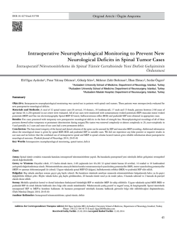
EXPERIMENTAL STUDY
Thymoquinone attenuates trauma induced spinal cord
damage in an animal model
Nilgün Üstün, M.D.,1 Mustafa Aras, M.D.,2 Tumay Ozgur, M.D.,3 Hamdullah Suphi Bayraktar, M.D.,4
Fatih Sefil, M.D.,5 Raif Ozden, M.D.,6 Abdullah Erman Yagiz, M.D.1
1
Department of Physical Medicine and Rehabilitation, Mustafa Kemal University Faculty of Medicine, Hatay;
2
Department of Neurosurgery, Mustafa Kemal University Faculty of Medicine, Hatay;
3
Department of Pathology, Mustafa Kemal University Faculty of Medicine, Hatay;
4
Department of Clinical Sciences, Mustafa Kemal University Faculty of Veterinary, Hatay;
5
Department of Physiology, Mustafa Kemal University Faculty of Medicine, Hatay;
6
Department of Orthopaedics and Traumatology, Mustafa Kemal University Faculty of Medicine, Hatay
ABSTRACT
BACKGROUND: Spinal cord injury (SCI) is one of the most devastating conditions leading to neurological impairment and disabilities. The aim of the study was to investigate the potential neuroprotective effect of thymoquinone (TQ) histopathologically in an
experimental model of traumatic spinal cord injury (SCI).
METHODS: Twenty-four male Wistar albino rats were randomly divided into 4 groups: control group; SCI group; SCI-induced and
10 mg/kg/day TQ administered group; SCI-induced and 30 mg/kg/day TQ administered group. TQ was given as intraperitoneal for three
days prior to injury and four days following injury. Spinal cord segment between T8 and T10 were taken for histopathologic examination. Hemorrhage, spongiosis and liquefactive necrosis were analyzed semiquantatively for histopathological changes.
RESULTS: Administration of TQ at a dose of 10 mg/kg did not cause any significant change on the histological features of neuronal
degeneration as compared to the SCI group (p=0.269); however, 30 mg/kg TQ significantly decreased the histological features of spinal
cord damage below that of the SCI group (p=0.011).
CONCLUSION: Data from this study suggest that TQ supplementation attenuates trauma induced spinal cord damage. Thus, TQ
needs to be taken into consideration, for it may have a neuroprotective effect in trauma induced spinal cord damage.
Key words: Experimental spinal cord injury; thymoquinone; histologic examination.
INTRODUCTION
Spinal cord injury (SCI) is one of the most devastating conditions leading to neurological impairment and disabilities.[1]
In SCIs, primary injury occurs after mechanical damage to
neuronal and vascular tissues at the time of trauma. Just after
mechanical injury, secondary injury occurs by the reduction
of spinal cord microvascular circulation and the deleterious
Address for correspondence: Nilgün Üstün, M.D.
Mustafa Kemal Üniversitesi Tıp Fakültesi, Fiziksel Tıp ve
Rehabilitasyon Anabilim Dalı, Hatay, Turkey
Tel: +90 326 - 229 10 00 E-mail: [email protected]
Qucik Response Code
Ulus Travma Acil Cerrahi Derg
2014;20(5):328-332
doi: 10.5505/tjtes.2014.05021
Copyright 2014
TJTES
328
biochemical effects of reactive oxygen species generation, cell
membrane lipid peroxidation and inflammation.[2,3] Therefore,
agents with antioxidant and anti-inflammatory properties are
proposed to be useful in trauma induced spinal cord damage.[4] None of the clinically available anti-oxidant therapies
has produced any clinically satisfactory intervention in trauma
induced SCI due to its complexity.[5] Therefore, there is still
a significant need for effective and safer agents for the treatment of spinal cord injury.
Thymoquinone (TQ), the main active constituent of Nigella
saliva seeds, is reported to have strong free radical scavenger
and antioxidant[6-9] and anti-inflammatory[10,11] properties in
different animal tissue models.
To the best of our knowledge, the neuroprotective effect of
TQ in SCI has not been investigated. Therefore, this study
was designed to investigate the potential protective effect of
TQ in a model of traumatic SCI.
Ulus Travma Acil Cerrahi Derg, September 2014, Vol. 20, No. 5
Yılmaz et al. Thymoquinone attenuates trauma induced spinal cord damage in an animal model
MATERIALS AND METHODS
Animals
Twenty-four adult male Wistar albino rats, weighing 300-350
g, were obtained from Mustafa Kemal University Laboratory
of Experimental Animals. The animals were fed with a standard rat chaw and allowed access to water. All rats were kept
in an air-conditioned room with 12-hour light and dark cycles,
where the temperature (22 ± 2ºC) and relative humidity (65–
70%) were kept constant. All experimental protocols were
approved by the Animal Care and Use Committee of Mustafa
Kemal University, School of Veterinary Medicine.
Chemical
TQ (2 isopropyl-5-methyl-1.4-benzoquinone) was obtained
from Sigma Chemical Co. (St. Louis, Missouri, USA) and dissolved in saline and heated at 60-80°C.
Experimental Design
Rats were randomly assigned into four groups of six rats each:
One group served as controls where the rats received sham
injury (laminectomy). Second group served as the SCI group
that underwent SCI after laminectomy. Third group under-
went SCI and was given intraperitoneal administration of TQ
aqueous solution at 10 mg/kg/day dose level for three days
prior to and four days after SCI. Fourth group underwent SCI
and was given intraperitoneal administration of TQ aqueous
solution at 30 mg/kg/day dose level for three days prior to
and four days after SCI.
Induction of SCI
The animals were anesthetized with intraperitoneal injection
of 75 mg/kg ketamine (Ketalar, Eczacıbaşı, Istanbul, Turkey) and
10 mg/kg xylazine (Rompun, Bayer, Istanbul, Turkey). Anesthetized rats were positioned in a prone position. Their dorsal
regions were shaved and cleaned with povidone-iodine. Under
sterile conditions, following T6-T12 midline skin incision and
paravertebral muscle dissection, spinous processes and laminar arcs of T8-T10 were removed. The dura was left intact. A
well characterized weight-drop technique was performed for
spinal cord trauma.[12] The animals were subjected to an impact of 100 g/cm (10 g weight from 10 cm height) to the dorsal
surface of the spinal cord. The force was applied via a stainless
steel rod (3 mm diameter tip) rounded at the surface. The rod
was dropped vertically through a 10 cm guide tube positioned
perpendicular to the center of the spinal cord. Afterwards, the
(a)
(b)
(c)
(d)
Figure 1. Light photomicrographs of the rats’ spinal cord tissue sections (H&E, x 200). Sham-operated control group with preserved morphology (a). Spinal trauma group; hemorrhage (h), severe vacuolic degeneration (v), liquefactive necrosis (ln) and
inflammatory infiltrate (i) (b). Degenerative changes remain with low doses of thymoquinone (TQ) (c). Improved spinal cord morphology with high doses of TQ (d).
Ulus Travma Acil Cerrahi Derg, September 2014, Vol. 20, No. 5
329
Yılmaz et al. Thymoquinone attenuates trauma induced spinal cord damage in an animal model
muscles and the incision were sutured. Rats in the sham injury
group underwent a similar surgical procedure as other groups;
however, no spinal cord trauma was performed. A week after
SCI induction, rats were sacrificed using an overdose of pentobarbital (200 mg/kg) and the same part of the injured spinal
cord tissue samples were excised. The sample of the spinal
cord tissue was stored in 10% buffered formaldehyde solution
for histologic examination.
Histologic Analysis
For histological examination, all tissue samples were fixed at
10% buffered formaldehyde solution for 24 hours and processed according to routine light microscopic tissue processing technique. Formalin-fixed paraffin-embedded tissue
sections (5 µm) were stained with hematoxylin and eosin
(H&E) and examined by an Olympus DP20 camera attachedOlympus CX41 photomicroscope. To prevent inter-individual
bias, all tissues were evaluated by the same pathologist (T.O.)
who was uninformed of the groups and test materials. Hemorrhage, spongiosis and liquefactive necrosis were analyzed
for histopathological changes. For histopathologic evaluation,
a semi-quantitative grading system modified by Malinovsky
et al.[13] was used on all specimens as following: No abnormal cells and change (grade 0), mild hemorrhage, spongiosis
(grade 1), moderate hemorrhage and spongiosis with liquefactive necrosis (grade 2), severe hemorrhage and spongiosis
with glial cell proliferation and liquefactive necrosis (grade 3).
Statistical Analysis
Statistical analyses were performed using SPSS software package program (SPSS Inc., Chicago, IL, USA), version 13.0 for
Windows. Kruskal-Wallis variance test was used to compare
differences among groups. When analysis of variance showed
significance, Mann-Whitney U-test was applied to determine
the difference. Data were expressed as mean± SD. Values of
p˂0.05 were accepted as statistically significant.
RESULTS
Light microscopic examination of the spinal cord tissue sections revealed a normal histological structure in the shamoperated control group (Fig. 1a). Histological examination
of the spinal cord tissue sections of the SCI group showed
moderate to severe hemorrhage, necrosis, inflammatory infiltrate and fibrosis (Fig. 1b). A statistically significant difference
was found among the groups (p=0.001). Neuronal degeneration in the SCI group was significant compared to the shamoperated control group (p=0.003, Table 1). Administration
of TQ at a dose of 10 mg/kg for three days prior to and four
days after SCI did not cause any significant change on histological features of neuronal degeneration as compared to the
SCI group (p=269, Table 1; Fig. 1c). However, 30 mg/kg TQ
significantly decreased the histological features of the spinal
cord damage below that of the SCI group (p=0.011, Table 1;
Fig. 1d).
330
Table 1. Histopathologic evaluation scores of the groups
Rat
Control
SCI
SCI+TQ10SCI+TQ30
1
0221
2
0321
3
0332
4
0321
5
0231
6
0321
Mean±SD 0.00±0.002.66±0.512.33±0.571.20±0.44
p0.003a0.269b0.011c
TQ: Thymoquinone; SCI: Spinal cord injured untreated group; SCI+TQ10: 10
mg/kg TQ treated spinal cord injured group; SCI+TQ30: 30 mg/kg TQ treated
spinal cord injured group; histopathologic grading scores: 0, no abnormal cells
and change; 1, mild hemorrhage, spongiosis; 2, moderate hemorrhage and spongiosis with liquefactive necrosis; 3, severe hemorrhage and spongiosis with glial
cell proliferation and liquefactive necrosis. ap, compared with C group group; bp
and cp, compared with SCI group.
DISCUSSION
This study was initiated to investigate whether TQ supplementation could reduce trauma induced spinal cord damage.
The results of the present study demonstrated that treatment with 30 mg/kg TQ for three days prior to and four
days after trauma had protective effects on trauma induced
spinal cord damage. This was shown by marked decrease in
the histopathological damage scores of the injured spinal cord
tissues. This neuroprotective effect of TQ could be explained
on the basis of its antioxidant[6-9] and anti-inflammatory effects.[10,11] To the best of our knowledge, this is the first report evaluating the neuroprotective effects of TQ on trauma
induced SCI.
Reactive oxygen species are continuously produced during
normal physiologic events and removed by antioxidant defense mechanism.[14] The imbalance between reactive oxygen
species and antioxidant defense mechanisms leads to lipid
peroxidation and oxidative damage in the lipid bilayers surrounding both the cell itself and membrane-bound organelles.
[15]
Recent studies have demonstrated that increase in reactive
oxygen species and the decrease in the antioxidant defense
mechanisms is a major contributor to the pathogenesis of
trauma induced spinal cord damage.[16,17] A variety of known
antioxidants and anti-inflammatory agents have shown protective effects on traumatic SCI in experimental models and
clinical trials.[18-24] Toklu et al.[18] have found that alpha-lipoic
acid reduces oxidative stress on traumatic SCI. Sahin et al.[19]
have shown significant protective effects of curcumin on traumatic spinal cord tissues against oxidative damage. Karalija et
al.[20] have demonstrated better pathological findings by Nacetyl-cysteine and acetyl-L-carnitine in the early treatment
of traumatic SCI by using quantitative immunohistochemistry
and western blotting for neuronal and glial cell markers, and
indicated a therapeutic potential for NAC and ALC in the
Ulus Travma Acil Cerrahi Derg, September 2014, Vol. 20, No. 5
Yılmaz et al. Thymoquinone attenuates trauma induced spinal cord damage in an animal model
early treatment of traumatic SCI. Cemil B et al.[21] have put
forward better pathological findings by aged garlic extract in
the traumatic SCI against oxidative damage. Erşahın et al.[22]
have reported that ghrelin could reduce SCI-induced oxidative stress and exert anti-inflammatory effects in the spinal
cord following trauma.
In the present study, histological examination of trauma induced spinal cord tissue sections revealed moderate to severe hemorrhage, necrosis, inflammatory infiltrate and fibrosis. TQ at a dose of 10 mg/kg for three days prior to and four
days after trauma did not cause any significant reduction in
the histopathologic damage scores as compared to the spinal
cord injured untreated group, while TQ at dose of 30 mg/kg
for three days prior to and four days after trauma significantly
reduced the histopathologic damage scores below that of the
spinal cord injured untreated group. These results are consistent with the data[25] demonstrating that TQ significantly
decreased lipid peroxidation and increased the antioxidant
levels in a model of hepatic ischemia reperfusion injury in a
dose dependent manner.
Neuroprotective effect of TQ in the present study could be
due to its free radical scavenging effect that could protect
cell membranes against trauma-induced lipid peroxidation. It
has been reported that TQ has strong antioxidant potentials
through scavenging ability of different free radicals, especially
superoxide scavenging activity that prevents oxidative injury
in several tissues with the ability to inhibit lipid peroxidation
and to preserve cell integrity.[15,26]
In an injured cord, overactivation of inflammatory response is
also a contributor to the trauma induced spinal cord damage.
[27]
Marked neuroprotective effect with TQ in this study could
also be explained by anti-inflammatory effect of TQ in addition to its antioxidant effects. TQ is reported to possess antiinflammatory effects by inhibition of eicosanoid generation.
[10,11,26,28]
The use of TQ has shown to have anti-inflammatory
effects in several inflammatory diseases, including experimental allergic encephalomyelitis,[29] colitis,[30] arthritis,[31] and bacterial prostatitis.[32]
One limitation of this study was the lack of measurement of
antioxidant enzyme activities and the degree of membrane
lipid peroxidation in the injured spinal cord tissue. The other
limitation of this study was the lack of motor testing of the
animals. Although SCI results in motor dysfunction in various
degrees among animals, these deficits were not unique for
each animal. Therefore, motor tests are not reliable to evaluate the effects of trauma on spinal cord tissue.
In conclusion, data from this study suggest that TQ supplementation attenuates trauma induced spinal cord damage.
Studies with presence of measurement of oxidant/antioxidant
status and motor testing of the animals are needed to make
inferences that are more reliable.
Ulus Travma Acil Cerrahi Derg, September 2014, Vol. 20, No. 5
Conflict of interest: None declared.
REFERENCES
1. Wu B, Ren X. Promoting axonal myelination for improving neurological
recovery in spinal cord injury. J Neurotrauma 2009;26:1847-56. CrossRef
2. Dumont RJ, Okonkwo DO, Verma S, Hurlbert RJ, Boulos PT, Ellegala
DB, et al. Acute spinal cord injury, part I: pathophysiologic mechanisms.
Clin Neuropharmacol 2001;24:254-64. CrossRef
3. Oyinbo CA. Secondary injury mechanisms in traumatic spinal cord
injury: a nugget of this multiply cascade. Acta Neurobiol Exp (Wars)
2011;71:281-99.
4. Slemmer JE, Shacka JJ, Sweeney MI, Weber JT. Antioxidants and free
radical scavengers for the treatment of stroke, traumatic brain injury and
aging. Curr Med Chem 2008;15:404-14. CrossRef
5. Bains M, Hall ED. Antioxidant therapies in traumatic brain and spinal
cord injury. Biochim Biophys Acta 2012;1822:675-84. CrossRef
6. Badary OA, Taha RA, Gamal el-Din AM, Abdel-Wahab MH. Thymoquinone is a potent superoxide anion scavenger. Drug Chem Toxicol
2003;26:87-98. CrossRef
7. Kruk I, Michalska T, Lichszteld K, Kładna A, Aboul-Enein HY. The effect of thymol and its derivatives on reactions generating reactive oxygen
species. Chemosphere 2000;41:1059-64. CrossRef
8. Mansour MA, Nagi MN, El-Khatib AS, Al-Bekairi AM. Effects of thymoquinone on antioxidant enzyme activities, lipid peroxidation and DTdiaphorase in different tissues of mice: a possible mechanism of action.
Cell Biochem Funct 2002;20:143-51. CrossRef
9. Nagi MN, Mansour MA. Protective effect of thymoquinone against
doxorubicin-induced cardiotoxicity in rats: a possible mechanism of protection. Pharmacol Res 2000;41:283-9. CrossRef
10. Al-Ghamdi MS. The anti-inflammatory, analgesic and antipyretic activity
of Nigella sativa. J Ethnopharmacol 2001;76:45-8. CrossRef
11. Houghton PJ, Zarka R, de las Heras B, Hoult JR. Fixed oil of Nigella
sativa and derived thymoquinone inhibit eicosanoid generation in leukocytes and membrane lipid peroxidation. Planta Med 1995;61:33-6. CrossRef
12. Allen AR. Surgery of experimental lesion of spinal cord equivalent to
crush injury of fracture dislocation of spinal column. A preliminary report. JAMA 1911;57:878-80. CrossRef
13. Malinovsky JM, Cozian A, Lepage JY, Mussini JM, Pinaud M, Souron
R. Ketamine and midazolam neurotoxicity in the rabbit. Anesthesiology
1991;75:91-7. CrossRef
14. Duffy S, So A, Murphy TH. Activation of endogenous antioxidant defenses in neuronal cells prevents free radical-mediated damage. J Neurochem 1998;71:69-77. CrossRef
15. Dal-Pizzol F, Klamt F, Benfato MS, Bernard EA, Moreira JC. Retinol
supplementation induces oxidative stress and modulates antioxidant enzyme activities in rat sertoli cells. Free Radic Res 2001;34:395-404. CrossRef
16. Hamann K, Durkes A, Ouyang H, Uchida K, Pond A, Shi R. Critical
role of acrolein in secondary injury following ex vivo spinal cord trauma. J
Neurochem 2008;107:712-21. CrossRef
17. Seligman ML, Flamm ES, Goldstein BD, Poser RG, Demopoulos HB,
Ransohoff J. Spectrofluorescent detection of malonaldehyde as a measure
of lipid free radical damage in response to ethanol potentiation of spinal
cord trauma. Lipids 1977;12:945-50. CrossRef
18. Toklu HZ, Hakan T, Celik H, Biber N, Erzik C, Ogunc AV, et al. Neuroprotective effects of alpha-lipoic acid in experimental spinal cord injury
in rats. J Spinal Cord Med 2010;33:401-9.
19. Sahin Kavaklı H, Koca C, Alıcı O. Antioxidant effects of curcumin in
spinal cord injury in rats. Ulus Travma Acil Cerrahi Derg 2011;17:148. CrossRef
331
Yılmaz et al. Thymoquinone attenuates trauma induced spinal cord damage in an animal model
20. Karalija A, Novikova LN, Kingham PJ, Wiberg M, Novikov LN. Neuroprotective effects of N-acetyl-cysteine and acetyl-L-carnitine after spinal
cord injury in adult rats. PLoS One 2012;7:e41086. CrossRef
21. Cemil B, Gökce EC, Erdamar H, Karabörk A, Onur O, Heper Okcu A,
et al. Effects of the aged garlic extract on spinal cord injury model in rat.
Ulus Travma Acil Cerrahi Derg 2012;18:463-8. CrossRef
22. Erşahın M, Toklu HZ, Erzık C, Akakin D, Tetık S, Sener G, et al. Ghrelin alleviates spinal cord injury in rats via its anti-inflammatory effects.
Turk Neurosurg 2011;21:599-605.
23. Hall ED. Antioxidant therapies for acute spinal cord injury. Neurotherapeutics 2011;8:152-67. CrossRef
24. Jia Z, Zhu H, Li J, Wang X, Misra H, Li Y. Oxidative stress in spinal cord
injury and antioxidant-based intervention. Spinal Cord 2012;50:264-74.
25. Abd El-Ghany RM, Sharaf NM, Kassem LA, Mahran LG, Heikal OA.
Thymoquinone triggers anti-apoptotic signaling targeting death ligand
and apoptotic regulators in a model of hepatic ischemia reperfusion injury. Drug Discov Ther 2009;3:296-306.
26. El-Dakhakhny M, Madi NJ, Lembert N, Ammon HP. Nigella sativa oil,
nigellone and derived thymoquinone inhibit synthesis of 5-lipoxygenase
products in polymorphonuclear leukocytes from rats. J Ethnopharmacol
2002;81:161-4. CrossRef
27. Fehlings MG, Nguyen DH. Immunoglobulin G: a potential treatment
to attenuate neuroinflammation following spinal cord injury. J Clin Immunol 2010;30 Suppl 1:109-12. CrossRef
28. Marsik P, Kokoska L, Landa P, Nepovim A, Soudek P, Vanek T. In vitro
inhibitory effects of thymol and quinones of Nigella sativa seeds on cyclooxygenase-1- and -2-catalyzed prostaglandin E2 biosyntheses. Planta
Med 2005;71:739-42. CrossRef
29. Mohamed A, Shoker A, Bendjelloul F, Mare A, Alzrigh M, Benghuzzi
H, et al. Improvement of experimental allergic encephalomyelitis (EAE)
by thymoquinone; an oxidative stress inhibitor. Biomed Sci Instrum
2003;39:440-5.
30. Mahgoub AA. Thymoquinone protects against experimental colitis in
rats. Toxicol Lett 2003;143:133-43. CrossRef
31. Tekeoglu I, Dogan A, Ediz L, Budancamanak M, Demirel A. Effects of
thymoquinone (volatile oil of black cumin) on rheumatoid arthritis in rat
models. Phytother Res 2007;21:895-7. CrossRef
32. Inci M, Davarci M, Inci M, Motor S, Yalcinkaya FR, Nacar E, et al. Antiinflammatory and antioxidant activity of thymoquinone in a rat model of
acute bacterial prostatitis. Hum Exp Toxicol 2013;32:354-61. CrossRef
DENEYSEL ÇALIŞMA - ÖZET
OLGU SUNUMU
Timokinon deneysel spinal kord yaralanmalı sıçanlarda spinal kord hasarını azaltır
Dr. Nilgün Üstün,1 Dr. Mustafa Aras,2 Dr. Tumay Ozgur,3 Dr. Hamdullah Suphi Bayraktar,4
Dr. Fatih Sefil,5 Dr. Raif Ozden,6 Dr. Abdullah Erman Yagiz1
Mustafa Kemal Üniversitesi Tıp Fakültesi, Fiziksel Tıp ve Rehabilitasyon Anabilim Dalı, Hatay;
Mustafa Kemal Üniversitesi Tıp Fakültesi, Beyin Cerrahisi Anabilim Dalı, Hatay;
3
Mustafa Kemal Üniversitesi Tıp Fakültesi, Patoloji Anabilim Dalı, Hatay;
4
Mustafa Kemal Üniversitesi Veterinerlik Fakültesi, Klinik Bilimler Anabilim Dalı, Hatay;
5
Mustafa Kemal Üniversitesi Tıp Fakültesi, Fizyoloji Anabilim Dalı, Hatay;
6
Mustafa Kemal Üniversitesi Tıp Fakültesi, Ortopedi ve Travmatoloji Anabilim Dalı, Hatay
1
2
AMAÇ: Spinal kord yaralanması (SKY) nörolojik bozukluk ve özürlülüğe yol açan en yıkıcı hastalık durumlarından biridir. Bu çalışmanın amacı deneysel SKY’lı sıçanlarda timokinonun (TQ) nöroprotektif etkilerinin histopatolojik olarak araştırılmasıdır.
GEREÇ VE YÖNTEM: Yirmi dört adet erkek Wistar albino sıçan dört gruba ayrıldı: Kontrol grubu; SKY grubu; SKY ve 10 mg/kg/gün TQ verilen
grup; SKY ve 30 mg/kg/gün TQ verilen grup. TQ intraperitoneal günde tek doz yaralanmadan 3 gün önce ve yaralanmayı takiben 4 gün olarak
verildi. T8-T10 spinal segmentleri histopatolojik inceleme için alındı. Segmentler histopatolojik olarak hemoraji, spongioz ve likefaksiyon nekrozu
açısından semikantitatif olarak analiz edildi.
BULGULAR: 10 mg/kg/gün TQ verilen SKY grubun spinal kord segmentlerinin histopatoljik incelemesinde anlamlı nöronal iyileşme saptanmazken
(p=0.269), 30 mg/kg/gün TQ verilen SKY grubunda anlamlı düzeyde nöronal iyileşme saptandı (p=0.011).
TARTIŞMA: Bu çalışmadan elde edilen sonuçlar TQ takviyesinin travmaya bağlı spinal kord hasarını azalttığını göstermektedir. Bu nedenle TQ travmatik SKY’da, nöroprotektif etkileri olabileceği için, dikkate alınmalıdır.
Anahtar sözcükler: Deneysel spinal kord yaralanması; histolojik inceleme; timokinon.
Ulus Travma Acil Cerrahi Derg 2014;20(5):328-332
332
doi: 10.5505/tjtes.2014.05021
Ulus Travma Acil Cerrahi Derg, September 2014, Vol. 20, No. 5
© Copyright 2026 Paperzz