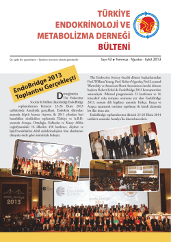
TÜRKİYE ENDOKRİNOLOJİ VE METABOLİZMA DERNEĞİ BÜLTENİ Üç ayda bir yayımlanır • Üyelere ücretsiz olarak gönderilir Sayı 46 l Nisan - Mayıs - Haziran 2014 36. TÜRKİYE ENDOKRİNOLOJİ ve METABOLİZMA HASTALIKLARI KONGRESİ TAMAMLANDI da Tiroid Ultrasonografi Kursu, 3. Ulusal Endokrin Hemşireliği Eğitim Kursu ve Adrenal Gonad Sempozyumu yapılmıştır. Kongre bilimsel programı içerisinde 4 yabancı konuşmacı konferansı olmak üzere 5 konferans, Genç Araştırıcı Konferansı, 9 uydu sempozyumu, 8 seçilmiş vaka sunumları, 13 panel, 5 İnteraktif Vaka, Sözel sunumlar ve çalışma grubu toplantıları yapılmıştır. 21-25 Mayıs 2014 tarihleri arasında Cornelia Dimond Hotel Belek Antalya’da yapılan 36. Ulusal Kongremiz başarı ile tamamlanmıştır. Kongremize yaklaşık 1.010 meslektaşımız katılmıştır. Kongre programı kapsamın- Her yıl olduğu gibi bu yılda genç araştırıcı, sözlü ve poster bildiri ödüllerimiz olmuştur. Bu yıl “En İyi Genç Araştırıcı” ödülünü almaya Uz. Dr. Neşe Çınar hak kazanmıştır. Kendisini tebrik ediyor başarılarının devamını diliyoruz. Poster ve Sözlü bildiri ödülü alan üyelerimiz ise aşağıdaki gibidir. Kendilerini tebrik ediyor, başarılarının devamını diliyoruz. 2 TÜRKİYE ENDOKRİNOLOJİ VE METABOLİZMA DERNEĞİ BÜLTENİ Sözel Bildiri Ödülleri Sözlü Bildiri Birincilik Ödülü S 15 - DHEASO4 düzeyinin Subklinik Cushing Sendromu için tanısal değeri var mı? Serkan Yener, Tevfik Demir, Seçil Özışık, Abdurrahman Çömlekçi Dokuz Eylül Üniversitesi Sözlü Bildiri İkincilik Ödülü S 17 - Obez kişilerde kromozom hasarı, sitostazi, sitotoksisite ve oksidatif DNA hasarının ve BKİ ile ilişkisinin değerlendirilmesi Fahri Bayram1, Hamiyet Dönmez Altuntaş2, Fatma Şahin2, Nazmiye Bitgen2, Zuhal Hamurcu2, Sülbiye Arıbaş1, Halit Diri1, Meral Mert3, Kürşat Gündoğan4 1Erciyes Üniversitesi Tıp Fakültesi Endokrinoloji ve Metabolizma Hastalıkları Bilim Dalı, Kayseri 2Erciyes Üniversitesi Tıp Fakültesi Tıbbi Biyoloji Anabilim Dalı, Kayseri 3Kayseri Eğitim ve Araştırma Hastanesi Endokrinoloji ve Metabo Lizma Hastalıkları Kliniği, Kayseri 4Erciyes Üniversitesi Tıp Fakültesi Dahiliye Yoğunbakım Üniversitesi, Kayseri Sözlü Bildiri Üçüncülük Ödülü S 19 - Türkiye'de erişkinlerde obezite ve fazla kiloluk prevalansını etkileyen faktörler Müge Özsan1, Fahri Bayram2, Ahmet Kaya3, Alper Sönmez4, Ali Osman Kalkan5, Tevfik Sabuncu6, Vedia Gedik7 1Niğde Devlet Hastanesi ,Niğde 2Erciyes Üniversitesi Tıp Fakü Ltesi Endokrinoloji Ve Metabolizma Bilim Dalı, Kayseri 3Necmettin Erbakan Üniversitesi Tıp Fakültesi Endokrinoloji Ve Metab. Bd, Konya 4Gülhane Askeri Tıp Akademisi Endokrinoloji Ve Metabo Lizma Bilim Dalı, Ankara 5Mevlana Üniversitesi Tıp Fakü Ltesi İç Hasta Lıkları Anabilim Dalı, Konya 6Harran Üniversitesi Tıp Fakü Ltesi Endok. Ve Metab. Bd, Şanlıurfa 7Ankara Üniversitesi Tıp Fakü Ltesi Endokrinoloji Ve Metab.bd, Ankara Poster Bildiri Ödülleri Poster Bildiri Birincilik Ödülü P 184 - Primer hiperparatiroidisi olan hastalarda parathormunun postural değişimi Cevdet Aydın, Şefika Burçak Polat, Ahmet Dirikoç, Neslihan Çuhacı, Berna Evranos, Reyhan Ersoy, Bekir Çakır Yıldırım Beyazıt Üniversitesi, Ankara Atatürk Eğitim ve Araştırma Hastanesi, Endokrinoloji ve Metabolizma Hastalıkları Kliniği Poster Bildiri İkincilik Ödülü P 134 - Yeni AGPAT2 mutasyonu 144 C>A HOM saptanan konjenital jeneralize lipodistrofi olgularında diyabetik nefropati dışı böbrek tutulumu Barış Akıncı1, Ilgın Yıldırım Şimşir2, Hüseyin Onay2, Sülen Sarıoğlu1, Sait Şen2, Tahir Atik2, Ali Çelik1, Tevfik Demir1 1Dokuz Eylül Üniversitesi Tıp Fakültesi 2Ege Üniversitesi Poster Bildiri Üçüncülük Ödülü P 039 - Renal Transplant hastalarında, farklı immunsupresif ilaçların inkretinler üzerine etkileri Nusret Yılmaz, Sebahat Özdem, Gültekin Süleymanlar, Ramazan Sarı Akdeniz Üniversitesi Tıp Fakültesi TÜRKİYE ENDOKRİNOLOJİ VE METABOLİZMA DERNEĞİ BÜLTENİ ECE 2014 Abstract Submission Geography Avrupa Endokrinoloji Derneği tarafından tüm ulusal endokrinoloji derneklerine iletilmiş olan ekteki grafikte ECE 2014'de sunulacak bildirilerin ülkelere göre dağılımı yer almaktadır. Buna göre ev sahibi Polonya ile aynı oranda olacak şekilde tüm bildirilerin %16'sı ülkemizden gönderilmiş bulunmaktadır. TİROİD HASTALIKLARI KURSLARI TEMD 11. Tiroid Hastalıkları Kursu 12.04.2014 tarihinde Eskişehir’de ve 12. Tiroid Hastalıkları Kursunun 12.si İstanbulda 03.05.2014 tarihinde gerçekleşmiştir. 3 4 TÜRKİYE ENDOKRİNOLOJİ VE METABOLİZMA DERNEĞİ BÜLTENİ Kongre ve Kurslarımız Bilimsel Kongreler ve Uluslararası Sempozyumlar Ayrıntılara ve 2014 yılına ait Bilimsel Toplantı Takvimine derneğimiz internet sayfasından (www.temd.org.tr) ulaşabilirsiniz. 06 - 10 Eylül 2014 38th Annual Meeting of the European Thyroid Association Santiago de Compostela, Spain http://www.eurothyroid.com/futureevents.html 12-14 Ekim 2014 10th International Congress of Endocrine Disorders (ICED) Tehran, IRAN http://www.iced2014.org 10 - 13 Eylül 2014 16th Congress of the European Neuroendocrine Association Sofia, Bulgaria http://www.eneassoc.org/ 23-26 Ekim 2014 EndoBridge 2014 Regnum Carya Golf & Spa Resort, Belek-Antalya http://endobridge.org/2014/ 15-19 Eylül 2014 50th EASD Annual Meeting Vienna, Austria www.easd.org 29 Ekim - 02 Kasım 2014 84th Annual Meeting of the American Thyroid Association Coronado, CA,USA http://www.thyroid.org 20 Eylül 2014 International Summit for Endocrine Gland Tumors Essentials of Thyroid Cancer Management Bornova, İzmir www.eso.net 28-29 Kasım 2014 6. Türkiye Tiroid Hastalıkları Kongresi Swissotel, Ankara www.tiroidkongresi2014.org 25-28 Eylül 2014 Diyabete Bakış 2014 Sempozyumu “Prof. Dr. Şazi İmamoğlu Onuruna " Dedeman Otel/Konya http://www.diyabetebakis.org/ 06-10 Mayıs 2015 37. Türkiye Endokrinoloji ve Metabolizma Hastalıkları Kongresi, Antalya www.temd.org.tr TÜRKİYE ENDOKRİNOLOJİ VE METABOLİZMA DERNEĞİ BÜLTENİ Üyelerimizden Literatür Seçmeleri Circulating glucagon to ghrelin ratio as a determinant of insulin resistance in hyperthyroidism. Plasma fetuin-A levels are reduced in patients with hypothyroidism. Bakiner O, Bozkirli E, Ertugrul D, Sezgin N, Ertorer E. Department of Endocrinology and Metabolic Diseases, Faculty of Medicine, Ibn-I Sina Hospital, Ankara University, Sihhiye, Ankara, Turkey. Endocrine. 2014 Feb;45(1):106-13. doi: 10.1007/s12020-013-9951-9. Epub 2013 Apr 10. Department of Endocrinology and Metabolism Diseases, Faculty of Medicine, Baskent University, Dadaloglu Mah. Serinevler 2591 Sk., No:4/A 01250 Yuregir/ADANA, Adana, Turkey. Eur J Endocrinol. 2014 Feb 4;170(3):411-8. doi: 10.1530/EJE-13-0831. Print 2014 Mar. Abstract Abstract Ağbaht K, Erdogan MF, Emral R, Baskal N, Güllü S. Due to stimulated overall metabolism, a state of nutritional inadequacy often ensues, during thyrotoxicosis. We aimed to investigate circulating levels of some major components of the system that regulates energy stores, glucose, and fat metabolism, during thyrotoxicosis compared to euthyroidism. Fasting serum ghrelin, leptin, adiponectin, insulin, glucagon, glucose, as well as body fat composition were analyzed during thyrotoxicosis in 40 hyperthyroid patients (50.5 ± 15.2 years old, 22 females, 31 with Graves disease, and 9 with toxic nodular goiter). The same measurements were repeated an average 3 months later, when all patients achieved euthyroidism. Compared to euthyroidism, in thyrotoxicosis, patients had lower ghrelin and fat mass; had comparable insulin, HOMA-IR, glucagon, and leptin levels; higher levels of circulating adiponectin. Fasting serum glucose tended to be higher during thyrotoxicosis. The unique correlation of HOMA-IR was with the-glucagon to ghrelin ratio-(r = 0.801, p < 0.001) in hyperthyrodism, and with glucagon itself in euthyroidism (r = -0.844, p < 0.001). Circulating levels of ghrelin are decreased; leptin, insulin, glucagon are unchanged; adiponectin are increased during hyperthyroidism. The fasting HOMA-IR tends to be higher, despite the decreased adiposity in hyperthyroidism. The-glucagon to ghrelin ratio-strongly correlates with fasting HOMA-IR in hyperthyroidism. Adrenocortical reserves in hyperthyroidism Agbaht K, Gullu S. Department of Endocrinology and Metabolic Diseases, Faculty of Medicine, Ibn-i Sina Hospital, Ankara University, Sihhiye, Ankara, Turkey. Endocrine. 2014 Feb;45(1):136-43. doi: 10.1007/s12020-013-9933-y. Epub 2013 Mar 27. Abstract Explicit data regarding the changes in adrenocortical reserves during hyperthyroidism do not exist. We aimed to document the capability (response) of adrenal gland to secrete cortisol and DHEA-S during hyperthyroidism compared to euthyroidism, and to describe factors associated with these responses. A standard-dose (0.25 mg/i.v.) ACTH stimulation test was performed to the same patients before hyperthyroidism treatment, and after attainment of euthyroidism. Baseline cortisol (Cor(0)), DHEA-S (DHEA-S(0)), cortisol binding globulin (CBG), ACTH, calculated free cortisol (by Coolen's equation = CFC), free cortisol index (FCI), 60-min cortisol (Cor(60)), and DHEA-S (DHEA-S(60)), delta cortisol (ΔCor), delta DHEA-S (ΔDHEA-S) responses were evaluated. Forty-one patients [22 females, 49.5 ± 15.2 years old, 32 Graves disease, nine toxic nodular goiter] had similar Cor(0), DHEA-S(0), CFC, FCI, and DHEA-S(60) in hyperthyroid and euthyroid states. Cor(60), ΔCor, and ΔDHEA-S were lower in hyperthyroidism. In four (10 %) patients the peak ACTH-stimulated cortisol values were lower than 18 μg/dL. When the test repeated after attainment of euthyroidism, all of the patients had normal cortisol response. Regression analysis demonstrated an independent association of Cor(60) with free T3 in hyperthyroidism. However, the predictors of CFC, FCI, and DHEA-S levels were serum creatinine levels in hyperthyroidism, and both creatinine and transaminase levels in euthyroidism. ACTH-stimulated peak cortisol, delta cortisol, and delta DHEA-S levels are decreased during hyperthyroidism, probably due to increased turnover. Since about 10 % of the subjects with hyperthyroidism are at risk for adrenal insufficiency, clinicians dealing with Graves' disease should be alert to the possibility of adrenal insufficiency during hyperthyroid stage. Objective: To determine plasma fetuin-A levels in hypothyroid patients before and after treatment with l-thyroxine (T4) and to determine the relation between plasma fetuin-A levels with cardiovascular risk factors. Design: A prospective, controlled, single-blind study. Methods: Forty-four treatment-naive female patients diagnosed with hypothyroidism and 39 age- and sex-matched control subjects were enrolled. Anthropometric measurements, blood pressure, plasma TSH, fetuin-A, free T4, LDL-cholesterol, triglyceride, C-reactive protein, fibrinogen levels, and brachial artery flow-mediated dilatation were measured. All measurements were repeated after 3 months in the control group and 3 months after the attainment of euthyroidism with l-T4 replacement in the hypothyroid group. Baseline data were compared between the two groups. Posttreatment plasma fetuin-A levels of hypothyroid patients were compared with baseline levels of both groups. The relationship between plasma fetuin-A, TSH levels, and other cardiovascular risk factors was evaluated. Results: Plasma fetuin-A levels were ∼20% lower in hypothyroid female patients compared with the controls (P=0.0001). Fetuin-A levels increased by ∼20% in hypothyroid patients after achievement of euthyroidism (P=0.0001) and were no longer different compared with controls (P=0.38). There was a negative correlation between plasma TSH and fetuin-A levels (r=-0.79; P=0.001). There was no significant correlation between plasma fetuin-A levels and cardiovascular risk factors within or between groups. The fetuin-A levels were normalized with thyroid hormone treatment. Conclusion: Plasma fetuin-A levels are reduced in female patients with hypothyroidism, which are restored to normal during restoration of euthyroidism. There was no relation with cardiovascular risk factors. Autoimmune thyroid disease in ankylosing spondylitis Emmungil H, Erdogan M, Kalfa M, Karabulut G, Kocanaogulları H, Inal V, Aksu K, Oksel F, Kabasakal Y, Keser G. Department of Internal Medicine, Division of Rheumatology, Mersin State Hospital, Nusratiye Hometown, Kuvayi Milliye Street, No: 32, 33050, Mersin, Turkey. Clin Rheumatol. 2014 Jul;33(7):955-61. doi: 10.1007/s10067-013-2466-1. Epub 2014 Jan 3. Abstract Although autoimmune thyroid disease is well known to be associated with primary Sjögren's syndrome (SjS) and with various autoimmune diseases, it is less clear whether a similar association also exists for ankylosing spondylitis (AS). Therefore, we investigated the frequency of autoimmune thyroid disease in patients with AS. In this cross sectional study, 80 patients with AS fulfilling the 1984 Modified New York Criteria and 80 healthy subjects, age and sex-matched with AS patients, were included. As the positive control group, 62 female patients with primary SjS were also studied. All cases underwent thyroid ultrasonography (USG) by a single endocrinologist. Thyroid function tests and thyroid autoantibodies were measured. The diagnosis of Hashimoto's thyroiditis (HT) was made if the patient had thyroid autoantibody positivity plus at least one of the following criteria: diffuse goiter with physical examination, abnormality in thyroid function tests, and parenchymal heterogeneity with USG. The chi-squared test and Fisher's exact test were used to compare cases and controls. The p values <0.05 were considered statistically significant. The frequencies of parenchymal heterogeneity with USG (30 vs 11.3 %, p = 0.045), thyroid autoantibody positivity (13.8 vs 2.5 %, p = 0.017), and concomitant diagnosis of HT (10 vs 1.3 %, p = 0.034) were significantly higher in AS group compared to healthy controls. Among AS patients having HT, subclinical hypothyroidism was detected only in a single patient. Frequency of autoimmune thyroid disease was significantly higher in AS group, compared to healthy controls. Prospective studies are needed to see the clinical relevance of these findings and outcome in the long term. 5 6 TÜRKİYE ENDOKRİNOLOJİ VE METABOLİZMA DERNEĞİ BÜLTENİ Diagnostic value of endoscopic ultrasonography for preoperative localization of parathyroid adenomas. Ersoy R, Ersoy O, Evranos Ogmen B, Polat SB, Kilic M, Yildirim N, Ozturk L, Cakir B. Is autoimmune thyroiditis a risk factor for early atherosclerosis in premenopausal women even if in euthyroid status? Department of Endocrinology and Metabolism, Faculty of Medicine, Yildirim Beyazit University, No: 2, Bilkent Yolu, Ankara, Turkey. Endocrine. 2014 Jan 11. [Epub ahead of print] Topaloglu O, Gokay F, Kucukler K, Burnik FS, Mete T, Yavuz HC, Berker D, Guler S. Abstract Abstract The most common cause of primary hyperparathyroidism (PHPT) is a single, sporadic parathyroid adenoma. Ultrasonography (US) and 99mTechnetium methoxyisobutylisonitrile (99mTc-MIBI) scintigraphy are the imaging methods most widely used to localize parathyroid adenomas. The purpose of the present study was to determine the diagnostic value and accuracy of endoscopic ultrasonography (EUS) for localizing parathyroid adenoma compared with those of US and 99mTc-MIBI scintigraphy. Forty-seven patients with a PHPT diagnosis and who were recommended for surgery were enrolled in this study. An endoscopist who was blinded to the previous US and 99mTc-MIBI scintigraphy results performed the EUS in each patient. Thirty-nine female and eight male patients with PHPT were evaluated. The presence of adenoma was confirmed by subsequent postsurgical pathology results. One case was excluded because the histopathological evaluation was compatible with a lymph node, although the lesion was detected using three different imaging modalities preoperatively. The locations of the parathyroid adenomas were correctly documented by US in 39 patients (84.7 %) by 99mTc-MIBI scintigraphy in 35 (76.0 %), and by EUS in 44 (95.6 %) of 46 patients. EUS located all 31 adenomas detected previously with US and 99mTc-MIBI scintigraphy. EUS also successfully located three adenomas that could not be identified by US and 99mTcMIBI scintigraphy. The positive predictive value and diagnostic accuracy of EUS, US, and 99mTc-MIBI were 97.7, 97.7, and 95.6 %; 88.6, 97.5, and 86.9 %; and 77.7, 97.2, 76.0 %, respectively. EUS was preferred as the second step imaging tool for detecting parathyroid adenomas that could not be localized by US and 99mTc-MIBI scintigraphy. Ultrasonographic visceral fat thickness in the first trimester can predict metabolic syndrome and gestational diabetes mellitus Gur EB, Ince O, Turan GA, Karadeniz M, Tatar S, Celik E, Yalcin M, Guclu S. Department of Obstetrics and Gynecology, Faculty of Medicine, Sifa University, Sanayi St. No:7, 35100, Bornova, Izmir, Turkey. Endocrine. 2014 Jan 23. [Epub ahead of print] Abstract The aim of this study is to evaluate whether ultrasonographic visceral fat thickness measurement in the early gestational period is useful for predicting the development of gestational diabetes mellitus (GDM) and metabolic syndrome (MS). The visceral fat thickness and subcutaneous fat thickness were measured via ultrasound at the first prenatal visit. The correlation between visceral and subcutaneous fat thickness and MS parameters, such as dyslipidemia, hypertension, and insulin resistance, was assessed. We also compared the use of visceral fat thickness measurement with body mass index (BMI) and waist circumference (WC) measurements for predicting the development of GDM. The subcutaneous fat thickness was found to be similar in the normal glucose metabolism and GDM groups at the first visit, whereas the visceral fat thickness was found to be considerably higher in the GDM groups (p = 0.04). The visceral fat thickness in the early stage of the gestation was correlated with hyperglycemia, dyslipidemia, high diastolic blood pressure, and insulin resistance. In contrast to subcutaneous fat thickness, BMI, and WC, only the visceral fat thickness was correlated with insulin resistance. The subcutaneous and visceral fat thicknesses at the first visit were significantly higher in the MS group (p = 0.02). There was a good correlation between visceral and subcutaneous fat thicknesses (r = 0.492, p < 0.001); however, there were poor correlations between visceral fat thickness and BMI and WC (r = 0.338, p = 0.01; r = 0.312, p = 0.02). The visceral fat thickness seemed to be a more sensitive predictor of GDM than WC and BMI. The optimal cutoff points for predicting GDM were visceral fat thickness 19.5 mm [area under curve (AUC) = 0.66, p = 0.043], WC 103.5 cm (AUC = 0.64, p = 0.079), and BMI 34.5 (AUC = 0.64, p = 0.069). Ultrasonographic visceral fat thickness measurement in the early period of gestation may be an easy, safe, and cost-effective scan test for predicting the development of metabolic diseases and GDM. Cayyolu Turkkonut Cevre Dostlari Sitesi, 3-B, Yenimahalle, Ankara, Turkey. Endocrine. 2013 Aug;44(1):145-51. doi: 10.1007/s12020-012-9842-5. Epub 2012 Nov 26. Autoimmune thyroiditis (AIT) is a systemic disease. It is well-known that overt thyroid dysfunction is a cardiovascular risk factor. However, the influence of euthyroid status is unclear. The aim of this study was to evaluate the metabolic parameters and carotid intima-media thickness (CIMT) in euthyroid premenopausal women with AIT. Fourtyeight premenopausal women and 18 age-matched healthy controls attending the Endocrinology and Metabolism Clinic from 2008 to 2009 were enrolled to this crosssectional study. Patients were divided into 2 groups according to TSH levels; patients in group 1 (n = 23) had TSH levels ≤ 2.5 μIU/mL and patients in group 2 had TSH levels > 2.5 μIU/mL (n = 25). All participants were evaluated by ultrasound for CIMT (mean of three segments in both carotid arteries) by the same experienced investigator. Fasting venous blood samples were collected to evaluate insulin resistance (HOMA-IR), TSH, FT4, plasma lipids, high-sensitive CRP (Hs-CRP), homocysteine, and fibrinogen. Carotid intima-media thickness was found to be significantly higher in patients than the controls (p < 0.001). However, there was no significant difference in average CIMT between group 1 and 2 (0.66 ± 0.08 vs 0.63 ± 0.09 mm). Anti-Tg levels were independently associated with CIMT in the patient group (p = 0.014). There were no significant correlations between serum TSH levels and BMI; waist circumference, serum lipids, and glucose levels. However, there was a positive significant correlation between TSH levels and blood pressure in the patients (for systolic blood pressure r = 0.466, p = 0.001, for diastolic blood pressure r = 0.372, p = 0.009). In the present study, it was shown that CIMT is increased in euthyroid premenopausal women with autoimmune thyroiditis compared to age-matched healthy controls. Comparative genotoxic and cytotoxic effects of the oral antidiabetic drugs sitagliptin, rosiglitazone, and pioglitazone in patients with type-2 diabetes: a cross-sectional, observational pilot study. Oz Gul O, Cinkilic N, Gul CB, Cander S, Vatan O, Ersoy C, Yılmaz D, Tuncel E. Uludag University Medical School, Department of Endocrinology and Metabolism, Bursa, Turkey. Mutat Res. 2013 Sep 18;757(1):31-5. doi: 10.1016/j.mrgentox.2013.04.024. Epub 2013 Jul 13. Abstract This cross-sectional, observational pilot study was designed to investigate the frequency of different endpoints of genotoxicity (sister-chromatid exchange, total chromosome aberrations, and micronucleus formation) and cytotoxicity (mitotic index, replication index, and nuclear division index) in the peripheral lymphocytes of patients with type-2 diabetes treated with different oral anti-diabetic agents for 6 months. A total of 104 patients who met the American Diabetes Association criteria for type-2 diabetes were enrolled in the study. Of the 104 patients, 33 were being treated with sitagliptin (100mg/day), 25 with pioglitazone (30mg/day), 22 with rosiglitazone (4mg/day), and 24 with medical nutrition therapy (control group). The results for all the genotoxicity endpoints were significantly different across the four study groups. Post hoc analysis revealed that the genotoxicity observed in the sitagliptin group was significantly higher than that observed in the medical nutrition therapy group, but lower than that occurring in subjects who received thiazolidinediones. All of the three cytotoxicity endpoints were significantly lower in patients treated by oral anti-diabetic agents compared with those who received medical nutrition therapy. However, the three indexes did not differ significantly in the sitagliptin, rosiglitazone, and pioglitazone groups. Taken together, these pilot data indicate that sitagliptin and thiazolidinediones may exert genotoxic and cytotoxic effects in patients with type-2 diabetes. Further investigations are necessary to clarify the possible long-term differences between oral anti-diabetic drugs in terms of genotoxicity and cytotoxicity, and how these can modulate the risk of developing diabetic complications in general and cancer in particular. TÜRKİYE ENDOKRİNOLOJİ VE METABOLİZMA DERNEĞİ BÜLTENİ Evaluation of central corneal and central retinal thicknesses and intraocular pressure in acromegaly patients. Polat SB, Ugurlu N, Ersoy R, Oguz O, Duru N, Cakir B. Endocrinology and Metabolism Department, Ataturk Training and Research Hospital, 6800, Ankara, Turkey. Pituitary. 2014 Aug;17(4):327-32. doi: 10.1007/s11102-013-0505-1. Introduction: Acromegaly is a disorder with increased morbidity which can involve many organs and the eye can be one of them which was investigated in few reports. Herein, we aimed to evaluate CCT, IOP and retinal thickness (RT), and their relationships with serum GH and IGF-1 levels and disease duration, in acromegaly patients. We compared the ocular parameters with those of a control group. This study included the largest number of patients of any comparable investigation to date. Material and Method: We enrolled 30 acromegaly patients (15 male, 15 female and age: 48.4 ± 12.8 years) and 21 age and gender matched controls. All participants underwent complete hormonal and ophtalmological evaluation including central corneal thickness (CCT), retinal thickness (RT) and intraocular pressure (IOP) values. RESULTS: There were no significant differences in median right and left CCTs and mean CCT (p = 0.646, p = 0.667 and p = 0.384, respectively). Nor were there statistically significant differences in median right and left RT, or mean central RT, between the acromegaly and control groups (p = 0.977, p = 0.738 and p = 0.811, respectively). However median right, left and mean IOPs were found to be significantly higher in the acromegaly group, despite there being no difference in the CCT values (p = 0.011, p = 0.028 and p = 0.047, respectively). When we analyzed two subgroups of acromegaly patients (active/inactive), we found that the median right, left and mean CCTs and RTs, were not significantly different between the groups. Although there was a significant difference in IOP between the acromegaly and control groups, the two subgroups of acromegaly patients had similar IOPs (p = 0.400, p = 0.621 and p = 0.451 for right, left and mean IOPs, respectively). IOP was not found to be correlated with serum IGF-I, GH or disease duration in acromegaly patients. Conclusion: Our study results suggest that acromegaly is associated with increased ocular pressures irrespective of disease activity. Therefore detailed ocular evaluation should be a routine component of the follow up visits of acromegaly patients. Epicardial adipose tissue thickness and NGAL levels in women with polycystic ovary syndrome Sahin SB, Cure MC, Ugurlu Y, Ergul E, Gur EU, Alyildiz N, Bostan M. Department of Endocrinology and Metabolism Disease, Recep Tayyip Erdogan University Medical School, Rize, Turkey. J Ovarian Res. 2014 Feb 16;7(1):24. doi: 10.1186/1757-2215-7-24. Abstract Background: Polycystic ovary syndrome (PCOS) is associated with an increased cardiovascular disease (CVD) risk and early atherosclerosis. Epicardial adipose tissue thickness (EATT) is clinically related to subclinical atherosclerosis. In the present study, considering the major role of neutrophil gelatinase-associated lipocalin (NGAL) which is an acute phase protein rapidly releasing upon inflammation and tissue injury, we aimed to evaluate NGAL levels and EATT in PCOS patients and assess their relationship with cardiometabolic factors. Methods: 64 patients with PCOS and 50 age- and body mass index-matched healthy controls were included in the study. We evaluated anthropometric, hormonal and metabolic parameters. EATT was measured by echocardiography above the free wall of the right ventricle. Serum NGAL and high-sensitive C- reactive protein (hsCRP) levels were measured by ELISA. Results: Mean EATT was 0,38 +/-0,16 mm in the PCOS group and 0,34 +/-0,36 mm in the control group (p = 0,144). In the obese PCOS group (n = 44) EAT was thicker compared to the obese control group (n = 41) (p = 0.026). Mean NGAL levels of the patients with PCOS were 101,98 +/-21,53 pg/ml, while mean NGAL levels were 107,40 +/-26,44 pg/ ml in the control group (p = 0,228). We found a significant positive correlation between EATT and age, BMI, waist circumference, fasting insulin, HOMA-IR, triglyceride and hsCRP levels in PCOS group. Conclusions: Thickness of the epicardial adipose tissue can be used to follow the risk of CVD development in obese PCOS cases. However serum NGAL levels do not differ in patients with PCOS and control group. Three noninvasive methods in the evaluation of subclinical cardiovascular disease in patients with acromegaly: epicardial fat thickness, aortic stiffness and serum cell adhesion molecules. Topaloglu O, Sayki Arslan M, Turak O, Ginis Z, Sahin M, Cebeci M, Ucan B, Cakir E, Karbek B, Ozbek M, Cakal E, Delibasi T. Department of Endocrinology and Metabolism, Diskapi Yildirim Beyazit Training and Research Hospital, Ankara, Turkey. Clin Endocrinol (Oxf). 2014 May;80(5):726-34. doi: 10.1111/cen.12356. Epub 2013 Dec 12. Context: Several studies have reported increased risk of cardiovascular disease due to early development of endothelial dysfunction and structural vascular changes in patients with acromegaly. Objective: The aim of this study was to evaluate subclinical cardiovascular disease with epicardial fat thickness (EFT), aortic stiffness and serum levels of cell adhesion molecules (CAMs) in patients with acromegaly. Design: Cross-sectional study. Patients: Twenty-seven patients with active acromegaly (AA), 13 patients with remission acromegaly (RA) and 37 age- and sex-matched healthy controls were studied. Measurements: Epicardial fat thickness was evaluated by transthoracic echocardiography (TTE). Aortic stiffness (β) index, aortic strain (AoS) and aortic distensibility (AoD) were calculated from the aortic diameters measured by TTE. Serum levels of CAMs such as intercellular adhesion molecule (ICAM)-1, vascular cell adhesion molecule (VCAM)-1 and E-selectin were measured. Results: Epicardial fat thickness was significantly increased in patients with RA and AA as compared to controls 9·71 ± 1·54 and 10·08 ± 1·95 mm vs 5·74 ± 0·92 mm, P < 0·001, respectively). A significant positive correlation was found between the EFT and growth hormone (GH) levels (r = 0·365, P = 0·024). β-index was similarly higher in patients with RA and AA than controls (15·68 ± 7·27 and 11·90 ± 8·24 vs 6·85 ± 2·87, P < 0·001, respectively). AoS and AoD were significantly decreased in patients with RA and AA as compared to the control group (3·81 ± 1·94 and 3·68 ± 1·99 vs 8·19 ± 4·19%, P < 0·001, respectively; and 1·21 ± 0·66 and 1·18 ± 0·63 vs 2·58 ± 1·50, 10(-6) cm(2) /dyn, P < 0·001, respectively). Serum ICAM-1 and VCAM-1 levels were significantly higher in patients as compared to the control group (P < 0·001 vs P = 0·032, respectively). There were no significant differences in EFT, AoD, AoS, β-index and serum CAMs between two patients groups (AA vs RA, P > 0·05). There was a significant negative correlation between E-selectin and AoD (r = -0·45, P = 0·008). In multiple linear regression analysis, EFT was found to be associated with GH levels (β-coefficient = 0·575, P = 0·008). Conclusion: This study suggests that EFT and risk of subclinical cardiovascular disease are increased in patients with acromegaly. Serum GH level is an independent risk factor for EFT. 7 8 TÜRKİYE ENDOKRİNOLOJİ VE METABOLİZMA DERNEĞİ BÜLTENİ Yeni Doçentlerimiz Yeni Profesörlerimiz Doç. Dr. Ayşe Kubat Üzüm Doç. Dr. Mehmet Ali Eren Prof. Dr. Taner Bayraktaroğlu Üyelerimizi tebrik eder, başarılar dileriz. Yeni Üyelerimiz Dr. Emre Arslan Dr. Mustafa Aydemir Dr. Rövşen Hasanov Gazi Üniversitesi Tıp Fakültesi, Ankara Cumhuriyet Üniversitesi Tıp Fakültesi, Sivas Hacettepe Üniversitesi Tıp Fakültesi, Ankara Üyelerimizi tebrik eder, başarılar dileriz. Türkiye Endokrinoloji ve Metabolizma Derneği Bülteni Türkiye Endokrinoloji ve Metabolizma Derneği’nce üç ayda bir yayımlanır. Yayın Türü: Yaygın süreli TEMD Adına Sahibi Prof. Dr. A. Sadi Gündoğdu Sorumlu Yazı Işleri Müdürü Prof. Dr. Mustafa Kemal Balcı Yayın Danışma Kurulu Prof. Dr. Abdurrahman Çömlekçi, Prof. Dr. Bülent Okan Yıldız, Prof. Dr. Mustafa Sait Gönen, Prof. Dr. Serdar Güler, Prof. Dr. Sevim Güllü Baskı tarihi: Ağustos 2014 TEMD bülteninde yayımlanacak derneğimiz ile ilgili haberlerin bekletilmeksizin ve en geç her ayın 1’ine kadar TEMD merkezine ulaşmış olması gerekmektedir. TEMD bülteni, www.temd.org.tr adresinden de PDF formatında görüntülenebilir. Yönetim Yeri: Meşrutiyet Cad. Ali Bey. Apt. 29/12, Kızılay 06420 Ankara Tel: (0312) 425 20 72 Faks: (0312) 425 20 98 E-posta: [email protected] Grafik Tasarım: BAYT Bilimsel Araştırmalar Basın Yayın ve Tanıtım Ltd. Şti. Tel: (0312) 431 30 62 • Faks (0312) 431 36 02 • E-posta: [email protected] Baskı: Miki Matbaacılık San. Tic. Ltd. Şti. Matbaacılar sitesi 560. Sk. No: 27 İvedik, Ankara • Tel: (312) 395 21 28
© Copyright 2026 Paperzz