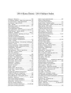
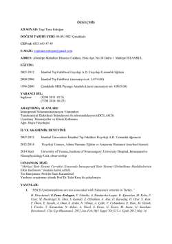
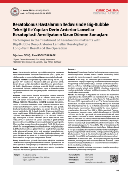
Optical Coherence Tomography Findings in Unilateral
Peripapillary Myelinated Retinal Nerve Fibers
Tek Taraflı Peripapiller Miyelinli Retina Sinir Liflerinde
Optik Koherens Tomografi Bulguları
Myelinli Sinir Liflerinde OCT Bulguları / OCT Findings in Myelinated Retinal Nerve Fibers
Alime Gunes1, Seden Demirci2, Mustafa Gunes3
Department of Ophthalmology, 2Department of Neurology, 3Department of Urology,
Süleyman Demirel University Faculty of Medicine, Isparta, Turkey
1
Özet
On dokuz yaşında bir erkek hasta, erken çocukluğundan beri olan sağ gözünde görme azlığı ile başvurdu. Düzeltilmiş en iyi görme keskinliği, sağ gözde 10 cm’ den
parmak sayma ve sol gözünde 1.0 idi. Relatif afferent pupil defekti yoktu; refraksiyon değerleri, sağ gözde -6.50 -1.25 x 110 ve sol gözde -0.25 değerindeydi. Biyomikroskobik mueyene ve göz içi basıncı her iki gözde normaldi. Fundus muayenesinde, sağ gözde geniş, çukurlaşmış disk, üst vasküler arkuat boyunca uzanan
aşırı myelinli peripapiller retina sinir lifleri mevcuttu. Optik koherens tomografi,
sağ gözde üst kadranda incelme ile birlikte normal ortalama peripapiller retina sinir lifi tabakası kalınlığı (RSLT) ve anormal foveal kontur göstermekteydi. Myelinli
retinal sinir lifleri, ortalama peripapiller RSLT kalınlığında değişikliklere neden olmasa da, anormal foveal morfoloji ve anizometrik miyopi ile ciddi görme kaybına neden olabilir.
Abstract
Anahtar Kelimeler
Myelinli Retina Sinir Lifleri; Optik Kohorens Tomografi; Retina Sinir Lifi Tabakası Kalınlığı
Keywords
A 19-year-old male presented with low vision in the right eye since early childhood. Best corrected visual acuity was counting fingers at 10 cm and 1.0 in the
right and left eyes. There was no relative afferent pupillary defect; refraction was
-6.50 -1.25 x 110 and -0.25 in the right and left eyes. Biomicroscopic examination
and intraocular pressures were normal in both eyes. On fundus examination, there
was large, cupped disk, extensive peripapillary myelinated retinal nerve fibers extending along superior vascular arcade in the right eye. Optical coherence tomography showed normal average peripapillary retinal nerve fiber layer (RNFL) thickness with thinning in superior quadrant and abnormal foveal contour in the right
eye. Although MRNF not cause changes in average RNFL thickness, may cause
to serious vision loss by abnormal foveal morphology and anisometropic myopia.
Myelinated Retinal Nerve Fibers; Optic Coherence Tomography; Retinal Nerve Fiber Layer Thickness
DOI: 10.4328/JCAM.2444
Received: 28.03.2014 Accepted: 11.04.2014 Publihed Online: 11.04.2014
Corresponding Author: Alime Gunes, Department of Ophthalmology, Süleyman Demirel University Faculty of Medicine, 32260, Isparta, Turkey.
GSM: +905054828345 E-Mail: [email protected]
1 | Journal of Clinical and Analytical Medicine
Myelinli Sinir Liflerinde OCT Bulguları / OCT Findings in Myelinated Retinal Nerve Fibers
Introduction
Myelination of the optic nerve begins at the 32nd pregnancy
week from the lateral geniculate nucleus and finishes at term. It
arrives the lamina cribrosa a short time after birth [1]. Myelinated retinal nerve fibers (MRNF) are not normally found in the
human retina, but histopathologic studies have demonstrated
that pieces of MRNF contain oligodendrocytes [2]. Williams
suggested that a defect in the lamina cribrosa would cause entry of oligodendrocytes into the retina [2]. It was supported by
extensive retinal myelination in animals with little or no lamina
cribrosa [2].
Anisometropic myopia, strabismus, abnormal foveal morphology, amblyopia and reduced vision may be associated with
MRNF [4,5].
In this report, we present optical coherence tomography (OCT)
findings in a patient with unilateral peripapillary myelinated
retinal nerve fibers.
Case Report
A 19-year-old male presented with low vision in the right eye
since early childhood. Best corrected visual acuity was counting fingers at 10 cm and 1.0 in the right and left eyes. Pupils
did not show relative afferent pupillary defect; refraction was
-6.50 -1.25 x 110 and -0.25 in the right and left eyes. Biomicroscopic examination and intraocular pressures were normal in
both eyes. On fundus examination, there was large, cupped disk,
extensive peripapillary MRNF extending along superior vascular
arcade in the right eye (Figure 1). Optical coherence tomography (OCT) (Spectral OCT SLO, OPKO/OTI Instrumentation, Miami, FL, USA) showed normal average retinal nerve fiber layer
(RNFL) thickness with thinning in superior quadrant (Figure 2)
and abnormal foveal contour in the right eye (Figure 3). In addition, our patient has had epilepsy and chronic renal failure
secondary to vesicoureteral reflux.
Figure 1. Fundus photography of the patient’s right eye showed large, cupped disk,
extensive peripapillary myelinated retinal nerve fibers extending along superior
vascular arcade.
Discussion
Peripapillary MRNF can be congenital or acquired. Congenital
retinal myelination is a developmental anomaly and the incidence in the population is 0.3-0.6%. Coexistence of MRNF and
ipsilateral high myopia, amblyopia, strabismus, optic disk hypo2 | Journal of Clinical and Analytical Medicine
Figure 2. Optical coherence tomography showed normal average retinal nerve
fiber layer thickness with thinning in superior quadrant in right eye and normal
average retinal nerve fiber layer thickness in left eye.
Figure 3. Optical coherence tomography showed abnormal foveal contour in right
eye.
plasia and vision loss has been reported in the literature [4,5].
Gharai et al. evaluated MRNF with OCT and reported that peripapillary MRNF involving the macula can cause foveal ectopia,
deprivation amblyopia and vision loss [6]. They suggested that
foveal position can be detected only by OCT.
In our case, OCT showed normal average RNFL thickness with
thinning in superior quadrant and abnormal foveal contour in
the right eye. Peripapillary MRNF extending along superior vascular arcade can explain the decreased thickness of superior
RNFL.
In addition, our patient has had epilepsy and chronic renal failure secondary to vesicoureteral reflux, but this association has
never been reported in the literature.
In this case, low vision in the right eye with MRNF may have
been due to foveal abnormality, anisometropic amblyopia, or
optic disk hypoplasia.
To the best of our knowledge, this is the first report of the OCT
findings including peripapillary RNFL in a patient with MRNF.
Although MRNF not cause changes in average RNFL thickness,
may cause to serious vision loss by abnormal foveal morphology and anisometropic myopia.
Competing interests
The authors declare that they have no competing interests.
References
1. Magoon EH, Robb RM. Development of myelin in human optic nerve and tract. A
light and electron microscopic study. Arch Ophthalmol 1981;99(4):655-9.
2. Straatsma BR, Foos RY, Heckenlively JR, Taylor GN. Myelinated retinal nerve
fibers. Am J Ophthalmol 1981;91(1):25-38.
3. Williams TD. Medullated retinal nerve fibers: Speculation son their cause and
presentation of cases. Am J Optom Physiol Opt 1986;63(2):142-51.
4. Lee MS, Gonzalez C. Unilateral peripapillary myelinizated retinal nerve fi-
Myelinli Sinir Liflerinde OCT Bulguları / OCT Findings in Myelinated Retinal Nerve Fibers
bers associated with strabismus, amblyopia, and myopia. Am J Ophthalmol
1998;125(4):554-6.
5. Yalcın E, Balcı O, Akıngol Z. Association of extensive myelinated nerve fibers and
high degree myopia: Case report. Indian J Ophthalmol 2013;61(10):606–7.
6. Gharai S, Prakash G, Ashok Kumar D, Jacob S, Agarwal A, Arora V. Spectral
domain optical coherence tomographic characteristics of unilateral peripapillary
myelinated retinal nerve fibers involving the macula. J AAPOS 2010;14(5):432-4.
3 | Journal of Clinical and Analytical Medicine
© Copyright 2026 Paperzz