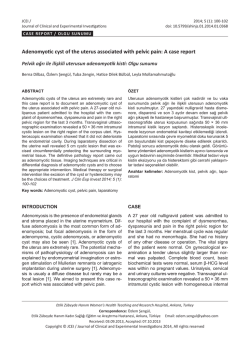
Pamukkale Tıp Dergisi
Pamukkale Medical Journal
Olgu Sunumu
doi: 10.5505/ptd.2015.94547
Infected mullerian adenosarcoma: report of a case and review of the
literature
Enfekte müllerian adenosarkoma: olgu sunumu ve literatürün gözden geçirilmesi
Banuhan Şahin*, Aysun Karabulut*, Nevzat Karabulut**, Mehmet Gündoğan***, Metin Akbulut***
*Pamukkale Üniversitesi Tıp Fakültesi, Kadın Hastalıkları ve Doğum AD, Denizli
**Pamukkale Üniversitesi Tıp Fakültesi, Radyoloji AD, Denizli
***Pamukkale Üniversitesi Tıp Fakültesi, Patoloji AD, Denizli
Özet
Müllerian adenosarkom daha çok postmenapozal dönemdeki kadınları etkileyen nadir bir tümördür. Patolojik
olarak düşük gradeli endometrial stromal sarkom ve çoğunlukla benign, ama bazen de atipik glandüler epitelden
oluşur. Hastalar çoğunlukla karın ağrısı, huzursuzluk ve uterin kanama şikayetiyle başvururlar. Bu yazıda abse
formasyonuyla komplike olan ve postoperatif dönemde sepsis gelişen bir Müllerian adenosarkom olgusunu
sunmayı amaçladık. Müllerian adenosarkomlar rölatif olarak iyi prognoza sahip nadir görülen tümörler olmakla
birlikte klinik tablo nekroza sekonder gelişen enfeksiyon, abse gelişimi ve postoperatif sepsisle komplike
olabilmektedir.
Pam Tıp Derg 2014;8(1): 79-81
Anahtar sözcükler:Mullerian adenosarkoma, abse, sepsis.
Abstract
Mullerian adenosarcoma is a rare tumor usually affecting postmenapousal women. Patients generally present
with abdominal pain, discomfort and uterine bleeding. A mixture of low grade endometrial stromal sarcoma,
and benign but sometimes atypical glandular epithelium is detected in the histopathologic examination. Herein,
we present an unusual case of Mullerian adenosarcoma complicated with abscess formation leading to sepsis
postoperatively. Mullerian adenosarcomas are uncommon tumors with relatively good prognosis, however
necrosis and secondary infection may lead to abscess formation and complicate the clinical picture with
postoperative sepsis.
Pam Med J 2014;8(1):79-81
Key words: Mullerian adenosarcoma, abscess, sepsis.
Introduction
Case report
Mullerian adenosarcoma is a rare tumor
usually affecting postmenapousal women [1,2].
It shows a mixture of low grade endometrial
stromal sarcoma, and benign but sometimes
atypical glandular epithelium. Patients generally
present with abdominal pain, discomfort and
uterine bleeding. Herein, we present an unusual
case of Mullerian adenosarcoma complicated
with abscess formation leading to sepsis
postoperatively.
A 66-year-old
female
presented
with
postmenopausal bleeding of two to three pads
and a foul smelling vaginal discharge for 2-3
weeks.
Sonographic examination showed
an enlarged uterus with two myomas around
1-2 cm, and a large intracavitary polypoid
mass. Pelvic magnetic resonance examination
demonstrated a 5x6 cm contrast enhancing
polypoid mass filling the endometrial cavity, and
an accompanying subendometrial hemorrhage
(Fig 1). Histopathologic examination of
endometrial
curettage
material
showed
endometrial polyp. Intraoperatively, the uterus
Banuhan Şahin
Yazışma Adresi: Pamukkale Üniversitesi Tıp Fakültesi, Kadın Hastalıkları ve Doğum AD, Denizli
e-mail: [email protected]
Gönderilme tarihi: 22.05.2014 Kabul tarihi: 08.12.2014
79
Pamukkale Tıp Dergisi 2015;8(1):79-81
was enlarged around 18-20 weeks of gestation,
and filled with purulant material during the
operation. Total abdominal hysterectomy
and
bilateral
salphyngooferectomy
was
performed along with peritoneal washing. In the
postoperative period, the patient had a fever
of 39oC, WBC increased to 16800/mm3 with
neutrophil predominance, CRP was 17 mg/
dl, and sedimentation rate was 39 mm/hour.
Intensive parenteral antibiotic therapy was
administered for both anaerobes and gramnegative bacteria for 10 days. Laboratory values
a
Şahin et al.
progressively turned to normal and the patient
was discharged on the 11th day postoperatively.
Histopathologic examination showed a
6x5x5cm mass filling the endometrial cavity with
areas of necrosis and hemorrhage. Hyperplastic
glandular endometrial cells were detected
between sarcomatous cell clusters leading
to a diagnosis of Mullerian adenosarcoma
(Fig 2).The tumoral mass was limited to the
endometrium without evidence of myometrial
invasion and lymphovascular involvement.
b
Figure 1. T1 weighted MR images before (A) and after contrast administration (B) demonstrate
enhancing polypoid mass in the endometrial cavity (arrows) with accompanying subendometrial
blood (arrowheads).
Figure 2. Müllerian adenosarcoma: sarcomatous areas formed by spindle shaped cells in form of
crevasse in some places and hyperplastic endometrial glandular structures. (H&E,x4; a,x10; b,x40).
80
Infected mullerian adenosarcoma
Discussion
Mullerian adenosarcoma is a slowly
progressive low grade neoplasm with benign
epithelial
and
malignant
sarcomatous
components [1,3]. The malignant component
is low grade endometrial stromal sarcoma
in almost all the cases [3]. The usual clinical
presentation is abdominal pain, discomfort
and uterine bleeding [3]. Similarly, our patient
also presented with postmenopausal bleeding,
but she also had a malodorous seroanginous
vaginal discharge originating from the abscess
within the endometrial cavity. Endometrial
sampling is important for the diagnosis, however,
in our case histopathologic examination of the
endometrial curettage material revealed an
endometrial polyp.
Magnetic resonance imaging (MRI) is
a useful modality providing information on
soft tissue intensity. However, MRI features
of adenosarcoma have been described in
only a few cases. Usually it manifests as a
heterogeneous mass encompassing solid
and cystic components filling the endometrial
cavity of an enlarged uterus. The mass
enhances similar to the myometrium [4]. In
our case, the mass was predominantly solid
and exhibited slightly lesser enhancement
compared to the myometrium. There was also
accompanying subendometrial blood. Bilateral
salphingooferectomy besides histerectomy
has traditionally been recommended in the
treatment of adenosarcoma. Recurrences are
rare in the absence of sarcomatous overgrowth
and myometrial invasion [5,6].
Conflict of interest: The authors declared no
conflict of interest.
References
1.
2.
3.
4.
5.
6.
Clement PB, Scully RE. Müllerian adenosarcoma of
the uterus. A clinicopathologic analysis of ten cases
of a distinctive type of müllerian mixed tumor. Cancer
1974;34:1138−1149.
Clement PB, Scully RE. Uterine tumors with mixed
epithelial mesenchymal elements. Semin Diagn Pathol
1988;5:199−222.
Gallardo A, Prat J. Mullerian Adenosarcoma; A
clinicopathologic and immunohistochemical study of
55 cases challenging the existence of adenofibroma.
Am J Surg Pathol 2009;33:278−288
Yoshizako T, Wada A, Kitagaki H, Ishikawa N, Miyazaki
K. MR imaging of uterine adenosarcoma: case
report and literature review. Magn Reson Med Sci
2011;10:251−254.
Nam JH. Surgical treatment of uterine sarcoma. Best
Pract Res Clin Obstet Gynaecol 2011; 25:751−760.
McCluggage WG. Mullerian adenosarcoma of the
female genital tract. Adv Anat Pathol 2010;17:122−129.
In conclusion, Mullerian adenosarcomas
are uncommon tumors with relatively good
prognosis. Because of intracavitary growth, they
usually generate symptoms and are diagnosed
at the early stages. Necrosis and secondary
infection may lead to abscess formation
complicating the clinical picture. Mullerian
adenosarcomas must be kept in mind in cases
with endometrial masses filling the enlarged
uterus and presenting with postmenapousal
bleeding, and they may also be associated with
postoperative sepsis.
81
© Copyright 2026 Paperzz