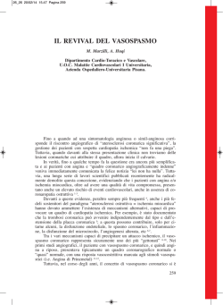
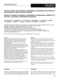
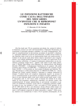
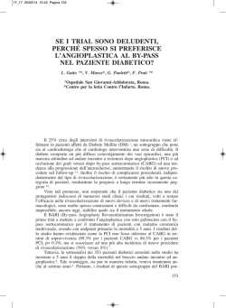
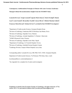
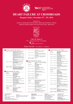
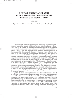
284
La Radiologia Medica - Radiol Med 106: 284-296, 2003
Edizioni Minerva Medica - Torino
Non-invasive angiography of the coronary arteries with multislice computed
tomography: state of the art and future
prospects
Angiografia coronarica non invasiva
con Tomografia Computerizzata spirale
multistrato
Stato dell’arte e prospettive future
L’angiografia convenzionale ha rappresentato fino ad oggi l’unico strumento
per la valutazione delle arterie coronarie. Tentativi sono stati effettuati durante
la scorsa decade per validare una tecnologia non invasiva per lo studio delle
coronarie. In particolare la Tomografia ad emissione di elettroni e la Risonanza
Magnetica sono state utilizzate a questo scopo senza però divenire strumenti
clinici. Di recente la tecnologia di tomografia computerizzata (TC) spirale,
migliorata dall’utilizzo di sistemi multistrato a 4 linee di detettori e da una
velocità di rotazione del tubo radiogeno di 500 ms, ha mostrato un ottimo
potenziale senza entrare nella pratica routinaria. L’introduzione della tecnologia multistrato a 16 canali, con tempi di rotazione del tubo radiogeno inferiori a 500 ms, ha reso possibile ottenere risultati di rilievo nella valutazione
delle stenosi coronariche significative (>50%), tali da proporne l’utilizzo clinico. In questo lavoro verranno descritti la tecnica di esecuzione dell’esame ed
i risultati, allo stato dell’arte, dell’angiografia con TC delle arterie coronarie.
Until now conventional angiography has represented the only technique for assessing the coronary arteries. During the last decade, attempts have been made
to validate a non-invasive technique for the study of
coronary arteries.
In particular, Electron-beam Tomography and
Magnetic Resonance Imaging have been used for
this purpose, even though they have not become clinical tools. More recently, spiral computed tomography (CT) technology, improved by the use of multiple slices and 500ms gantry rotation times, has
shown a good potential without entering routine clinical practice.
The introduction of multislice technology with 16
rows and rotation times below 500ms has yielded
such good results in detecting significant (>50%)
coronary artery stenosis that it has been proposed as
a clinical tool. This paper describes the examination
technique and the results achieved in CT angiography of the coronary arteries.
PAROLE CHIAVE: Coronarografia - Imaging non invasivo - Tomografia computerizzata
multistrato.
KEY WORDS: Coronary angiography - Non-invasive coronary imaging - Multislice Computed Tomography.
Filippo CADEMARTIRI1 - Giacomo LUCCICHENTI2
Riccardo MARANO3 - Koen NIEMAN1, 4 - Nico MOLLET1, 4
Pim DE FEYTER1,4 - Gabriel P KRESTIN1 - Lorenzo BONOMO3
A
Introduzione
Introduction
Dalla sua introduzione nella pratica clinica, l’angiografia
coronarica convenzionale rappresenta lo strumento più accurato per la valutazione dell’emodinamica delle arterie coronarie [1, 2]. Questa tecnica è soggetta a quegli inconvenienti che sono caratteristici delle metodiche angiografiche
convenzionali come la natura proiettiva delle immagini e
l’impossibilità di visualizzare la parete del vaso. Inoltre,
invasività, rischi e costi impediscono l’impiego di questa
tecnica su larga scala se non in pazienti con precise indicazioni cliniche.
Nell’ultima decade vi sono stati alcuni tentativi di ovviare
a questi inconvenienti. In particolare la risonanza magnetica (RM) e la tomografia computerizzata ad emissione di elettroni (EBT), hanno fornito risultati interessanti ma non sufficienti per estenderne l’utilizzo alla pratica clinica routinaria [1, 3-8].
L’imaging delle arterie coronarie necessita, a causa delle
dimensioni delle strutture studiate e del movimento indotto
dalle pulsazioni cardiache, di risoluzioni spaziale e tempo-
Ever since its introduction, Coronary Angiography (CA)
has been considered the gold standard in the hemodynamic evaluation of coronary arteries [1, 2].
Drawbacks of this technique are the inner projective nature
of the images, and the inability to directly visualize the vessel walls. The invasive nature, the risks and the costs limit
the use of CA strictly to patients with a well-defined clinical indication. In the last decade attempts have been made
to overcome these limitations with Magnetic Resonance (MR)
and Electron-Beam Computed Tomography (EBCT), with
interesting results but not sufficient to extend the use of these
diagnostic tools to routine clinical practice [1, 3-8]. Imaging
of the coronary arteries requires high spatial and temporal
resolution because of the small size of the vessels and of
motion due to the heartbeat. Multislice Computed tomography (MSCT) technology provides high spatial and temporal
resolution. Four-row MSCT has shown encouraging results
although the rate of non-assessable vessels is high [9-13].
The introduction of 16-row MSCT scanners allows the spa-
Dipartimento di Radiologia1 - Erasmus Medical Center - Rotterdam, Olanda - Dipartimento di Radiologia2 - Università degli Studi di Parma - Italia Dipartimento di Radiologia3 - Università di Chieti - Italia - Dipartimento di Cardiologia4 - Thoraxcentre - Erasmus Medical Center - Olanda.
Pervenuto alla Redazione il 03.04.2003; revisionato il 05.05.2003; restituito corretto il 23.05.2003; accettato per la pubblicazione il 31.05.2003.
Indirizzo per la richiesta di estratti: Dott F. Cademartiri, Viale Rustici, 2 - 43100 Parma PR - Italia - Tel. 0521/961833.
E-mail:[email protected]
F. Cademartiri et al: Angiografia coronarica non invasiva con Tomografia Computerizzata spirale multistrato
rale elevate. Queste ultime sono state ottenute con l’impiego
della tecnologia multistrato. Le apparecchiature multistrato
a 4 canali hanno fornito risultati incoraggianti ma insufficienti a causa di una elevata percentuale di vasi non valutabili
[9-13].
Lo sviluppo di scanner a 16 canali ha permesso un ulteriore miglioramento delle risoluzioni spaziale e temporale.
La scansione coronarica può essere, infatti, completata in
un tempo inferiore ai 20 s (contro i 35-40s precedentemente
necessari), e con una risoluzione spaziale di 0,75mm (contro il valore di 1-1,25 mm delle precedenti generazioni [1417]. Gli ottimi risultati ottenuti con le apparecchiature a 16
strati hanno indotto a proporre l’impiego di questa tecnica
nella routine clinica [18, 19].
Lo scopo di questo lavoro è di illustrare il razionale clinico-diagnostico, le basi tecniche, i risultati e le prospettive
future della tecnologia multistrato applicata alle arterie coronarie. In particolare verrà focalizzata l’attenzione sul paragone
tra apparecchiature a 4 e 16 strati.
Materiale e metodi
Preparazione del paziente
I pazienti con fibrillazione atriale, allergia nota al mezzo
di contrasto iodato e insufficienza renale (creatinina sierica
>120 mmol/L-1) vengono esclusi [18].
È stato descritto come la frequenza cardiaca elevata influenzi negativamente le prestazioni ed il successo della scansione in termini di qualità diagnostica [8, 13, 20, 21]. Ai
pazienti con frequenza cardiaca superiore a 65 battiti per
minuto (bpm) viene somministrato, 45-60 minuti prima della scansione, metoprolo-tartrato per os con dosaggio compreso tra 50 e 200 mg a seconda della frequenza cardiaca.
La bradicardizzazione è più difficoltosa in pazienti con frequenza >90 bpm. Per uno studio ottimale delle arterie coronarie è necessario ottenere una frequenza cardiaca al di sotto di 65 bpm. Nel caso in cui si stia valutando il muscolo
cardiaco, il pericardio, l’anatomia dei grossi vasi o la funzione miocardica è possibile studiare anche pazienti con frequenze cardiache superiori o con fibrillazione atriale a condizione che sia a bassa risposta ventricolare.
Al fine di ottenere una traccia elettrocardiografica (ECG),
tre elettrodi vengono posizionati per ottenere una sola
derivazione ECG corrispondente a D1. Il vettore della
derivazione infatti va dalla spalla destra alla spalla sinistra.
Gli elettrodi vengono posizionati nel cavo ascellare destro
e sinistro (la terra sulla superifice addominale o sul fianco
sinistro) in modo che non siano a contatto con i grandi muscoli del torace (grande pettorale e grande dorsale). Questo
evita irregolarità della linea basale dell’ECG, dovute a contrattura muscolare, quando le braccia vengono posizionate
al di sopra della testa per la scansione.
Il mezzo di contrasto iodato viene somministrato per via
endovenosa antecubitale mediante un’agocannula da 18-20
G ed un iniettore automatico. Il lato da scegliere quando possibile è il destro. Da questo lato infatti l’anatomia della vena
anonima è più favorevole e gli artefatti da indurimento legati
alla elevata densità del contrasto a livello venoso non compromettono eccessivamente la visualizzazione dell’arco aor-
285
tial and temporal resolution to be increased. Coronary CT
imaging can be performed in less than 20 sec (versus 3540s of 4-row MSCT), with a spatial resolution of 0.75mm
(versus 1-1.25mm of 4-row MSCT) [14-17]. Sixteen-row
MSCT has already shown good results and it is suggested
as a tool for routine clinical practice [18, 19].
The aim of this paper is to illustrate the clinical and diagnostic rationale, the basics of the technique and future prospects of multislice technology applied to coronary artery
imaging. Particular focus will be given to the comparison
between 4-row and 16-row multislice scanners.
Materials and methods
Patient preparation
Exclusion criteria are atrial fibrillation, previous contrast material (CM) adverse reaction, renal insufficiency
(serum creatinine >120mmol/L-1) [18].
High heart rates have been reported to reduce diagnostic quality of MSCT scans [8, 13, 20, 21]. Patients with a
heart rate >65bpm are given an oral dose of metoprol-tartrate (50-200mg depending on the heart rate), 45-60 minutes before the start of the scan. It is more difficult to slow
down the heart rate when its value is >90bpm. When the
heart rate is <65bpm it is usually possible to perform an
optimal MSCT of the coronary arteries.
For the evaluation of the myocardium, the pericardium,
the anatomy of the great intra-thoracic vessels or cardiac
function, it is possible to scan patients with higher heart
rates or patients with atrial fibrillation and low ventricular
response. The patient is positioned in the supine position,
and then the three leads are connected to record the patient’s
ECG. The leads are positioned on the middle axillary lines
and on the left flank, to avoid irregularity of the baseline
ECG track due to muscle contraction when both arms are
positioned above the head during the scan.
Contrast material is administered intravenously (i.v.) with
an automatic power injector using a 18-20G cannula; the
right arm is preferred because of the favorable anatomy of
the right brachio-cephalic vein and because it produces less
beam hardening artifacts at the level of the aortic arch.
CT scan
Angiographic CT is performed with a 4-16 row multislice
scanner (Sensation 4-16 – Siemens Medical Solutions,
Forchheim, Germany), after i.v. administration of 100-125
ml of non ionic contrast material (300-400 mgI/ml) at a flow
rate of 4-5 ml/s with an automatic power injector (EnVision
CT – MedRad, Pittsburgh, USA). The administration of a
saline bolus-chaser of 40-50ml at the same flow rate, with
a double head injector (Stellant - MedRad, Pittsburgh, USA)
can be useful to push the contrast medium after the end of
its administration while the scan is still underway.
The patient’s ECG is recorded at the same time as CT
scan is performed. The scan time is 40s for 4-row MSCT
scanners and <20s for 16-row scanners. Scan parameters
for 4-row MSCT are: detector number 4, detector collimation 1mm, rotation time 500ms, feed/rotation 1.5mm (pitch
0.375), kVp 120, mAs 400-500, field of view 150mm, effec-
286
F. Cademartiri et al: Angiografia coronarica non invasiva con Tomografia Computerizzata spirale multistrato
Fig. 1. — Schema del sistema di modulazione del milliamperaggio.
Modulazione prospettica della dose somministrata al paziente attraverso
la sincronizzazione della corrente del tubo radiogeno con il tracciato ECG.
The prospective modulation of the X-ray exposure through the synchronization of the tube current with the ECG.
tico e dell’origine dei tronchi sovra-aortici (quando la scansione viene estesa per coprire per esempio l’origine delle
arterie mammarie interne).
Scansione
La scansione angiografica viene effettuata con scanner
spirali a 4-16 linee di detettori (Sensation 4-16 - Siemens
Medical Solutions, Forchheim, Germania), dopo la somministrazione endovenosa per via antecubitale di un bolo di
mezzo di contrasto non ionico (300-400 mgI/ml) di 100-125
ml con una velocità di 4-5 ml/s mediante un iniettore automatico (EnVision CT - MedRad, Pittsburgh, USA). La somministrazione di un bolo da 40-50 ml di soluzione salina alla
stessa velocità del bolo principale è di ausilio per sospingere il bolo di mezzo di contrasto dalle vene del braccio. In
questo caso è necessario impiegare un iniettore a doppia testa (Stellant - MedRad, Pittsburgh, USA).
Contemporaneamente alla scansione viene registrata una
traccia elettrocardiografica (ECG). La durata complessiva
della scansione è di circa 40 s per i tomografi a 4 linee e
inferiore ai 20 s per quelli a 16 linee. I parametri di scansione per la generazione a 4 linee sono: numero di detettori/collimazione 4/1 mm, tempo di rotazione del tubo radiogeno 500 ms, avanzamento/rotazione 1,5 mm (pitch 0,375),
kVp 120, mAs 400, campo di vista 150 mm, spessore effettivo dello strato 1,25 mm, incremento di ricostruzione 0,7
mm, filtro di convoluzione per tessuti molli.
I parametri di scansione per la generazione a 16 linee
sono: numero di detettori/collimazione 16/0,75 mm, tempo
di rotazione del tubo radiogeno 420 ms, avanzamento/
rotazione 3,0 mm (pitch 0,25), kVp 120, mAs 400-500, campo di vista 150 mm, spessore effettivo dello strato 0,75 mm,
incremento di ricostruzione 0,4 mm, filtro di convoluzione
per tessuti molli. Nella tabella I sono riassunti i principali
parametri di scansione, irradiazione, ricostruzione e somministrazione del mezzo di contrasto.
tive slice width 1.25 mm, reconstruction increment 0.7mm,
soft tissue kernel. Scan parameters for 16-row MSCT are:
detector number 16, detector collimation 0.75mm, rotation
time 420ms, feed/rotation 3.0mm (pitch 0.25), kVp 120, mAs
400-500, field of view 150mm, effective slice width 0.75mm,
reconstruction increment 0.4mm, soft tissue kernel.
Table I summarises the main scan parameters, radiation
dose, reconstruction and contrast medium administration
parameters.
Dose
Radiation exposure
La dose somministrata al paziente varia a seconda del sesso, del volume e dei parametri di scansione. Per il maschio
Radiation exposure depends on the patient’s sex, the
study volume and scan parameters. For males the dose is
Fig. 2. — Schema dei metodi di sincronizzazione retrospettiva. Modalità
di sincronizzazione retrospettiva della ricostruzione basata sul segnale
ECG. La finestra di ricostruzione può essere posizionata utilizzando un
metodo con ritardo fisso detto «assoluto inverso» (A), o con ritardo percentuale detto «relativo» (B). Nel primo caso la distanza dall’onda R
rimane costante quindi a frequenze elevate (>70bpm) la fase che piu’
favorevolmente sarà definita tele-diatsolica sarà prossima a –350 ms,
mentre a frequenze inferiori la finestra a –400 ms è ottimale. Nel secondo caso invece, la posizione delle finestre di ricostruzione varia proporzionalmente alla variazione dell’intervallo RR e, di conseguenza, alla
frequenza cardiaca.
Retrospective ECG synchronisation of the reconstruction. The reconstruction window can be positioned with a fixed delay so called «absolute
reverse method» (A), or with a percentage delay so called «relative
method» (B). In the first case, the distance from the R-wave is constant,
then at higher heart rates (>70bpm) the most favourable window will be
at –350 ms, while at lower heart rates –400 ms is almost always optimal.
In the second case, the position of reconstruction window varies proportionally with the variation of RR interval and heart rate.
F. Cademartiri et al: Angiografia coronarica non invasiva con Tomografia Computerizzata spirale multistrato
287
è di circa 6,82 mSv mentre la femmina è di circa 10,12 mSv,
senza l’utilizzo di modulazione prospettica del milliamperaggio [22, 23]. Questi valori possono essere ulteriormente
ridotti sincronizzando l’emissione di raggi X all’ECG prospetticamente mediante la modulazione della corrente del
tubo radiogeno [22]. Con questa tecnica è possibile ridurre
la dose fino al 50% in meno a seconda della frequenza cardiaca (fig. 1).
Ricostruzione delle immagini
Le immagini vengono ricostruite in modo da corrispondere ad una particolare fase del ciclo cardiaco. Ciò
viene ottenuto sincronizzando retrospettivamente i dati
acquisiti all’ECG registrato durante la scansione. La fase
del ciclo cardiaco alla quale appartengono i dati grezzi
impiegati per la ricostruzione dell’immagine viene espressa come percentuale dell’intervallo R-R, oppure come distanza temporale dalla successiva onda R espressa in milli-secondi (Fig. 2). Qualunque fase del ciclo cardiaco può
essere ricostruita per ottenere informazioni morfologiche
delle strutture vascolari o funzionali (frazione d’eiezione,
perfusione miocardica e motilità delle pareti miocardiche).
Le coronarie possono essere valutate impiegando ad
esempio tre finestre di ricostruzione della fase tele-diastolica a -350ms, -400ms, e -450ms dalla successiva onda
R. Utilizzando il metodo percentuale relativo le finestre da
utilizzare sono 65%, 70% e 75%. La necessità di ricostruire diverse finestre per la visualizzazione delle coronarie
deriva dal movimento desincronizzato dei differenti segmenti coronarici. Alcuni tratti dei vasi infatti possono
mostrare minore motilità in una fase del ciclo mentre altri
in fasi leggermente diverse [8, 24]. Gli strati così ricostruiti possono essere definiti isocardiofasici, poiché appartengono alla stessa fase del ciclo cardiaco. Nel caso in cui la
finestra di ricostruzione sia di durata insufficiente per fornire
tutti i dati necessari, è possibile ottenere le informazioni
da due cicli cardiaci adiacenti con una metodo definito di
ricostruzione multi-segmentaria mostrato in figura 3 [25].
Per la ricostruzione di circa 250 immagini sono necessari
circa 42s con una velocità di ricostruzione che può arrivare
fino a 6 immagini al secondo.
Analisi delle immagini
La valutazione delle immagini viene effettuata su una
stazione di lavoro (PC). Le ridotte dimensioni e l’anatomia
complessa delle strutture studiate impediscono la loro valutazione sul solo piano di scansione. È fondamentale
l’impiego di algoritmi di riformattazione multiplanare lungo il decorso delle arterie coronarie, e Maximum Intensity
Projection (MIP, proiezione di massima intensità) che fornisce immagini di tipo angiografico. Le ricostruzioni tridimensionali con Volume Rendering (VR) permettono di rappresentare in modo sintetico la sede e l’entità della patologia (fig. 4).
Esistono degli strumenti (software dedicati) semi-automatici ed automatici per la segmentazione dei vasi (Vessel
View - Siemens Medical Solutions, Forchheim, Germania)
che hanno lo scopo di velocizzare l’analisi delle immagini.
L’asse centrale del vaso viene ottenuto a partire dalle sue
due estremità. Da questo asse centrale è possibile ottenere
Fig. 3. — Schema della modalita’ di ricostruzione bi-segmentale.
Ricostruzione delle immagini nella quale tutte le proiezioni in un arco di
180° sono ottenute durante la stessa diastole (A). All’aumentare della frequenza cardiaca, con la tecnica multisegmentaria, le proiezioni dell’arco
di 180° vengono ottenute da due o più fasi diastoliche (B).
Image reconstruction in which all the 180° projections are obtained in
the same diastole (A). When the heart frequency increases, with the segmented reconstruction technique, projections through 180° are obtained
from the two or more diastolic phases (B).
6.82 mSv whereas for females it is 10.12 mSv, without the
use of prospective tube current modulation [22, 23]. These
values can be reduced by 50% using prospective synchronization between ECG and tube current modulation [22]
(fig. 1).
Image reconstruction
All images are reconstructed in a defined phase of the
cardiac cycle, by means of retrospective synchronization
with the ECG track. This phase of the cardiac cycle is
expressed as a percentage of the R-R interval (Relative
Delay) or as temporal distance (ms) before the next R wave
(fig. 2).
Images can be also reconstructed in other phases of the
cardiac cycle to obtain morphological or functional information (ejection fraction, myocardial perfusion and cardiac
wall motion).
The coronary arteries can be evaluated in the late diastolic phase with three different temporal windows at -350ms,
-400ms, and -450ms before the next R wave. When the Relative
Delay technique is applied, 65%, 70% and 75% are usually preferred.
Several different reconstruction windows are normally
required to visualize the coronary arteries because of their
non synchronous motion [8, 24]. All the reconstructed images of one dataset can be defined as iso-cardio-phasic, because
they belong to the same cardiac phase.
When the temporal window is too short to provide all the
data, the information can be obtained from two contiguous
cardiac cycles with a multi-segmental reconstruction algorithm (fig. 3) [25].
To reconstruct 250 images, 42s are needed, with a maximum reconstruction speed of 6 image/sec.
288
F. Cademartiri et al: Angiografia coronarica non invasiva con Tomografia Computerizzata spirale multistrato
Fig. 4. — Esempio di scansione coronarica con apparecchiatura TC multistrato a 16 canali. La ricostruzione tridimensionale VR (A), mostra una
panoramica anatomica della regione del cuore e dei grossi vasi. Isolando
nel volume tridimensionale il cuore dalle altre strutture toraciche (B, C,
e D), si può apprezzare il tipico decorso dei 3 vasi principali. La arteria
discendente anteriore (ADA) che decorre nel solco interventricolare anteriore (B e C), il ramo circonflesso dell’ arteria coronaria di sinistra (Cx)
che decorre nel solco atrio-ventricolare sinistro (D), e la arteria coronaria
destra (CDx) che decorre nel solco atrio-ventricolare destro (B e C).
Chiaramente dimostrabile anche la posizione del tronco comune (TC)
della coronaria sinistra (D). Una analisi dettagliata del lume vascolare
presuppone l’utilizzo di ricostruzione multiplanari e curvate di tipo centro-luminale (E, F, G, e H). È possibile infatti visualizzare l’origine di
entrambe le coronarie (TC-ADA e CDx) in un solo piano para-coronale
curvato (E). La ADA ricostruita lungo il suo intero decorso è dimostrata in F. Lo stesso tipo di ricostruzione viene applicato alla Cx in G ed
alla CDx in H. Nelle immagini sono anche dimostrati due rami diagonali (dalla ADA in B e C), e ben 5 rami marginali (dalla Cx in D). Nel
caso presentato si osserva una dominanza sinistra, ossia quando la Cx
prevale sulla CDx nella parete postero-inferiore del cuore e dà origine
alla arteria discendente posteriore (D, G per la Cx e C, H per mostrare
la ridotta estensione di CDx nel solco atrio-ventricolare destro). A livello del tratto distale di Cx si osserva una occlusione totale (freccia in D e
G). Non sono presenti calcificazioni lungo il decorso delle coronarie e
dei loro rami.
CT angiography with 16 row scanner of the coronary arteries. Threedimensional reconstruction with VR (A), shows a panoramic view of the
heart and the main vessels. The path of the vessels can be better depicted by separating the heart from adjacent structures (B, C and D).
Descending anterior artery ADA courses in the anterior interventricular groove (B and C), the left circumflex coronary artery (Cx) courses in
the left atrio-ventricular groove (D), and the right circumflex coronary
artery (CDx) in the right atrio-ventricular groove (B and C). The common trunk of the left coronary artery is clearly visible (D). Multiplanar
and curved reformatted images through the center-lumen allow detailed
analysis of the vessel (E,F,G and H). The origin of both coronary arteries (TC-ADA and CDx) can be displayed in a single curved plane (E).
The ADA is displayed along its path (F). The same kind of reconstruction has been performed in for Cx (G) and CDx (H). Two diagonal (from
the ADA in D) and 5 marginal (from Cx in D) branches are also visible.
In this case there is a left dominance, with Cx dominating over the CDx
in the postero-inferior wall and generating the posterior descendent
artery (D, G for the Cx, and H to show the limited length of CDx in the
right atrio-ventricular sulcus). There is a total occlusion of the Cx in its
distal tract (arrow in D and G). No calcifications are visible in the coronary arteries and their branches.
immagini riformattate perpendicolari o lungo il decorso di
questo asse.
Risultati
In tabella II sono riassunti i dati della letteratura ove la
TC multistrato a 4 e a 16 canali è stata paragonata alla coronarografia convenzionale.
Gli studi di coronarografia con TC a 4 strati sono stati effettuati su popolazioni comprese tra i 28 ed i 100 pazienti. La valutazione ha riguardato solo i vasi di diametro
uguale o superiore a 2 mm (o 1,5 mm in [26]), e sono stati considerate le riduzioni del lume uguali o superiori al
50%. Con questi criteri di valutazione, sono stati ottenuti valori di sensibilità tra 75% ed 91% e di specificità tra
76% e 99% per la visualizzazione delle stenosi vascolari
[9, 11, 12, 21, 26-29]. Nonostante questi valori appaiano
Image analysis
All images are evaluated at the workstation (PC). The
small size of the coronary vessels and their complex anatomy prevent the evaluation on the native axial plane. It is
therefore mandatory to use Multiplanar reconstructions
(MPR) along the path of coronary arteries and Maximum
Intensity Projection (MIP) reconstruction algorithms are
normally used to create angiographic images.
Three-dimensional (3D) Volume Rendered (VR) reconstructions allow the panoramic visualization of the site and
grade of atherosclerotic disease (fig. 4).
Dedicated software (Vessel View - Siemens Medical
Solutions, Forchheim, Germany) can be used for semi-automatic or automatic segmentation of the vessels to increase
the speed of vessel analysis. The extremities of the vessel are
manually targeted and then a central lumen line is reconstructed.
F. Cademartiri et al: Angiografia coronarica non invasiva con Tomografia Computerizzata spirale multistrato
289
TABELLA I. — Protocolli di scansione angiografica coronarica con TC multistrato.
Scanner
Produttore
Siemens
Siemens
Modello
Volume zoom
Sensation 16
Tipo
4 strati
16 strati
Detettori
4
16
Collimazione
1 mm
0.75 mm
KiloVolt
120
120
Scansione
MilliAmpere/s
400
400-500
Tempo di rotazione
500 ms
420 ms
Modulazione mAs
Sistolica
Sistolica
Risoluzione temporale effettiva
250 ms
210 ms
Risoluzione temporale massima*
125 ms
105 ms
Avanzamento/rotazione
1.5 mm
3.0 mm
Avanzamento/secondo
3.0 mm
7.1 mm
Pitch del detettore
1.5
4.0
Pitch conico
0.375
0.25
Tempo di scansione (120-140mm)
40-45s
17-20s
5.71 mSv
6.82 mSv
Dose
Dose effettiva – maschio
Dose effettiva – femmina
8.49 mSv
10.12 mSv
Dose effettiva – maschio (modulazione)
2.8 mSv
3.41 mSv
Dose effettiva – femmina (modulazione)
4.24 mSv
5.06 mSv
Ricostruzione
Spessore effettivo dello strato
1.25 mm
0.75-1.00mm
Intervallo di ricostruzione
0.8 mm
0.5-0.6mm
Finestre temporali di ricostruzione
-350 ms; -400 ms ; -450 ms
-350ms; -400ms ; -450ms
Campo di vista - FOV
140-180 mm
140-180 mm
Filtro di convoluzione
medio
medio
Finestra (ampiezza/centro)
600/200
600/200
140 ml
100 ml
Mezzo di contrasto
Volume
Velocità di somministrazione
3-4 ml/s
4-5 ml/s
Tempo di somministr. Mdc
30-25s
25-20s
Concentrazione Iodica
300-400 mgI/ml
300-400 mgI/ml
Bolus chaser
40ml @ 4 ml/s
40ml @ 4 ml/s
Accesso venoso
Antecubitale
Antecubitale
Tempo totale somministr.
30/42.5 s
25/37.5 s
elevati, tra il 5% ed il 32% dei segmenti non erano valutabili [9, 11,12,21,26-29]. Una serie di 102 pazienti si
distingue da quelle citate riportando con due osservatori
separati, una sensibilità, specificità pari a 86-93% e 9697%, rispettivamente senza esclusione di segmenti [30].
Il valore predittivo positivo, il valore predittivo negativo
Results
Table II summarises the data from the literature on the
comparison between 4-row and 16-row MSCT and conventional coronary angiography in the evaluation of coronary
artery stenosis. Four-row MSCT coronary angiography has
290
F. Cademartiri et al: Angiografia coronarica non invasiva con Tomografia Computerizzata spirale multistrato
TABLE I.—Scan protocols for coronary angiography with multislice CT.
Scanner
Manufacturer
Siemens
Siemens
Model
Volume zoom
Sensation 16
Type
4 slice
16 slice
Detectors
4
16
Collimation
1 mm
0.75 mm
KiloVolt
120
120
Scan
MilliAmpere/s
400
400-500
Rotation time
500 ms
420 ms
MAs modulation
Systolic
Systolic
Actual temporal resolution
250 ms
210 ms
Maximum termporal resolution*
125 ms
105 ms
Feed/rotation
1.5 mm
3.0 mm
Feed/second
3.0 mm
7.1 mm
Detector pitch
1.5
4.0
Beam-cone pitch
0.375
0.25
Scan time (120-140mm)
40-45s
17-20s
5.71 mSv
6.82 mSv
Effective dose – females
8.49 mSv
10.12 mSv
Effective dose – males (modulation)
2.8 mSv
3.41 mSv
Effective dose – females (modulation)
4.24 mSv
5.06 mSv
Dose
Effective dose – males
Reconstruction
Effective slice width
1.25 mm
0.75-1.00mm
Reconstruction interval
0.8 mm
0.5-0.6mm
Reconstruction time window
-350 ms; -400 ms ; -450 ms
-350ms; -400ms ; -450ms
Field of view – FOV
140-180 mm
140-180 mm
Convolution filter
medium
medium
Window (amplitude/center)
600/200
600/200
Volume
140 ml
100 ml
Administration speed
3-4 ml/s
4-5 ml/s
Administration time (CM)
30-25s
25-20s
Contrast medium
Iodine concentration
300-400 mgI/ml
300-400 mgI/ml
Bolus chaser
40ml @ 4 ml/s
40ml @ 4 ml/s
Venous access
Antecubital
Antecubital
Total administration time.
30/42.5 s
25/37.5 s
e l’accuratezza diagnostica complessiva, sono rispet-tivamente tra 76-92%, 94-98%, e 79-98% [9, 11, 12, 21, 2629].
Gli studi di coronarografia con TC a 16 strati hanno fornito risultati migliori. In uno studio [18] nessun vaso di
diametro superiore a 2 mm è stato escluso dall’analisi, al
been performed on populations between 28 and 100 patients
(pts), evaluating all vessels with a diameter ≥2 mm (or 1.5
mm in [26]; stenoses ≥50% were considered. In the visualization of coronary artery stenosis, the sensitivity ranged
between 75% and 91% with a specificity between 76% and
99% [9, 11, 12, 21, 26-29]. Even though these diagnostic
291
F. Cademartiri et al: Angiografia coronarica non invasiva con Tomografia Computerizzata spirale multistrato
TABELLA II. — Letteratura sulle casistiche di angiografia coronarica non invasiva mediante TC multistrato.
N° paz.
Excl. (%)
Se. (%)
Sp. (%)
VPP (%)
VPN (%)
Acc. (%)
Nieman K(12)
Lancet ‘01
31
27
81
97
81
97
95
Achenbach S(9)
Circulation ‘01
64
32
85
76
56
94
79
Knez A(38)
AJC ‘01
44
6
78
98
84
96
94
Vogl TJ(28)
Radiology ‘01
38
19
75
99
92
98
98
Giesler T(29)
AJR ‘02
100
29
91
89
66
98
96
Nieman K(27)
AJC ‘02
53
30
82
93
66
97
90
Kopp AF(30)*
EHJ ‘02
102
0
93
97
81
99
96
Nieman K(21)
Heart ‘02
78
32
84
95
67
98
93
Becker CR(26)
JCAT ‘02
28
5
81
90
57
97
89
Nieman K(18)
Circulation ‘02
58
0
95
86
80
97
97
Ropers D(19)
Circulation ‘03
77
12
92
93
79
97
93
4-strati
16-strati
Accuratezza diagnostica valutata nella rilevazione di stenosi significative, con riduzione del diametro uguale o superiore al 50%, e comparata con la coronarografia
convenzionale come «gold standard».
N° paz.: numero di pazienti inclusi nello studio; Excl.: percentuale di segmenti esclusi considerati non valutabili; Se.: sensibilità; Sp.: specificità; VPP: valore predittivo positivo; VPN: valore predittivo negativo; Acc.: accuratezza; AJR: American Journal of Roentgenology; EHJ: European Heart Journal; AJC: American Journal
of Cardiology; JCAT: Journal of Computed Assisted Tomography; *Risultati dell’ osservatore con la migliore performance.
contrario degli studi precedenti [9, 11, 12, 21, 27-30]. La
sensibilità, la specificità, il valore predittivo positivo e
negativo sono risultati rispettivamente 95%, 86%, 80%,
e 97%. L’accuratezza complessiva nella valutazione del
tronco comune della coronaria sinistra, della arteria discendente anteriore, della coronaria destra e della circonflessa, sono risultate 100%, 91%, 86%, e 81%, rispettivamente [18]. In un altro studio, solo il 12% dei segmenti
di diametro uguale o superiore a 2 mm è stato escluso.
Questo valore, apparentemente basso in senso assoluto,
lo è sicuramente per il gruppo da cui proviene che era uso
escludere tra il 29% ed il 32% dei vasi esaminati con la
precedente generazione di scanner a 4 strati [9, 29]. I valori di sensibilità, specificità, valore predittivo positivo e
negativo sono risultati 92%, 93%, 79%, e 97%, rispettivamente [19].
Discussione
Le arterie coronarie e quindi le lesioni ad esse riferite sono
di piccole dimensioni. Inoltre la pulsazione cardiaca concede una finestra temporale relativamente breve durante la
quale il movimento è minimo. Le risoluzioni spaziale e tem-
values may appear good, between 5% and 32% of all available segments was considered not assessable [9, 11, 12, 21,
26-29]. One series on 102 patients reports different results,
with two readers’ sensitivity and specificity of 86-93% and
96-97%, respectively, without exclusion of any segment [30].
The positive and negative predictive value and the diagnostic accuracy were 76-92%, 94-98%, and 79-98%, respectively [9, 11, 12, 21, 26-29].
Studies performed using 16-row MSCT reported better
results. In one study [18] no vessel with a diameter ≥2mm
was excluded, differently from most previous studies [9, 11,
12, 21, 27-30]. The sensitivity, the specificity, the positive
and negative predictive values were 95%, 86%, 80% and
97%, respectively. The overall accuracy in the evaluation of
the left main (LM) artery, left anterior descending artery
(LAD), right coronary artery (RCA) and circumflex (CX)
were 100%, 91%, 86%, and 81%, respectively [18].
In another study, 12% of the vessels with diameter ≥2mm
were excluded; this exclusion rate is low but it is even more
important because it comes from the group that excluded
29% to 32% of the available vessels with 4-row MSCT [9,
29]. The sensitivity, specificity, positive and negative predictive values were 92%, 93%, 79%, and 97%, respectively [19].
292
F. Cademartiri et al: Angiografia coronarica non invasiva con Tomografia Computerizzata spirale multistrato
TABLE II.—Literature reports on series of non-invasice coronary angiography with multislice CT.
N° paz.
Excl. (%)
Se. (%)
Sp. (%)
VPP (%)
VPN (%)
Acc. (%)
Nieman K(12)
Lancet ‘01
31
27
81
97
81
97
95
Achenbach S(9)
Circulation ‘01
64
32
85
76
56
94
79
Knez A(38)
AJC ‘01
44
6
78
98
84
96
94
Vogl TJ(28)
Radiology ‘01
38
19
75
99
92
98
98
Giesler T(29)
AJR ‘02
100
29
91
89
66
98
96
Nieman K(27)
AJC ‘02
53
30
82
93
66
97
90
Kopp AF(30)*
EHJ ‘02
102
0
93
97
81
99
96
Nieman K(21)
Heart ‘02
78
32
84
95
67
98
93
Becker CR(26)
JCAT ‘02
28
5
81
90
57
97
89
Nieman K(18)
Circulation ‘02
58
0
95
86
80
97
97
Ropers D(19)
Circulation ‘03
77
12
92
93
79
97
93
4-strati
16-strati
Diagnostic accuracy for the detection of significant stenosis, with diameter reduction equal or more than 50%, compared with conventional coronary angiography
as «gold standard».
N° paz.: patient included in the study; Excl.: percentage of excluded segments; Se.: sensitivity; Sp.: specificity; VPP: positive predictive value; VPN: negative predictive value; Acc.: accuracy; AJR: American Journal of Roentgenology; EHJ: European Heart Journal; AJC: American Journal of Cardiology; JCAT: Journal of
Computed Assisted Tomography; *Results of the observer with better performance.
porale delle apparecchiature rappresentano quindi i principali fattori limitanti nell’imaging non invasivo delle arterie
coronarie. Per questo motivo l’angiografia convenzionale ha
rappresentato per molti decenni l’unica tecnica disponibile
per lo studio di questi vasi.
La EBT è dotata di elevata risoluzione temporale (50-100
ms), ma ha scarsa risoluzione spaziale lungo l’asse longitudinale (strati da 3 mm). Inoltre la scansione viene sincronizzata prospetticamente sul segnale ECG. Ciò rende la
durata della scansione (e dell’apnea) dipendente dalla frequenza cardiaca. Quando questa è troppo bassa è necessario
somministrare atropina per permettere di acquisire l’intero
cuore durante una sola apnea.
La RM è dotata di elevata risoluzione temporale (~50ms),
ma caratterizzata da una risoluzione spaziale insufficiente a
dimostrare le porzioni distali dei vasi coronarici.
Lo sviluppo della tecnologia multistrato ha reso possibile
l’impiego della TC per l’imaging vascolare non invasivo
delle arterie coronarie. Ciò è stato possibile aumentando la
risoluzione spaziale mediante l’impiego di spessori di strato di 1 mm. La risoluzione temporale è stata aumentata
riducendo i tempi di rotazione del gantry al di sotto di 500
ms e con l’introduzione di algoritmi di ricostruzione delle
immagini in grado di sfruttare solo 180 gradi della rotazione
Discussion
The coronary arteries and their lesions are small in size.
Moreover, the heartbeat determines a relatively short temporal window during which motion is minimized. Therefore,
spatial and temporal resolution represent the main limiting
factors in the non-invasive evaluation of coronary artery
imaging. For these reasons, conventional coronary angiography has remained for decades the only available technique for the study of the coronary tree.
EBT is characterized by a high temporal resolution (50100ms) but with a low spatial resolution in the z-axis (3mm
slice). Moreover the scan is performed using prospective
ECG-triggering. For this reason, the scan time (and apnea)
depends on the heart rate. When the heart rate is too low it
may be necessary to administer atropine in order to scan
the heart within a single breath-hold.
Magnetic Resonance has a high temporal resolution
(~50 ms), but it has an inadequate spatial resolution to visualize the distal segment of the coronary vessels.
The development of multislice technology has enabled noninvasive coronary artery imaging with spiral CT, with an
increased spatial resolution due to thinner collimations (1mm).
The improvement of the temporal resolution is obtained with
F. Cademartiri et al: Angiografia coronarica non invasiva con Tomografia Computerizzata spirale multistrato
293
Fig. 5. — Esempio di placca aterosclerotica soft a livello della coronaria sinistra. Ricostruzione 3D con VR delle coronarie (A-D). Le ricostruzioni
multiplanari con algoritmo MIP mostrano l’arteria coronaria di sinistra
(E), la sua suddivisione nel ramo discendente anteriore (E-G) e circonflesso (H) e la coronaria di destra (I). A livello del tratto prossimale della
ADA si osserva un ispessimento omogeneo della parete arteriosa settale,
in assenza di lesioni calcifiche, da riferirsi a placca aterosclerotica «soft»
(freccia).
Example of soft aterosclerotic plaque of the left coronary artery. Threedimensional VR of the coronary arteries (A-D). Multiplanar MIP show
the left coronary artery (E), its bifurcation in left anterior descending
artery (E-G) and circumflex artery (H), and right coronary artery (I). In
proximal ADA there is an homogeneous thickening of the septal wall of
the artery, without calcified lesions, to be referred as “soft” atherosclerotic plaque (arrow).
del tubo radiogeno.
I risultati ottenuti con apparecchiature a 4 canali sono
stati incoraggianti [9, 12], ma erano gravati da una elevata
percentuale di vasi esclusi dalla valutazione a causa di artefatti. In molti studi la valutazione era limitata alle localizzazioni molto prossimali (LM e segmenti prossimali di
ADA, Cx, e CDx) [31-35]. In particolare la visualizzazione
delle porzioni intermedie di CDx e di Cx era difficoltosa a
causa del movimento presente anche in fase telediastolica
(fasi precoci della sistole atriale) e per la loro direzione
cranio-caudale [27]. Ciò ha impedito l’impiego di questa
tecnica nella routine. Questa tecnica è inoltre soggetta a
artefatti da movimento che dipendono dalla frequenza cardiaca, e dagli artefatti legati alla presenza di calcificazioni
coronariche [21-27].
Lo sviluppo di apparecchiature a 16 strati ha ovviato a
questi inconvenienti proponendo la TC per la valutazione
non invasiva delle arterie coronarie su larga scala (fig. 5). I
risultati ottenuti indicano che è possibile effettuare una valutazione completa e accurata di tutti i vasi coronarici nella
maggior parte dei pazienti [18, 19]. Nei pazienti con fibrillazione atriale o con frequenza cardiaca superiore a 70 bpm
sono state introdotte le ricostruzioni multi-segmentarie (già
disponibili anche sulle apparecchiature a 4 strati). Con questo
algoritmo di ricostruzione, invece di utilizzare tutti i dati dal
medesimo ciclo cardiaco, una immagine viene ricostruita
impiegando segmenti provenienti da cicli cardiaci contigui.
Questa tecnica è tuttavia influenzata da alcuni fattori. In
essa si assume che la posizione del cuore sia identica tra
due o più cicli cardiaci adiacenti. Questa precisione millimetrica tra un ciclo e l’altro è meno probabile in caso di
frequenza cardiaca elevata. Inoltre, la ricostruzione multisegmentaria presuppone l’impiego di dati più distanti tra
loro lungo l’asse longitudinale a causa dello spostamento
del tavolo tra un ciclo e l’altro. Una interpolazione di scansioni più vicine può essere ottenuta aumentando la collimazione o riducendo la velocità del tavolo con un aumento del tempo di scansione ed in ultima analisi delle radiazioni somministrate al paziente.
Un aumento della velocità di rotazione del tubo radiogeno
sarebbe la soluzione ideale per ottenere risoluzioni temporali effettive pari o inferiori ai 150 ms (pari ad un tempo di
rotazione del tubo radiogeno di 300 ms). Ciò ridurrebbe
inoltre la necessità di somministrazione di farmaci beta-bloccanti prima dell’esame.
Tra i limiti attuali della tecnica ricordiamo la difficile visu-
a faster rotation time (<500ms) and with dedicated reconstruction algorithm, which uses information from half rotation (180°).
Promising results have been reported with 4-row MSCT
[9, 12], but a high rate of vessel exclusion due to the presence
of artifacts was also reported. In many reports, the evaluation was limited to the proximal portion of the coronary arteries [31-35]. In particular, the visualization of mid portion of
RCA and CX was difficult because of motion artifacts during
294
F. Cademartiri et al: Angiografia coronarica non invasiva con Tomografia Computerizzata spirale multistrato
Fig. 6. – Esempio di follow-up di bypass aorto-coronarico. In A è mostrata la vista anteriore di un bypass coronarico confezionato con arteria mammaria interna sinistra (AMIS) per un ramo diagonale della coronaria discendente anteriore (freccia). Il ramo diagonale appare lievemente stirato verso l’alto dal bypass. Nel medesimo paziente (B e C) è presente anche un graft venoso (VGS) che decorre tra la vena cava
superiore e atrio destro, per anastomizzarsi distalmente sulla faccia posteriore del cuore sulla arteria discendente posteriore (ramo della coronaria destra che decorre nel solco interventricolare posteriore. Si nota chiaramente il flusso retrogrado nel tratto distale della CDx (C).
Follow-up of a coronary by-pass. A by-pass from the left internal mammary artery (AMIS) to a diagonal branch of the anterior descendent
artery (arrow) is shown is A. The diagonal branch is slightly stretched upwards from the by-pass. In the same patient (B and C) a venous graft
(VGS) is also present running between the superior vena cava and the right atrium, flowing into the posterior descendent artery in the posterior aspect of the heart. Retrograde flow is visible in the distal tract of the CDx (C).
alizzazione degli stent e delle calcificazioni. La valutazione
degli stent è soggetta, infatti, ad artefatti derivanti dal materiale e dal diametro dello stent utilizzato. Il materiale impiegato ha generalmente elevato coefficiente di attenuazione.
Ciò lo fa apparire più grande di quanto effettivamente non
sia, determinando il cosiddetto effetto «blooming». Questo
effetto può impedire la visualizzazione di iperplasie intimali
o del flusso all’interno di stent di piccolo calibro. L’impiego
di adeguati filtri di ricostruzione può attenuare l’effetto
«blooming». Un’alternativa definitiva sarebbe quella di impiegare materiali che non provochino artefatti. Per ciò che
concerne le calcificazioni, si ha lo stesso problema citato
per gli stent. Ossia, quando la calcificazione del vaso è estesa e di dimensioni notevoli, a causa del “blooming”, il lume
vascolare non è valutabile in modo appropriato. L’utilizzo
di filtri più elevati può mitigare questo effetto soprattutto se
abbinato all’utlizzo di collimazioni di spessore inferiore. Le
clip chirugiche, rilevate lungo il decorso dei bypass (soprattutto arteriosi) pongono problematiche simili (fig. 6).
Le prospettive future di sviluppo sono rappresentate
sostanzialmente da un ulteriore miglioramento delle
risoluzioni spaziale e temporale. L’aumento del numero di
detettori, pur comportando una riduzione del tempo di scansione, non implicherebbe un significativo miglioramento
della qualità dell’immagine e introdurrebbe ulteriori sforzi
nella correzione del fascio conico. I cosiddetti “flat panel
detector” hanno invece dimostrato in studi effettuati in laboratorio di produrre un evidente miglioramento dell’immagine ovviando a tutti gli inconvenienti sopra descritti. Le
the late diastolic phase (early phase of atrial systolic contraction) and for their vertical direction [27]. Furthermore, this
technique was limited by cardiac motion artifacts and by the
presence of consistent coronary artery calcifications [21, 27].
All these reasons have limited its routine use.
The introduction of 16-row MSCT overcomes several limitations proposing it as a clinical tool for the non-invasive
evaluation of coronary arteries (fig. 5). The reported data
show the capability of the 16-row MSCT scanner to evaluate
all coronary artery segments in most of the patients [18, 19].
In the case of high heart rate (>70bpm) and atrial fibrillation multi-segmental reconstruction algorithms are available. Unfortunately, multi-segmental reconstructions have
some limitations: between contiguous heart beats it is
assumed that the heart position is exactly the same, but this
is less probable at higher heart rates.
Multisegmental reconstruction requires data that are far
apart from each other along the z-axis because of table’s
motion between two or more contiguous cardiac cycles. Data
interpolation can be achieved by increasing collimation or
reducing the table feed, but with an increased scan time and
exposure radiation of the patient.
An increased rotation time should be the right solution in
order to obtain an effective temporal resolution of 150 ms
(rotation time of 300 ms). Then, it could be possible to reduce
the administration of beta-blockers prior to the scan. Stents
and calcifications still remain a field where the evaluation
capability of MSCT is limited.
The evaluation of stents is limited by artifacts due to the
F. Cademartiri et al: Angiografia coronarica non invasiva con Tomografia Computerizzata spirale multistrato
dosi elevate, e i tempi di rotazione (~30 s) non permettono
ancora il loro impiego clinico.
In conclusione, l’angiografia coronarica non invasiva con
tomografia computerizzata spirale a 16 detettori si aggiunge
alla coronarografia convenzionale nella diagnosi e nel follow-up del paziente con patologia coronarica sospetta o
nota. La coronarografia TC può trovare impiego nei pazienti a rischio elevato e/o con sintomatologia aspecifica, nei
quali non sia possibile eseguire una angiografia coronarica
convenzionale per i rischi eccessivi nei quali incorrerebbero. Un ulteriore impiego di questa tecnica è rappresentato dal follow-up di pazienti sottoposti a procedure invasive
come il posiziona-mento di stent o di bypass coronarico.
Con tale metodica è inoltre attualmente in corso la valutazione dell’effetto sulla placca delle terapie farmacologiche
ipo-lipemizzanti orali, delle cavità cardiache (frazione di
eiezione e motilità del ventricolo sinistro) e delle valvole
native e protesiche [36, 37].
Infine è comunque fondamentale che questo tipo di esame
sia gestito da una equipe dedicata basata sulla collaborazione
tra specialisti radiologo e cardiologo.
Abbreviazioni nelle figure
ADA
AD
AS
AMIS
Ao
AP
CDx
Cx
Arteria Discendente
anteriore
Atrio destro
Atrio sinistro
Arteria mammaria
interna sinistra
Aorta
Arteria Polmonare
Arteria Coronaria
Destra
Arteria circonflessa
Bibliografia/References
1) Passariello R, De Santis M: Coronary
artery disease. Update and prospects of
radiologic imaging with CT and MR.
Radiol Med 101: 411-423, 2001.
2) Traversi E, Aldrovandi A, Barazzoni
G et al: Non-invasive coronary angiography by multislice computed tomography: a new diagnostic method? Ital Heart
J 3: 665-668, 2002.
3) Achenbach S, Kessler W, Moshage WE
et al: Visualization of the coronary arteries in three-dimensional reconstructions
using respiratory gated magnetic resonance imaging. Coron Artery Dis 8: 441448, 1997.
4) Achenbach S, Moshage W, Bachmann
K: Noninvasive coronary angiography by
contrast-enhanced electron beam computed tomography. Clin Cardiol 21: 323330, 1998.
5) van Geuns RJ, Wielopolski PA, de
Bruin HG et al: MR coronary angiography with breath-hold targeted volumes:
preliminary clinical results. Radiology
217: 270-277, 2000.
D1
M1
Feg
SC
TC
TEVD
VD
VGS
VS
Primo ramo diagonale
Primo ramo marginale
Fegato
Seno coronario
Tronco comune
Tratto di efflusso
del ventricolo destro
Ventricolo destro
Vena grande safena
Ventricolo sinistro
6) Rensing BJ, Bongaerts A, van Geuns
RJ et al: Intravenous coronary angiography by electron beam computed tomography: a clinical evaluation. Circulation
98: 2509-2012, 1998.
7) Kim WY, Danias PG, Stuber M et al:
Coronary magnetic resonance angiography for the detection of coronary stenoses.
N Engl J Med 345: 1863-1869, 2001.
8) Romagnoli A, Nisini A, Gandini R et al:
Multidetector row CT coronary an-giography: technique and preliminary experience.
Radiol Med 103: 443-455, 2002.
9) Achenbach S, Giesler T, Ropers D et
al: Detection of coronary artery stenoses
by contrast-enhanced, retrospectively electrocardiographically-gated, multislice spiral computed tomography. Circulation
103: 2535-2538, 2001.
10) Achenbach S, Ulzheimer S, Baum U
et al: Noninvasive coronary angiography
by retrospectively ECG-gated multislice
spiral CT. Circulation 102: 2823-2828,
2000.
11) Knez A, Becker C, Ohnesorge B et
al: Noninvasive detection of coronary
artery stenosis by multislice helical com-
295
material of the stent (high density) and to its diameter. Because
of the so-called “blooming” artifact the stent walls appear
larger than they really are. This artifact can prevent the optimal evaluation of intimal hyperplasia and stent endoluminal
flow. The use of dedicated kernel can reduce the blooming artifact. An ideal alternative could be the use of different stent
material with lower attenuation values. Calcifications of coronary arteries can also determine blooming artifacts, in particular when they are large, therefore limiting the evaluation
of vessel lumen. This problem can be reduced by using a higher reconstruction kernel and thinner collimation. Similarly,
surgical clips around the arterial coronary bypass can cause
the same artifacts (fig. 6).
The future developments of MSCT technology are towards
a higher spatial and temporal resolution. In fact, an increased
number of detectors allows a shorter scan time but it does
not improve image quality (e.g. in-plane temporal resolution), with more problems regarding cone beam correction.
The new «flat panel detectors» have shown to improve
image quality in laboratory tests, overcoming many previous limitations. The high radiation exposure and slow rotation time (~30s) do not yet allow their clinical use.
In conclusion, non-invasive coronary angiography with
16-row MSCT is an alternative diagnostic tool in the evaluation of coronary artery and in the follow-up of patients
with known or suspected coronary artery disease.
Coronary MSCT angiography can be applied in patients
with high risk and/or non-specific symptoms, in which conventional coronary angiography is considered too far because
of its inner risk.
A further role of this technique could be in the follow-up
of patients who have undergone endo-vascular procedures,
such as stent implantation and surgical myocardial re-vascularization. The evaluation of oral lipid-lowering drugs on
atherosclerotic plaques, of cardiac chambers (ejection fraction and left ventricle motion) and native or graft valves with
MSCT is currently in progress [36, 37]. This diagnostic tool
requires close co-operation between the radiologist and cardiologist in order to perform appropriately.
puted tomography. Circulation 101: E221222, 2000.
12) Nieman K, Oudkerk M, Rensing BJ
et al: Coronary angiography with multislice computed tomography. The Lancet
357: 599-603, 2001.
13) Hong C, Becker CR, Huber A et al:
ECG-gated reconstructed multi-detector
row CT coronary angiography: effect of
varying trigger delay on image quality.
Radiology 220: 712-717, 2001.
14) Heuschmid M, Kuttner A, Flohr T et
al: Visualization of coronary arteries in
CT as assessed by a new 16 slice technology and reduced gantry rotation time:
first experiences. Rofo Fortschr Geb
Rontgenstr Neuen Bildgeb Verfahr 174:
721-724, 2002.
15) Flohr T, Stierstorfer K, Bruder H et
al: New technical developments in multislice CT-Part 1: Approaching isotropic
resolution with sub-millimeter 16-slice
scanning. Rofo Fortschr Geb Rontgenstr
Neuen Bildgeb Verfahr 174: 839-845,
2002.
16) Flohr T, Bruder H, Stierstorfer K et
al: New Technical Developments in
Multislice CT, Part 2: Sub-Millimeter 16Slice Scanning and Increased Gantry
Rotation Speed for Cardiac Imaging. Rofo
Fortschr Geb Rontgenstr Neuen Bildgeb
Verfahr 174: 1022-1027, 2002.
17) Cademartiri F, Nieman K, Raaymakers RH et al: Non-invasive demonstration of coronary artery anomaly performed using 16-slice multidetector spiral
computed tomography. Ital Heart J 4: 5659, 2003.
18) Nieman K, Cademartiri F, Lemos PA
et al: Reliable noninvasive coronary
angiography with fast submillimeter multislice spiral computed tomography.
Circulation 106: 2051-2054, 2002.
19) Ropers D, Baum U, Pohle K et al:
Detection of coronary artery stenoses
with thin-slice multi-detector row spiral computed tomography and multiplanar reconstruction. Circulation 107:
664-666, 2003.
20) Kachelriess M, Ulzheimer S, Kalender WA: ECG-correlated image reconstruction from subsecond multi-slice spiral CT scans of the heart. Med Phys 27:
1881-1902, 2000.
296
F. Cademartiri et al: Angiografia coronarica non invasiva con Tomografia Computerizzata spirale multistrato
21) Nieman K, Rensing BJ, van Geuns
RJ et al: Non-invasive coronary angiography with multislice spiral computed
tomography: impact of heart rate. Heart
88: 470-474, 2002.
22) Jakobs TF, Becker CR, Ohnesorge B
et al: Multislice helical CT of the heart
with retrospective ECG gating: reduction
of radiation exposure by ECG-controlled
tube current modulation. Eur Radiol 12:
1081-1086, 2002.
23) Flohr T, Kuttner A, Bruder H et al:
Performance Evaluation of a Multi-Slice
CT System with 16-Slice Detector and
Increased Gantry Rotation Speed for
Isotropic Submillimeter Imaging of the
Heart. Herz 28: 7-19, 2003.
24) Kopp AF, Schroeder S, Kuettner A et
al: Coronary arteries: retrospectively
ECG-gated multi-detector row CT
angiography with selective optimization
of the image reconstruction window.
Radiology 221: 683-688, 2001.
25) Halliburton SS, Stillman AE, Flohr
T et al: Do segmented reconstruction
algorithms for cardiac multi-slice computed tomography improve image quality? Herz 28: 20-31, 2003.
26) Becker CR, Knez A, Leber A et al:
Detection of coronary artery stenoses with
multislice helical CT angiography. J
Comput Assist Tomogr 26: 750-755,
2002.
27) Nieman K, Rensing BJ, van Geuns
RJ et al: Usefulness of multislice computed tomography for detecting obstructive coronary artery disease. Am J Cardiol
89: 913-918, 2002.
28) Vogl TJ, Abolmaali ND, Diebold T
et al: Techniques for the detection of coronary atherosclerosis: multi-detector row
CT coronary angiography. Radiology 223:
212-220, 2002.
29) Giesler T, Baum U, Ropers D et al:
Noninvasive visualization of coronary
arteries using contrast-enhanced multidetector CT: influence of heart rate on
image quality and stenosis detection. AJR
179: 911-916, 2002.
30) Kopp AF, Schroeder S, Kuettner A
et al: Non-invasive coronary angiography with high resolution multidetectorrow computed tomogra-phy. Results in
102 patients. Eur Heart J 23: 1714-1725,
2002.
31) Schroeder S, Kopp AF, Ohnesorge B
et al: Accuracy and reliability of quantitative measurements in coronary arteries
by multi-slice computed tomography:
experimental and initial clinical results.
Clin Radiol 56: 466-474, 2001.
32) Schroeder S, Kopp AF, Baumbach
A et al: Noninvasive detection and evaluation of atherosclerotic coronary
plaques with multislice computed tomography. J Am Coll Cardiol 37: 1430-1435,
2001.
33) Schroeder S, Flohr T, Kopp AF et al:
Accuracy of density measurements within plaques located in artificial coronary
arteries by X-ray multislice CT: results
of a phantom study. J Comput Assist
Tomogr 25: 900-906, 2001.
34) Schroeder S, Kopp AF, Baumbach A
et al: Non-invasive characterisation of
coronary lesion morphology by multislice computed tomography: a promising
new technology for risk stratification of
patients with coronary artery disease.
Heart 85: 576-578, 2001.
35) Kopp AF, Schroeder S, Baumbach A
et al: Non-invasive characterisation of
coronary lesion morphology and composition by multislice CT: first results in
comparison with intracoronary ultrasound.
Eur Radiol 11: 1607-1611, 2001.
36) Willmann JK, Kobza R, Roos JE et
al: ECG-gated multi-detector row CT for
assessment of mitral valve disease: initial experience. Eur Radiol 12: 2662-2669,
2002.
37) Willmann JK, Weishaupt D, Lachat
M et al: Electrocardiographically gated
multi-detector row CT for assessment of
valvular morphology and calcification in
aortic stenosis. Radiology 225: 120-128,
2002.
38) Knez A, Becker CR, Leber A et al:
Usefulness of multislice spiral computed tomography angiography for determina-tion of coronary artery stenoses.
Am J Cardiol 88: 1191-1194, 2001.
Dott. F. Cademartiri
Viale Rustici, 2
43100 Parma PR
Tel. 0521/961833
E-mail:[email protected]
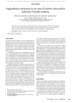
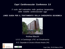
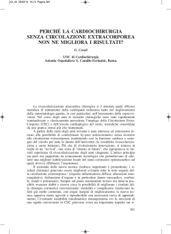
© Copyright 2026 Paperzz