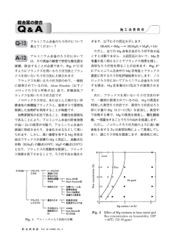
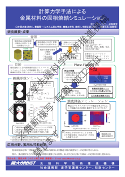
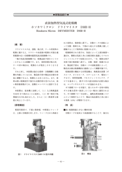
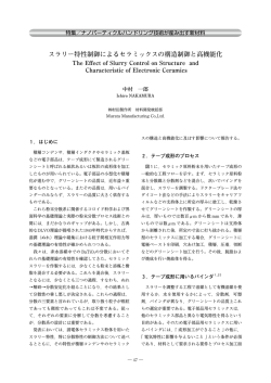
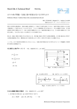
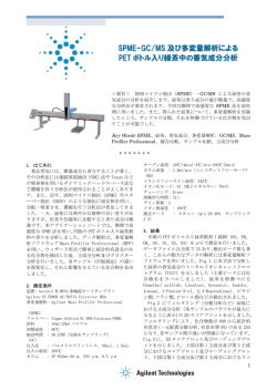
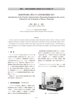
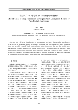
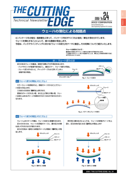
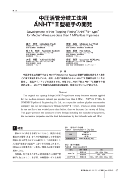
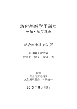
76 日本小児放射線学会雑誌
特集 腹部の最新・機能画像
1 . 腹腔鏡下大腸癌手術のための 3D−CT angiography
松木 充,村上卓道
近畿大学医学部放射線医学教室 放射線診断学部門
3D CT angiography for laparoscopic colorectal surgery
Mitsuru Matsuki, Takamichi Murakami
Department of Radiology, Kinki University Faculty of Medicine
Abstract
Laparoscopic surgery has gained clinical acceptance as a minimally invasive technique for
colorectal cancer. However, it takes a long time to resect a tumor and dissect lymph nodes, and
identify and ligate the proper vessels under laparoscopic guidance. Moreover, vessels and organs
can be injured during lymph node dissection and vessel ligation under laparoscopic guidance.
Therefore, we utilized 3D CT angiography for preoperative assessment of vascular anatomy
before laparoscopic surgery. And, USPIO, a lymphographic contrast medium of iron-containing
nanoparticles, has been shown to be useful in the characterization of lymph nodes. Commonly,
systematic mesocolonic lymph node dissection is performed according to the regional staging of
colon cancer. However, the 3D-CT angiography fusing nodes, which were diagnosed as metastases on USPIO-enhanced MRI, may enable us to perform individual lymph node dissection.
Keywords:3D-CT angiography, Laparoscopic surgery, Colorectal cancer, Ultrasmall superparamagnetic iron oxide particles(USPIO)
はじめに
す危険性がある.よって,われわれはバリエーショ
ンに富む大腸に関与する動静脈を非侵襲的に描出
腹腔鏡下手術は,内視鏡による拡大視効果に
する 3D-CT angiography を大腸癌の術前マッピン
よって細かい作業が可能になり,小さな術創のた
グとして活用し,これを Virtual CT laparoscopy と
め術後疼痛や運動制限を軽減し,美容上も優れて
呼んでいる1,2).本稿では,Virtual CT laparoscopy
いるといった利点を有する.さらに,病変部以外
の活用の実際について述べる.
の腸管露出がほとんどないことも加わって,腸蠕
動が術後早期に回復し,経口摂取も早く開始でき,
腹腔鏡下大腸癌手術の適応と術式
癒着のリスクも減少する.これらによって,入院
腹腔鏡下大腸癌手術は,早期癌では,内視鏡的
期間の短縮と早期の社会復帰といった大きな恩恵
粘膜切除術(EMR)適応外の病変を適応とし,全
がもたらされた.しかし一方,腹腔鏡下の操作の
部位で施行可能である.進行癌に対しては適切な
ため術野全体を捉えることが困難で,直接臓器に
手技や創部再発,長期予後の問題があるため,施
触ることができないといった欠点を有し,解剖学
設により適応が異なっている.われわれの施設で
的誤認による血管,周囲臓器への損傷を引き起こ
は減圧不能な腸閉塞例,高度他臓器浸潤例,巨大
4
Vol.29 No.2, 2013 77
腫瘍例などを除いた進行癌に対しても腹腔鏡下手
と venography,urography を 融 合 し,multiphase
術を施行している.リンパ節郭清は,EMR 困難な
fusion 画像を得る1,2).対応すべき血管である上腸
M 癌では 1 群,SM 癌では 2 群,進行癌では 3 群リ
間膜動脈,下腸間膜動脈およびそれらの分枝には
ンパ節郭清を原則としている.
バリエーションが多く,同時に上腸間膜静脈,下
腸間膜静脈,性腺静脈,尿管との多彩な位置関係
CT 撮影方法
が腹腔鏡下での動脈根部,静脈の処理,リンパ節
前処置として,腫瘍部位のマーキングのために
郭清を困難なものとする.よって,術前に 3D-CT
手術前に施行される大腸内視鏡検査の直後に,大
angiography によって大腸に関与する動脈,静脈,
腸全体に適度に空気が送気された状態で撮影す
尿管の位置関係を知ることは,安全かつ迅速な手
る.使用装置として 64 列検出器マルチスライス
術の遂行に有用と考えられた.
CT(Aquilion64)を用い,管電圧 120 kV,管電流
300 mA のもと撮影条件は,0.5 秒ローテーション,
Virtual laparoscopy 活用法の実際
コリメーション 0.5 ㎜,ヘリカルピッチ 53(ビーム
領域別の大腸癌に対する Virtual laparoscopy の
ピッチ 0.828),再構成間隔 1 ㎜とする.造影方法
活用を述べる.
は,非イオン性造影剤 300 mgI/㎖を用い,総量体
重(㎏)×2 ~ 2.5 ㎖を自動注入器にて 5 ㎖/秒で急
1)盲腸癌,上行結腸癌に対して
速注入する.その後,Real Prep 法を用いて肝上縁
進行癌の 3 群リンパ節郭清では surgical trunk に
から恥骨結合まで動脈相(造影約 20 秒後)を撮影
沿って回結腸動脈根部から中結腸動脈根部までの
し,造影 50 秒後より恥骨結合から肝上縁に向かっ
リンパ節を郭清して血管を処理する必要がある.
て静脈相を撮影し
(Go and Return)
,S 状結腸,直
その郭清時に注意すべきポイントとして,a)上腸
腸癌症例に対しては造影 5 分後の排泄相を追加す
間膜動脈から直接分岐する右結腸動脈の有無と
る.これによって得られた 3D-CT arteriography
b)回結腸動脈と上腸間膜静脈の位置関係がある.
MCA-rt
MCA-rt
MCA
MCA
RCA
ICA
Tumor
Tumor
ICA
a b
Fig.1 3D−CT arteriography
a:3D−CT arteriography shows no right colic artery(RCA)directly branching from
the superior mesenteric artery(SMA)
. Therefore, in right hemicolectomy, the
origins of the iliocolic artery(ICA)and right branch of middle colic artery
(MCA−rt)are ligated.
b:3D−CT arteriography shows RCA directly branching from the SMA. Therefore,
in right hemicolectomy, it is necessary to carefully isolate and ligate the proximal portion of RCA during the dissection of the intermediate lymph nodes(N2)
nodes along the SMA.
5
78 日本小児放射線学会雑誌
上腸間膜動脈から直接分岐する右結腸動脈を認め
理の際,その背側を走行する回結腸静脈枝や上腸
ない場合(Fig.1a)は,回結腸動脈根部を郭清した
間膜静脈本幹の損傷に注意して正確に回結腸動脈
後,上腸間膜動脈に沿って中結腸動脈根部まで迅
根部を処理する必要がある.しかし,回結腸動
速に郭清を進めることができる.しかし上腸間膜
脈が上腸間膜静脈の背側を走行する場合(type B)
動脈より直接分岐する右結腸動脈を認める場合
(Fig.3)
,上腸間膜静脈背側の回結腸動脈に沿った
(Fig.1b),回結腸動脈,右結腸動脈分岐部を同定,
綿密なリンパ節郭清が必要となる.3D-CT arte-
切離した後,中結腸動脈根部まで en bloc に郭清
riography,multiphase fusion 画像は,上腸間膜動
する.また回結腸動脈が上腸間膜静脈の腹側を走
脈から直接分岐する右結腸動脈の有無,回結腸動
行する場合(type A)
(Fig.2)
,回結腸動脈根部の処
脈と上腸間膜静脈の位置関係の把握に有用である.
ARCV
GCT
MCA
ICA
MCA-rt
ICV
ICA
ICV
SMV
a b
Fig.2 Multiphase fusion image(a)and intraoperative view(b)
a:Multiphase fusion image shows clearly the iliocolic artery(ICA)running ventral to the superior mesenteric vein(SMV[type A]
)
.
b:In a reference to the multiphase fusion image, it is necessary to carefully
isolate and ligate the proximal side of ICA without injuring the iliocolic vein
(ICV)and SMV dorsal to ICA.
MCA : middle colic artery, MCA-rt : right branch of the middle colic artery,
GCT : gastrocolic trunk of Henle, ARCV : accessory right colic vein
MCA
ICA
ICV
Tumor
6
Fig.3
Multiphase fusion image
Multiphase fusion image shows clearly
the ICA running dorsal to the SMV
(type B)
. Therefore, the isolation of ICA
requires meticulous dissection of the
intermediate lymph nodes(N2)along
the ICA dorsal to SMV.
ICV : Iliocolic vein, MCA : middle colic
artery
Vol.29 No.2, 2013 79
2)
横行結腸癌に対して
S 状結腸動脈の本数,分岐パターンにはバリエー
左側結腸曲進行癌は通常中結腸動脈左枝より栄
ションがあり,左結腸動脈より分岐,左結腸動脈と
養を受けているため,左半結腸切除に際し中結腸
同時に下腸間膜動脈より分岐,上直腸動脈より分
動脈あるいは中結腸動脈左枝を結紮,処理する.
6)
岐することがある
(Fig.5)
.例えば S 状結腸進行癌
しかし,まれに副左結腸動脈によって支配され
3 群リンパ節郭清症例(Fig.6)で,第 1S 状結腸動脈
ている場合,副左結腸動脈のみを処理することに
が左結腸動脈(LCA)から分岐し,腫瘍を支配して
よって中結腸動脈を温存することができる.副左
いることを術前に把握することができれば,中枢側
結腸動脈は,中結腸動脈分岐部より中枢側の上腸
リンパ節郭清を下腸間膜動脈根部より第 1S 状結腸
間膜動脈より分岐し,脾彎曲部の結腸に流入する
動脈まで en bloc に行い,第 1S 状結腸動脈根部の
動脈で,その頻度は欧米で 4 ~ 8%,本邦で 33 ~
みを処理し,左結腸動脈,上直腸動脈を温存する
.3D-CT arteriographyは,
3~5)
49%といわれている
副左結腸動脈の同定に有用である
(Fig.4)
.
ことができる.さらに下腸間膜静脈を処理する際,
近傍を走行する左結腸動脈を損傷させないよう注
意する必要がある(Fig.7)
.また S 状結腸動脈根部
3)S 状結腸~直腸進行癌に対して
処理の際,近傍を走行する尿管を(Fig.8)
,また
S 状結腸~直腸進行癌の 3 群リンパ節郭清に対
下腸間膜動脈に沿った2群リンパ節を郭清する際,
し,本邦では S 状結腸が長い人が多く,正常腸管
近傍を走行する性腺静脈を損傷させないように注
を長く温存しつつ残存腸管や吻合部への血流を維
意する必要がある(Fig.9)
.3D-CT arteriography
持して縫合不全を予防するため,われわれは左結
は腫瘍の支配血管,S 状結腸動脈の分岐パターン
腸動脈などを温存したリンパ節郭清を行っている.
を明瞭に描出し,multiphase fusion 画像は下腸間
膜動脈,その主要分枝と下腸間膜静脈,性腺静脈,
尿管との位置関係を明瞭に描出する.
Tumor
MCA-lt
将来展望について
大腸癌のリンパ節転移に関するCT でのサイズ診
断として①長径 1~1.5 ㎝以上のリンパ節,② 1 ㎝
LCA
accessory LCA
以下でも 3 個以上集合したものなどが挙げられる.
また形態診断として辺縁の不明瞭なリンパ節,短
MCA
Fig.4 3D−CT arteriography
3D−CT arteriography shows clearly an
accessory left colic artery(accessory
LCA)
, which branches from the SMA and
runs toward the splenic flexure along the
lower border of the pancreatic body, feeding the left transverse colon cancer. With
reference to the 3D−CT arteriography,
it is necessary to isolate and ligate the
accessory LCA with preservation of the
MCA.
MCA−lt : left branch of the middle colic
artery
SA
SA
SA
SA
SA
SA
SA
SA
Fig.5 Variation of origin and branches of the
inferior mesenteric artery
7
80 日本小児放射線学会雑誌
Tumor
LCA
IMA
LCA
S1
S1
SRA
SRA
IMA
a b
Fig.6 3D−CT arteriography(a)and intraoperative view(b)
a:3D−CT arteriography shows 1st sigmoid colon artery(S1)branching from
the LCA and feeding a sigmoid colon cancer.
b:With reference to the 3D−CT arteriography, it is necessary to isolate and
ligate accurately the proximal side of the S1, and preserve the LCA and
superior rectal artery(SRA)
.
IMA : Inferior mesenteric artery
LCA
SRA
IMV
S1
Fig.7
Multiphase fusion image
Multiphase fusion image shows clearly the IMV running
near the origin of the LCA. With reference to the multiphase fusion image, it is necessary to isolate and ligate the
superior mesenteric vein without injuring the LCA.
Tumor
LCA
Ureter
S1
S2
8
Fig.8
Multiphase fusion image
Multiphase fusion image shows clearly the ureter running near the origin
of the S1. With reference to the multiphase fusion image, it is necessary
to isolate and ligate the S1 without
injury to the ureter.
S2 : 2nd sigmoid artery
Vol.29 No.2, 2013 81
LCA
gonadal
vein
Lymph node
Macrophage
S1
Tumor
Fig.9 Multiphase fusion image
Multiphase fusion image shows clearly
the anatomical relationship between the
SMA and gonadal vein. With reference
to the multiphase fusion image, it is necessary to dissect safely the lymph nodes
along the SMA without injury to the gonadal vein.
径 / 長径比の大きいものを転移として考える.し
かし,これらの診断基準では感度はせいぜい約
45%と決して満足できるものではない.そこで
Fillippone ら7)は,①長径 1 ㎝以上の結腸辺縁,結
腸傍リンパ節あるいは 1 ㎝以下でも 3 個以上集合
USPIO
Fig.10 Lymph node accumulation mechanisms
of ultrasmall superparamagnetic iron oxide particles(USPIO)
After intravenous injection of USPIO, the
nanoparticles circulate systemically and
access the interstitium. These nanoparticles in the interstitium are phagocytized
by components of the reticuloendothelial
system such as macrophages or histiocytes, and are drained through lymphatic vessels to the lymph nodes. These
iron-containing nanoparticles accumulate in normal lymph nodes, enhancing
susceptibility effects and reducing the
nodal T2* signal. Conversely, metastatic
lymph nodes lack reticuloendothelial activity, and are thus unable to take up the
iron-containing nanoparticles that cause
local field heterogeneity.
した結腸辺縁,結腸傍リンパ節を認めた場合 N1,
② 3 個以上の内臓辺縁リンパ節を認めた場合 N2,
③長径 1 ㎝以上の後腹膜リンパ節を認めた場合 N3
よって貪食能が低下したリンパ節は信号の低下が
と診断すると成績が向上すると報告しているが,
乏しく,リンパ節の質的診断に非常に有用である
サイズあるいは形態評価での診断能には限界があ
(Fig.11)
.USPIO を用いた精度の高いリンパ節転
る.現在,欧米では MRI のリンパ節特異性造影剤
移の情報が得られれば,3D-CT angiography に転
である USPIO(ultra-small superparamagnetic iron
移リンパ節の情報を付加することによって,個々
oxide)の有用性が報告されている8).USPIO は,
の症例に合ったテーラーメードのリンパ節郭清も
低分子量のデキストランで被覆された酸化鉄製剤
可能になり
(Virtual CT laparoscopy with individual
で平均粒子径は 30 nmより小さく,静脈内投与する
lymph nodes dissection)
(Fig.12),より低侵襲な
と USPIO は血管壁から間質に漏出され,その漏出
腹腔鏡下手術に寄与するものとして期待される.
した USPIO をマクロファージが貪食し,リンパ節
に運ぶことによってリンパ節の網内系に広く分布
する(Fig.10).取り込まれたリンパ節は,USPIO
Virtual CT laparoscopy は,腹腔鏡下大腸癌手術
によって T2* 強調画像で信号が低下し,転移に
を安全かつ迅速に遂行する上で必要不可欠な情報
8)
おわりに
9
82 日本小児放射線学会雑誌
Fig.11 USPIO−enhanced MRI in the patient with descending colon cancer
a:Precontrast T2* weighted image shows enlarged paracolic lymph node(arrow).
b:On postcontrast T2* weighted image, paracolic lymph node(arrow)shows high
signal, which reflects metastatic node. Paraaortic lymph nodes show complete
darkening, which reflects nonmetastatic nodes.
LCA
SRA
S1
Fig.12 Virtual CT laparopscopy with individual lymph node dissection
Commonly, systematic mesocolonic lymph node dissection is
performed up to the root of the IMA in patients with advanced
sigmoid colon cancer. However, USPIO-enhanced MRI shows
lymph node metastases at the paracolic region and along the
S1. Using the 3D-CT angiography fusing metastatic nodes,
surgeon can perform individual lymph node dissection.
10
a b
Vol.29 No.2, 2013 83
となっている.今後,リンパ節特異性造影剤を用
いた MRI による精度の高いリンパ節診断が Virtual
CT laparoscopy に付加されるとテーラーメードの
リンパ節郭清も可能になり,より低侵襲な腹腔鏡
下手術に寄与するものとして期待される.
●文献
1) Matsuki M, Okuda J, Kanazawa S, et al : Virtual
CT colectomy by three-dimensional imaging using
multidetector-row CT for laparoscopic colorectal
surgery. Abdominal imaging 2005 ; 30 : 698 - 708.
2) Kanamoto T, Matsuki M, Okuda J, et al : Preoperative evaluation of local invasion, metastatic lymph
nodes of colorectal cancer and mesenteric vascular
variations using MDCT before laparoscopic surgery. J Comput Assist Tomogr 2007 ; 31 : 831 - 839.
3) 矢田裕一, 沢井清司, 大原都桂, 他 : 動脈の分岐
走行とリンパ節転移状況からみた結腸癌の部位別
D2 郭清術.日消外会誌 1996 ; 29 : 710 - 716.
4) 齊藤修治, 衣笠祐介, 塩見明生, 他 : 副中結腸動脈
周囲リンパ節郭清を要する脾彎曲部横行結腸癌に
対する腹腔鏡下手術. 手術 2009 ; 63 : 1691 - 1695.
5) Rusu MC, Vlad M, Voinea LM, et al : Detailed anatomy of a left accessory aberrant colic artery. Surg
Radiol Anat 2008 ; 30 : 595 - 599.
6) Zebrowski W, Augustyniak E, Zajac S, et al : Variation of origin and branches of the inferior mesenteric arter y and its anastomoses. Folia Morphol
1971 ; 30 : 510 - 517.
7) Filippone A, Ambrosini R, Fuschi M, et al : Preoperative T and N staging of colorectal cancer : accuracy of contrast-enhanced multi-detector row CT
colonography-initial experience. Radiology 2004 ;
231 : 83 - 90.
8) Harinsinghani MG, Barentsz J, Hahn PF, et al :
Noninvasive detection of clinically occult lymphnode metastases in prostate cancer. N Eng J Med
2003 ; 348 : 2491 - 2499.
11
© Copyright 2026 Paperzz