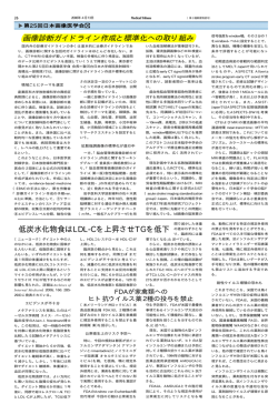
Longitudinal Changes of Resting-State Functional Connectivity During Motor
Recovery After Stroke
Chang-hyun Park, Won Hyuk Chang, Suk Hoon Ohn, Sung Tae Kim, Oh Young
Bang, Alvaro Pascual-Leone and Yun-Hee Kim
Stroke 2011, 42:1357-1362: originally published online March 24, 2011
doi: 10.1161/STROKEAHA.110.596155
Stroke is published by the American Heart Association. 7272 Greenville Avenue, Dallas, TX 72514
Copyright © 2011 American Heart Association. All rights reserved. Print ISSN: 0039-2499. Online
ISSN: 1524-4628
The online version of this article, along with updated information and services, is
located on the World Wide Web at:
http://stroke.ahajournals.org/content/42/5/1357
Data Supplement (unedited) at:
http://stroke.ahajournals.org/content/suppl/2011/05/02/STROKEAHA.110.596155.DC1.html
http://stroke.ahajournals.org/content/suppl/2012/02/28/STROKEAHA.110.596155.DC2.html
Subscriptions: Information about subscribing to Stroke is online at
http://stroke.ahajournals.org//subscriptions/
Permissions: Permissions & Rights Desk, Lippincott Williams & Wilkins, a division of Wolters
Kluwer Health, 351 West Camden Street, Baltimore, MD 21202-2436. Phone: 410-528-4050. Fax:
410-528-8550. E-mail:
[email protected]
Reprints: Information about reprints can be found online at
http://www.lww.com/reprints
Downloaded from http://stroke.ahajournals.org/ at Harvard University on April 17, 2012
Longitudinal Changes of Resting-State Functional
Connectivity During Motor Recovery After Stroke
Chang-hyun Park, PhD; Won Hyuk Chang, MD; Suk Hoon Ohn, MD; Sung Tae Kim, MD;
Oh Young Bang, MD, PhD; Alvaro Pascual-Leone, MD, PhD; Yun-Hee Kim, MD, PhD
Background and Purpose—Functional MRI (fMRI) studies could provide crucial information on the neural mechanisms
of motor recovery in patients with stroke. Resting-state fMRI is applicable to patients with stroke who are not capable
of proper performance of the motor task. In this study, we explored neural correlates of motor recovery in patients with
stroke by investigating longitudinal changes in resting-state functional connectivity of the ipsilesional primary motor
cortex (M1).
Methods—A longitudinal observational study using repeated fMRI experiments was conducted in 12 patients with stroke.
Resting-state fMRI data were acquired 4 times over a period of 6 months. Patients participated in the first session of
fMRI shortly after onset and thereafter in subsequent sessions at 1, 3, and 6 months after onset. Resting-state functional
connectivity of the ipsilesional M1 was assessed and compared with that of healthy subjects.
Results—Compared with healthy subjects, patients demonstrated higher functional connectivity with the ipsilesional
frontal and parietal cortices, bilateral thalamus, and cerebellum. Instead, functional connectivity with the contralesional
M1 and occipital cortex were decreased in patients with stroke. Functional connectivity between the ipsilesional and
contralesional M1 showed the most asymmetry at 1 month after onset to the ipsilesional side. Functional connectivity
of the ipsilesional M1 with the contralesional thalamus, supplementary motor area, and middle frontal gyrus at onset was
positively correlated with motor recovery at 6 months after stroke.
Conclusions—Resting-state fMRI elicited distinctive but comparable results with previous task-based fMRI, presenting
complementary and practical values for use in the study of patients with stroke. (Stroke. 2011;42:1357-1362.)
Key Words: functional connectivity 䡲 motor recovery 䡲 resting-state fMRI 䡲 stroke
F
unctional MRI (fMRI) has played an integral role in
defining the neural substrates and mechanisms underlying recovery after brain disease such as stroke at the system
level of the brain. Cortical reorganization has been characterized by observation of changes in brain activation during
motor recovery after stroke.1– 6 fMRI studies using motor
activation tasks have been conducted for investigation of the
effects of specific therapeutic interventions, including
constraint-induced movement therapy,7 treadmill training,8
and repetitive transcranial magnetic stimulation9; these studies focused on recovery mechanisms associated with these
interventions.
On the other hand, longitudinal studies have been conducted
for assessment of changes in brain activation that are related to
recovery after stroke. The initial contralesional shift of activation
and evolution to later ipsilesional activation,1,2 recruitment of
additional regions that are not activated in healthy subjects,10 and
importance of ipsilesional surviving regions11 during motor
recovery have been demonstrated using task-based fMRI. However, these reports showed certain variability in brain activation
results; one reason for this diversity originated from use of
diverse activation paradigms, which prevent adequate comparison between results, although passive movement4 and motor
imagery5 have been proposed as alternative methods. In addition, longitudinal studies using task-based fMRI are limited in
their application for patients with stroke with severe impairment,
and results may be confounded by changes in performance
during recovery as well.
Resting-state fMRI is a recently evolving method from
which functional connectivity between distant brain regions
is extracted based on low-frequency fluctuations. Although
the meaning of the resting-state fMRI signal has been debated
since its initial trial,12 evidence has suggested that resting
fluctuations correspond to neuronal activation during task
Received July 12, 2010; final revision received November 24, 2010; accepted November 30, 2010.
From the Samsung Biomedical Research Institute (C.-H.P.), Samsung Medical Center, Seoul, Korea; the Departments of Physical and Rehabilitation
Medicine (W.H.C., Y.-H.K.), Diagnostic Radiology and Imaging Science (S.T.K.), and Neurology (O.Y.B.), Samsung Medical Center, Sungkyunkwan
University School of Medicine, Seoul, Korea; the Department of Physical Medicine and Rehabilitation (S.H.O.), Hallym University College of Medicine,
Seoul, Korea; and the Berenson-Allen Center for Noninvasive Brain Stimulation (A.P.-L.), Beth Israel Deaconess Medical Center, Boston, MA.
The online-only Data Supplement is available at http://stroke.ahajournals.org/cgi/content/full/STROKEAHA.110.596155/DC1.
Correspondence to Yun-Hee Kim, MD, PhD, Professor and Chairperson, Department of Physical and Rehabilitation Medicine, Stroke and
Cerebrovascular Center, Samsung Medical Center, Sungkyunkwan University School of Medicine, 50 Irwon-dong, Gangnam-gu, Seoul, 135-710,
Republic of Korea. E-mail [email protected]; [email protected]
© 2011 American Heart Association, Inc.
Stroke is available at http://stroke.ahajournals.org
DOI: 10.1161/STROKEAHA.110.596155
Downloaded from http://stroke.ahajournals.org/
1357 at Harvard University on April 17, 2012
1358
Stroke
May 2011
performance.13 The methodological advantage of resting state
is that it can be performed without an overt task or external
input; therefore, it is applicable to unconscious patients,
infants,14 and even to experimental animals.15
In healthy subjects, resting-state fMRI has shown remarkable consistency in functional connectivity16,17; however,
significant differences were observed within the aged population18 or after interventions such as acupuncture.19 Restingstate fMRI has demonstrated unique changes in patients with
various neurological disorders, including Alzheimer disease,20 attention deficit hyperactivity disorder,21 depression,22
and schizophrenia.23
For patients with stroke with severe motor impairment who
could not perform the fMRI activation task at the early stage of
onset, it is expected to be achieved through long-term follow-up
by use of resting-state fMRI. Therefore, in this study, we aimed
to carry out long-term follow-up of resting-state fMRI in patients
with stroke for delineation of the neural substrates of motor
recovery after stroke. We analyzed functional connectivity of the
ipsilesional primary motor cortex (M1) in patients with stroke
and compared it with that of healthy subjects. To propose a
plausible underlying mechanism for successful stroke recovery,
we also investigated neural correlates associated with long-term
motor recovery at 6 months after stroke.
Methods
Subjects
A total of 51 patients who had their first-ever stroke were assessed
for their eligibility. Inclusion criteria were as follows: (1) ⬍2 weeks
from the onset of ischemic stroke; (2) unilateral supratentorial
lesions; (3) moderate to severe motor deficits of the contralesional
upper and lower extremities; and (4) age ⬎18 years and ⬍75 years.
Exclusion criteria were as follows: (1) any clinically significant or
unstable medical disorder; (2) any neuropsychiatric comorbidity
other than stroke; and (3) any contraindication to MRI. Twenty-five
patients out of 51 were excluded and 26 patients were enrolled in this
study. Fourteen patients dropped out during the follow-up period.
Finally, 12 patients with ischemic stroke (5 males and 7 females,
58.4⫾6.9 years) with supratentorial lesions completed longitudinal
fMRI experiments, and their image data were included in the
analysis (Figure 1; Table 1). Also, 11 healthy subjects (3 males and
8 females, 52.1⫾9.4 years) who reported no history of psychiatric or
neurological problems were included as an age-matched control
group. Experiments were conducted with the understanding and
written consent of each participant, and ethics approval was provided
by the Institutional Review Board.
Experimental Design
This study was designed as a longitudinal observational study for
conduct of repeated fMRI experiments. A cross-sectional controlled
study design was also applied for comparison of data from patients
with stroke with those of healthy subjects.
fMRI Data Acquisition
Resting-state fMRI data were longitudinally acquired 4 times over a
period of 6 months in patients with stroke. Patients participated in the
first session of fMRI shortly after onset (10.5⫾4.3 days) and
thereafter in subsequent sessions at 1, 3, and 6 months after onset. In
healthy subjects, we obtained one time resting-state fMRI data.
During the resting state, subjects were instructed to keep their eyes
closed and to remain motionless. fMRI data were acquired using a
Philips ACHIEVA MR scanner (Philips Medical Systems, Best, The
Netherlands) operating at 3 T. At each session, a total of 100
whole-brain images was collected using a T2*-weighted gradient-
Figure 1. Patient enrollment process for a longitudinal observational study conducting repeated functional MRI (fMRI) experiments. A total of 51 patients with first-ever stroke were
assessed for their eligibility. Twenty-five patients were excluded
and 14 patients dropped out during the follow-up fMRI experiments. Finally, 12 patients with ischemic stroke completed longitudinal fMRI experiments. Acquisition of resting-state fMRI
data, accompanied by behavioral assessment using Fugl-Meyer
assessment, was performed within 2 weeks after onset and then
at 1, 3, and 6 months after onset.
echo echoplanar imaging sequence (repetition time⫽3000 ms, echo
time⫽35 ms, number of slices⫽35, slice thickness⫽4 mm, matrix
size⫽128⫻128, field of view⫽220⫻220 mm).
Behavioral Assessment
Degree of motor impairment was scored using the Fugl-Meyer
assessment for upper and lower extremities24 on the same day as
fMRI data acquisition.
fMRI Data Analysis
fMRI data were preprocessed using SPM8 (Wellcome Trust Centre
for Neuroimaging, University College London, London, UK) and
AFNI (Scientific and Statistical Computing Core, National Institute of
Mental Health, Bethesda, MD) software. Preprocessing steps included
spatial realignment to the mean volume of a series of images, normalization into the same coordinate frame as the MNI template brain,
band-pass filtering between 0.01 and 0.08 Hz, and smoothing using a
Gaussian filter of 8 mm full width at half maximum.
Correlation analysis between the reference time course of the M1
and the time course of every voxel in the brain was performed for
acquisition of a map of correlation coefficients that revealed functional connectivity of the M1. The reference time course was
extracted from the ipsilesional M1 in patients with stroke and the left
M1 in healthy subjects. M1 was defined to include voxels covering
approximately the caudal half of the precentral gyrus along the
anterior wall of the central sulcus. Correction of time courses was
made by regressing out the time courses that corresponded to head
motions and global fluctuations.
A map of correlation coefficients was converted to a map of
Gaussian distributed values through Fisher z-transformation defined
by z⫽tanh⫺1 r or z⫽(1/2)ln[(1⫹r)/(1⫺r)], where r is a correlation
coefficient, z is an approximately Gaussian distributed value, tanh⫺1
is the inverse hyperbolic tangent function, and ln is the natural
logarithm function.25 The lesion side of the correlation map was set
Downloaded from http://stroke.ahajournals.org/ at Harvard University on April 17, 2012
Park et al
Table 1.
Resting-State fMRI in Stroke Recovery
1359
Patient Characteristics and Motor Function
FMA Scores
Patient No.
Gender
Age, Years
Lesion
Onset
1 Month
3 Months
6 Months
FMA Change
1
2
3
4
5
6
7
8
9
10
11
12
Mean⫾SD
F
F
F
M
F
F
M
M
M
F
M
F
M⫽5; F⫽7
66
61
55
74
58
47
55
62
59
52
57
55
58.4⫾6.9
L MCA infarction
L MCA infarction
R MCA infarction
L CR infarction
L MCA infarction
L MCA infarction
L ACA infarction
L MCA infarction
R MCA infarction
R CR infarction
L MCA infarction
R SC infarction
8
20
30
16
36
44
19
19
24
52
13
9
24.2⫾13.8
8
22
55
22
42
59
42
22
24
52
13
9
30.8⫾18.3
19
27
70
17
52
100
60
52
24
99
52
34
50.5⫾28.5
27
33
73
21
52
100
73
57
24
99
52
34
53.8⫾27.7
19
13
43
5
16
56
54
38
0
47
39
25
29.6⫾19.1
FMA indicates Fugl-Meyer assessment; F, female; M, male; L, left; R, right; MCA, middle cerebral artery; CR, corona radiata; ACA,
anterior cerebral artery; SC, striatocapsular; FMA change, FMA total scores at 6 months⫺FMA total scores at onset.
to the left side by flipping the map from right to left about the
midsagittal line for patients with lesions on the right side.
Fisher z-transformed and flipped correlation maps were used for
random-effects analysis. Two-sample t tests were performed to find
areas that showed significant differences in functional connectivity
between patients and healthy subjects. Also, to search for brain
regions correlated with motor improvement, correlation maps of
patients at onset were regressed with increases in the Fugl-Meyer
assessment score at 6 months after stroke. We determined the
significance using height (uncorrected P⬍0.001 at the voxel level)
and extent (uncorrected P⬍0.05 at the cluster level) thresholds.
6 months. Figure 2B shows comparisons of connectivity between patients with stroke and healthy subjects at 4 time points.
Significant differences of connectivity in the SMN are summarized (Supplemental Table I, http://stroke.ahajournals.org). Pa-
Lateralization Index
As a quantitative measure of functional connectivity, the lateralization index (LI) was calculated for each correlation map. The LI was
introduced for the purpose of providing a specific description of the
asymmetry of functional connectivity between the ipsilesional and
contralesional M1 according to the following definition: (number of
connected voxels in the ipsilesional M1/total number of voxels in the
ipsilesional M1)⫺(number of connected voxels in the contralesional
M1/total number of voxels in the contralesional M1). If functional
connectivity of the ipsilesional M1 with any voxel had a value ⬎95th
percentile of the Gaussian distribution when considering all Gaussian
distributed values in a map, the voxel was determined to be
connected. This approach yielded LIs that ranged between ⫺1 and 1,
in which ⫺1 referred to contralesional connectivity only, 1 ipsilesional connectivity only, and values close to 0 referred to symmetrical connectivity. The LI of patients was assessed at each time point
and compared with that of healthy subjects.
Results
Differences in Connectivity Between Patients and
Healthy Subjects
Correlation analysis of data acquired from 11 healthy subjects
demonstrated the discrete network, namely sensorimotor network (SMN), which is displayed in Figure 2A. SMN of healthy
subjects included motor–sensory-related regions such as the
primary sensorimotor cortex, premotor cortex, supplementary
motor area (SMA), cingulate motor area, secondary somatosensory cortex, cerebellum, basal ganglia, thalamus, frontal and
parietal cortices, and striate and extrastriate cortices. SMN in
patients with stroke showed asymmetrical involvement, and
other regions were additionally included throughout a period of
Figure 2. A, Sensorimotor networks acquired by resting-state
functional connectivity of the ipsilesional primary motor cortex in
healthy subjects. B, Significant differences in resting-state functional connectivity between patients and healthy subjects over 4
time points of onset (B1), 1 month (B2), 3 months (B3), and 6
months (B4) after onset. Red–yellow blobs and blue– green
blobs indicate increased and decreased functional connectivity
in patients compared with healthy subjects, respectively. The
left side of the brain is the ipsilesional hemisphere. SMC indicates sensorimotor cortex; SMA, supplementary motor area;
PPC, posterior parietal cortex; OC, occipital cortex; Cbll, cerebellum; MFG, middle frontal gyrus; Th, thalamus.
Downloaded from http://stroke.ahajournals.org/ at Harvard University on April 17, 2012
1360
Stroke
May 2011
of patients was larger at onset and even larger at 1 month after
onset compared with that of healthy subjects. At 3 months
and 6 months after onset, the LI of patients had decreased so
that it did not differ significantly from that of healthy
subjects. Corresponding maps of functional connectivity also
showed that asymmetry of functional connectivity between
ipsilesional and contralesional M1 increased until 1 month
after onset and then decreased.
Correlation of Connectivity at Onset With Later
Motor Improvement
Figure 3. Time-dependent changes in resting-state functional
connectivity. Quantitative changes were exhibited by the lateralization index (LI) and corresponding maps of functional connectivity were also displayed. The LI was compared between
patients and healthy subjects over 4 time points, including
onset, 1 month, 3 months, and 6 months after onset. In the
graph of the LI, points represent means, error bars represent
SDs, and stars represent significant differences between
patients and healthy subjects at a threshold of P⬍0.05.
tients with stroke displayed decreased connectivity of the ipsilesional M1 with the sensorimotor cortex, occipital cortex, middle
frontal gyrus (MFG), and posterior parietal cortex since onset.
On the other hand, patients with stroke showed increased
connectivity of the ipsilesional M1 with the cerebellum, thalamus, MFG, and posterior parietal cortex since onset. In particular, decreased connectivity with the sensorimotor cortex and
increased connectivity with the cerebellum persisted throughout
a period of 6 months after onset. In general, it is conceivable that
connectivity of the ipsilesional M1 increased within ipsilesional
brain regions, whereas it decreased within contralesional brain
regions.
Time-Dependent Changes in Connectivity
Figure 3 shows time-dependent changes in the LI together
with corresponding maps of functional connectivity. The LI
Figure 4 shows brain regions in which functional connectivity at
onset was positively correlated with later motor improvement, as
measured by increases in the Fugl-Meyer assessment score at 6
months after onset. Brain areas demonstrating significant correlation with Fugl-Meyer assessment changes are summarized in
Table 2. Connectivity of the ipsilesional M1 with the contralesional thalamus, SMA, and MFG showed positive correlation
with later motor improvement. R2 statistics were 0.8400, 0.7821,
and 0.7111 for the thalamus, SMA, and MFG, respectively, in
linear regression analysis or partial correlation coefficients were
0.8998, 0.8822, and 0.8311 for the thalamus, SMA, and MFG,
respectively, in partial correlation analysis with control of
Fugl-Meyer assessment scores at onset.
Discussion
In the current study, we investigated (1) differences in
resting-state functional connectivity between patients and
healthy subjects during the period after stroke; and (2) a
prognostic value of initial resting-state functional connectivity for assessment of later motor improvement. Our results
demonstrated characteristic asymmetry of resting-state functional connectivity of the ipsilesional M1 in patients with
stroke, which lasted until 6 months after onset. Connectivity
with subcortical SMN areas such as the cerebellum and
thalamus increased at the early stage of stroke. On the other
hand, connectivity with ipsilesional cortical areas increased
and connectivity with contralesional cortical areas decreased.
Preservation of connectivity with the contralesional thalamus,
SMA, and MFG at an early stage of stroke was meaningful
for later motor recovery in these patients.
If resting-state fMRI activity reflects neuronal baseline
activation, changes in resting-state connectivity may be
Figure 4. A, Significant positive correlations of patients’ resting-state functional
connectivity at onset with later motor
improvement, as indexed by changes in
the Fugl-Meyer assessment score for 6
months after onset. B, Linear regression
of functional connectivity in the thalamus
(B1), SMA (B2), and MFG (B3) on
increases in the Fugl-Meyer assessment
score. The goodness of fit for each linear regression was given by the R2 statistic. Th indicates thalamus; SMA, supplementary motor area; MFG, middle
frontal gyrus.
Downloaded from http://stroke.ahajournals.org/ at Harvard University on April 17, 2012
Park et al
Resting-State fMRI in Stroke Recovery
1361
Table 2. Cluster Maxima Showing a Significant Positive Correlation Between Patients’
Resting-State Functional Connectivity at Onset and Later Motor Improvement as Indexed by
Changes in the Fugl-Meyer Assessment Score for 6 Months After Onset
Peak MNI
Coordinates, mm
Brain Region
BA
Thalamus
Side
x
y
z
Voxel Count
Z-Score
P
C
8
⫺26
12
18
3.7726
0.0001
SMA
6
C
10
⫺6
54
15
3.5941
0.0002
MFG
48
C
34
16
26
16
3.1698
0.0008
MNI indicates Montreal Neurological Institute; BA, Brodmann area; SMA, supplementary motor area; MFG, middle
frontal gyrus; C, contralesional.
related to functional changes in the brain. Previous studies
using resting-state fMRI have demonstrated differences in the
default-mode network in Alzheimer disease20 and connectivity of the dorsal anterior cingulate cortex in attention deficit
hyperactivity disorder,26 implying pathophysiology of disease. Correspondence of the regions involved in the current
resting-state connectivity study with previous motor task
activation studies implies that stroke also influences restingstate connectivity in reference to functional impairment. In
previous task-based fMRI studies, activation of the contralesional sensorimotor cortex showed an initial increase and
then decreased or vanished in correspondence with functional
restoration of the perilesional cortex and the ipsilesional M1.2
In the current study, decreased connectivity between the
ipsilesional M1 and contralesional hemispheric cortex was
demonstrated after unilateral ischemic injury of the motor
network. This finding implies that breakdown of harmonious
interaction between two hemispheres at resting state may lead
to alteration of the activity of the contralesional hemisphere
in response to ipsilesional M1 activity.
Specifically, breakdown of harmonious interaction between
both M1 could be quantitatively characterized in terms of the LI.
Patients’ functional connectivity between the ipsilesional and
contralesional M1 was more highly lateralized to the ipsilesional
M1 at onset, compared with healthy subjects, and showed the
greatest asymmetry at 1 month after onset. Restoration of
relatively symmetrical connectivity since 3 months after onset
may be achieved after widespread reorganization in the sensorimotor system. That is, in the process of recovery after stroke,
increased asymmetry in functional connectivity between both
hemispheres in resting-state fMRI is considered to correspond to
rearrangements of activation over the bihemispheric sensorimotor system in task-based fMRI.
Changes in connectivity of the ipsilesional M1 with the
nonprimary SMN regions such as the frontal and parietal
cortices and occipital cortex were observed; these may reflect
plastic changes to compensate for impaired connectivity with the
contralesional hemisphere or response to disconnection of transcallosal inhibition. These findings coincide with previous taskbased fMRI studies that reported increased activation of the
frontoparietal cortex10 and other nonmotor brain areas such as
the occipital cortex6 in association with motor tasks in patients
with stroke. Changes in involvement of the cerebellum and
thalamus after stroke have also been demonstrated in previous
task-based fMRI studies of motor recovery.2,6,10 In particular,
activation of the cerebellum was correlated with later motor
recovery.27 Taken together, resting-state SMN connectivity appears to reflect abnormalities of motor network interaction after
stroke as well as plastic changes in response to motor network
impairment. In addition, these changes appear to have an
association with changes in brain activation provoked by performance of overt motor tasks.
In addition, regression analysis showed that preservation of
connectivity of the ipsilesional M1 with the contralesional
thalamus, SMA, and MFG at an early stage of stroke was
positively correlated with later motor improvement at 6 months
after stroke. The crucial role of the SMA in motor recovery has
been demonstrated in previous task-based fMRI studies of
patients with stroke in which early involvement of the SMA in
the process of stroke recovery2 and correlation of initial activation of the SMA with motor recovery28 were described. The
MFG is not regarded as a primary SMN region; however,
recruitment of the MFG may be helpful in reinforcement of the
management of cognitive load required for motor performance.10 In the case of the thalamus, despite its important
contribution to processing and relay of sensorimotor information, the role of the thalamus in recovery of motor function has
not yet been established. Strong recruitment of regions related to
sensory integration such as the thalamus at an early stage of
stroke, as shown in the current study, may suggest a beneficial
effect of sensory-related areas on later motor restoration in
patients with stroke. For detailed clarification of the role of those
regions, further investigation should be invited.
With a view that motor recovery corresponds to reorganization of surviving neuronal networks over the bihemispheric
sensorimotor system, overall patterns of use of neuronal
resources should be examined with respect to functional
specialization and integration. Results of the current study are
distinctive; however, they are comparable with those of
previous task-based fMRI studies by a plausible association
between resting-state connectivity and motor task activation.
Despite its novel results, the current study has some limitations in presenting results that cover various patterns of stroke
recovery. Due to a high dropout rate in long-term follow-up over
a period of 6 months, we only had final resting-state fMRI data
for 12 patients. Most dropouts were due to patients’ circumstances. Still, with resting-state fMRI, recruitment of different
subgroups of patients with uniform characteristics and careful
control during follow-up appear to be requirements for successful explanation of different stroke recovery patterns.
Another limitation is that, in the current study, we did not
specifically measure physiological noise such as cardiac and
Downloaded from http://stroke.ahajournals.org/ at Harvard University on April 17, 2012
1362
Stroke
May 2011
respiratory cycles. It has previously been proclaimed that cardiac29 and respiratory30 cycles can obscure detection of lowfrequency fluctuations in resting-state fMRI and, thus, induce
changes in resting-state connectivity, although resting-state connectivity cannot be explained by cardiorespiratory effects
alone.31 Therefore, investigation of resting-state connectivity
corrected for cardiorespiratory effects would provide us with
better information and is recommended for future study.
Conclusions
Stroke recovery might be time-dependent and affected according to task parameters. In this study, we attempted to
overcome these critical issues through longitudinal restingstate fMRI. Although the implications of resting-state fMRI
are still under dispute, systematic assessment of initial
resting-state functional connectivity may provide prognostic
insight for later motor recovery. In addition, practical values
of the resting-state fMRI study, free from a number of
confounds that are associated with task performances, may
enable thorough long-term follow-up in patients with severe
motor impairment at onset of stroke.
Sources of Funding
This study was supported by a Korean Science and Engineering
Foundation (KOSEF) grant funded by the Korean government
(MOST; No. M10644000022-06N4400-02210) and by a grant from
the Samsung Biomedical Research Institute (#SBRI C-A7-407-1).
A.P.L. was supported in part by Grant UL1 RR025758, Harvard
Clinical and Translational Science Center, from the National Center
for Research Resources and National Institutes of Health grant K 24
RR018875. The funders had no role in the design and conduct of the
study; in collection, management, analysis, and interpretation of the
data; or in preparation, review, or approval of the manuscript.
Disclosures
None.
References
1. Kim YH, You SH, Kwon YH, Hallett M, Kim JH, Jang SH. Longitudinal
fMRI study for locomotor recovery in patients with stroke. Neurology.
2006;67:330 –333.
2. Tombari D, Loubinoux I, Pariente J, Gerdelat A, Albucher JF, Tardy J,
Cassol E, Chollet F. A longitudinal fMRI study: in recovering and then in
clinically stable sub-cortical stroke patients. Neuroimage. 2004;23:
827– 839.
3. Loubinoux I, Dechaumont-Palacin S, Castel-Lacanal E, De Boissezon X,
Marque P, Pariente J, Albucher JF, Berry I, Chollet F. Prognostic value
of fMRI in recovery of hand function in subcortical stroke patients. Cereb
Cortex. 2007;17:2980 –2987.
4. Ward NS, Brown MM, Thompson AJ, Frackowiak RS. Longitudinal
changes in cerebral response to proprioceptive input in individual patients
after stroke: an fMRI study. Neurorehabil Neural Repair. 2006;20:
398 – 405.
5. Nair DG, Fuchs A, Burkart S, Steinberg FL, Kelso JA. Assessing
recovery in middle cerebral artery stroke using functional MRI. Brain Inj.
2005;19:1165–1176.
6. Ward NS, Brown MM, Thompson AJ, Frackowiak RS. Neural correlates
of motor recovery after stroke: a longitudinal fMRI study. Brain. 2003;
126:2476 –2496.
7. Sheng B, Lin M. A longitudinal study of functional magnetic resonance
imaging in upper-limb hemiplegia after stroke treated with constraintinduced movement therapy. Brain Inj. 2009;23:65–70.
8. Enzinger C, Dawes H, Johansen-Berg H, Wade D, Bogdanovic M, Collett
J, Guy C, Kischka U, Ropele S, Fazekas F, Matthews PM. Brain activity
changes associated with treadmill training after stroke. Stroke. 2009;40:
2460 –2467.
9. Nowak DA, Grefkes C, Dafotakis M, Eickhoff S, Kust J, Karbe H, Fink
GR. Effects of low-frequency repetitive transcranial magnetic stimulation
of the contralesional primary motor cortex on movement kinematics and
neural activity in subcortical stroke. Arch Neurol. 2008;65:741–747.
10. Puh U, Vovk A, Sevsek F, Suput D. Increased cognitive load during simple
and complex motor tasks in acute stage after stroke. Int J Psychophysiol.
2007;63:173–180.
11. Ward NS. Functional reorganization of the cerebral motor system after
stroke. Curr Opin Neurol. 2004;17:725–730.
12. Biswal B, Yetkin FZ, Haughton VM, Hyde JS. Functional connectivity in
the motor cortex of resting human brain using echo-planar MRI. Magn
Reson Med. 1995;34:537–541.
13. Bianciardi M, Fukunaga M, van Gelderen P, Horovitz SG, de Zwart JA,
Duyn JH. Modulation of spontaneous fMRI activity in human visual
cortex by behavioral state. Neuroimage. 2009;45:160 –168.
14. Fransson P, Skiold B, Horsch S, Nordell A, Blennow M, Lagercrantz H,
Aden U. Resting-state networks in the infant brain. Proc Natl Acad Sci
U S A. 2007;104:15531–15536.
15. Pawela CP, Biswal BB, Cho YR, Kao DS, Li R, Jones SR, Schulte ML,
Matloub HS, Hudetz AG, Hyde JS. Resting-state functional connectivity
of the rat brain. Magn Reson Med. 2008;59:1021–1029.
16. Chen S, Ross TJ, Zhan W, Myers CS, Chuang KS, Heishman SJ, Stein
EA, Yang Y. Group independent component analysis reveals consistent
resting-state networks across multiple sessions. Brain Res. 2008;1239:
141–151.
17. Damoiseaux JS, Rombouts SA, Barkhof F, Scheltens P, Stam CJ, Smith
SM, Beckmann CF. Consistent resting-state networks across healthy
subjects. Proc Natl Acad Sci U S A. 2006;103:13848 –13853.
18. Damoiseaux JS, Beckmann CF, Arigita EJ, Barkhof F, Scheltens P, Stam
CJ, Smith SM, Rombouts SA. Reduced resting-state brain activity in the
‘default network’ in normal aging. Cereb Cortex. 2008;18:1856 –1864.
19. Dhond RP, Yeh C, Park K, Kettner N, Napadow V. Acupuncture modulates resting state connectivity in default and sensorimotor brain
networks. Pain. 2008;136:407– 418.
20. Sorg C, Riedl V, Muhlau M, Calhoun VD, Eichele T, Laer L, Drzezga A,
Forstl H, Kurz A, Zimmer C, Wohlschlager AM. Selective changes of
resting-state networks in individuals at risk for Alzheimer’s disease. Proc
Natl Acad Sci U S A. 2007;104:18760 –18765.
21. Zang YF, He Y, Zhu CZ, Cao QJ, Sui MQ, Liang M, Tian LX, Jiang TZ,
Wang YF. Altered baseline brain activity in children with ADHD
revealed by resting-state functional MRI. Brain Dev. 2007;29:83–91.
22. Anand A, Li Y, Wang Y, Gardner K, Lowe MJ. Reciprocal effects of
antidepressant treatment on activity and connectivity of the mood regulating circuit: an fMRI study. J Neuropsychiatry Clin Neurosci. 2007;19:
274 –282.
23. Liang M, Zhou Y, Jiang T, Liu Z, Tian L, Liu H, Hao Y. Widespread
functional disconnectivity in schizophrenia with resting-state functional
magnetic resonance imaging. Neuroreport. 2006;17:209 –213.
24. Fugl-Meyer AR, Jaasko L, Leyman I, Olsson S, Steglind S. The poststroke hemiplegic patient. 1. A method for evaluation of physical performance. Scand J Rehabil Med. 1975;7:13–31.
25. Fisher RA. Frequency distribution of the values of the correlation coefficient in samples from an indefinitely large population. Biometrika.
1915;10:507–521.
26. Tian LX, Jiang TZ, Wang YF, Zang YF, He Y, Liang M, Sui MQ, Cao
QJ, Hu SY, Peng M, Zhuo Y. Altered resting-state functional connectivity
patterns of anterior cingulate cortex in adolescents with attention deficit
hyperactivity disorder. Neurosci Lett. 2006;400:39 – 43.
27. Johansen-Berg H, Dawes H, Guy C, Smith SM, Wade DT, Matthews PM.
Correlation between motor improvements and altered fMRI activity after
rehabilitative therapy. Brain. 2002;125:2731–2742.
28. Loubinoux I, Carel C, Pariente J, Dechaumont S, Albucher JF, Marque P,
Manelfe C, Chollet F. Correlation between cerebral reorganization and motor
recovery after subcortical infarcts. Neuroimage. 2003;20:2166–2180.
29. Bhattacharyya PK, Lowe MJ. Cardiac-induced physiologic noise in tissue
is a direct observation of cardiac-induced fluctuations. Magn Reson Imag.
2004;22:9 –13.
30. Birn RM, Diamond JB, Smith MA, Bandettini PA. Separating
respiratory-variation-related fluctuations from neuronal-activity-related
fluctuations in fMRI. Neuroimage. 2006;31:1536 –1548.
31. Van Buuren M, Gladwin TE, Zandbelt BB, Van Den Heuvel M, Ramsey
NF, Kahn RS, Vink M. Cardiorespiratory effects on default-mode
network activity as measured with fMRI. Human Brain Mapping. 2009;
30:3031–3042.
Downloaded from http://stroke.ahajournals.org/ at Harvard University on April 17, 2012
SUPPLEMENTAL MATERIAL.
Supplemental Table. Cluster maxima showing the significant differences in resting-state
functional connectivity between patients and healthy subjects.
Time
Onset
Brain region
BA
Side
Peak MNI
coordinates (mm)
x
y
z
Voxel
count
Z-score
p-value
Patients > healthy subjects
MFG
9, 45, 46
I
-38
48
18
251
4.3523
0.0000
Thalamus
I
-16
-18
10
111
4.3705
0.0000
Cerebellum
I
-36
-78
-44
78
4.6893
0.0000
I
-18
-80
-34
58
3.9953
0.0000
C
18
-82
-48
68
3.6710
0.0001
27, 30, 37
C
10
-38
-4
143
4.6346
0.0000
18, 19
I
-34
-86
8
67
4.2580
0.0000
19, 37
I
-30
-60
-18
36
3.9377
0.0000
18
I
-24
-82
-6
81
3.8865
0.0001
19
C
28
-80
26
123
3.7013
0.0001
18
C
22
-86
-8
30
3.5099
0.0002
PPC
7, 40
C
28
-54
52
39
3.8708
0.0001
SMC
3, 4
I
-52
-12
42
31
3.5819
0.0002
7, 39, 40
I
-46
-56
38
316
4.6086
0.0000
39, 40, 48
C
56
-50
40
155
4.1949
0.0000
7, 40
I
-40
-62
56
62
3.7263
0.0001
Patients < healthy subjects
Occipital cortex
1 month
Patients > healthy subjects
PPC
Cerebellum
I
-38
-78
-40
35
4.0493
0.0000
C
44
-70
-36
41
3.6536
0.0001
19, 37
C
22
-52
4
88
4.4688
0.0000
27, 30, 37
C
12
-36
-2
99
4.3705
0.0000
18, 19
C
36
-86
16
63
4.1732
0.0000
19
C
18
-86
46
43
3.8681
0.0001
7, 19
C
26
-74
42
30
3.7282
0.0001
SMC
3, 4, 48
C
52
-20
34
70
4.2323
0.0000
PPC
2, 3, 40
C
30
-42
52
60
3.8407
0.0001
MFG
11, 38, 48
C
24
20
-20
58
4.6086
0.0000
10
I
-8
64
4
33
3.7786
0.0001
10, 11
I
-8
50
-14
57
3.6305
0.0001
C
26
-58
-54
40
4.2989
0.0000
C
20
-48
-40
48
4.2593
0.0000
I
-34
-78
-40
39
4.1426
0.0000
C
16
-80
-50
60
4.1243
0.0000
Patients < healthy subjects
Occipital cortex
3 months
Patients > healthy subjects
Cerebellum
Downloaded from http://stroke.ahajournals.org/ at Harvard University on April 17, 2012
SUPPLEMENTAL MATERIAL.
MFG
I
-20
-34
-48
77
4.0609
0.0000
I
-42
-54
-52
29
3.7547
0.0001
I
-38
38
36
35
3.5571
0.0002
C
18
-18
8
33
4.3602
0.0000
37, 39
I
-52
-64
16
26
3.7334
0.0001
3
C
42
-30
60
27
3.7876
0.0001
I
-36
-78
-40
101
4.2081
0.0000
I
-2
-72
-10
33
3.9546
0.0000
I
-28
-52
-42
26
3.7464
0.0001
22, 40, 48
I
-66
-40
22
74
4.1617
0.0000
10, 11
I
-10
50
-14
474
4.7508
0.0000
11, 38, 48
C
22
18
-20
37
4.3602
0.0000
2, 3, 4
C
48
-20
46
189
4.3543
0.0000
6, 43, 48
C
50
-2
20
39
4.1862
0.0000
4, 6
I
-50
-2
36
37
3.8871
0.0001
18, 19
C
18
-90
26
178
4.5855
0.0000
18, 19
C
36
-88
12
37
4.0220
0.0000
5, 7, 40
C
22
-58
50
160
3.6941
0.0001
9, 46
Thalamus
Temporal cortex
6 months
Patients < healthy subjects
SMC
Patients > healthy subjects
Cerebellum
PPC
Patients < healthy subjects
MFG
SMC
Occipital cortex
PPC
BA, Brodmann’s area; MNI, Montreal Neurological Institute; MFG, Middle frontal gyrus;
PPC, Posterior parietal cortex; SMC: sensorimotor cortex; I, Ipsilesional; C, Contralesional
Downloaded from http://stroke.ahajournals.org/ at Harvard University on April 17, 2012
12
Stroke 日本語版 Vol. 6, No. 2
Full Article
脳卒中後の運動回復過程における MRI 安静時機能的
結合の長期的変化
Longitudinal Changes of Resting-State Functional Connectivity During Motor Recovery
After Stroke
Chang-hyun Park, PhD1; Won Hyuk Chang, MD2; Suk Hoon Ohn, MD5; Sung Tae Kim, MD3; Oh
Young Bang, MD, PhD4; Alvaro Pascual-Leone, MD, PhD6; Yun-Hee Kim, MD, PhD2
1
Samsung Biomedical Research Institute, Samsung Medical Center, Seoul, Korea; 2 Departments of Physical and Rehabilitation Medicine,
Diagnostic Radiology and Imaging Science, and 4 Neurology, Samsung Medical Center, Sungkyunkwan University School of Medicine,
Seoul, Korea; 5 Department of Physical Medicine and Rehabilitation, Hallym University College of Medicine, Seoul, Korea; and
6
Berenson-Allen Center for Noninvasive Brain Stimulation, Beth Israel Deaconess Medical Center, Boston, MA
3
背景および目的:機能的 MRI( fMRI )検査により,脳卒中
患者における運動回復の神経メカニズムに関する重要な
情報を得ることができる。安静時 fMRI は,運動課題を正
しく遂行できない脳卒中患者に適用できる。本研究では,
病変と同側の一次運動野( M1 )の安静時機能的結合の長期
的変化を調べることにより,脳卒中患者における運動回
復の神経系相関を探索した。
方法:12 例の脳卒中患者を対象に,fMRI を反復した縦断
的観察研究を実施した。安静時 fMRI データを 6 カ月間に
4 回取得した。患者は発症後間もなく最初の fMRI セッショ
ンに参加し,その後,発症から 1 カ月,3 カ月,6 カ月の
時点でセッションに参加した。病変と同側の M1 の安静
時機能的結合を評価し,健康被験者と比較した。
結果:健康被験者と比べて,患者は,病変と同側の前頭
皮質および頭頂皮質,両側視床,および小脳との高い機
能的結合を示した。一方,病変の対側の M1 および後頭
皮質との機能的結合は脳卒中患者で低かった。同側と対
側の M1 の間の機能的結合は,発症後 1 カ月で同側との
非対称性が最も高かった。発症時における同側の M1 と
対側の視床,補足運動野,および中前頭回との機能的結
合は,脳卒中から 6 カ月後における運動回復と正の相関
を示した。
結論:安静時 fMRI の結果は特有のものであるが,従来の
課題ベースの fMRI と類似しており,脳卒中患者の研究に
おける補完的かつ実用的な価値が示された。
Stroke 2011; 42: 1357-1362
KEYWORDS 機能的結合,運動回復,安静時 fMRI,脳卒中
機能的 MRI( fMRI )は,脳のシステムレベルにおける
が代替法として提唱されているが,このばらつきの 1 つ
脳卒中などの脳疾患後の回復の根底をなす神経基質およ
の理由は,多様な賦活パラダイムの使用が結果の適切な
びメカニズムを明確にするうえで不可欠な役割を果たし
比較を妨げることによる。さらに,課題ベースの fMRI を
てきた。皮質の再構築は,脳卒中後の運動回復期に脳活
用いた縦断的研究は,重度の障害を有する脳卒中患者に
動の変化が観察されることを特徴とするとされている
対してはその適用が限られ,また,回復中の課題処理能
。
1-6
運動賦活課題を用いた fMRI 検査は,強制誘導運動療法 7,
力の変化も結果と交絡する可能性がある。
トレッドミル訓練 ,反復経頭蓋磁気刺激 などの,特定
安静時 fMRI は最近発展をみせている方法であり,離れ
の治療的介入の効果を調べるために行われている。これ
た脳領域間の機能的結合が低周波変動に基づいて抽出さ
らの検査は,こうした介入に関連する回復のメカニズム
れる。安静時の fMRI 信号の意義については最初の試み以
に焦点をあてたものである。
来議論がなされているが 12,安静時の変動は課題遂行中
8
9
一方,脳卒中後の回復に関連した脳活動の変化を評価
の神経細胞活動に対応していることがこれまでのエビデ
するための縦断的研究が行われている。脳活動は当初,
ンスから示唆されている 13。安静状態の方法論的利点は,
病変の対側に移行し,その後,病変と同側の活動に進展
明白な課題や外部からの入力なしに実施できることであ
すること
る。このため,意識不明の患者,乳幼児 14,さらには実
,健康被験者では賦活されない領域も動員さ
1,2
れること 10,また,運動回復期には同側の生存領域が重
験動物 15 にさえ適用できる。
要であることが 11,課題ベースの fMRI により証明されて
健康被験者においては,安静時 fMRI は機能的結合に
いる。しかし,これらの報告では,脳の賦活結果に一定
ついて顕著な整合性を示すが 16,17,高齢集団内や 18,鍼
のばらつきが認められる。受動的運動 4 や運動イメージ 5
治療 19 などの介入後には有意な差が認められた。安静時
Downloaded from http://stroke.ahajournals.org/ at Harvard University on April 17, 2012
stroke6-2.indb 12
11.9.27 10:53:44 AM
脳卒中後の運動回復過程における MRI 安静時機能的結合の長期的変化
fMRI はアルツハイマー病 20,注意欠陥多動性障害 21,う
51 例の患者の適格性を評価
つ病 22,統合失調症 23 など,さまざまな神経障害を有す
25 例を除外
24 例は選択基準を満たさなかった
1 例は参加を希望しなかった
る患者において独特な変化を示している。
重度の運動障害を有し発症の初期段階で fMRI の賦活
課題を遂行できなかった脳卒中患者の場合でも,安静時
26 例を登録
fMRI の使用によって長期追跡調査が実施可能になると予
想される。このため,本研究では,脳卒中後における運
動回復の神経系基盤を描くため,脳卒中患者を対象に安
静時 fMRI の長期追跡調査を行うことを目的とした。脳卒
中患者における病変と同側の一次運動野(M1)
の機能的結
第 1 回 fMRI および
運動評価
26 例を評価
第 2 回 fMRI および
運動評価
2 例が脱落
1 例は脳卒中再発
1 例は参加を希望しなかった
24 例を評価
第 3 回 fMRI および
運動評価
7 例が脱落
1 例は家族が同意を撤回
6 例は参加を希望しなかった
17 例を評価
第 4 回 fMRI および
運動評価
5 例が脱落
1 例は予期しない健康問題
4 例は参加を希望しなかった
12 例を評価
合を解析し,健康被験者と比較した。また,脳卒中から
の回復の根底となるメカニズムとして可能性の高い提案
をするため,脳卒中から 6 カ月後の長期的な運動回復に
関連する神経系相関を検討した。
方 法
被験者
12 例を解析に組み入れた
初発脳卒中を発症した合計 51 例の患者について適格
性を評価した。選択基準は以下の通りとした:
(1)虚血性
脳卒中の発症から 2 週未満である;
(2)一側性のテント上
病変;
(3)病変と対側の上下肢の中等度から重度の運動障
害;
(4)年齢 18 歳超かつ 75 歳未満。除外基準は以下の通
りとした:
(1)臨床的に重要な,もしくは不安定な医学的
状態;
(2)脳卒中以外の精神神経系併存疾患;
(3)MRI の
禁忌。51 例中 25 例の患者が除外され,26 例が本研究に
13
反復機能的 MRI( fMRI )検査を行う縦断的観察研究の患者
登録プロセス。初発の脳卒中を発症した合計 51 例の患者
について適格性を評価した。51 例中 25 例の患者が除外
され,14 例がフォローアップ fMRI 検査中に脱落した。
図 1 最終的に,12 例の虚血性脳卒中患者が縦断的 fMRI 検査
を完了した。安静時 fMRI データの取得は,Fugl-Meyer
評価を用いた行動評価とともに発症から 2 週間以内に実施
し,その後は発症から 1 カ月,3 カ月,6 カ月の時点で実
施した。
登録されたが,14 例が追跡調査期間中に脱落した。最終
的に,テント上病変を有する 12 例の虚血性脳卒中患者
(男
性 5 例,女性 7 例,58.4 ± 6.9 歳)が縦断的 fMRI 検査を
完了し,これらの患者の画像データを解析に組み入れた
から 1 カ月,3 カ月,6 カ月の時点でさらにセッションに
(図 1,表 1 )
。また,精神医学的または神経学的に問題
参加した。健康被験者については,安静時 fMRI データを
となる既往歴の報告がない 11 例の健康被験者(男性 3 例,
1 回取得した。
女性 8 例,52.1 ± 9.4 歳)
を,年齢が一致した対照群とし
安静状態時に,目を閉じて動かずにいるように被験者
て組み入れた。研究は各参加者の理解と同意書を得て行
に指示した。3 T で動作する Philips ACHIEVA MR スキャ
われ,治験審査委員会から倫理的承認を得た。
ナー( Philips Medical Systems, オランダ,ベスト市)
を用
いて fMRI データを取得した。各セッションで,T2* 強調
実験デザイン
本研究は,反復 fMRI 実験の実施のための縦断的観察研
グラディエントエコー・エコープラナーイメージングシー
ケンス(繰り返し時間= 3,000 ms,エコー時間= 35 ms,
究としてデザインされた。また,脳卒中患者のデータと
スライス数= 35,スライス厚= 4 mm,マトリックスサ
健康被験者のデータを比較するための横断的比較試験デ
イズ= 128 × 128,視野= 220 × 220 mm )を用いて合計
ザインも適用した。
100 枚の画像を撮影した。
fMRI データの取得
行動評価
脳卒中患者において,安静時 fMRI データを 6 カ月間
fMRI データの取得と同じ日に,上肢および下肢の
に 4 回,
長期的に取得した。患者は発症後間もなく
(10.5 ±
Fugl-Meyer 評価 24 を用いて運動障害の程度のスコアをつ
4.3 日)最初の fMRI セッションに参加し,その後,発症
けた。
Downloaded from http://stroke.ahajournals.org/ at Harvard University on April 17, 2012
stroke6-2.indb 13
11.9.27 10:53:45 AM
14
Stroke 日本語版 Vol. 6, No. 2
表 1 患者背景および運動機能
FMA スコア
患者番号
性別
年齢(歳)
病変
発症
1 カ月
3 カ月
6 カ月
1
F
66
L MCA 梗塞
8
8
19
27
FMA の変化
19
2
F
61
L MCA 梗塞
20
22
27
33
13
3
F
55
R MCA 梗塞
30
55
70
73
43
4
M
74
L CR 梗塞
16
22
17
21
5
5
F
58
L MCA 梗塞
36
42
52
52
16
6
F
47
L MCA 梗塞
44
59
100
100
56
7
M
55
L ACA 梗塞
19
42
60
73
54
8
M
62
L MCA 梗塞
19
22
52
57
38
9
M
59
R MCA 梗塞
24
24
24
24
0
10
F
52
R CR 梗塞
52
52
99
99
47
11
M
57
L MCA 梗塞
13
13
52
52
39
12
F
55
R SC 梗塞
平均値± SD M = 5; F = 7
58.4 ± 6.9
9
9
34
34
25
24.2 ± 13.8
30.8 ± 18.3
50.5 ± 28.5
53.8 ± 27.7
29.6 ± 19.1
FMA:Fugl-Meyer 評価,F:女性,M:男性,L:左,R:右,MCA:中大脳動脈,CR:放射冠,ACA:前大脳動脈,SC:線条体内包,
FMA の変化:6 カ月時の FMA 総スコア-発症時の FMA 総スコア。
fMRI データの解析
康被験者との間で機能的結合に有意差が認められる領域
fMRI データは SPM8(ウェルカムトラスト神経画像セ
を探した。また,
運動の改善と相関した脳領域を探すため,
ンター,ユニヴァーシティ・カレッジ・ロンドン,英国ロ
発症時の患者の相関マップを脳卒中から 6 カ月後の Fugl-
ンドン)
および AFNI( Scientific and Statistical Computing
Meyer 評価スコアの上昇により回帰推定した。有意性の
Core,米国国立精神保健研究所,メリーランド州ベセス
決定には,高さ(ボクセルレベルで未補正の p < 0.001)
お
ダ)
ソフトウェアを用いて前処理した。前処理の手順には,
よび程度(クラスタレベルで未補正の p < 0.05 )
の閾値を
一連の画像の平均容積への空間再構成,MNI テンプレー
使用した。
ト脳と同じ座標フレームへの標準化,0.01 ~ 0.08 Hz の
バンドパスフィルタリング,半値全幅 8 mm のガウスフィ
ルターを用いた平滑化が含まれた。
Lateralization Index
機能的結合の定量的尺度として,各相関マップについ
M1 の機能的結合を明らかにする相関係数のマップを取
て Lateralization Index(LI:優位側の指標)
を算出した。
得するため,基準となる M1 の経時的推移と脳のすべての
LI を取り入れたのは,
( 病変と同側 M1 の結合ボクセル数 /
ボクセルの経時的推移との相関解析を行った。基準とな
病変と同側 M1 の総ボクセル数)-(対側 M1 の結合ボク
る経時的推移は,脳卒中患者の病変と同側の M1 および健
セル数 / 対側 M1 の総ボクセル数)
という定義に従い,同
康被験者の左 M1 から抽出した。M1 は,中心溝の前壁に
側と対側の M1 間の機能的結合の非対称性を明確に記述す
沿った中心前回の尾側半分をほぼカバーするボクセルを
るためである。同側 M1 と評価するボクセルとの機能的結
含むものと定義した。頭部の動きと全体的変動に対応す
合が,マップ内のすべてのガウス分布の値を考慮した時
る経時的推移を回帰推定することにより,経時的推移の
にガウス分布の 95 パーセンタイルを超える場合,そのボ
修正を行った。
クセルは結合されていると判断した。この方法により得
z = tanh
-1
r または z =
(1/2)
ln
[(1 + r)
(
/ 1 - r)(
]r:
相関係数,z :近似的なガウス分布の値,tanh
:逆双曲
-1
られた LI は- 1 から 1 の範囲であり,ここでは- 1 は対
側の結合のみを表し,1 は同側の結合のみを表し,0 に近
正接関数,ln:自然対数関数)
で定義される Fisher の z 変
い値は対称的な結合を表す。患者の LI を各時点で評価し,
換により,相関係数マップをガウス分布の値のマップに
健康被験者の LI と比較した。
変換した 。右側に病変を有する患者については正中矢
25
状線を中心にして右から左へマップを反転させることに
より,相関マップの病変側を左側に設定した。
Fisher の z 変換を行い反転させた相関マップを使用し
て変量効果解析を行った。二標本 t 検定を行い,患者と健
結 果
患者と健康被験者との結合の差
11 例の健康被験者から取得したデータの相関解析の結
Downloaded from http://stroke.ahajournals.org/ at Harvard University on April 17, 2012
stroke6-2.indb 14
11.9.27 10:53:46 AM
15
脳卒中後の運動回復過程における MRI 安静時機能的結合の長期的変化
A
SMC
SMA
Cbll
OC
B1
MFG
Th
10
10
8
8
6
6
4
4
2
2
0
0
B2
PPC
Cbll
Cbll
SMC
SMC
0.6
*
*
0.5
Lateralization Index
PPC
0.4
0.3
0.2
0.1
0.0
PPC
B3
MFG
SMC
PPC
OC
OC
B4
Th
-0.1
健康被験者
PPC
Cbll
SMC
PPC
Cbll
PPC
A,健康被験者における病変と同側の一次運動野の安静時
機能的結合により得られた感覚運動ネットワーク。B,発
症時(B1),発症から 1 カ月後(B2),3 カ月後( B3 ),6
カ月後( B4 )の 4 つの時点における患者と健康被験者の安
静時機能的結合の有意な差。赤‐黄色の小塊は健康被験者
図2
と比較して患者の機能的結合が高いことを示し,青‐緑色
の小塊は健康被験者と比較して患者の機能的結合が低いこ
とを示す。脳の左側が病変と同側の半球である。SMC:感
覚運動野,SMA:補足運動野,PPC:後頭頂皮質,OC:
後頭皮質,Cbll:小脳,MFG:中前頭回,Th:視床。
発症
1 カ月
3 カ月
6 カ月
安静時機能的結合の経時的変化。Lateralization Index
( LI )による定量的変化と,対応する機能的結合のマップを
示す。発症時,発症から 1 カ月後,3 カ月後,6 カ月後の
図 3 4 つのタイムポイントで患者と健康被験者の LI を比較し
た。この LI のグラフの丸印は平均値を表し,エラーバーは
標準偏差を,星印は p < 0.05 の閾値での患者と健康被験
者との有意差を表す。
続した。全般に,同側の M1 の結合は同側の脳領域内で増
加し,病変と対側の脳領域内で減少したと考えられる。
結合の経時的変化
図 3 に,LI の経時的変化と,対応する機能的結合のマッ
プを示す。患者の LI は健康被験者と比べて発症時に高く,
果は分散したネットワーク,すなわち感覚運動ネットワー
発症から 1 カ月後にはさらに高かった。発症から 3 カ月
ク(sensorimotor network:SMN)
を示した。これを図 2A
後および 6 カ月後には患者の LI は低下し,健康被験者と
に示す。健康被験者の SMN には,一次感覚運動野,運動
の有意差はなくなった。対応する機能的結合のマップで
前野,補足運動野(supplementary motor area:SMA)
,帯
も,発症から 1 カ月後までは病変と同側および対側の M1
状皮質運動野,二次体性感覚皮質,小脳,基底核,視床,
間の機能的結合に非対称性が増加し,その後減少した。
前頭皮質および頭頂皮質,線条皮質および外線条皮質など
の運動‐感覚に関連した領域が含まれた。脳卒中患者の
発症時の結合とその後の運動改善との相関
SMN は非対称的な関与を示し,6 カ月の期間全体を通じ
図 4 に,発症から 6 カ月後の Fugl-Meyer 評価スコア
てその他の領域がさらに含まれた。図 2B に,4 つの時点
の上昇を指標として,発症時の機能的結合とその後の運
での脳卒中患者と健康被験者の結合の比較を示す。SMN
動の改善との間に正の相関が認められた脳領域を示す。
における結合の有意差は supplement に要約している(補足
Fugl-Meyer 評価の変化と有意な相関を示す脳領域を表
の表 I,http://stroke.ahajournals.org)
。脳卒中患者は発症
2 に要約する。病変と同側の M1 と対側の視床,SMA,
以降,病変と同側の M1 と感覚運動野,後頭皮質,中前頭
MFG との結合は,その後の運動の改善と正の相関を示し
回(middle frontal gyrus:MFG)
,後頭頂皮質との結合の
た。線形回帰分析での R 2 統計量は視床,SMA,MFG に
減少を示した。その一方で,脳卒中患者は発症以降,病変
ついてそれぞれ 0.8400,0.7821,0.7111 であり,また,
と同側の M1 と小脳,視床,MFG,後頭頂皮質との結合
発症時の Fugl-Meyer 評価スコアを対照とした偏相関分析
の増加を示した。特に,感覚運動野との結合の減少と小脳
での偏相関計数は視床,SMA,MFG についてそれぞれ
との結合の増加は発症後 6 カ月間の期間全体を通して持
0.8998,0.8822,0.8311 であった。
Downloaded from http://stroke.ahajournals.org/ at Harvard University on April 17, 2012
stroke6-2.indb 15
11.9.27 10:53:48 AM
16
Stroke 日本語版 Vol. 6, No. 2
A
MFG
15
10
SMA
Th
5
0
B1
B2
R 2 = 0.8400
B3
R 2 = 0.7821
R 2 = 0.7111
0.6
0.6
0.6
0.4
パラメータ推定値
0.8
パラメータ推定値
0.8
パラメータ推定値
0.8
0.4
0.2
0.4
0.2
0
-0.2
0.2
0
-0.2
-0.4
-0.4
-0.6
-0.4
-0.6
-0.8
-0.6
-0.8
0
10
20
30
FMA の上昇
40
50
60
0
-0.2
-0.8
0
10
20
30
FMA の上昇
視床
40
50
60
0
10
SMA
20
30
FMA の上昇
40
MFG
考 察
50
60
A, 発 症 か ら 6 カ 月 間 の FuglMeyer 評価スコアの変化を指標と
する,発症時の患者の安静時機能的
結合とその後の運動の改善との有意
な正の相関。B,Fugl-Meyer 評価
図 4 スコアの上昇に対する視床( B1 ),
SMA( B2 ),MFG( B3 )における
機能的結合の線形回帰。各線形回
帰の適合度を R 2 統計量により示し
た。Th:視床,SMA:補足運動野,
MFG:中前頭回。
安静時の結合に関する領域が,以前の研究における運動
課題の脳賦活に関する領域と対応していることから,脳
本研究では,
(1)脳卒中後の期間中の患者と健康被験者
卒中は機能障害に関しても安静時の結合に影響を及ぼす
との安静時機能的結合の差,および(2)
その後の運動改善
ことが示唆される。課題ベースの fMRI を用いた以前の研
を評価するための初期の安静時機能的結合の予後予測能
究では,病変と対側の感覚運動野の活動が最初は上昇し,
を調べた。本研究の結果,脳卒中患者において病変と同
続いて,病変周囲の皮質および病変と同側の M1 の機能回
側の M1 の安静時機能的結合に特徴的な非対称性が示さ
復に対応して低下または消失した 2。本研究では,
運動ネッ
れ,これは発症から 6 カ月後まで持続した。小脳や視床
トワークの一側性虚血傷害後に,同側の M1 と対側大脳半
などの皮質下 SMN 領域との結合は,脳卒中の初期段階で
球皮質の間に結合の減少が示された。この所見は,安静
増加した。他方,病変と同側の皮質領との結合は増加し,
状態における 2 つの大脳半球間の調和した相互作用が途
対側の皮質領との結合は減少した。脳卒中の初期段階に
絶えると,同側 M1 の活動に呼応して対側大脳半球の活動
おける対側の視床,SMA,MFG との結合の保持は,これ
が変化する可能性があることを示唆している。
らの患者におけるその後の運動回復にとって意味をもっ
特に,両側 M1 間の調和した相互作用の途絶は,LI に
たものであった。
関して定量的に特徴付けることができると思われる。患
安静時 fMRI 活動が神経細胞のベースラインの活動を
者において同側と対側の M1 間の機能的結合は,健康被験
反映しているならば,安静時機能的結合の変化は脳の機
者と比べて,発症時には同側 M1 がより優位であり,発症
能的変化に関連している可能性がある。安静時 fMRI を用
から 1 カ月後に最大の非対称性を示した。発症後 3 カ月
いたこれまでの研究から,アルツハイマー病におけるデ
以降の比較的対称的な結合の回復は,感覚運動系の広範
フォルト・モード・ネットワーク
の差や ,注意欠陥
な再構築の後で達成されると思われる。すなわち,脳卒
多動性障害における背側前帯状皮質の結合 26 の差が示さ
中後の回復過程で,安静時 fMRI にみられる両半球間の機
れ,疾患の病態生理が示唆されている。本研究における
能的結合の非対称性の増加は,課題ベースの fMRI におけ
注)
表2
20
発症から 6 カ月間の Fugl-Meyer 評価スコアの変化を指標とする,発症時の患者の安静時機能的結合とその後の運
動の改善との有意な正の相関を示すクラスター最大値。
ピーク MNI 座標,mm
脳領域
BA
視床
側面
x
z
ボクセル数
Z スコア
p値
C
8
− 26
12
18
3.7726
0.0001
y
SMA
6
C
10
−6
54
15
3.5941
0.0002
MFG
48
C
34
16
26
16
3.1698
0.0008
MNI:モントリオール神経学研究所,BA:ブロードマン領野,SMA:補足運動野,MFG:中前頭回,C:病変の対側。
注:本論文で紹介されている Resting-State Functional Connectivity には,その概念が生まれた初期から Resting-State Network や Default
Mode Network という用語も用いられている。
Downloaded from http://stroke.ahajournals.org/ at Harvard University on April 17, 2012
stroke6-2.indb 16
11.9.27 10:53:50 AM
脳卒中後の運動回復過程における MRI 安静時機能的結合の長期的変化
17
る両半球の感覚運動系活動の再構築に対応していると考
がある。6 カ月間にわたる長期追跡調査での脱落率が高
えられる。
かったため,12 例の患者についてのみ最終的な安静時
病変と同側の M1 と,前頭皮質と頭頂皮質や後頭皮質
fMRI データが得られた。ほとんどの脱落例は患者の事情
などの非一次 SMN との結合の変化が認められた。これ
によるものであった。やはり,安静時 fMRI では,均一な
らは,病変と対側の大脳半球との結合の低下を補うため,
特徴をもつさまざまなサブグループの患者を集め,追跡
あるいは経脳梁抑制の断絶に対応するための可塑的変化
調査中は綿密に管理することが,脳卒中回復のさまざま
を反映したものと思われる。これらの所見は,脳卒中患
なパターンを説明するための要件と思われる。
や後頭皮
もう 1 つの限界として,本研究では,心拍周期や呼吸
質などのその他の非運動野 6 の活動の上昇を報告した課
周期などの生理的ノイズを特に測定しなかったことであ
題ベースの fMRI を用いた以前の研究と一致している。脳
る。安静時結合は心呼吸系の影響のみで説明することは
卒中後の小脳および視床の関与の変化も,運動回復に関
できないが 31,これまでに心拍周期 29 および呼吸周期 30
する課題ベースの fMRI を用いた以前の研究で示されてい
は安静時 fMRI での低周波変動の検出を不明瞭にする可能
る
性があり,このため,安静時結合の変化を誘発する可能
者における運動課題と関連して前頭頭頂皮質
10
。特に,小脳の活動はその後の運動回復と相関し
2,6,10
ていた 。これらを総合すると,安静時の SMN の結合は,
性があることが明らかにされている。したがって,心呼
脳卒中後の運動ネットワークの相互作用の異常ならびに
吸系の影響について補正した安静時結合によってよりす
運動ネットワークの障害に反応した可塑的変化を反映し
ぐれた情報が得られると思われ,将来の研究での補正が
ていると思われる。さらに,これらの変化は,明白な運
推奨される。
27
動課題の遂行により誘発される脳活動の変化と関連があ
結 論
ると思われる。
さらに,回帰分析の結果,脳卒中の初期段階における
病変と同側の M1 と対側の視床,SMA,MFG との結合の
脳卒中の回復は時間依存性であり,課題パラメータの
保持は,脳卒中から 6 カ月後の運動の改善と正の相関を
影響を受ける可能性がある。本研究では,縦断的な安静
示した。脳卒中患者を対象とした課題ベースの fMRI を用
時 fMRI により,これらの重要な問題を克服しようと試み
いた以前の研究では,脳卒中回復のプロセスへの SMA の
た。安静時 fMRI の意味するところについては依然として
早期の関与 および初期の SMA の活動と運動回復との相
議論があるが,初期の安静時機能的結合の系統的評価に
関
2
が観察され,運動回復における SMA の重要な役割
より,その後の運動回復に対する予後的な洞察が得られ
が示されている。MFG は一次 SMN 領域とはみなされな
る可能性がある。さらに,安静時 fMRI 検査の実用的な価
い。しかし,MFG の動員は運動遂行に必要な認知的負荷
値は,課題処理能力に関連したいくつかの交絡因子とは
の管理を強化するのに役立つ可能性がある 。視床の場合,
関係なく,脳卒中発症時に重度の運動障害を有する患者
感覚運動情報の処理および中継への重要な貢献にもかか
における詳細な長期追跡調査を可能にするかもしれない。
28
10
わらず,運動機能の回復における視床の役割は確立され
ていない。本研究で示されたように,脳卒中の初期段階
研究費財源
における視床などの感覚統合に関連した領域の強い動員
本研究は,韓国政府が資金提供する Korean Science
は,脳卒中患者におけるその後の運動回復に対する感覚
and Engineering Foundation( KOSEF )
の助成金(MOST;
関連領域の有益な影響を示唆する可能性がある。これら
No. M10644000022-06N4400-02210 ) お よ び Samsung
の領域の役割を詳細に解明するため,さらなる研究が推
Biomedical Research Institute の助成金(#SBRI C-A7-407-
奨される。
1)の支援を受けて行われた。A.P.L. は National Center
運動回復は両側大脳半球にまたがった感覚運動系の残
for Research Resources の Grant UL1 RR025758, Harvard
存神経ネットワークの再構築に対応すると考えると,存
Clinical and Translational Science Center および National
在する神経細胞を使う全体的パターンを機能の分化と統
Institutes of Health の助成金 K 24 RR018875 による支援
合に関して検討するべきである。本研究の結果は他と一
も一部受けている。資金提供者は,本研究のデザインお
線を画すものであるが,安静時結合と運動課題の脳賦活
よび実施,データの収集,管理,解析および解釈,論文
との間の妥当と思われる関連性の点では,課題ベースの
原稿の準備,レビューおよび承認に一切関与していない。
fMRI を用いた以前の研究と類似している。
斬新な結果にもかかわらず,本研究は脳卒中回復のさ
情報開示
まざまなパターンに対応した結果を提示する点では限界
なし。
Downloaded from http://stroke.ahajournals.org/ at Harvard University on April 17, 2012
stroke6-2.indb 17
11.9.27 10:53:50 AM
Clinical
Translational
Center, from the
National
Center
Stroke and
recovery
mightScience
be time-dependent
and
affected
acfor
Research
and National
Institutes
of Health
grant K 24
cording
to Resources
task parameters.
In this
study,
we attempted
to
RR018875. The funders had no role in the design and conduct of the
overcome
these
critical
issues
through
longitudinal
restingstudy; in collection, management, analysis, and interpretation of the
stateorfMRI.
Althoughreview,
the implications
ofthe
resting-state
data;
in preparation,
or approval of
manuscript.fMRI
are still under dispute, systematic assessment of initial
resting-state functionalDisclosures
connectivity may provide prognostic
18 Stroke
Vol.recovery.
6, No. 2
日本語版
None.
insight
for later
motor
In addition, practical values
of the resting-state fMRI study, free from a number of
References
confounds that are associated
with task performances, may
1. Kim YH,
You SH,long-term
Kwon YH, Hallett
M, KiminJH,
Jang SH.with
Longitudinal
enable
thorough
follow-up
patients
severe
fMRI study for locomotor recovery in patients with stroke. Neurology.
motor
impairment
at
onset
of
stroke.
2006;67:330 –333.
Duyn H,
JH.Kurz
Modulation
of spontaneous
fMRIAM.
activity
in human
visual
Forstl
A, Zimmer
C, Wohlschlager
Selective
changes
of
cortex by behavioral
state.
Neuroimage.
–168.disease. Proc
resting-state
networks in
individuals
at risk2009;45:160
for Alzheimer’s
14. Natl
Fransson
Horsch S, Nordell
A, Blennow M, Lagercrantz H,
Acad P,
SciSkiold
U S A.B,2007;104:18760
–18765.
AdenYF,
U. He
Resting-state
infant
brain.
ProcLX,
NatlJiang
AcadTZ,
Sci
21. Zang
Y, Zhu CZ,networks
Cao QJ, in
Suithe
MQ,
Liang
M, Tian
U S A.YF.
2007;104:15531–15536.
Wang
Altered baseline brain activity in children with ADHD
15. revealed
Pawela CP,
Biswal BB, functional
Cho YR, Kao
DS,
Li R,Dev.
Jones
SR, Schulte ML,
by resting-state
MRI.
Brain
2007;29:83–91.
Matloub
Hudetz
JS. K,
Resting-state
22. Anand
A, HS,
Li Y,
WangAG,
Y, Hyde
Gardner
Lowe MJ. functional
Reciprocalconnectivity
effects of
of the rat brain.
Magn Reson
Med. and
2008;59:1021–1029.
antidepressant
treatment
on activity
connectivity of the mood regu16. lating
Chen circuit:
S, RossanTJ,
Zhan
W, Myers
CS, Chuang Clin
KS, Neurosci.
Heishman2007;19:
SJ, Stein
fMRI
study.
J Neuropsychiatry
EA,–282.
Yang Y. Group independent component analysis reveals consistent
274
resting-state
networks
2008;1239:
23. Liang
M, Zhou
Y, Jiangacross
T, Liumultiple
Z, Tiansessions.
L, Liu H,Brain
Hao Res.
Y. Widespread
141–151. disconnectivity in schizophrenia with resting-state functional
functional
17. magnetic
Damoiseaux
JS, Rombouts
Barkhof F,2006;17:209
Scheltens P,–213.
Stam CJ, Smith
resonance
imaging.SA,
Neuroreport.
SM, Beckmann
CF. Consistent
resting-state
networks
24. Fugl-Meyer
AR, Jaasko
L, Leyman
I, Olsson S,
Steglindacross
S. Thehealthy
postsubjects.
Proc Natlpatient.
Acad Sci
S A. 2006;103:13848
stroke
hemiplegic
1. AU method
for evaluation–13853.
of physical per18. formance.
Damoiseaux
JS, Beckmann
CF, Arigita
EJ, Barkhof F, Scheltens P, Stam
Scand
J Rehabil Med.
1975;7:13–31.
CJ, Smith
Rombouts
SA. Reduced
activity coefin the
25. Fisher
RA. SM,
Frequency
distribution
of theresting-state
values of thebrain
correlation
‘defaultinnetwork’
normal
Cereblarge
Cortex.
2008;18:1856
–1864.
ficient
samples infrom
an aging.
indefinitely
population.
Biometrika.
19. 1915;10:507–521.
Dhond RP, Yeh C, Park K, Kettner N, Napadow V. Acupuncture modulates
stateWang
connectivity
default
26. Tian
LX,resting
Jiang TZ,
YF, ZanginYF,
He Y, and
Liangsensorimotor
M, Sui MQ, brain
Cao
networks.
2008;136:407–
418. resting-state functional connectivity
QJ,
Hu SY, Pain.
Peng M,
Zhuo Y. Altered
20. patterns
Sorg C, of
Riedl
V, Muhlau
M, cortex
Calhoun
Eichele T,
Laer
L, Drzezga
A,
anterior
cingulate
in VD,
adolescents
with
attention
deficit
Forstl H, Kurz
A, Zimmer
C, Wohlschlager
AM.– Selective
changes of
hyperactivity
disorder.
Neurosci
Lett. 2006;400:39
43.
resting-state networks
in H,
individuals
at riskSM,
for Wade
Alzheimer’s
disease. PM.
Proc
27. Johansen-Berg
H, Dawes
Guy C, Smith
DT, Matthews
Natl Acad Sci
U S A.
2007;104:18760
–18765.
Correlation
between
motor
improvements
and altered fMRI activity after
21. rehabilitative
Zang YF, Hetherapy.
Y, Zhu CZ,
Cao2002;125:2731–2742.
QJ, Sui MQ, Liang M, Tian LX, Jiang TZ,
Brain.
Wang YF.I, Carel
Altered
activity S,inAlbucher
childrenJF,with
ADHD
28. Loubinoux
C, baseline
Pariente J,brain
Dechaumont
Marque
P,
revealedC,by
resting-state
functional
MRI.
Brainreorganization
Dev. 2007;29:83–91.
Manelfe
Chollet
F. Correlation
between
cerebral
and motor
22. recovery
Anand A,
Lisubcortical
Y, Wang infarcts.
Y, Gardner
K, Lowe2003;20:2166–2180.
MJ. Reciprocal effects of
after
Neuroimage.
antidepressantPK,
treatment
on activity
and connectivity
of the
mood
regu29. Bhattacharyya
Lowe MJ.
Cardiac-induced
physiologic
noise
in tissue
circuit:
an fMRIofstudy.
J Neuropsychiatry
Clin Magn
Neurosci.
2007;19:
islating
a direct
observation
cardiac-induced
fluctuations.
Reson
Imag.
274 –282.–13.
2004;22:9
23. Birn
LiangRM,
M, Zhou
Y, Jiang
Liu Z, MA,
Tian L,
Liu H, Hao
Widespread
30.
Diamond
JB,T,Smith
Bandettini
PA.Y. Separating
functional disconnectivity influctuations
schizophrenia
with
resting-state functional
respiratory-variation-related
from
neuronal-activity-related
magnetic resonance
Neuroreport.
2006;17:209
fluctuations
in fMRI. imaging.
Neuroimage.
2006;31:1536
–1548. –213.
24. Van
Fugl-Meyer
AR,
Jaasko TE,
L, Leyman
Olsson
S, Steglind
post31.
Buuren M,
Gladwin
ZandbeltI,BB,
Van Den
Heuvel S.
M,The
Ramsey
stroke
hemiplegic
patient.
1. A method for evaluation
physical perNF,
Kahn
RS, Vink
M. Cardiorespiratory
effects onofdefault-mode
formance.
ScandasJ measured
Rehabil Med.
network
activity
with 1975;7:13–31.
fMRI. Human Brain Mapping. 2009;
25. 30:3031–3042.
Fisher RA. Frequency distribution of the values of the correlation coefficient in samples from an indefinitely large population. Biometrika.
1915;10:507–521.
26. Tian LX, Jiang TZ, Wang YF, Zang YF, He Y, Liang M, Sui MQ, Cao
by MICHAEL
BRUNKE on July 29, 2011
QJ, Hu SY, Peng M, Zhuo Y. Altered resting-state functional connectivity
patterns of anterior cingulate cortex in adolescents with attention deficit
hyperactivity disorder. Neurosci Lett. 2006;400:39 – 43.
27. Johansen-Berg H, Dawes H, Guy C, Smith SM, Wade DT, Matthews PM.
Correlation between motor improvements and altered fMRI activity after
rehabilitative therapy. Brain. 2002;125:2731–2742.
28. Loubinoux I, Carel C, Pariente J, Dechaumont S, Albucher JF, Marque P,
Manelfe C, Chollet F. Correlation between cerebral reorganization and motor
recovery after subcortical infarcts. Neuroimage. 2003;20:2166–2180.
29. Bhattacharyya PK, Lowe MJ. Cardiac-induced physiologic noise in tissue
is a direct observation of cardiac-induced fluctuations. Magn Reson Imag.
2004;22:9 –13.
30. Birn RM, Diamond JB, Smith MA, Bandettini PA. Separating
respiratory-variation-related fluctuations from neuronal-activity-related
fluctuations in fMRI. Neuroimage. 2006;31:1536 –1548.
31. Van Buuren M, Gladwin TE, Zandbelt BB, Van Den Heuvel M, Ramsey
NF, Kahn RS, Vink M. Cardiorespiratory effects on default-mode
network activity as measured with fMRI. Human Brain Mapping. 2009;
30:3031–3042.
2. Tombari D, Loubinoux I, Pariente J, Gerdelat A, Albucher JF, Tardy J,
Cassol E, Chollet F.Sources
A longitudinal
study: in recovering and then in
offMRI
Funding
clinically
stablesupported
sub-cortical
patients.
Neuroimage.
2004;23:
This
study was
by stroke
a Korean
Science
and Engineering
827– 839.
Foundation
(KOSEF) grant funded by the Korean government
3. Loubinoux I, Dechaumont-Palacin S, Castel-Lacanal E, De Boissezon X,
(MOST; No. M10644000022-06N4400-02210) and by a grant from
Marque P, Pariente J, Albucher JF, Berry I, Chollet F. Prognostic value
theofSamsung
Biomedical Research Institute (#SBRI C-A7-407-1).
fMRI in recovery of hand function in subcortical stroke patients. Cereb
A.P.L.
was
supported –2987.
in part by Grant UL1 RR025758, Harvard
Cortex.
2007;17:2980
Clinical
Science Center,
from the RS.
National
Center
4. Ward and
NS, Translational
Brown MM, Thompson
AJ, Frackowiak
Longitudinal
for changes
Research
Resources
and National
Institutes
grant
K 24
in cerebral
response
to proprioceptive
inputofinHealth
individual
patients
RR018875.
Theanfunders
no Neurorehabil
role in the design
conduct
of the
after stroke:
fMRI had
study.
Neuraland
Repair.
2006;20:
study;
collection, management, analysis, and interpretation of the
398 –in405.
data;
or DG,
in preparation,
review,S,orSteinberg
approvalFL,
of the
manuscript.
5. Nair
Fuchs A, Burkart
Kelso
JA. Assessing
recovery in middle cerebral artery stroke using functional MRI. Brain Inj.
2005;19:1165–1176.
Disclosures
6. Ward NS, Brown MM, Thompson AJ, Frackowiak RS. Neural correlates
None.
of motor recovery after stroke: a longitudinal fMRI study. Brain. 2003;
126:2476 –2496.
References
7. Sheng B, Lin M. A longitudinal
study of functional magnetic resonance
1. imaging
Kim YH,inYou
SH, Kwon
YH, Hallett
M, stroke
Kim JH,
Jang SH.
upper-limb
hemiplegia
after
treated
withLongitudinal
constraintfMRI study
for locomotor
patients with stroke. Neurology.
induced
movement
therapy. recovery
Brain Inj.in2009;23:65–70.
2006;67:330
–333. H, Johansen-Berg H, Wade D, Bogdanovic M, Collett
8. Enzinger
C, Dawes
2. J,Tombari
D, Loubinoux
I, Pariente
J, Gerdelat
A, Albucher
JF, activity
Tardy J,
Guy C, Kischka
U, Ropele
S, Fazekas
F, Matthews
PM. Brain
Cassol E,
Chollet F.with
A longitudinal
fMRI study:
in recovering
then in
changes
associated
treadmill training
after stroke.
Stroke.and
2009;40:
clinically
2460
–2467.stable sub-cortical stroke patients. Neuroimage. 2004;23:
827– 839.
9. Nowak
DA, Grefkes C, Dafotakis M, Eickhoff S, Kust J, Karbe H, Fink
aimed that car3. GR.
Loubinoux
I, Dechaumont-Palacin
S, Castel-Lacanal
E, De Boissezon
X,
Effects of
low-frequency repetitive
transcranial magnetic
stimulation
tection of lowofMarque
the contralesional
primary
motor
movement
kinematicsvalue
and
P, Pariente J,
Albucher
JF,cortex
Berry on
I, Chollet
F. Prognostic
Downloaded
from
http://stroke.ahajournals.org/
nd, thus, induce
neural
activity
in subcortical
stroke. Arch
Neurol. 2008;65:741–747.
of fMRI
in recovery
of hand function
in subcortical
stroke patients. Cereb
esting-state con10. Puh
U, Vovk
A, Sevsek–2987.
F, Suput D. Increased cognitive load during simple
Cortex.
2007;17:2980
complex
motor tasks
acute stageAJ,
after
stroke. Int RS.
J Psychophysiol.
4. and
Ward
NS, Brown
MM, inThompson
Frackowiak
Longitudinal
piratory effects
2007;63:173–180.
changes in cerebral response to proprioceptive input in individual patients
ate connectivity
11. Ward
Functional
reorganization
of the cerebral
system
after
after NS.
stroke:
an fMRI
study. Neurorehabil
Neuralmotor
Repair.
2006;20:
provide us with
stroke.
Curr Opin Neurol. 2004;17:725–730.
398 – 405.
12.5. Biswal
B, Yetkin
Hyde JS. FL,
Functional
in
Nair DG,
FuchsFZ,
A,Haughton
Burkart VM,
S, Steinberg
Kelso connectivity
JA. Assessing
re study.
the
motor in
cortex
of cerebral
resting human
brain using
using functional
echo-planar
MRI.
Magn
recovery
middle
artery stroke
MRI.
Brain
Inj.
Reson
Med. 1995;34:537–541.
2005;19:1165–1176.
13.6. Bianciardi
Fukunaga
M, van Gelderen
P, Horovitz
de Zwart
JA,
Ward NS, M,
Brown
MM, Thompson
AJ, Frackowiak
RS.SG,
Neural
correlates
nd affected acDuyn
JH. recovery
Modulation
spontaneous
fMRI activity
in human
of motor
afterofstroke:
a longitudinal
fMRI study.
Brain.visual
2003;
cortex
by behavioral
state. Neuroimage. 2009;45:160 –168.
126:2476
–2496.
e attempted to
14.7. Fransson
Skiold
Horsch S, Nordell
Blennow magnetic
M, Lagercrantz
H,
Sheng B,P,Lin
M. AB,longitudinal
study ofA,functional
resonance
udinal restingAden
U. Resting-state
in the
infant
brain.
Procwith
Natl constraintAcad Sci
imaging
in upper-limbnetworks
hemiplegia
after
stroke
treated
ing-state fMRI
Uinduced
S A. 2007;104:15531–15536.
movement therapy. Brain Inj. 2009;23:65–70.
ment of initial
15.8. Pawela
CP,C,Biswal
Cho YR, Kao H,
DS,Wade
Li R,D,Jones
SR, Schulte
ML,
Enzinger
DawesBB,
H, Johansen-Berg
Bogdanovic
M, Collett
Matloub
HS,
HudetzU,AG,
HydeS,JS.
Resting-state
functional
connectivity
J, Guy C,
Kischka
Ropele
Fazekas
F, Matthews
PM. Brain
activity
vide prognostic
ofchanges
the rat associated
brain. Magn
Reson
Med. training
2008;59:1021–1029.
with
treadmill
after stroke. Stroke. 2009;40:
practical values
16. Chen
Ross TJ, Zhan W, Myers CS, Chuang KS, Heishman SJ, Stein
2460 S,
–2467.
a number of
EA, Yang Y. Group independent component analysis reveals consistent
resting-state networks across multiple sessions. Brain Res. 2008;1239:
ormances, may
141–151.
nts with severe
Downloaded from http://stroke.ahajournals.org/ by MICHAEL BRUNKE on July 29,
17. Damoiseaux JS, Rombouts SA, Barkhof F, Scheltens P, Stam CJ, Smith
SM, Beckmann CF. Consistent resting-state networks across healthy
subjects. Proc Natl Acad Sci U S A. 2006;103:13848 –13853.
18. Damoiseaux JS, Beckmann CF, Arigita EJ, Barkhof F, Scheltens P, Stam
CJ, Smith SM, Rombouts SA. Reduced resting-state brain activity in the
and Engineering
‘default network’ in normal aging. Cereb Cortex. 2008;18:1856 –1864.
ean government
19. Dhond RP, Yeh C, Park K, Kettner N, Napadow V. Acupuncture modby a grant from
ulates resting state connectivity in default and sensorimotor brain
RI C-A7-407-1).
networks. Pain. 2008;136:407– 418.
025758, Harvard
20. Sorg C, Riedl V, Muhlau M, Calhoun VD, Eichele T, Laer L, Drzezga A,
National Center
Forstl H, Kurz A, Zimmer C, Wohlschlager AM. Selective changes of
Health grant K 24
resting-state networks in individuals at risk for Alzheimer’s disease. Proc
nd conduct of the
Natl Acad Sci U S A. 2007;104:18760 –18765.
erpretation of the
21. Zang YF, He Y, Zhu CZ, Cao QJ, Sui MQ, Liang M, Tian LX, Jiang TZ,
manuscript.
Wang YF. Altered baseline brain activity in children with ADHD
revealed by resting-state functional MRI. Brain Dev. 2007;29:83–91.
22. Anand A, Li Y, Wang Y, Gardner K, Lowe MJ. Reciprocal effects of
antidepressant treatment on activity and connectivity of the mood regulating circuit: an fMRI study. J Neuropsychiatry Clin Neurosci. 2007;19:
274 –282.
23. Liang M, Zhou Y, Jiang T, Liu Z, Tian L, Liu H, Hao Y. Widespread
functional disconnectivity in schizophrenia with resting-state functional
g SH. Longitudinal
magnetic resonance imaging. Neuroreport. 2006;17:209 –213.
stroke. Neurology.
24. Fugl-Meyer AR, Jaasko L, Leyman I, Olsson S, Steglind S. The poststroke hemiplegic patient. 1. A method for evaluation of physical perbucher JF, Tardy J,
formance. Scand J Rehabil Med. 1975;7:13–31.
overing and then in
25. Fisher RA. Frequency distribution of the values of the correlation coefroimage. 2004;23:
ficient in samples
from an indefinitely
large population. Biometrika. at Harvard University on April 17, 2012
Downloaded
from http://stroke.ahajournals.org/
1915;10:507–521.
E, De Boissezon X,
26. Tian LX, Jiang TZ, Wang YF, Zang YF, He Y, Liang M, Sui MQ, Cao
F. Prognostic value
oke patients. Cereb
stroke6-2.indb QJ,
18 Hu SY, Peng M, Zhuo Y. Altered resting-state functional connectivity
2011
11.9.27 10:53:51 AM
© Copyright 2026 Paperzz