ISSN 1881-901X
INCLUDED IN THIS ISSUE
Full Articles
パーキンソン病患者にみられる中枢神経系以外の病変
多系統萎縮症の診断を目的とした神経画像の基準案
Abstracts
初期パーキンソン病患者に対する 6-[18F]Fluoro-L-dopa 取り込みの
追跡調査で示された被殻における非直線性の進行パターン
他 7 本収載
日本語版 Vol.3 No.2 November 2009
e-mail: [email protected]
日本語版
Vol.3 No.2 November 2009
監修:
水野 美邦
順天堂大学医学部附属
順天堂越谷病院院長
編集委員: 宇川 義一
福島県立医科大学医学部
髙橋 良輔
京都大学医学研究科臨床神経学教授
(五十音順)
神経内科学講座教授
野元 正弘
愛媛大学大学院医学系研究科
病態治療内科教授
梶 龍兒
徳島大学医学部神経内科教授
近藤 智善
公立大学法人和歌山県立医科大学
服部 信孝
順天堂大学医学部脳神経内科教授
神経内科教授
山本 光利
香川県立中央病院神経内科主任部長
Full Articles
パーキンソン病患者にみられる中枢神経系以外の病変
2
多系統萎縮症の診断を目的とした神経画像の基準案
10
Abstracts
初期パーキンソン病患者に対する6-[18F]Fluoro-L-dopa
パーキンソン病患者の死亡率:20年間の追跡調査研究
34
パーキンソン病患者の嗅上皮生検
36
取り込みの追跡調査で示された被殻における非直線性の
進行パターン
26
パーキンソン病患者のQOLに対する淡蒼球または
視床下部深部脳刺激の長期効果
28
進行期パーキンソン病に対するレボドパの十二指腸内投与:
患者の特徴,適応,有効性,安全性
38
パーキンソン病患者にみられる体重減少と1日の
エネルギー消費量および身体活動量との関連性
30
進行期パーキンソン病患者の運動機能および筋電図での
サイレントピリオドに対するDBS,前運動野rTMS,
レボドパの効果
多系統萎縮症患者の拡散強調画像:
臨床サブタイプ間の比較
40
32
表紙:第13回Movement Disorder Society国際学会の2009年パリ開催記念。
1889年のパリ万博のために新たに建てられた建造物や記念碑がたち並ぶ
1900年当時のパリ市街地図。
(New York Public Library.出典:Garnier Freres, 1900)
Selected from Movement Disorders Vol. 24 No. 5-8, 2009
パーキンソン病患者にみられる中枢神経系以外の
病変
Lesions Outside the CNS in Parkinson’s Disease
*, **
Ruth Djaldetti, MD, Nirit Lev, MD, and Eldad Melamed, MD
*
Department of Neurology, Rabin Medical Center, Beilinson Campus, Petah Tiqva, Israel
The Norma and Alan Aufzien Chair for Research of Parkinson’s Disease, Sackler Faculty of Medicine, Tel Aviv
University, Tel Aviv, Israel
*
*
パーキンソン病(Parkinson’
s disease; PD)は,黒質
いる。この脳以外の器官には,主に消化管,心臓,腎臓,
緻密部のドパミン作動性ニューロンの消失だけで誘発さ
泌尿生殖器系,皮膚がある。そのほか,PD で障害され
れるような単純な運動障害ではない。おそらく,黒質は
る CNS 以外の器官として眼や副腎がある。本稿では,
PD における唯一の脳損傷領域でもなければ,最初に損
PD で認められる脳以外の所見について,解剖学的・生
傷を受ける領域でもないと考えられる。また,過去およ
理学的・臨床的特徴を見直し,PD の病因および発症機
び最近の研究では,PD の神経変性過程は非常に広範に
序との関連性を報告する。本稿では PD を全身性の
わたり,中枢神経系(central nervous system; CNS)
CNS・末梢障害と捉え,PD の発症原因と進行に関する
だけでなく末梢自律神経系や,末梢自律神経系の支配下
新しい仮説の妥当性を示した。
にある脳以外の器官にも影響が及ぶことが証明されて
Movement Disorders, Vol. 24, No. 6, 2009, pp. 793–800
Key Word パーキンソン病,自律神経系,末梢器官
こ の 10 年 の 間 に, パ ー キ ン ソ ン 病(Parkinson’
s
最近の研究では,PD の神経変性過程は非常に広範で,
disease; PD)の臨床像と進行に対する解釈は革新的な変
中枢神経系(central nervous system; CNS)だけでなく,
化を遂げた。当初,PD は黒質緻密部(substantia nigra
末梢の自律神経系回路にも影響することが報告されてい
pars compacta; SNpc)のドパミン作動性ニューロンの消失
る。PD 患者では自律神経症状が高率にみられることが膨
を特徴とする,単純な運動障害と考えられていた。現在
大な証拠から確認されているが,その理由は広範な神経
では,運動性・非運動性症状を伴う複雑な多巣性神経変
変性によって説明できる。CNS 以外で障害が最も顕著に
性疾患であることが広く認められている。おそらく,黒
認められるのは,末梢の自律神経系(autonomic nervous
質は PD における唯一の脳損傷領域でもなければ,最初
system; ANS)に支配される器官と,カテコールアミン作
に損傷を受ける領域でもないと考えられる。Braak ら は
動性ニューロンを含む器官である。
1
PD 進行過程の病理学的病期を詳細に報告し,重要かつ
本稿では,PD で障害が認められる CNS 以外の器官に
大きな影響力をもつ研究成果をあげた。Braak らは,PD
ついて,病変の解剖学的・生理学的・臨床的特徴に焦点
の特徴であるニューロン内のα - シヌクレイン凝集は下
を当て,
PD の病因および発症機序との関連性を報告する。
位脳幹,特に迷走神経背側核と嗅球に始まり,徐々に脳
幹上部に広がり,疾患の後期(第 3 期)になってようや
く SNpc に至ることを確認した。Saito ら 2 は,PD 患者の
α- シヌクレイノパチーが延髄に始まることを確認した。
2
PD における自律神経系の障害
ANS は解剖学的に副交感神経系と交感神経系に分けら
R.Djaldetti et al.
れる。副交感神経系の中心部分は,迷走神経背側核と仙
特に response fluctuation がみられる患者で胃排出時間の
髄部の側角細胞である。これらの構造から出る節前線維
短縮が報告された 17,18。胃排出時間が延長した場合でも,
は,胸壁,腹壁,骨盤内臓器の神経節に到達する。節後
薬物動態学的な影響を伴う。胃が収縮不良になると,レ
線維は心臓,食道,胃,結腸,膀胱,腎臓の平滑筋を活
ボドパ(L—ドパ)の十二指腸への到達が妨げられ,吸収
性化する役割をもつ。交感神経系の中心部分は,脊髄の
が遅延し,阻害されることさえある。その結果,
「delayed
中間外側細胞柱に始まる。節前神経は末梢の脊椎傍神経
on」
(L—ドパ投与から臨床効果発現までの時間が長くなる
節に入り,節後線維は心臓,消化管,膀胱などの末梢器
こと)や「no on」
(L—ドパを投与しても患者を「薬効 on」
官まで伸びている。
状 態 に で き な い こ と ) な ど, い く つ か の タ イ プ の
PD では一部の末梢器官に広範な障害が認められる。
fluctuation が生じる 19。胃不全麻痺は運動促進剤で治療
これらの器官には,ANS の支配を受ける食道,胃,結腸,
できるが,副作用のためにその使用は制限される。この
心臓,膀胱,皮膚のほか,カテコールアミン作動性ニュー
ほか,上部消化管での L—ドパ滞留を回避する方法として,
ロンを含む眼,副腎がある。
Duodopa の投与(L—ドパの十二指腸内持続投与)20,ア
ポモルフィンの皮下注射 21,rotigotine パッチ剤の使用 22
消化管
がある。
消化管の末梢コリン作動性副交感神経支配は,アウエ
結腸
ルバッハ筋層間神経叢とマイスネル粘膜下神経叢の 2 つ
PD 患者の結腸の筋層間神経叢および粘膜下神経叢に
の神経叢に由来する。これらの神経叢は蠕動波に関与し
Lewy 小体が存在することは,1987 年に最初に報告され 23,
ており,主な神経伝達物質はアセチルコリンである。
他の研究でも確認されている 4,24。また,筋層間神経叢の
ドパミン作動性ニューロンの数が少なく,残存ニューロ
食道
ンには Lewy 小体が存在することも報告されている 24。神
研究初期には,消化管症状のない PD 患者と嚥下障害
経変性疾患のない患者から得た腹部・骨盤部の 100 手術
患者の食道のアウエルバッハ神経叢に Lewy 小体の所見
標本の研究では,9%の標本で腹部・骨盤部神経叢にα -
が認められたことで
,PD における自律神経障害の関
シヌクレイン凝集を認めたが,Lewy 小体は検出されな
与が証明された。PD 患者のほぼ 80%に嚥下障害がみら
かった。α - シヌクレイン凝集陽性の患者には心臓交感
れる 。バリウム嚥下検査では,口腔期,咽頭期ともに
神経症状が認められ,1 例では線条体の [123I]Ioflupane 取
患者の 79 ~ 95%に異常がみられる。口腔期の嚥下障害
り込みが減少しており,パーキンソニズムであることが
は PD に関連する物理的障害の影響を受け,咽頭期には
示された 25。
3–6
7,8
咽頭と食道に運動障害がみられるのが特徴である 9–12。
多数の電気生理学的試験および結腸通過試験では,PD
PD 患者の 78%で,嚥下障害が一因と考えられる口腔内
患者の 80%超で結腸通過時間が異常に長いことが明らか
の唾液過剰や流涎が報告されている
にされている 26,27。この所見と臨床的に相関する症状は便
。唾液分泌量は
13,14
ボツリヌス毒素の耳下腺内注射によって減らすことがで
秘であり,患者の最大 30 ~ 60%で報告されている 14,28–30。
きるが,一方で嚥下障害を悪化させる可能性がある 15。
一部の研究者は,便秘が PD の運動症状に先行する場合
もあることを示唆している。PD 患者 12 名を対象とした
胃
研究では,7 名が PD と診断されるまでの平均 16 年間便
胃に Lewy 小体が存在するという報告は 1 件しかない 5。
秘を訴えていた 31。若年時の便秘も PD の危険因子であ
しかし,最近,PD 患者の胃のマイスネル粘膜下神経叢
ると思われる。男性 6,790 名のコホート研究では,重度
のニューロンで,α - シヌクレイン免疫反応陽性封入体
の便秘を呈する者は,平均 12 年間の追跡調査終了後に
が検出された 。胃の自律神経障害による主な臨床症状
PD を発症するリスクが 4.5 倍高かった 32。便秘が PD の
は,胃排出障害(胃不全麻痺)である。患者は,悪心,
危険因子であるのか,または便秘が PD の基礎にある病
早期満腹感,
腹部不快感,
腹部膨満感を訴えることが多い。
理生理学的プロセスに関連する初期徴候であるのかを判
胃の核吸収試験(nuclear absorption study)では,
PD 患者,
断するには,さらなる研究が必要である。
16
3
パーキンソン病患者にみられる中枢神経系以外の病変
便秘はドパミン作動薬にはあまり反応せず,L—ドパ投
末梢 ANS が,MSA では中枢 ANS が障害されることで説
与によって悪化する場合もある。ドパミンアゴニストや
明できる。したがって,心イメージングは PD を早期発
抗コリン薬による悪化はさらに多くみられる。このため,
見し,2 種類のシヌクレイノパチーを鑑別するためのバ
排便回数を減らす作用のある薬剤の投与中止が第一の治
イオマーカーとして有用である。
療戦略となる。このほか,患者の食事・運動習慣の改善や,
浸透圧性緩下剤の投与という選択肢もある。
PD では,副交感神経の心臓活動が障害されても症状
が現れない場合がある。異常を検出できるのは,バルサ
ルバ法,深呼吸に対する心拍数変動の検査,head-up tilt
心 臓
試験などの客観的検査だけである。しかし,このような
変化はおそらく心臓副交感神経の機能低下というよりも,
PD 患者の心臓解剖標本の研究では,チロシン - ヒドロ
年齢や投薬によるものである 40。交感神経循環不全に伴
キシラーゼ陽性および陰性の神経突起において,Lewy
う心臓コリン作動性交感神経支配を評価するために,PD
小体およびα - シヌクレイン陽性の神経突起が認められ
患 者 12 例を 対 象とし て 定 量 的 軸 索 反 射 性 発 汗 試 験
た。この研究結果を受け,PD 患者の心臓における交感神
(quantitative sudomotor axon reflex test; QSART)が行われ
経障害が確認された
。食道や結腸とは対照的に,PD
33,34
たが,全例で異常は認められなかった。この結果から,
患者の心臓では副交感神経系の障害を示す病理学的証拠
PD の交感神経系病変がコリン作動性神経ではなく,節後
は認められていない。
のカテコールアミン作動性神経の消失によるものである
PD では心臓交感神経の異常がよくみられる。食後低
ことが示唆された 51。
血圧,運動誘発性低血圧,心拍数変動の障害,不整脈に
加え,患者の 20 ~ 50%には起立性低血圧がみられる。
このような心臓所見は,きわめて初期の PD 患者でも報
告されており 35,36,必ずしも投薬による副作用には関連し
ないと考えられる
。
PD の尿路症状は,末梢自律神経ニューロンの損傷によ
り説明できると考えられる。健康成人の腹部骨盤部自律
37,38
交感神経の除神経に伴う生化学的特徴として,ノルア
神経叢のα - シヌクレイン凝集を検討した横断的病理学
ドレナリン注入に対する過敏性,head-up tilt 試験※におけ
的研究では,膀胱前立腺標本の 26%が陽性であった 25。
るアルギニンバソプレッシン反応の保持,起立性低血圧
排尿障害は各研究によって 27 ~ 71%と幅があるもの
患者での血中ノルアドレナリン濃度の低下がある
。臨
39,40
の,PD 患者に高率にみられ 52–56,刺激症状が主体である。
床で日常的に利用される自律神経機能検査で異常が検出
尿路症状のうち最も多いのが夜間頻尿で 55,57,尿意切迫
されなかった一方,ノルアドレナリンの生理的アナログ
(33 ~ 54%)
,頻尿(16 ~ 36%)
,切迫性尿失禁 53,55,56 と続
である MIBG およびフルオロドパミンを用いた心臓の
く。臨床症状は,膀胱充満の初期にみられる不随意性排尿
6-[ F]-PET 試験で,心臓のリガンド取り込みが初期 PD
筋収縮(排尿筋反射亢進)の尿流動態所見と相関する 58。
患者で既に減少していたとの報告がある 41–44。画像所見
PD における尿路症状と運動症状との関係は,まだ明ら
18
は,寡動,発症時年齢,罹病期間と相関したが,性別,
かにされていない。尿路症状が PD の進行ではなく年齢
筋強剛,姿勢安定性とは相関しなかった
と相関することを示した研究もあるが 55,59,神経学的機能
。以上の結
45,46
果は,自律神経症状が四肢・体幹の寡動の重症度と相関
することを示した臨床試験と合致するが,振戦または硬
直とは合致しない 47,48。
MSA)の患者では,心臓の MIBG および [ F]- ドパミン
18
取り込みに異常はみられない 49,50。この違いは,PD では
※
障害と PD 病期との相関を報告した研究もある 53,60,61。
PD 患者の尿路機能障害に関してはいくつかの理由が
考えられるが,最も広く受け入れられているのは,大脳
PD と異なり,多系統萎縮症(multisystem atrophy;
4
膀 胱
日本語版注釈:傾けた台の上に立ってもらい,下肢に血液を貯め
て心臓への血流を低下させて自律神経調節機構を刺激し,徐脈・
血圧低下を促すことで失神の要因を調べる検査。
基底核の出力が排尿反射を抑制し,PD における SNpc の
細胞消失が排尿筋を不安定にするというものである 58。
神経性の膀胱症状,特に尿意切迫と頻尿の治療には抗
コリン作動薬が用いられる。膀胱機能に対する L—ドパお
よびアポモルフィンの作用を検討した複数の研究では,
相反する結果が得られている 58,62,63。視床下核に対する深
R.Djaldetti et al.
部脳刺激(deep brain stimulation of the subthalamic nucleus;
(interplexiform cell)の主要神経伝達物質である 81,82。こ
DBS-STN)も,膀胱機能を改善する可能性がある 64–66。
のため,目立った視覚異常は存在しないものの,PD 患者
両側 DBS-STN を施行した患者を対象とした膀胱充満時
の網膜および視覚伝導路については広範な研究が行われ
の尿流動態検査では,膀胱充満感の正常化とともに感覚
ている。
障害の改善が認められた
PD 患者に最もよくみられる視覚異常は,色の識別能力,
。
67,68
時空間コントラスト感度,色の輪郭知覚の低下である 83–85。
皮 膚
色の識別能力低下と PD の重症度および罹病期間との関
係については,依然として不明なままである。それは,
皮膚の無髄自律神経線維は交感神経節鎖に始まり,末
ほとんどの研究でサンプルサイズが小さく,また試験時
梢神経内を進んで最終標的(血管,汗腺,立毛筋,毛嚢)
点で患者が既に抗 PD 治療薬を投与されていることが結
に到達する。この神経線維はノルアドレナリン作動性で,
果に影響すると考えられるからである。色覚障害と線条
例外として汗腺を支配する神経はコリン作動性である。
体の [123I] β -CIT SPECT 取り込みとの間に相関がなかっ
皮膚はアクセスが容易な組織であるが,
これまでのところ,
たことから,色覚障害はドパミン作動性黒質線条体変性
PD における皮膚障害を検討した試験はごくわずかであ
とは直接関係せず,おそらく黒質以外の病変が関与して
る。PD 患者 22 例を対象として,汎神経マーカー PGP 9.5
いると考えられる 86。網膜レベルでは,明所視,暗所視,
で免疫染色した皮膚生検検体の試験では,ANS 支配下の
パターン刺激網膜電図に変化がみられ,いずれも PD の
あらゆる皮膚要素で著明な除神経が認められた。興味深
臨床病期との相関が認められている 87。
いことに,汗腺と血管が最も影響を受けていたが,この所
このような異常は,CNS のドパミン欠乏だけでなく,
見は多汗症や起立性低血圧の訴えとは相関しなかった 。
網膜のドパミン欠乏に起因するものと考えられる。PD 患
別の研究では,PD 患者 16 例の上臀部の皮膚生検検体の
者(8 眼)の網膜を対象とした研究では,対照群に比し
一部でα - シヌクレインが検出されたものの,PD の重症
てドパミン量の有意な減少が認められたが,ドパミン代
度との相関は認められなかった 。
謝 産 物 の ジ ヒ ド ロ キ シ フ ェ ニ ル 酢 酸
69
70
多汗症および体温調節障害は,PD 患者にみられる自律
(dihydroxyphenylacetic acid; DOPAC)
,ホモバリニン酸
神経機能障害の非運動症状に含まれる。発汗異常の発現
(homovanillic acid; HVA)の減少はみられなかった 88。死
頻度は 30 ~ 50%と推定され 71,一部の患者では多量の
後に L—ドパを投与した剖検例 3 例では,対照群と比較し
発汗が最も厄介な症状の 1 つであり,日中や夜間に数回
てドパミン量に差はみられず,L—ドパの全身投与により
の着替えが必要となる。多汗症は motor fluctuation を呈す
網膜でドパミン合成が誘導されることが示唆された。網
る患者で報告されており ,
「off」時に顕著にみられる 。
膜におけるα - シヌクレイン凝集の有無に関する報告は
好発部位は顔面と体幹部であるが,必ずしもジスキネジ
ない。これが明らかになれば,PD 患者の著明な色識別異
ア発現部位とは一致しない。発汗増加と罹病期間および
常や他の視機能障害が,全身性ドパミン欠乏に続発する
71
進行との間に相関性はない 。発汗障害を検討した研究
網膜内ドパミン作動性シナプス活性の変化によるものな
では,PD 患者,特に多汗を訴える患者で交感神経性発
のか 88,または PD の神経変性過程の一部であるのかに
汗 皮 膚 反 射 反 応(sympathetic sudomotor skin reflex
ついて,新しい情報が得られるであろう。
72
73
response; SSR)が消失または低下していることが報告さ
れた 74–78。SSR の異常パターンから,汗腺のコリン作動
性神経支配の減弱が示唆された。PD または MSA の患者
では,障害の強い側で交感神経のコリン作動性機能障害
がより顕著に認められる
。
79,80
副 腎
副腎は進化的に交感神経節と関係があり,その被膜に
は自律神経および神経節が含まれる。PD に伴い,副腎の
末梢 ANS に障害がみられるとの証拠が示されている 89–91。
眼
ドパミンは,網膜のアマクリン細胞および網状間細胞
ある研究グループは,一部の患者に副腎髄質封入体(副
腎小体)があることを報告し,これを Lewy 小体に関連
するものと考えた(他の神経疾患とも関連する)92。また,
5
パーキンソン病患者にみられる中枢神経系以外の病変
全身剖検例 783 例のうち 11%では,副腎の交感神経節細
濃 度 が 低 い こと, ス ー パ ー オ キ シ ド ジ ス ム タ ー ゼ
胞および神経束に Lewy 小体関連のα - シヌクレイノパ
(superoxide dismutase; SOD)の活性が高いこと 105,脳ミ
チーが認められている。ただし,副腎の典型的な封入体
トコンドリアの複合体ⅠおよびⅠV の活性が低いことが挙
である副腎小体の所見は陰性であった 。この PD 患者
げられる 106。神経以外の組織にも同様の変化が認められ
18 名のサンプルでは,全例で副腎に Lewy 小体が認めら
ている。ある比較対照試験では,PD 患者で赤血球 SOD
れた。副腎の病理学的所見は,起立性低血圧を除き,副
活性 107,ヒドロキシルラジカル(特に早期発症例)
,血漿
腎不全の症状と一致せず,他の部位の交感神経病変にも
SOD 活性が有意に高く,赤血球 SOD/SOD1 が有意に低
起因する可能性が考えられる。
値であった(高齢患者)108。ヒドロキシルラジカルの増
93
加は,ミトコンドリアの電子伝達系の損傷によるものと考
末梢血
えられる。実際に,別の研究では PD 患者の骨格筋 109,110,
リンパ球 111,112,血小板ミトコンドリア 113 でミトコンドリ
PD 患者の SNpc に反応性ミクログリアの所見が認めら
ア呼吸酵素の減少が確認されている。PD 患者 18 例の検
れること ,および線条体ドパミン作動性ニューロンに
体による皮膚線維芽細胞培養では,呼吸鎖複合体Ⅰおよ
おいて炎症性サイトカインの増加が認められること
か
びⅠV の欠乏が確認され,全身性のミトコンドリア欠損で
ら,免疫反応による炎症が,PD の発症機序の 1 つである
あることが示された 114。呼吸鎖欠損はおそらく環境毒素
可能性が示唆される。Kim らが報告した末梢血単核細胞
によるものであり,この毒素は脳選択的ではなく他の組
でのα - シヌクレインの過剰発現は ,CNS と末梢免疫
織にも影響を及ぼす。ただし,PD における酸化ストレス,
系とを結びつける所見と考えられる。さらにマウス骨髄
特に SOD 活性の末梢血マーカーに関する他の諸試験で
では,赤芽球,網赤血球,赤血球をはじめとする赤血球
は矛盾する結果が報告されており 115–117,黒質は末梢組織
系細胞にα - シヌクレインの高発現がみられるとの報告
よりも酸化ストレスを受けやすいことが示唆されている。
94
95
96
もある 。しかし,患者群と対照群との間で,α - シヌク
97
レインの量や血小板中の変性型蛋白質の有無に差がない
との報告もある 69,98。
結 論
PD に伴い,末梢血のサイトカイン産生にも変化がみら
今は PD を運動症状優位のドパミン介在性大脳基底核
れる。PD 患者 33 名を対象とした末梢血 T リンパ球数に
疾患として捉えることはできない。CNS 以外の器官,特
関する長期試験では,CD4 T 細胞と CD8 T 細胞の比が
に広範な自律神経支配を受ける器官も障害を受けること
減 少し,CD5 T 細 胞が 少なく,IFN- γ産 生 T 細 胞と
は明白である。本稿の知見に基づき,PD を多系統の運動
IL-4 産生 T 細胞の比が増加していた 。このような免疫
自律神経疾患として捉え,PD に対するアプローチを変更
学的所見は,リンパ球の産生と増殖に影響を及ぼす末梢
していくのは理にかなっている。実際,PD においては黒
ドパミン
質だけが影響を受けるのではなく,初期の PD では黒質
+
+
+
99
,または IFN- γ産生 T 細胞数を減少させ
100,101
る L- ドパの連用によって誘発された可能性がある
102
。高
が影響を受けない場合さえある。おそらく PD の神経変
濃度の IL-6 も PD のリスクとなる可能性がある
。異常
性過程は中枢 ANS,場合によっては末梢 ANS に起始す
T リンパ球の血液脳関門を越えた脳内への侵入が,PD 患
ると考えられる。興味深いことに,一部の研究者は PD
者の脳にみられる免疫組織化学的変化の一因であると思
が腸神経系に始まり,投射ニューロンを介して迷走神経
われる。
背側核に進み,CNS の他部位に進むという説を提唱して
103
いる 16。もし,実際に自律神経節が最初に影響を受ける
その他
6
のであれば,鉄,神経メラニン,ドパミンが神経変性過
程のトリガーであるという病因論や,PD では嗅球が主に
PD の発病には,酸化ストレスが関与している可能性が
影響を受けるという病因論は,もはや解決すべき唯一の
ある。黒質は,いくつかの原因により酸化障害を受けや
研究課題とは言えないであろう。このような病因論に代
すい(総説
参照)
。PD で酸化ストレスが高いことを示
わる理論として,有毒物質の摂取による PD の発症が考
す証拠として,黒質の鉄濃度が高いこと,グルタチオン
えられる。摂取した有毒物質は,自律神経節に損傷を与
104
disorders in Parkinson’s disease. Parkinsonism Relat Disord
2003;9:349–353.
15. Lipp A, Trottenberg T, Schink T, Kupsch A, Arnold G. A
randomized trial of botulinum toxin A for treatment of drooling.
R.Djaldetti et al.
Neurology 2003;61:1279–1281.
16. Braak H, de Vos RA, Bohl J, Del Tredici K. Gastric synuclein
immunoreactive inclusions in Meissner’s and Auerbach’s
plexuses in cases staged for Parkinson’s disease-related brain
えた後,腸内自律神経叢から迷走神経を通って脳に到達
pathology. Neurisci Lett 206;396:67–72.
しうる。黒質は酸化ストレスに曝されているため,ドパ
17. Kurlan R, Rothfield KP, Woodward WR Nutt JG, Miller C,
798
R. DJALDETTI ETLichter
AL. D, Shoulson I. Erratic gastric emptying of levodopa
ミン作動性ニューロンは,脳の他の部位よりもこのよう
may cause ‘‘random’’ fluctuations of parkinsonian motility.
Neurology 1988;38:419–421.
な神経毒性に脆弱であると考えられる。この理論を用い
18. Djaldetti R, Baron J, Ziv I, Melamed E. Gastric emptying in
involvement of peripheral organs is exciting not only
Parkinson’s disease: patients with and without response fluctuaれば,自律神経症状,特に消化管症状(便秘)が運動症
because it changes our understanding of the disease,
tions. Neurology 1996;46:1051–1054.
798
R. DJALDETTI
ET
AL.
but also because peripheral tissues (e.g., における末梢
skin,
colon)
19. Djaldetti
R, Melamed E. Management of response fluctuations:
状前に現れる理由を説明できる。また,PD
Movement
Disorders,
Vol. 24,Neurology
No. 6, 20091998;51:S36–S40.
practical
guidelines.
are more accessible for pathological investigation. This
器官の障害が興味深く感じられるのは,PD
20. Samanta J, Hauser RA. Duodenal levodopa infusion for the
could increase the closure of discoveringに対する我々
novel bio18. Djaldetti
J, Ziv I,disease.
Melamed
E. Gastric
emptying in
treatmentR,ofBaron
Parkinson’s
Expert
Opin Pharmacother
involvement of peripheral organs is exciting not only
の解釈を変化させたという理由からだけではない。すな
markers for early disease detection. Therefore, more
Parkinson’s
disease: patients with and without response fluctua2007;8:657–664.
because it changes our understanding of the disease,
21.tions.
KollsNeurology
BJ, Stacy 1996;46:1051–1054.
M. Apomorphine: a rapid rescue agent for the
efforts should be invested to investigate this route.
わち,末梢組織(皮膚,結腸など)がアクセスしやすい
but
also because peripheral tissues (e.g., skin, colon)
19. Djaldetti
R, Melamed
Management
responseParkinson
fluctuations:
management
of motorE.fluctuations
in of
advanced
dispractical
guidelines.
Neurology
1998;51:S36–S40.
ease.
Clin
Neuropharmacol
2006;29:292–301.
are
more
accessible
for
pathological
investigation.
This
部位であり,病理学的検討を行いやすいこともその理由
20.
J, Hauser
Duodenal levodopa
for the
22.Samanta
Steiger M.
ConstantRA.
dopaminergic
stimulationinfusion
by transdermal
could increase the closure of discovering novel biotreatment
of dopaminergic
Parkinson’s disease.
Opin Pharmacother
delivery of
drugs: a Expert
new treatment
paradigm in
である。これによって
PD
早期発見のための新規バイオ
REFERENCES
markers for early disease detection. Therefore, more
2007;8:657–664.
Parkinson’s disease. Eur J Neurol 2008;15:6–15.
21.
BJ,WJ,
Stacy
M. Apomorphine:
a rapid
rescue
for Parthe
23.Kolls
Kupsky
Grimes
MM, Sweeting
J, Bertsch
R, agent
Cote LJ.
1. Braak
H, Del
K, Rub
U, de Vos RA,
Jansen
Steur EN,
マーカー開発を早める可能性がある。したがって,末梢
efforts
should
beTredici
invested
to investigate
this
route.
management
of motor
fluctuations concentric
in advanced
Parkinson
diskinson’s disease
and megacolon:
hyaline
inclusions
Braak E. Staging of brain pathology related to sporadic Parkin器官の研究にさらに努力を重ねる必要がある。
ease.
Clinbodies)
Neuropharmacol
(Lewy
in enteric 2006;29:292–301.
ganglion cells. Neurology 1987;37:
son’s disease. Neurobiol Aging 2003;24:197–211.
22. Steiger
M. Constant dopaminergic stimulation by transdermal
1253–1255.
2. Saito Y, Kawashima A, Ruberu NN, et al. Accumulation of
of C,
dopaminergic
drugs: a new
24.delivery
Singaram
Ashraf W, Gaumnitz
EA, treatment
Torbey C, paradigm
Sengupta in
A,
phosphorylated a-synuclein
in aging human brain. J NeuropaREFERENCES
REFERENCES
Parkinson’s
Neurol 2008;15:6–15.
Pfeiffer R, disease.
Quigley Eur
EM.JDopaminergic
defect of enteric nervous
thol Exp Neurol 2003;62:644–654.
23. Kupsky
Grimes MM,disease
Sweeting
J, Bertsch
Cote LJ.
Par1.3.Braak
H, Del
RubYang
U, deP,Vos
RA, Jansen
Steur EN,
system WJ,
in Parkinson’s
patients
with R,
chronic
constipaQualman
SJ, Tredici
Haupt K,
HM,
Hamilton
SR. Esophageal
kinson’s
disease
and megacolon: concentric hyaline inclusions
Braak
Staging
of brain pathology
related
sporadic
Parkintion. Lancet
1995;346:861–864.
Lewy E.bodies
associated
with ganglion
cellto loss
in achalasia.
bodies) in enteric
ganglion cells.
1987;37:F,
son’s
disease.toNeurobiol
Aging
2003;24:197–211.
25.(Lewy
Minguez-Castellanos
A, Chamorro
CE, Neurology
Escamilla-Sevilla
Similarity
Parkinson’s
disease.
Gastroenterology 1984;87:
1253–1255.
2. Saito
Y, Kawashima A, Ruberu NN, et al. Accumulation of
et al. Do a-synuclein aggregates in autonomic plexuses predate
848–856.
24. Singaram
C, Ashraf
W, Gaumnitz
Torbey
C, Sengupta
A,
a-synuclein
human
brain.F. JParkinson’s
NeuropaLewy body
disorders?
A cohortEA,
study.
Neurology
2007;68:
4.phosphorylated
Wakabayashi K,
Takahashi inH,aging
Ohama
E, Ikuta
Pfeiffer
R, Quigley EM. Dopaminergic defect of enteric nervous
thol
Exp Neurol
2003;62:644–654. study of Lewy body-contain201:2–8.
disease:
an immunohistochemical
in LL,
Parkinson’s
patientsRF.
withGastrointestinal
chronic constipa3. Qualman
SJ, in
Haupt
HM, Yang
P, Hamilton
SR. Esophageal
26.system
Edwards
Quigley disease
EM, Pfeiffer
dysing neurons
the enteric
nervous
system. Acta
Neuropathol
tion.
Lancet
Lewy
bodies associated with ganglion cell loss in achalasia.
function
in 1995;346:861–864.
Parkinson’s disease: frequency and pathophysiology.
1990;79:581–583.
25. Minguez-Castellanos
A, Chamorro CE, Escamilla-Sevilla F,
to Parkinson’s
Gastroenterology
Neurology 1992;42:726–732.
5.Similarity
Wakabayashi
K, Takahashidisease.
H, Ohama
E, Takeda S,1984;87:
Ikuta F.
al. Do
a-synuclein
aggregatesmotility
in autonomic
plexuses
848–856.
27.etJost
WH.
Gastrointestinal
problems
in predate
patients
Lewy bodies in the visceral autonomic nervous system in ParLewy
body disorders?
A Effects
cohort of
study.
Neurology 2007;68:
4. Wakabayashi
K, Takahashi
H,1993;60:609–612.
Ohama E, Ikuta F. Parkinson’s
with Parkinson’s
disease.
antiparkinsonian
treatment
kinson’s disease.
Adv Neurol
201:2–8.
an immunohistochemical
study ofS,Lewy
body-containand guidelines for management. Drugs Aging 1997;10:249–
6.disease:
Wakabayashi
K, Takahashi H, Takeda
Ohama
E, Ikuta F.
26. Edwards
LL, Quigley EM, Pfeiffer RF. Gastrointestinal dysing
neurons in
the enteric
nervous
Acta inNeuropathol
258.
Parkinson’s
disease:
the presence
of system.
Lewy bodies
Auerbach’s
Parkinson’s
disease:
frequency
and pathophysiology.
1990;79:581–583.
28.function
EdwardsinLL,
Pfeiffer RF,
Quigley
EM, Hofman
R, Balluff M.
and Meissner’s plexuses. Acta Neuropathol 1988;76:217–221.
Neurology
1992;42:726–732.
5.7.Wakabayashi
K, Takahashi
H, OhamaS, E,
Takeda
S, IkutacriteF.
Gastrointestinal
symptoms in Parkinson’s disease. Mov Disord
Clarke CE, Gullaksen
E, Macdonald
Lowe
F. Referral
27. Jost
WH. Gastrointestinal motility problems in patients
Lewy
in and
the visceral
nervous system
in Par1991;6:151–156.
ria forbodies
speech
languageautonomic
therapy assessment
of dysphagia
Parkinson’s
Effects of antiparkinsonian
kinson’s
disease.
Adv Neurol
1993;60:609–612.
29.with
Singer
C, Weinerdisease.
WJ, Sanchez-Ramos
JR. Autonomictreatment
dysfunccaused by
idiopathic
Parkinson’s
disease. Acta Neurol Scand
and
forParkinson’s
management.
Drugs
6. Wakabayashi
K, Takahashi H, Takeda S, Ohama E, Ikuta F.
tion guidelines
in men with
disease.
EurAging
Neurol1997;10:249–
1992;32:134–
1998;97:27–35.
258.
disease:
presence
of Lewy bodies
in Auerbach’s
140.
8.Parkinson’s
Leopold NA,
KageltheMC.
Prepharyngeal
dysphagia
in Parkin28.
Pfeiffer RF,
Quigley
EM, Rooden
HofmanSM,
R, Balluff
M.
and
Meissner’s
plexuses. Acta
Neuropathol 1988;76:217–221.
30.Edwards
Verbaan LL,
D, Marinus
J, Visser
M, van
Stiggelbout
son’s
disease. Dysphagia
1996;11:14–22.
Gastrointestinal
symptoms
in Parkinson’s
disease. Mov
Disordin
7.9.Clarke
CE,LL,
Gullaksen
Macdonald
S, Lowe
F. Referral
criteAM, van Hilten
JJ. Patient-reported
autonomic
symptoms
Edwards
Quigley E,
EM,
Harned RK,
Hofman
R, Pfeiffer
RF.
1991;6:151–156.
ria
for speech andoflanguage
therapy
of Parkinson’s
dysphagia
Parkinson disease. Neurology 2007;69:333–341.
Characterization
swallowing
and assessment
defecation in
29.
WJ,
Autonomic
caused
idiopathic
Parkinson’s
disease. Acta Neurol Scand
31.Singer
AshrafC,W,Weiner
Pfeiffer
RF,Sanchez-Ramos
Park F, Lof J, JR.
Quigley
EMM. dysfuncConstipadisease.byAm
J Gastroenterol
1994;89:15–25.
tion
with Parkinson’s
Neurol 1992;32:134–
tionininmen
Parkinson’s
disease: disease.
objectiveEur
assessment
and response
10.1998;97:27–35.
Leopold NA, Kagel MC. Pharyngo-esophageal dysphagia in
140.
8. Leopold
NA,disease.
Kagel Dysphagia
MC. Prepharyngeal
dysphagia in Parkinto psyllium. Mov Disord 1997;12:946–951.
Parkinson’s
1997;12:11–18.
30.
D, Marinus
J, Visser
M, LR,
van et
Rooden
SM, Stiggelbout
disease.
Dysphagia
32.Verbaan
Abbott RD,
Petrovitch
H, White
al. Frequency
of bowel
11.son’s
Castell
JA, Johnston
BT,1996;11:14–22.
Colcher A, Li Q, Gideon RM, Castell
AM,
van Hilten
JJ. future
Patient-reported
autonomicdisease.
symptoms
in
9. Edwards
LL, Quigley
EM, HarnedofRK,
R, Pfeiffer
RF.
movements
and the
risk of Parkinson’s
NeurolDO. Manometric
abnormalities
theHofman
oesophagus
in patients
Parkinson
disease. Neurology 2007;69:333–341.
Characterization
swallowing
and defecation in
Parkinson’s
ogy 2001;57:456–462.
with Parkinson’sof disease.
Neurogastroenterol
Motil
2001;13:
31.
ParkY,F, Takahashi
Lof J, Quigley
EMM. Constipadisease.
Am J Gastroenterol 1994;89:15–25.
33.Ashraf
AminoW,
T, Pfeiffer
Orimo RF,
S, Itoh
A, Uchihara
T, Mizu361–364.
tion
Parkinson’s
assessment
and occurs
responsein
10.
Kagel MC.JA,Pharyngo-esophageal
dysphagia
in
sawain H.
Profound disease:
cardiac objective
sympathetic
denervation
12.Leopold
Blonsky NA,
ER, Logemann
Boshes B, Fisher HB.
Comparison
toParkinson
psyllium.disease.
Mov Disord
Parkinson’s
Dysphagiafunction
1997;12:11–18.
Brain1997;12:946–951.
Pathol 2005;15:29–34.
of speech disease.
and swallowing
in patients with tremor
32.
RD,
H, White
LR, et al.M,
Frequency
of bowel
11. Castell
JA, and
Johnston
BT, Colcher
Li Q, Gideon
RM, Castell
34.Abbott
Iwanaga
K,Petrovitch
Wakabayashi
K, Yoshimoto
et al. Lewy
bodydisorders
in normal
geriatricA,patients:
a cinefluorographic.
movements
and theinfuture
riskplexus
of Parkinson’s
disease.
DO.
Manometric
abnormalities of the oesophagus in patients
type degeneration
cardiac
in Parkinson’s
and NeurolincidenJ Gerontol
1975;30:299–303
ogy
2001;57:456–462.
disease.
Neurogastroenterol
Motil 2001;13:
tal Lewy
body diseases. Neurology 1999;52:1269–1271.
13.with
EadieParkinson’s
MJ, Tyrer JH.
Alimentary
disorder in parkinsonism.
Aust
33.
S, Itoh
Y, Takahashi
A, F,
Uchihara
361–364.
35.Amino
Orimo T,
S, Orimo
Takahashi
A, Uchihara
T, Mori
Kakita T,
A, MizuWakaAnn Med 1965;14:23–27.
sawa
H. K,
Profound
cardiac
sympathetic denervation
occurs in
12.
JA,Królicki
Boshes L,
B, Spychala
Fisher HB.A.Comparison
bayashi
Takahashi
H. Degeneration
of cardiac sympathetic
14.Blonsky
PotulskaER,
A, Logemann
Friedman A,
Swallowing
Parkinson
disease.
2005;15:29–34.
ofdisorders
speech in
andParkinson’s
swallowingdisease.
functionParkinsonism
in patients with
nerve begins
in theBrain
earlyPathol
disease
process of Parkinson’s disease.
Relat tremor
Disord
34. Iwanaga
K, Wakabayashi
K, Yoshimoto M, et al. Lewy bodydisorders
and in normal geriatric patients: a cinefluorographic.
Brain Pathol
2007;17:24–30.
2003;9:349–353.
degeneration
Parkinson’s
and inciden-of
Gerontol
1975;30:299–303
36.type
Barbic
F, PeregoinF,cardiac
Canesiplexus
M, etinal.
Early abnormalities
15.J Lipp
A, Trottenberg
T, Schink T, Kupsch A, Arnold G. A
tal
Lewy body
diseases.autonomic
Neurologycontrol
1999;52:1269–1271.
13. Eadie
MJ, Tyrer
Alimentarytoxin
disorder
parkinsonism.
Aust
vascular
and cardiac
in Parkinson’s disease
randomized
trialJH.
of botulinum
A forintreatment
of drooling.
35. Orimo
Takahashi A,
Uchihara T,Hypertension
Mori F, Kakita
A, WakaAnn
Med 1965;14:23–27.
withoutS, orthostatic
hypotension.
2007;49:120–
Neurology
2003;61:1279–1281.
bayashi
14.
Królicki
Spychala
A. Swallowing
126. K, Takahashi H. Degeneration of cardiac sympathetic
16.Potulska
Braak H,A,deFriedman
Vos RA, A,
Bohl
J, Del L,
Tredici
K. Gastric
synuclein
beginsDS,
in the
early disease
processC,
of Pechnik
Parkinson’s
disorders
in Parkinson’s
disease.
ParkinsonismandRelat
Disord
37.nerve
Goldstein
Eldadah
BA, Holmes
S, disease.
Moak J,
immunoreactive
inclusions
in Meissner’s
Auerbach’s
Brain
Pathol
2007;17:24–30.
2003;9:349–353.
Saleem
A, Sharabi
Y. Neurocirculatory abnormalities in Parkinplexuses in cases staged for Parkinson’s disease-related brain
36. Barbic
F, Perego
Canesi M,
et al. Earlyindependence
abnormalitiesfrom
of
15. Lipp
A, Trottenberg
T, 206;396:67–72.
Schink T, Kupsch A, Arnold G. A
son disease
with F,orthostatic
hypotension:
pathology.
Neurisci Lett
vascular
cardiac Hypertension
autonomic control
in Parkinson’s disease
of botulinum
toxin A for
treatment
of drooling.
levodopaand
treatment.
2005;46:1333–1339.
17.randomized
Kurlan R, trial
Rothfield
KP, Woodward
WR
Nutt JG,
Miller C,
orthostatic
hypotension.
Hypertension
2007;49:120–
Neurology
38.without
Goldstein
DS, Holmes
C, Li ST, Bruce
S, Metman
LV, Cannon
Lichter D,2003;61:1279–1281.
Shoulson I. Erratic gastric emptying of levodopa
126.
16. Braak
de Vos
RA, Bohl
J, Del Tredici
K. Gastric synuclein
RO. Cardiac sympathetic denervation in Parkinson disease. Ann
may H,
cause
‘‘random’’
fluctuations
of parkinsonian
motility.
37. Goldstein
DS,
Eldadah BA, Holmes C, Pechnik S, Moak J,
immunoreactive
inclusions in Meissner’s and Auerbach’s
Intern Med
2000;133:338–347.
Neurology 1988;38:419–421.
Saleem A, Sharabi Y. Neurocirculatory abnormalities in Parkinplexuses in cases staged for Parkinson’s disease-related brain
son disease with orthostatic hypotension: independence from
pathology. Neurisci Lett 206;396:67–72.
levodopa treatment. Hypertension 2005;46:1333–1339.
17. Kurlan R, Rothfield KP, Woodward WR Nutt JG, Miller C,
38. Goldstein DS, Holmes C, Li ST, Bruce S, Metman LV, Cannon
Lichter D, Shoulson I. Erratic gastric emptying of levodopa
Movement
Vol. 24, No.fluctuations
6, 2009
RO. Cardiac sympathetic denervation in Parkinson disease. Ann
may Disorders,
cause ‘‘random’’
of parkinsonian motility.
Intern Med 2000;133:338–347.
Neurology 1988;38:419–421.
nerve
Brain
36. Barbic
vascul
withou
126.
37. Goldst
Saleem
son d
levodo
38. Goldst
RO. C
Intern
7
OUTSIDE
kinson’s
asegawa
istics of
ton Res
Cardiac
ac symrd 2003;
awa H.
phy in
1999;67:
8
39. Koike Y, Takahashi A. Autonomic dysfunction in Parkinson’s
disease. Eur Neurol 1997;38:8–12.
40. Niimi Y, Ieda T, Hirayama M, Koike Y, Sobue G, Hasegawa
LESIONS
OUTSIDE
パーキンソン病患者にみられる中枢神経系以外の病変
Y, Takahashi A. Clinical and physiological
characteristics
of
autonomic failure with Parkinson’s disease. Clin Auton Res
1999;9:139–144.
41.Koike
Courbon
F, Brefel-Courbon
C, Thalamas
C, inet Parkinson’s
al. Cardiac
39.
Y, Takahashi
A. Autonomic
dysfunction
MIBG Eur
scintigraphy
is a sensitive tool for detecting cardiac symdisease.
Neurol 1997;38:8–12.
pathetic
in Parkinson’s
MovG,Disord
2003;
40. Niimi
Y, denervation
Ieda T, Hirayama
M, Koikedisease.
Y, Sobue
Hasegawa
Y,18:890–897.
Takahashi A. Clinical and physiological characteristics of
42.autonomic
Orimo S, failure
Ozawa with
E, Nakade
S, Sugimoto
T, Mizusawa
Parkinson’s
disease. Clin
Auton ResH.
[123]I-metaiodobenzylguanidine myocardial scintigraphy in
1999;9:139–144.
Parkinson’s
disease. J Neurol
1999;67:
41. Courbon
F, Brefel-Courbon
C, Neurosurg
Thalamas Psychiatry
C, et al. Cardiac
189–194.
MIBG
scintigraphy is a sensitive tool for detecting cardiac sym43.pathetic
Takatsudenervation
H, Nishida in
H, Parkinson’s
Matsuo H, etdisease.
al. Cardiac
deMovsympathetic
Disord 2003;
nervation from the early stage of Parkinson’s disease: clinical
18:890–897.
and experimental
with S,
radiolabeled
J Nucl Med
42. Orimo
S, Ozawa studies
E, Nakade
Sugimoto MIBG.
T, Mizusawa
H.
2000;41:71–77.
[123]I-metaiodobenzylguanidine
myocardial scintigraphy in
44.Parkinson’s
Oka H, Mochio
S, Onouchi
Morita M,Psychiatry
Yoshioka 1999;67:
M, Inoue
disease.
J NeurolK,Neurosurg
K. Cardiovascular dysautonomia in de novo Parkinson’s dis189–194.
ease. J H,
Neurol
Sci H,
2006;241:59–65.
43. Takatsu
Nishida
Matsuo H, et al. Cardiac sympathetic de45.nervation
Suzuki M,
Urashima
M,stage
Oka of
H, Parkinson’s
Hashimoto M,
Taira clinical
K. Carfrom
the early
disease:
diacexperimental
sympatheticstudies
denervation
in bradykinesia-dominant
and
with radiolabeled
MIBG. J NuclParkinMed
son’s disease. Neuroreport 2007;18:1867–1870.
2000;41:71–77.
46.Oka
Spiegel
J, Hellwig
D, Farmakis
G, etM,al.Yoshioka
Myocardial
44.
H, Mochio
S, Onouchi
K, Morita
M, sympaInoue
degenerationdysautonomia
correlates withinclinical
phenotype
of ParkinK.thetic
Cardiovascular
de novo
Parkinson’s
disson’sJ disease.
Mov
Disord 2007;22:1004–1008.
ease.
Neurol Sci
2006;241:59–65.
47.Suzuki
Haapaniemi
TH, Pursiainen
V, Hashimoto
KorpelainenM,JT,Taira
Huikuri
HV,
45.
M, Urashima
M, Oka H,
K. CarSotaniemi
KA, Myllylä
VV. inAmbulatory
ECG and analysis
diac
sympathetic
denervation
bradykinesia-dominant
Parkin-of
heartdisease.
rate variability
in Parkinson’s
disease. J Neurol Neurosurg
son’s
Neuroreport
2007;18:1867–1870.
Psychiatry
2001;70:305–310.
46. Spiegel
J, Hellwig
D, Farmakis G, et al. Myocardial sympa48.thetic
Devos
D, Kroumova
M, Bordet
R, Vodougnon
H, ofGuieu
JD,
degeneration
correlates
with clinical
phenotype
ParkinLibersa
C, Destee
A. Heart
rate variability and Parkinson’s disson’s
disease.
Mov Disord
2007;22:1004–1008.
ease severity.
J Neural
Transm
47. Haapaniemi
TH,
Pursiainen
V, 2003;110:997–1011.
Korpelainen JT, Huikuri HV,
49.Sotaniemi
Taki J, Nakajima
K, Hwang
EH, Matsunari
I, and
Komai
K, YoshKA, Myllylä
VV. Ambulatory
ECG
analysis
of
ita M,
K, Tonami
N. Peripheral
dysfuncheart
rateSakajiri
variability
in Parkinson’s
disease. sympathetic
J Neurol Neurosurg
tion in patients
with Parkinson’s disease without autonomic failPsychiatry
2001;70:305–310.
ure isD,heart
selectiveM,and
disease
specific. EurH,J Guieu
Nucl JD,
Med
48. Devos
Kroumova
Bordet
R, Vodougnon
2000;27:566–573.
Libersa
C, Destee A. Heart rate variability and Parkinson’s dis50.ease
Braune
S, Reinhardt
M, Schnitzer
R, Riedel A, Lücking CH.
severity.
J Neural Transm
2003;110:997–1011.
Cardiac
uptake of
[123I]MIBG
separates I,Parkinson’s
disease
49. Taki
J, Nakajima
K, Hwang
EH, Matsunari
Komai K, Yoshatrophy.
Neurology sympathetic
1999;53:1020–1025.
itafrom
M, multiple
Sakajiri system
K, Tonami
N. Peripheral
dysfunc51.tion
Sharabi
Y, Li with
ST, Parkinson’s
Dendi R, Holmes
Goldstein
DS. Neuroin patients
diseaseC,
without
autonomic
failtransmitter
denervation
Parkinson’s
ure
is heart specificity
selective of
andsympathetic
disease specific.
Eur Jin Nucl
Med
disease. Neurology 2003;60:1036–1039.
2000;27:566–573.
52.Braune
Winge S,
K, Reinhardt
Skau AM, M,
Stimpel
H, Nielsen
KK, A,
Werdelin
L. CH.
Prev50.
Schnitzer
R, Riedel
Lücking
alence of
bladder
Parkinson’s
disease. Neurourol
Cardiac
uptake
of dysfunction
[123I]MIBGin separates
Parkinson’s
disease
Urodyn
2006;25:116–122.
from
multiple
system atrophy. Neurology 1999;53:1020–1025.
53.Sharabi
Araki I,Y,Kuno
S. Assessment
of voiding
dysfunction
Parkin51.
Li ST,
Dendi R, Holmes
C, Goldstein
DS.inNeuroson’s disease
by the
Prostate Symptom
Score.
transmitter
specificity
of International
sympathetic denervation
in Parkinson’s
J Neurol
Neurosurg
Psychiatry 2000;68:429–433.
disease.
Neurology
2003;60:1036–1039.
54.Winge
BergerK,Y,Skau
Blaivas
DeLaRocha
ER, Salinas
JM. Urodynamic
52.
AM,JG,
Stimpel
H, Nielsen
KK, Werdelin
L. Prevfindings
Parkinson’s
disease.
Urol 1987;138:836–838.
alence
of in
bladder
dysfunction
in JParkinson’s
disease. Neurourol
55.Urodyn
Campos-Sousa
RN, Quagliato E, da Silva BB, De Carvalho
2006;25:116–122.
RM, I,
Jr,Kuno
Ribeiro
de Carvalho
DF. Urinary
symptoms
in Par53. Araki
S. SC,
Assessment
of voiding
dysfunction
in Parkinkinson’s
disease:
and associated
Arq Score.
Neuroson’s
disease
by prevalence
the International
Prostate factors.
Symptom
2003;61:359–363.
J psiquiatr
Neurol Neurosurg
Psychiatry 2000;68:429–433.
56.Berger
Lemack
Dewey
RB, Roehrborn
CG, O’Suilleabhain
PE,
54.
Y, GE,
Blaivas
JG, DeLaRocha
ER, Salinas
JM. Urodynamic
Zimmern
PE. Questionnaire-based
of bladder dysfindings
in Parkinson’s
disease. J Urolassessment
1987;138:836–838.
function in patients
with mild E,
to moderate
disease.
55. Campos-Sousa
RN, Quagliato
da Silva Parkinson’s
BB, De Carvalho
Urology
2000;56:250–254.
RM,
Jr, Ribeiro
SC, de Carvalho DF. Urinary symptoms in Par57.kinson’s
Winge disease:
K, Fowler
CJ. Bladder
dysfunction
in Parkinsonism:
prevalence
and associated
factors.
Arq Neuromechanisms,
prevalence, symptoms, and management. Mov
psiquiatr
2003;61:359–363.
Disord 2006;21:737–745.
56. Lemack
GE, Dewey RB, Roehrborn CG, O’Suilleabhain PE,
58.Zimmern
Singer C.PE.
Urinary
dysfunction in Parkinson’s
Clin dysNeuQuestionnaire-based
assessment disease.
of bladder
rosci 1998;5:78–86.
function
in patients with mild to moderate Parkinson’s disease.
59.Urology
Gray R,2000;56:250–254.
Stern G, Malone-Lee J. Lower urinary tract dysfunction
in Parkinson’s
disease:
changes dysfunction
relate to agein and
not disease.
57. Winge
K, Fowler
CJ. Bladder
Parkinsonism:
Age Ageing 1995;24:499–504.
mechanisms,
prevalence, symptoms, and management. Mov
Disord 2006;21:737–745.
58. Singer C. Urinary dysfunction in Parkinson’s disease. Clin Neurosci 1998;5:78–86.
THE
CNSR,INStern
PDG, Malone-Lee J. Lower urinary tract dysfunction
799
59. Gray
in Parkinson’s disease: changes relate to age and not disease.
Age Ageing 1995;24:499–504.
60. Araki I, Kitahara M, Oida T, Kuno S. Voiding dysfunction and
Parkinson’s disease: urodynamic abnormalities and urinary
symptoms. J Urol 2000;164:1640–1643.
61. Sakakibara R, Shinotoh H, Uchiyama T, Yoshiyama M, Hattori
T, Yamanishi T. SPECT imaging of the dopamine transporter
with [(123)I]-b-CIT reveals marked decline of nigrostriatal dopaminergic function in Parkinson’s disease with urinary dysfunction. J Neurol Sci 2001;187:55–59.
62. Uchiyama T, Sakakibara R, Hattori T, Yamanishi T. Short-term
effect of a single levodopa dose on micturition disturbance in
Parkinson’s disease patients with the wearing-off phenomenon.
Mov Disord 2003;18:573–578.
63. Aranda B, Cramer P. Effects of apomorphine and L-dopa on
60. Araki I, Kitahara M, Oida T, Kuno S. Voiding dysfunction and
Parkinson’s disease: urodynamic abnormalities and urinary
symptoms. J Urol 2000;164:1640–1643.
THE
IN PD
799
61.CNS
Sakakibara
R, Shinotoh H, Uchiyama T, Yoshiyama M, Hattori
T, Yamanishi T. SPECT imaging of the dopamine transporter
with [(123)I]-b-CIT reveals marked decline of nigrostriatal dopaminergic
function
in Parkinson’s
urinary and
dys60. Araki
I, Kitahara
M, Oida
T, Kuno S. disease
Voidingwith
dysfunction
function. J Neurol
2001;187:55–59.
Parkinson’s
disease:Sciurodynamic
abnormalities and urinary
62.symptoms.
UchiyamaJT,Urol
Sakakibara
R, Hattori T, Yamanishi T. Short-term
2000;164:1640–1643.
effect of aR,single
levodopa
dose on T,
micturition
disturbance
61. Sakakibara
Shinotoh
H, Uchiyama
Yoshiyama
M, Hattoriin
patients
with of
thethe
wearing-off
T,Parkinson’s
Yamanishidisease
T. SPECT
imaging
dopaminephenomenon.
transporter
Mov[(123)I]-b-CIT
Disord 2003;18:573–578.
with
reveals marked decline of nigrostriatal do63.paminergic
Aranda B, function
Cramer P.
of apomorphine
L-dopa
in Effects
Parkinson’s
disease withand
urinary
dys-on
the parkinsonian
Neurourol Urodyn 1993;12:203–209.
function.
J Neurol bladder.
Sci 2001;187:55–59.
64.Uchiyama
Winge K,T,Nielsen
KK, R,
Stimpel
A, Short-term
Jensen SR,
62.
Sakakibara
HattoriH,T,Lokkegaard
Yamanishi T.
Werdelin
Lower
urinary dose
tract on
symptoms
and disturbance
bladder control
effect
of a L.
single
levodopa
micturition
in
in advanceddisease
Parkinson’s
disease:
effects
of deep phenomenon.
brain stimulaParkinson’s
patients
with the
wearing-off
tion Disord
in the subthalamic
nucleus. Mov Disord 2007;22:220–225.
Mov
2003;18:573–578.
65.Aranda
Seif C,B,
Herzog
J, van
der HC,ofet apomorphine
al. Effect of subthalamic
63.
Cramer
P. Effects
and L-dopa deep
on
brain
stimulation
on theNeurourol
function Urodyn
of the urinary
bladder. Ann
the
parkinsonian
bladder.
1993;12:203–209.
NeurolK,2004;55:118–120.
64. Winge
Nielsen KK, Stimpel H, Lokkegaard A, Jensen SR,
66.Werdelin
Finazzi-Agrò
E, Peppe
D’Amico
A, Petta
F, Mazzone
L. Lower
urinaryA,tract
symptoms
and bladder
controlP,
Micali F, disease:
Caltagirone
C. of
Effects
of subthalamic
inStanzione
advanced P,Parkinson’s
effects
deep brain
stimulanucleus
urodynamic
findings
in patients with
tion
in thestimulation
subthalamicon
nucleus.
Mov Disord
2007;22:220–225.
Parkinson’s
disease.
Urol
2003;169:1388–1391.
65. Seif
C, Herzog
J, van Jder
HC,
et al. Effect of subthalamic deep
67.brain
Herzog
J, Weiss on
PH,the
Assmus
A, of
et al.
stimulation
stimulation
function
theSubthalamic
urinary bladder.
Ann
modulates
cortical control of urinary bladder in Parkinson’s disNeurol
2004;55:118–120.
ease. Brain 2006;129:3366–3375.
66. Finazzi-Agrò
E, Peppe A, D’Amico A, Petta F, Mazzone P,
68.Stanzione
Herzog J,P,Weiss
PH,F,Assmus
A, et C.
al. Improved
gating
Micali
Caltagirone
Effects ofsensory
subthalamic
of urinary
bladder on
afferents
in Parkinson’s
disease
following
nucleus
stimulation
urodynamic
findings in
patients
with
subthalamicdisease.
stimulation.
2008;131:132–145.
Parkinson’s
J UrolBrain
2003;169:1388–1391.
69.Herzog
Dabby J,R,Weiss
Djaldetti
R, Shahmurov
Treves TA,
Gabai B,
67.
PH, Assmus
A, et al.M,
Subthalamic
stimulation
Melamed cortical
E, Sadeh
M, Avinoach
Skin biopsy
for assessment
modulates
control
of urinaryI. bladder
in Parkinson’s
disof autonomic
denervation in Parkinson’s disease. J Neural
ease.
Brain 2006;129:3366–3375.
TransmJ,2006;113:1169–1176.
68. Herzog
Weiss PH, Assmus A, et al. Improved sensory gating
70.ofMichell
Luheshi
LM, Barker
RA. Skin disease
and platelet
a-synurinaryAW,
bladder
afferents
in Parkinson’s
following
uclein as peripheral
biomarkers
of Parkinson’s disease. Neurosci
subthalamic
stimulation.
Brain 2008;131:132–145.
Lett 2005;381:294–298.
69. Dabby
R, Djaldetti R, Shahmurov M, Treves TA, Gabai B,
71.Melamed
HirayamaE, M.
Sweating
dysfunction
Parkinson’s
disease.
Sadeh
M, Avinoach
I. Skininbiopsy
for assessment
2006;253(suppl
ofJ Neurol
autonomic
denervation7):42–47.
in Parkinson’s disease. J Neural
72.Transm
Sage JI,2006;113:1169–1176.
Mark MH. Drenching sweats as an off phenomenon in
Parkinson’s
andRA.
relation
to plasma
70. Michell
AW, disease:
Luheshi treatment
LM, Barker
Skin and
plateletlevodopa
a-synprofile.
Neurolbiomarkers
1995;37:120–122.
uclein
as Ann
peripheral
of Parkinson’s disease. Neurosci
73.Lett
Goetz
CG, Lutge W, Tanner CM. Autonomic dysfunction in
2005;381:294–298.
Parkinson’s
Neurology
1986;36:73–75.
71. Hirayama
M.disease.
Sweating
dysfunction
in Parkinson’s disease.
74.J Mano
Nakamuro T, 7):42–47.
Takayanagi T, Mayer RF. Sweat funcNeurolY,2006;253(suppl
tion JI,
in Parkinson’s
disease. J Neurol
72. Sage
Mark MH. Drenching
sweats 1994;241:573–576.
as an off phenomenon in
75.Parkinson’s
Bordet R, Benhadjali
J, Destee
Hurtevent
JF, Bourriez
JL,
disease: treatment
andA,relation
to plasma
levodopa
Guieu Ann
JD. Sympathetic
skin response and R-R interval variabiliprofile.
Neurol 1995;37:120–122.
ty in CG,
multiple
atrophyCM.
and Autonomic
idiopathic Parkinson’s
73. Goetz
Lutgesystem
W, Tanner
dysfunction disin
ease. Mov disease.
Disord 1996;11:268–272.
Parkinson’s
Neurology 1986;36:73–75.
76.Mano
Braune
Korchounov
AM, Schipper
HI. Autonomic
74.
Y, HJ,
Nakamuro
T, Takayanagi
T, Mayer
RF. Sweatdysfuncfunctionin in
Parkinson’s
disease
assessed
by sympathetic skin
tion
Parkinson’s
disease.
J Neurol
1994;241:573–576.
response:
a prospective
clinical A,
andHurtevent
neurophysiological
trialJL,on
75. Bordet
R, Benhadjali
J, Destee
JF, Bourriez
50 patients.
Acta Neurol
Scand
1997;95:293–297.
Guieu
JD. Sympathetic
skin
response
and R-R interval variabili77.tySchestatsky
Valls-Solé
J, Ehlers
JA, Rieder
CR, Gomes
in multipleP,system
atrophy
and idiopathic
Parkinson’s
dis-I.
Hyperhidrosis
Parkinson’s disease. Mov Disord 2006;21:
ease.
Mov Disordin1996;11:268–272.
41744–1748.
76. Braune
HJ, Korchounov AM, Schipper HI. Autonomic dysfunc78.tion
Schestatsky
P, Ehlersdisease
JA, Rieder
CR, Gomes
I. Evaluation
in Parkinson’s
assessed
by sympathetic
skinof
sympathetic
skin response
in Parkinson’s
disease. Parkinsonism
response:
a prospective
clinical
and neurophysiological
trial on
Disord
2006;12:486–491.
50Relat
patients.
Acta
Neurol Scand 1997;95:293–297.
79.Schestatsky
De Marinis P,
M,Valls-Solé
Stocchi F, J,Gregori
Accornero
N. Sympathetic
77.
EhlersB,JA,
Rieder CR,
Gomes I.
skin response inandParkinson’s
cardiovascular
autonomic
function2006;21:
tests in
Hyperhidrosis
disease.
Mov Disord
Parkinson’s disease and multiple system atrophy with auto41744–1748.
nomic failure.
Mov Disord
2000;15:1215–1220.
78. Schestatsky
P, Ehlers
JA, Rieder
CR, Gomes I. Evaluation of
80.sympathetic
Bordet R, Benhadjali
J, Destee
A, Hurtevent
Bourriez JL,
skin response
in Parkinson’s
disease.JF,Parkinsonism
800Relat
R.variabiliDJALDETTI ET AL.
Guieu
JD. Sympathetic
skin response and R-R interval
Disord
2006;12:486–491.
ty Marinis
in multiple
systemF,atrophy
idiopathic N.
Parkinson’s
dis79. De
M, Stocchi
Gregoriand
B, Accornero
Sympathetic
ease.response
Mov Disord
skin
and 1996;11:268–272.
cardiovascular autonomic function tests in
81.Parkinson’s
Kramer SG.disease
Dopamine:
a retinalsystem
neurotransmitter.
I. Retinal
100. Bergqu
and multiple
atrophy with
autouptake,
storage,
and
light-stimulated
release of H3-dopamine in
endoge
nomic
failure.
Mov
Disord
2000;15:1215–1220.
vivo. Invest
Ophthalmol
1971;10:438–452.
catech
80. Bordet
R, Benhadjali
J, Destee
A, Hurtevent JF, Bourriez JL,
82.Guieu
Brandies
R, Yehuda S.skin
Theresponse
possibleand
roleR-R
of interval
retinal dopaminercrine l
JD. Sympathetic
variabiliMovement
Disorders,
24, No. 6,dis2009
visualatrophy
performance.
NeurosciVol.
Biobehav
Rev
101. Cooktygicin system
multipleinsystem
and idiopathic
Parkinson’s
2008;32:611–656.
tion of
ease.
Mov Disord 1996;11:268–272.
83. Bodis-Wollner I. Visual electrophysiology in Parkinson’s disnon-cl
ease: PERG, VEP and visual P300. Clin Electroencephalogr
1995;8
1997;28:143–147.
102. Carr L
84. Price MJ, Feldman RG, Adelberg D, Kayne H. Abnormalities
of L-d
Movement
Disorders,
Vol. 24, No. 6,
2009
in color vision and contrast
sensitivity
in Parkinson’s
disease.
produc
Neurology 1992;42:887–890.
103. Chen
85. Buttner T, Kuhn W, Muller T, Patzold T, Heidbrink K, Przuneral in
tek H. Distorted color discrimination in ‘de novo’ parkinsonian
Am J
patients. Neurology 1995;45:386–387.
104. Schapi
86. Muller T, Kuhn W, Buttner T, Eising E, Coenen H, Haas M,
thol A
Przuntek. Colour vision abnormalities do not correlate with
105. Jenner
dopaminergic nigrostriatal degeneration in Parkinson’s disease.
disease
J Neurol 1998;245:659–664.
106. Schapi
2008;32:611–656.
83.
I. Visual electrophysiology
in Parkinson’s
dis81.Bodis-Wollner
Kramer SG. Dopamine:
a retinal neurotransmitter.
I. Retinal
ease:
PERG,
VEPandand
visual P300. release
Clin Electroencephalogr
uptake,
storage,
light-stimulated
of H3-dopamine in
1997;28:143–147.
vivo. Invest Ophthalmol 1971;10:438–452.
84.
MJ, R,
Feldman
Adelberg
D, role
Kayne
Abnormalities
82.Price
Brandies
YehudaRG,
S. The
possible
of H.
retinal
dopamineringic
color
visioninand
contrast
sensitivity Neurosci
in Parkinson’s
disease.
system
visual
performance.
Biobehav
Rev
Neurology
1992;42:887–890.
2008;32:611–656.
85.
T, Kuhn I.W,Visual
Mullerelectrophysiology
T, Patzold T, Heidbrink
K, Przun83.Buttner
Bodis-Wollner
in Parkinson’s
distek
H. Distorted
colorand
discrimination
in Clin
‘de novo’
parkinsonian
ease:
PERG, VEP
visual P300.
Electroencephalogr
patients.
Neurology 1995;45:386–387.
1997;28:143–147.
86.
T, Kuhn
W, Buttner
T, Eising
Coenen
H, Haas M,
84.Muller
Price MJ,
Feldman
RG, Adelberg
D, E,
Kayne
H. Abnormalities
Przuntek.
Colour and
vision
abnormalities
correlatedisease.
with
in color vision
contrast
sensitivity do
in not
Parkinson’s
dopaminergic
nigrostriatal degeneration in Parkinson’s disease.
Neurology 1992;42:887–890.
Neurol 1998;245:659–664.
85.J Buttner
T, Kuhn W, Muller T, Patzold T, Heidbrink K, Przun87. Sartucci
F, Orlandicolor
G, Lucetti
C, Bonuccelli
Murri
L, Orsini
tek H. Distorted
discrimination
in ‘deU,
novo’
parkinsonian
C,patients.
Porciatti
V. Changes
in pattern electroretinograms to
Neurology
1995;45:386–387.
red-green
and blue-yellow
86.equiluminant
Muller T, Kuhn
W, Buttner
T, Eising E,gratings
Coenen in
H, patients
Haas M,
with
early Parkinson’s
disease.
J Clin Neurophysiol
2003;20:
Przuntek.
Colour vision
abnormalities
do not correlate
with
375–381.
dopaminergic nigrostriatal degeneration in Parkinson’s disease.
88. Harnois
Di Paolo T. Decreased dopamine in the retinas of
J NeurolC,1998;245:659–664.
Parkinson’s
disease.
Invest Ophthalmol
Sci
87.patients
Sartucciwith
F, Orlandi
G, Lucetti
C, Bonuccelli
U, Murri Vis
L, Orsini
1990;31:2473–2475.
C, Porciatti V. Changes in pattern electroretinograms to
89. Wakabayashi
Takahashi
Neuropathology
of autonomic
equiluminant K,red-green
andH.blue-yellow
gratings
in patients
nervous
system
in Parkinson’s
disease.
EurNeurophysiol
Neurol 1997;38:2–7.
with early
Parkinson’s
disease.
J Clin
2003;20:
90. den
Jager WA. Histochemistry of adrenal bodies in Parkinson’s
375–381.
Neurol
88.disease.
HarnoisArch
C, Di
Paolo1970;23:528–533.
T. Decreased dopamine in the retinas of
91. Sugie
M, with
Goto Parkinson’s
J, Kawamuradisease.
M, OtaInvest
H. Increased
norepinephpatients
Ophthalmol
Vis Sci
rine-associated
adrenomedullary inclusions in Parkinson’s dis1990;31:2473–2475.
Pathol Int K,
2005;55:130–136.
89.ease.
Wakabayashi
Takahashi H. Neuropathology of autonomic
92. Hart
MN, system
Cyrus A,
Jr. Hyaline globules
of the
adrenal
medulla.
nervous
in Parkinson’s
disease. Eur
Neurol
1997;38:2–7.
Clin Pathol
1968;49:387–391.
90.Am
denJ Jager
WA. Histochemistry
of adrenal bodies in Parkinson’s
93. Fumimura
Y, Ikemura
Saito Y, et al. Analysis of the adrenal
disease. Arch
Neurol M,
1970;23:528–533.
useful
the peripheral
auto91.gland
SugieisM,
GotoforJ, evaluating
Kawamurapathology
M, Ota H.ofIncreased
norepinephnomic
nervous system
in Lewy body
disease.
J Neuropathol
rine-associated
adrenomedullary
inclusions
in Parkinson’s
disExp
Neurol
ease.
Pathol2007;66:354–362.
Int 2005;55:130–136.
94.
PL,Cyrus
ItagakiA,S,Jr.
Boyes
BE, McGeer
Reactive
microglia
92.McGeer
Hart MN,
Hyaline
globulesEG.
of the
adrenal
medulla.
are
positive
HLA-DR
in the substantia nigra of Parkinson’s and
Am
J Clinfor
Pathol
1968;49:387–391.
disease
brains.
93.Alzheimer’s
Fumimura Y,
Ikemura
M,Neurology
Saito Y, et1988;38:1285–1291.
al. Analysis of the adrenal
95. Mogi
HaradaforM,evaluating
Narabayashi
H, Inagaki
Minami autoM,
glandM,
is useful
pathology
of theH,
peripheral
Nagatsu
T. Interleukin
(IL)-1
b, IL-2,
IL-4,
IL-6 and
transformnomic nervous
system
in Lewy
body
disease.
J Neuropathol
ing
levels are elevated in ventricular cerebroExpgrowth
Neurolfactor-a
2007;66:354–362.
fluid
juvenile
parkinsonism
andEG.
Parkinson’s
disease.
94.spinal
McGeer
PL,inItagaki
S, Boyes
BE, McGeer
Reactive microglia
Neurosci
Lettfor
1996;211:13–16.
are positive
HLA-DR in the substantia nigra of Parkinson’s and
96. Kim
S, Seo JH,
Suh UH.
a-synuclein
Parkinson’s disease, and
Alzheimer’s
disease
brains.
Neurology, 1988;38:1285–1291.
disease.M,
Park
Relat Disord
95.Alzheimer’s
Mogi M, Harada
Narabayashi
H,2004;10:S9–S13.
Inagaki H, Minami M,
97. Nakai
M, T.
Fujita
M, Waragai
al. Expression
of alpha-synNagatsu
Interleukin
(IL)-1M,b,etIL-2,
IL-4, IL-6 and
transformuclein,
a presynaptic
implicated
ing growth
factor-a protein
levels are
elevatedininParkinson’s
ventricular disease,
cerebroinspinal
erythropoietic
lineage.parkinsonism
Biochem Biophys
Res Commun
fluid in juvenile
and Parkinson’s
disease.
2007;358:104–110.
Neurosci Lett 1996;211:13–16.
98.
A, Uitti
Wszolek , ZK,
Yamada disease,
T. Altera96.Baba
Kim Y,
S, Kuroiwa
Seo JH, Suh
UH. RJ,
a-synuclein
Parkinson’s
and
tions
of T-lymphocyte
populations
in Parkinson’s
disease. ParAlzheimer’s
disease. Park
Relat Disord
2004;10:S9–S13.
2005;11:493–498.
97.kinsonism
Nakai M,Relat
FujitaDisord
M, Waragai
M, et al. Expression of alpha-syn. DJALDETTI 99.
ETLiAL.
QX, aCampbell
BC,protein
McLean
CA, etinal.Parkinson’s
Platelet aand
uclein,
presynaptic
implicated
disease,
g-synucleins
in Parkinson’s
and normal
in erythropoietic
lineage.disease
Biochem
Biophyscontrol
Res subjects.
Commun
J 2007;358:104–110.
Alzheimer’s Dis 2002;4:309–315.
. Retinal
100.
J, Tarkowski
A, Ekman
R, Ewing
A. Discovery
of
98.Bergquist
Baba Y, Kuroiwa
A, Uitti
RJ, Wszolek
ZK, Yamada
T. Alteraamine in
endogenous
catecholamines
in lymphocytes
and evidence
tions of T-lymphocyte
populations
in Parkinson’s
disease. for
Parcatecholamine
regulation
of lymphocyte function via an autokinsonism Relat
Disord 2005;11:493–498.
opaminerloop. Campbell
Proc Natl Acad
Sci USA CA,
1994;91:12912–12916.
99.crine
Li QX,
BC, McLean
et al. Platelet a- and
hav Rev
101. Cook-Mills
JM,in Cohen
RL, Perlman
Chambers
DA. subjects.
Inhibig-synucleins
Parkinson’s
disease RL,
and normal
control
tion
of lymphocyte
by catecholamines: evidence for a
J Alzheimer’s
Dis activation
2002;4:309–315.
on’s disnon-classical mechanism of catecholamine action. Immunology
cephalogr
1995;85:544–549.
102. Carr L, Tucker A, Fernandez-Botran R. In vivo administration
ormalities
of L-dopa or dopamine decreases the number of splenic IFN gs disease.
producing cells. J Neuroimmunol 2003;142:166–169.
103. Chen H, O’Reilly EJ, Schwarzschild MA, Ascherio A. PeriphK, Przuneral inflammatory biomarkers and risk of Parkinson’s disease.
Movement
Vol.2008;167:90–95.
24, No. 6, 2009
kinsonian
Am Disorders,
J Epidemiol
104. Schapira AH. Oxidative stress in Parkinson’s disease. NeuropaHaas M,
thol Appl Neurobiol 1995;21:3–9.
late with
105. Jenner P, Olanow CW. Understanding cell death in Parkinson’s
s disease.
disease. Ann Neurol 1998;44:72–84.
106. Schapira AH, Cooper JM, Dexter D, Clark JB, Jenner P, MarsMovement
Disorders,
Vol. 24, No.
6, 2009I deficiency in Parkinson’s disL, Orsini
den CD.
Mitochondrial
complex
grams to
ease. J Neurochem 1990;54:823–827.
patients
107. Younes-Mhenni S, Frih-Ayed M, Kerkeni A, Bost M, Chazot
2003;20:
G. Peripheral blood markers of oxidative stress in Parkinson’s
disease. Eur Neurol 2007;58:78–83.
retinas of
108. Ihara Y, Chuda M, Kuroda S, Hayabara T. Hydroxyl radical
Vis Sci
and superoxide dismutase in blood of patients with Parkinson’s
disease: relationship to clinical data. J Neurol Sci 1999;170:90–
utonomic
95.
7;38:2–7.
109. Schapira AH, Mann VM, Cooper JM, et al. Anatomic and disrkinson’s
ease specificity of NADH CoQ1 reductase (complex I) deficiency in Parkinson’s disease. J Neurochem 1990;55:2142–
repineph2145.
on’s dis110. Mizuno Y, Ohta S, Tanaka M, et al. Deficiencies in complex I
subunits of the respiratory chain in Parkinson’s disease. Bio-
tion of lymphocyte activation by catecholamines: evidence for a
action.
100.non-classical
Bergquist J, mechanism
Tarkowski of
A, catecholamine
Ekman R, Ewing
A. Immunology
Discovery of
1995;85:544–549.
endogenous catecholamines in lymphocytes and evidence for
102. Carr
L, Tucker A,
Fernandez-Botran
R. Infunction
vivo administration
catecholamine
regulation
of lymphocyte
via an autoofcrine
L-dopa
dopamine
decreases
the number
ofR.Djaldetti
splenic IFNetg-al.
loop.orProc
Natl Acad
Sci USA
1994;91:12912–12916.
cells.
Neuroimmunol
2003;142:166–169.
101.producing
Cook-Mills
JM,J Cohen
RL, Perlman
RL, Chambers DA. Inhibi103. Chen
H, lymphocyte
O’Reilly EJ,activation
Schwarzschild
MA, Ascherio
A. Periphtion of
by catecholamines:
evidence
for a
eral
inflammatory
biomarkers
and risk of Parkinson’s
disease.
non-classical
mechanism
of catecholamine
action. Immunology
Am
J Epidemiol 2008;167:90–95.
1995;85:544–549.
104.
Oxidative
stress in Parkinson’s
disease.
Neuropa102.Schapira
Carr L, AH.
Tucker
A, Fernandez-Botran
R. In vivo
administration
thol
Appl Neurobiol
1995;21:3–9.
of L-dopa
or dopamine
decreases the number of splenic IFN g105. Jenner
P, Olanow
Understanding
cell death in Parkinson’s
producing
cells. JCW.
Neuroimmunol
2003;142:166–169.
Ann
Neurol EJ,
1998;44:72–84.
103.disease.
Chen H,
O’Reilly
Schwarzschild MA, Ascherio A. Periph106. Schapira
AH, Cooperbiomarkers
JM, Dexterand
D, risk
Clark
Jenner P, disease.
Marseral inflammatory
of JB,
Parkinson’s
den
Mitochondrial
complex I deficiency in Parkinson’s disAmCD.
J Epidemiol
2008;167:90–95.
J Neurochem
1990;54:823–827.
104.ease.
Schapira
AH. Oxidative
stress in Parkinson’s disease. Neuropa107. Younes-Mhenni
S, Frih-Ayed
M, Kerkeni A, Bost M, Chazot
thol Appl Neurobiol
1995;21:3–9.
Peripheral
bloodCW.
markers
of oxidative
105.G.Jenner
P, Olanow
Understanding
cellstress
deathininParkinson’s
Parkinson’s
disease.
disease.Eur
AnnNeurol
Neurol2007;58:78–83.
1998;44:72–84.
108.
Y, Chuda
M, Kuroda
S, Hayabara
Hydroxyl
radical
106.Ihara
Schapira
AH, Cooper
JM, Dexter
D, ClarkT.JB,
Jenner P,
Marsand
dismutasecomplex
in bloodI of
patients in
with
Parkinson’s
densuperoxide
CD. Mitochondrial
deficiency
Parkinson’s
disdisease:
to clinical data. J Neurol Sci 1999;170:90–
ease. J relationship
Neurochem 1990;54:823–827.
107.95.
Younes-Mhenni S, Frih-Ayed M, Kerkeni A, Bost M, Chazot
109. Schapira
AH, Mann
Cooper
JM, et al. stress
Anatomic
and disG. Peripheral
bloodVM,
markers
of oxidative
in Parkinson’s
ease
specificity
of NADH
CoQ1 reductase (complex I) defidisease.
Eur Neurol
2007;58:78–83.
in Chuda
Parkinson’s
disease.S, JHayabara
Neurochem
1990;55:2142–
108.ciency
Ihara Y,
M, Kuroda
T. Hydroxyl
radical
2145.
and superoxide dismutase in blood of patients with Parkinson’s
110. Mizuno
Ohta S, Tanaka
M, etdata.
al. Deficiencies
complex I
disease:Y,relationship
to clinical
J Neurol Sciin1999;170:90–
subunits
of the respiratory chain in Parkinson’s disease. Bio95.
Biophys
Commun
1989;163:1450–1455.
109.chem
Schapira
AH,Res
Mann
VM, Cooper
JM, et al. Anatomic and dis111. Shinde
S, Pasupathy
K. Respiratory-chain
enzyme
activities
in
ease specificity
of NADH
CoQ1 reductase
(complex
I) defiisolated
of disease.
lymphocytes
from patients
with Parciency mitochondria
in Parkinson’s
J Neurochem
1990;55:2142–
kinson’s
2145. disease: preliminary study. Neurol India 2006;54:390–
110.393.
Mizuno Y, Ohta S, Tanaka M, et al. Deficiencies in complex I
112. Barroso
Y, Huertas
al. Respiratory
subunitsN,of Campos
the respiratory
chainR,in etParkinson’s
disease.chain
Bioenzyme
activities
in Commun
lymphocytes
from untreated patients with
chem Biophys
Res
1989;163:1450–1455.
disease. Clin
1993;39:667–669.
111.Parkinson’s
Shinde S, Pasupathy
K. Chem
Respiratory-chain
enzyme activities in
113. Parker
WD,
Jr, Boyson SJ,
Parks JK. Abnormalities
of the
isolated
mitochondria
of lymphocytes
from patients
withelecPartron
transport
chainpreliminary
in idiopathic
Parkinson’s
Ann Neukinson’s
disease:
study.
Neurol disease.
India 2006;54:390–
rol
1989;26:719–723.
393.
114.
K, Wiedemann
FR, R,
Wallesch
CW, Kunz WS.
112.Winkler-Stuck
Barroso N, Campos
Y, Huertas
et al. Respiratory
chain
Effect
of coenzyme
on the mitochondrial
function
of skin
enzyme
activities inQ10
lymphocytes
from untreated
patients
with
fibroblasts
from
Parkinson
patients.
J Neurol Sci 2004;220:41–
Parkinson’s
disease.
Clin Chem
1993;39:667–669.
113.48.
Parker WD, Jr, Boyson SJ, Parks JK. Abnormalities of the elec115. Mann
VM, Cooper
Krige D,Parkinson’s
Daniel SE,
Schapira
tron transport
chain JM,
in idiopathic
disease.
Ann AH,
NeuMarsden
CD. Brain, skeletal muscle and platelet homogenate
rol 1989;26:719–723.
in Parkinson’s
disease. Brain
114.mitochondrial
Winkler-Stuckfunction
K, Wiedemann
FR, Wallesch
CW, 1992;115:
Kunz WS.
333–342.
Effect of coenzyme Q10 on the mitochondrial function of skin
116. Yoshino
H, from
Nakagawa-Hattori
Y, Kondo
T, Mizuno
Y. Mitofibroblasts
Parkinson patients.
J Neurol
Sci 2004;220:41–
chondrial
complex I and II activities of lymphocytes and plate48.
in Parkinson’s
disease.
NeuralD,Transm
115.lets
Mann
VM, Cooper
JM, JKrige
Daniel1992;4:27–34.
SE, Schapira AH,
117. Ihara
Y, Chuda
M, Kuroda
Hayabara
Hydroxyl
radical
Marsden
CD. Brain,
skeletalS, muscle
andT.platelet
homogenate
and
superoxide function
dismutaseininParkinson’s
blood of patients
Parkinson’s
mitochondrial
disease.with
Brain
1992;115:
disease:
relationship to clinical data. J Neurol Sci 1999;170:75–
333–342.
116.76.
Yoshino H, Nakagawa-Hattori Y, Kondo T, Mizuno Y. Mitochondrial complex I and II activities of lymphocytes and platelets in Parkinson’s disease. J Neural Transm 1992;4:27–34.
117. Ihara Y, Chuda M, Kuroda S, Hayabara T. Hydroxyl radical
and superoxide dismutase in blood of patients with Parkinson’s
disease: relationship to clinical data. J Neurol Sci 1999;170:75–
76.
9
多系統萎縮症の診断を目的とした神経画像の
基準案
Proposed Neuroimaging Criteria for the Diagnosis of Multiple System Atrophy
*
David J. Brooks, MD, Dsc, FRCP and Klaus Seppi MD for the Neuroimaging Working Group on MSA
*
Division of Neuroscience, Faculty of Medicine, Imperial College London, London, United Kingdom
本稿では,多系統萎縮症(multiple system atrophy;
比較・検討する。本研究の結果,
神経画像バイオマーカー
MSA)の形態画像と機能画像に関する最新の知見をレ
は臨床評価と並んで貴重な裏付けとなるデータを提示
ビューする。また,MSA と他のパーキンソン症候群と
するものの,診断のための代替マーカーとしては使用で
の鑑別診断における各種画像診断法の有用性について
きないとの結論に達した。
Movement Disorders, Vol. 24, No. 7, 2009, pp. 949–964
Key Word 多系統萎縮症,パーキンソン,MRI,PET,SPECT
略 語
本論文中の略語一覧は P25 をご参照下さい。
多系統萎縮症(multiple system atrophy; MSA)の疾患
MSA 患者 10 名以上,対照群 10 名以上を対象としている。
スペクトルには,シャイ ・ ドレーガー症候群,線条体黒
研究を選択する際の要件として,診断の感度および特異
質変性症(striatonigral degeneration; SND または MSA-P)
,
度を算出できるよう,患者は確立された診断基準と詳細
孤 発 性 オ リ ー ブ 橋 小 脳 萎 縮 症(sporadic
なデータに基づいて診断されていることとした。また,
olivopontocerebellar atrophy; sOPCA または MSA-C)が含
試験期間を通じ,同一の磁気共鳴(magnetic resonance;
まれる。病理学的には,黒質(substantia nigra; SN)
,線
MR)プロトコルか,少なくとも同一の MR 装置または同
条体,
脳幹・小脳核,
脊髄中間外側柱のグリアおよびニュー
一の磁場強度を用いていることを要件とした。試験期間
ロンに,嗜銀性のα—シヌクレイン陽性封入体が出現する
を通じて異なる MR プロトコルまたは磁場強度を用いた
ことを特徴とする。MSA の診断に関する現行のコンセン
研究は採用せず,表に記載しなかった。
サス・ガイドラインは画像所見には触れておらず,診断
は純粋に臨床基準に基づいている。本稿では,神経画像
従来型 MRI
の潜在的な価値と役割を提示し,考察した。実際に,コ
従来の 1.5T MRI で認められる MSA 患者の異常所見に
ン セン サ ス 基 準 の 改 訂 版には,
「MSA の 可 能 性 あり
は,萎縮,T2 低信号,
「スリット様」辺縁部高信号などの
(possible MSA)
」の診断カテゴリーのために神経画像基
被 殻 異 常 だ け で なく,下 位 脳 幹・中 小 脳 脚(middle
準が追加されている。
cerebellar peduncle; MCP)
・小脳の萎縮のほか,脳橋・
MCP・小脳の高信号も含まれる 1,2,7,12,24–28。MSA とパーキ
MSA における形態画像
10
ンソン病(Parkinson’
s disease; PD)は被殻萎縮によって鑑
別できると考えられるが,PD 患者に被殻の T2 低信号や
MSA の領域では,過去 20 年間に形態画像検査の診断
被殻の辺縁部高信号が認められる場合もある(Table 1 参
精度に関する研究成果がいくつか報告されている。これ
照)5,7,29–31。実際に,3.0T MRI の T2 強調画像における被
らの研究は Table 1 ~ 4 にまとめたが,いずれの試験も
殻の辺縁部高信号は,非特異的かつ正常な所見である 32。
1
MSA 52/PD 88/HC 29
MSA-P 24/PD 27
MSA-P 10/PD 11/HC 7
MSA-P 36/MSA-C
27/PD 30
MSA-P 14/MSA-C 4/PD 21
MSA-C 30/IDCA-P 11
Righini et al., 20024
Schocke et al., 20025
Lee et al., 20046
Bhattacharya et al., 20027
Burk et al., 20058
MSA-P 15/PD 65/PSP 10
MSA 28/PD 25
Cohort size
von Lewinski et al., 20073
Kraft et al., 1999
2
Konagaya et al., 1994
Author/Year
Putaminal rim
Putaminal rim
Putaminal signal changes at T2
Putaminal rim and hypointense putamen
Hypointense putamen
Putaminal signal changes using T2* GE sequences
Signal loss dorsolateral putamen
Signal loss dorsolateral putamen and hyperintense putaminal rim
SIPUT/CAUD
Putaminal signal changes on FSE and CSE
FSE
CSE (proton density—putaminal hyperintensity)
CSE (T2–putaminal hypointensity)
Putaminal abnormalities at T2
Putaminal atrophy
Putaminal rim
10 supra- and infratentorial MRI findings characteristic for MSA;
findings with the highest sensitivity (>70%) are given below;
specificity values vs. PD are given
Putaminal hypointensity
Putaminal atrophy
Putaminal rim
Dilatation 4th ventricle
MCP atrophy
Pontine atrophy
Cerebellar atrophy
Signal increase cerebellum
Signal increase MCP
Several putaminal and infratentorial abnormalities on MRI; significant
MRI findings differentiating MSA-P from PD are given below
Putaminal atrophy
Putaminal hypointensity (moderate/severe)
Putaminal rim (moderate/severe)
Midbrain atrophy
Pontine atrophy
Atrophy medulla oblongata
Signal increase MCP
Hot cross bun sign
Several supra- and infratentorial MRI findings characteristic for MSA;
significant MRI findings differentiating MSA-C from IDCA-P are
given below
MCP atrophy
Brainstem atrophy
Hypointensity dentate nuclei
Hot cross bun sign
Hyperintensity MCP
Diagnostic predictor
60
80
45
88
83
69
42
65
64
36
61
61
Sensitivity (%)
93
100
57
97
87
43
43
36
36
43
43
21
7
82
82
100
100
100
100
100
95
100
100
100
100
100
70
87
90
87
87
87
90
97
100
100
91 vs. PD/100 vs. HC
100
88
100
91 vs. PD
97 vs. PD
95 vs. PD
100
91 vs. PD/60 vs. PSP
100
100
Specificity (%)
950
90 (MSA-P)/82 (MSA-C)
83 (MSA-P)
72 (MSA-P)
78 (MSA-P)/93 (MSA-C)
75 (MSA-P)/100 (MSA-C)
100 (MSA-C)
96 (MSA-C)
82 (MSA-C)
85 (MSA-C)
1 MSA
の診断における従来型
MRI
TABLE 1.Table
Diagnostic
accuracy
of conventional MRI
forの診断精度
the diagnosis of MSA
D.J. Brooks and K. Seppi
D.J. BROOKS AND K. SEPPI
Movement Disorders, Vol. 24, No. 7, 2009
11
多系統萎縮症の診断を目的とした神経画像の基準案
MSA-P 15/PD 20/HC 11
Seppi et al., 200611
MSA =多系統萎縮症,PD
=パーキンソン病,PSP
=進行性核上性麻痺,HC
=健常対照,GE
=グラディエントエコー,SI
=背外側被殻の信号強度(signal
intensity;
PUT/CAUD
(SI) SI)/尾状核頭部の
dorsolateral
MSA, multiple system
atrophy; PD, Parkinson’s
disease; PSP, progressive
supranuclear palsy;
HC, healthy controls; GE,
gradient
echo; SIPUT/CAUD, signal intensity
putamen/SI head of caudate =従来型スピンエコー,MCP
nucleus; FSE, fast spin echo; CSE,
conventional spin =
echo;
MCP,に一致しない小脳外の症状を伴う孤発性小脳性運動失調症,SCP
middle cerebellar peduncle; IDCA-P, idiopathic cerebellar ataxia with
extracerebellar presenSI,FSE =高速スピンエコー,CSE
=中小脳脚,IDCA-P
MSA-C
=上小脳脚
tation not corresponding to MSA-C; SCP, superior cerebellar peduncle.
1.0T で実施した 1 件 3 を除き,他の試験はすべて
1.5T で実施。
All but one study3, which was performed at 1.0 T, were performed at 1.5 T.
100 (vs. PD and HC)
100 (vs. PD and HC)
100 (vs. PD and HC)
MSA-P 11/PD 12/PSP 20/HC 7
Nicoletti et al., 2006
Paviour et al., 200710
MSA 16 (MSA-P in 13)/PD
26/ HC 14
Putaminal atrophy, putaminal hypointensity, putaminal rim, brainstem
atrophy, hyperintensity of the MCP, and cruciform hyperintensity of
the pons; significant MRI findings differentiating MSA-P from PD
and HC are given below
Putaminal hypointensity
Putaminal rim
Hot cross bun sign
Presence of at least one of the abnormal features mentioned earlier
Putaminal rim, putaminal hypointensity, signal increases in MCP
and pons
Putaminal hypointensity
Putaminal rim
Signal increase pons
Signal increase MCP
Putaminal abnormalities
Putaminal rim (moderate/severe)
Putaminal hypointensity (moderate/severe)
Putaminal atrophy
27
55
45
36
38
56
50
100
47
47
67
100
96 vs. PD/100 vs. HC
100
96 vs. PD/100 vs. HC
Specificity (%)
Sensitivity (%)
Diagnostic predictor
Author/Year
9
Cohort size
TABLE
1.(つづき)
(Continued)
Table 1 100 vs. PD/80 vs. PSP
83 vs. PD/90% vs. PSP
100 vs. all groups
100 vs. all groups
NEUROIMAGING CRITERIA FOR
THE
DIAGNOSIS
MSA
951
MSA
では,
脳橋およびOF
MCP
が高信号を呈し
「十字サイン」
が認められる場合がある(Figure 1)
。ただし,この MSA
5,7,29–31
Table 1).
In fact, on T2-weighted images at 3.0
の特徴は,他の原因によりパーキンソニズム患者にもみ
T, a hyperintense putaminal rim is a nonspecific, normal
33,34
られる場合がある
。
within the pons and
finding.32 Signal hyperintensities
全体的にみると,1.5T
MRI の場合,MSA
MCPs
in MSA may occasionally
result in theに関連して
‘‘hot-cross
bun’’ sign (Fig. 1) and, while a feature of MSA,
may
2,5,7,12,24–28
認められる被殻・テント下構造の信号変化と萎縮
occur also in patients with Parkinsonism because of other
は,PD
または健常対照被験者との比較において特異性
causes.33,34
5–7,26
Overall, the specificity
of the MRI signal MSA
changes
が高い所見である
。しかし,報告されている
の
and atrophy at 1.5 T associated with MSA in putamen
compared with
and infratentorial structures
その感度は至適レベルには達していない(Figure
2 およ
PD or healthy controls is high,5–7,26 but their reported
5,6,28,35,36to
sensitivity
detect
MSA
is
inconsistent
in
the
び 3)
。実際に,最近の研究では,MSA の罹病期
literature and suboptimal in the early disease stages
間と脳橋・被殻の信号異常との間には明らかに関連性の
(Fig. 2 and 3).5,6,28,35,36 A recent study in fact suggests
37
あることが示唆されている
。MSAdisease
の検出感度は,スラ
a positive association between
duration and
signal
abnormalities
in
the
pons
as
well
as
in the putaイス厚などの撮影技術面の変更や,従来型スピンエコー
men in MSA.37 Sensitivity can be somewhat improved
(conventional spin-echo; CSE)
,または T2* 強調グラディ
by modifying technical aspects
such as slice thickness
エントエコーを用いることで,ある程度向上させること
and the use of conventional spin-echo (CSE) or T2*3–5,38
The specificity
weighted3–5,38
gradient
echo sequences.
ができる
。ただし,MSA
と他のタイプの非定型パー
of conventional MRI for discriminating MSA from
キンソン症候群(APS)との鑑別において,従来型
other types of APS is suboptimal27,30 (Table 1). MRI
27,30
Evidence supporting a role for conventional
MRI
の特異度は至適レベルには達していない
(Table
1)
。in
the diagnosis of MSA-C is scarce. Burk et al.8 investiMSA-C の診断において,従来型 MRI の有用性を裏付
gated the discriminative validity of conventional MRI
8
ける証拠はごくわずかである。Burk
は,MSA-C
患者
(cMRI) at 1.5 T in patients withらsporadic
cerebellar
ataxia including patients・パーキンソニズム(idiopathic
with MSA-C and patients
と孤発性小脳性運動失調症
with idiopathic cerebellar ataxia and Parkinsonism
cerebellar ataxia and Parkinsonism; IDCA-P)患者を含む散
(IDCA-P). Patients with MSA-C were characterized by a
発性小脳性運動失調症患者を対象に,従来の
1.5T MRI
higher frequency and severity of MRI abnormalities
(atrophic
changes
and
additional
hyperintense
signal
による鑑別の妥当性を検討した。MSA-C 患者では,MCP
changes) of the MCP and pons. The presence of these
と脳橋の MRI 異常所見(萎縮性変化とさらなる高信号変
MRI features thus points to the diagnosis of MSA-C and
化)の頻度および重症度が高いという特徴がある。した
helps differentiate MSA-C from other types of sporadic
cerebellar
with extracerebellar
が
っ て,こataxia
の ような
MRI 所 見 が 認features
め ら れ(Table
る 場 合1).
は
検出感度は文献間で一致しておらず,
初期の MSA の場合,
2,5,7,12,24–28
MSA-C
と診 断され
る。また,これ
らの MRI
所 見 は,
Quantitative
Assessment
of Regional
Cerebral
Atrophy
Using MRI Planimetry and Volumetry
MSA-C
と小脳外所見を伴う他のタイプの散発性小脳性運
Using MRI volumetry (MRV) with semiautomatic
segmentation techniques with a region-of-interest
(ROI) approach at 1.5 T, volume loss of different supratentorial
and infratentorial brain structures, includMRI
を用いた面積測定および容積測定による局所
ing striatum, brainstem, and cerebellum, has been
的脳萎縮の定量的評価
reported in patients with MSA12,39. On the basis of
関心領域(region-of-interest;
ROI)法および半自動領域
individual
structure volumetry,
differentiation of MSA
12
Applicafrom
other
parkinsonian
disorders
is scarce.volumetry;
抽出法を用いた 1.5T MRI 容 積 測定(MRI
tion of a stepwise discriminant analysis in patients
MRV)に関する研究では,MSA 患者において線条体,
with MSA, PSP, PD, and healthy controls allowed dis脳幹,小脳をはじめとする様々なテント上およびテント
crimination of all 12 patients with MSA-P and 15 of
17 patients with MSA-C from the 12,39
PD 。個々の脳構
patients and
下脳構造の容積減少が報告されている
healthy controls, whereas differentiation of patients
造の容積測定結果に基づいて,MSA
と他のパーキンソン
2).
with MSA from PSP was poor12 (Table
動失調症との鑑別に役立つ(Table 1)
。
障害とを鑑別することはほとんどできない 12。MSA 患者,
進行性核上性麻痺(progressive supranuclear palsy; PSP)
Movement Disorders, Vol. 24, No. 7, 2009
12
952
D.J. Brooks and K. Seppi
D.J. BROOKS AND K. SEPPI
Table 2 MSA の診断を目的とした MRI による面積測定および容積測定による局所脳萎縮の定量的評価における MRI の
TABLE 2. Diagnostic accuracy of MRI methods for the quantitative assessment of regional cerebral atrophy including MR診断精度
planimetry/-volumetry
for the diagnosis of MSA
Author/Year
Cohort size
Diagnostic predictor
MSA-P 12/MSA-C 17/PD Atrophy of the caudate nucleus, putamen,
Schulz
11/PSP 6/HC 16
brainstem, and cerebellum using MRV
et al., 199912
Model including volumes of brainstem,
caudate nucleus, putamen, cerebellum
(using stepwise linear discrimination
model)a
MSA-C 33/IDCA-P
Burk
13/HC 16
et al., 200413
Nicoletti
et al., 20069
Atrophy of the caudate nucleus, putamen,
brainstem, and cerebellum using MRV.
Model including volumes of cerebellum,
brainstem, caudate nucleus and putamen,
(using stepwise linear discrimination
model)b
MCP width on T1
MSA 16 (MSA-P
in 13)/PD 26/HC 14
MCP with <8 mm
Single and combined quantitative imaging
measurements of brain structures on T1:
midbrain area (M), pontine area (P),
MCP width (MCP), SCP width (SCP).
Sensitivity and specificity given only
compared with PSP.
P/M ratio
MCP/SCP ratio
MR parkinsonism index 5 (P/M)*(MCP/
SCP)
MSA-P 19/PD
Quattrone
108/PSP 33/HC 50
et al., 200814
Sensitivity (%)
67 (MSA-P)/
76 (MSA-C)
Specificity (%)
100 (MSA-P vs. PD)/
100 (MSA-P vs. HC)/
67 (MSA-P vs. PSP/
100 (MSA-C vs. PD)/
82 (MSA-C
vs. HC)/100
(MSA-C vs. PSP)
82
100
100
100
95
90
100
97
94
100
a
No PD
patient was classified
as having
MSA-P or vice-versa;
3 of the PD patients
were classified
havingまたは
MSA-C
orに分類された。
PSP; discrimMSA-P
に分類された
PD 患者または
PD に分類された
MSA-P however,
患者はいなかったが,PD
患者のうち
3 名は as
MSA-C
PSP
ination of patients with MSA from PSP was poor.
b 患者と PSP 患者との鑑別は十分にできなかった。
MSA
There was substantial cerebellar atrophy in both cerebellar groups, whereas additional brainstem atrophy was significantly more pronounced in
b
小脳疾患の両患者群で実質的な小脳萎縮が認められたが,脳幹萎縮は
MSA-C 患者の方が有意に顕著であった。尾状核および被殻の絶対的
MSA-C
patients. Absolute caudate and putamen atrophy was found to be restricted
to single MSA-C individuals, whereas group comparisons of
mean volumes did not yield
significant
differences from
controls. One control subject (6%) and 6 MSA-C patients (18%) were classified
as
な萎縮が認められたのは
MSA-C
患者のみであったが,
平均容積の群間比較では対照群との間に有意差はみられなかった。対照群の
1 名(6%)
IDCA-P.
および
MSA-C
群のsystem
6 名(18%)は
IDCA-P
に分類された。
MSA,
multiple
atrophy; PD,
Parkinson’s
disease; PSP, progressive supranuclear palsy; HC, healthy controls; IDCA-P, idiopathic cereMSAataxia
=多系統萎縮症,PD
=パーキンソン病,PSP
=進行性核上性麻痺,HC
= MSA-C
bellar
with extracerebellar
presentation not corresponding
to MSA-C; MCP, =健常対照,IDCA-P
middle cerebellar peduncle;
SCP, に一致しない小脳外の症状を
superior cerebellar peduncle.
All studies were performed at 1.5 T.
伴う孤発性小脳性運動失調症,MCP
=中小脳脚,SCP =上小脳脚
試験はすべて 1.5T
13 で実施。
a
Burk et al. used MRV with ROIs to study the atrophy of the cerebellum, brainstem, caudate nucleus, and
患者,PD
putamen,患者,健常対照被験者を対象にステップワイ
in patients with sporadic cerebellar ataxia
(including
patients with MSA-C and
comズ判別分析を行ったところ,MSA-P
患者IDCA-P)
12 名全ておよ
pared with control subjects. By application of stepwise
び MSA-C 患者 17 名中 15 名と,PD 患者および健常対照
discriminant analysis, diagnosis could be correctly pre被験者とを鑑別することができたが,MSA
PSP 患
dicted in 94% of the controls, 82% of患者と
the MSA-C
12 patients (Table 2).
patients,
and
100%
of
the
IDCA-P
者との十分な鑑別はできなかった (Table 2)
。
More recently,
studies using voxel-based morphome13
Burk
ら は,ROI
法を用いた
により,散発性小
try
(VBM)
have been
performedMRV
in patients
with MSA
confirming previous ROI-based
volumetric
studies
脳性運動失調症患者(MSA-C
患者および
IDCA-P 患者を
showing basal ganglia and infratentorial volume loss in
含む)と対照被験者の小脳,脳幹,尾状核,被殻の萎縮
MSA patients40–45 with structural abnormalities in
を比較・検討した。ステップワイズ判別分析では,対照
infratentorial brain regions being more pronounced in
44
Furthermore,
these
MSA-C
than
in MSA-P
patients.
群の
94%,
MSA-C
患者の 82%,
IDCA-P
患者の 100%で,
data also revealed volume loss in several cortical
正確に診断を予測することができた(Table 2)
。
regions in patients with MSA-P and MSA-C. However,
つい最近,MSA
患者を対象にボクセル単位形態計測
VBM
is not appropriate
for routine diagnostic work-up
of individual patients.
法(voxel-based
morphometry; VBM)を用いた試験が行
As an indirect measure of brain structures known to
be atrophic in MSA, groups have applied simple quanROI
法による容積測定試験結果が確認された。さらに,
titative
measures of diameters and areas of different
structures
on MSA-C
MRI for患者では,いくつかの皮質領域の
the differential diagnosis of
MSA-P 患者と
MSA from other neurodegenerative parkinsonian disor容積が減少していることも明らかになった。しかし,
ders. The average MCP width was shown to be signifiVBM
cantlyは個々の患者を対象としたルーチンの診断検査には
smaller in patients with MSA than in those with
PD
or
control subjects without any overlap between
適していない。
MSA patients and PD patients or healthy subjects.9
MSA
において萎縮が確認されている脳構造の間接的
The
separation
of MSA from PSP patients, however,
withと他の神経
the wellwas suboptimal.9,14 (Table 2) Consistent
測定法として,複数の研究グループが
MSA
known atrophy of the pons in MSA, the area of the
変性パーキンソン障害との鑑別診断を目的に,様々な構
pons measured on midsagittal T1-weighted MR images
造の
MRI 画像上の直径と面積を単純に定量してきた。平
has been
shown to be significantly smaller in MSA-P
patients
compared
with patients
PD and PSP as
均
MCP 幅は
MSA 患者の方が
PD with
患者または対照被験者
well as healthy controls with overlapping individual
よりも有意に小さく,MSA
患者と PD 患者または健常被
values.14,46 On the other hand, midbrain areas meas験者との間に重複はみられないことが明らかにされてい
ured on midsagittal T1-weighted MR images are sig9
nificantly
smaller 患者と
in PSPPSPpatients
compared with
る
。ただし,MSA
患者との鑑別精度は至適
われ,MSA 患者では大脳基底核およびテント下の容積が
レベルには達していない 9,14(Table 2)
。正中矢状断 T1 強
減少し 40–45,MSA-C 患者は MSA-P 患者よりもテント下
調 MRI 画像で測定した脳橋の面積は,MSA でよく知ら
脳領域の構造異常が顕著である
れている脳橋の萎縮と一致して,MSA-P 患者の方が健常
Movement Disorders, Vol. 24, No. 7, 2009
44
ことを示した以前の
13
NEUROIMAGING CRITERIA FOR THE DIAGNOSIS OF MSA
953
多系統萎縮症の診断を目的とした神経画像の基準案
TABLE 3. Diagnostic accuracy of quantitative structural MR-based techniques including diffusion-weighted
Table 3 MSA の診断における拡散強調画像と磁化移動画像を含む定量的形態
MR and magnetization transfer imaging for the diagnosis of MSA MRI の診断精度
Author/Year
Cohort size
Schocke et al., 20025
MSA-P 10/PD 11/HC 7
Seppi et al., 200415
MSA-P 15/PD 17/HC 10
including all patients
studied by Schocke et al.,
2002
MSA-P 11/PD 17/HC 10
Schocke et al., 200416
11
Seppi et al., 2006
Nicoletti et al., 200617
Paviour et al., 200710
Ito et al., 200718
Eckert et al., 200419
MSA-P 15/PD 20/HC 11
including all patients
studied by Schocke et al.,
2004
MSA-P 16/PD 16/PSP 16/
HC 15
MSA-P 11/PD 12/PSP 20/
HC 7
MSA 20 (MSA-P 10)/PD
21/HC 20
MSA 12/PD 15/PSP
10/HC 20
Diagnostic predictor
ADCs in the basal ganglia, pons, white
matter; sensitivity and specificity values
given for putaminal ADCs (best
discriminator)
Putaminal ADCs > 0.760 3 103 mm2/s
Striatal ADCs
Striatal ADCs > 0.795 3 103 mm2/s
Putaminal ADC values
Putaminal ADC > 0.80 3 103 mm2/s
Putaminal ADC values of the whole,
anterior and posterior putamen
Putaminal ADC > 0.80 3 103 mm2/s
Posterior putaminal ADC > 0.80 3 103
mm2/s
ADC values in the basal ganglia, pons,
white matter; sensitivity and specificity
values given for putaminal and MCP
diffusivity (best discriminators)
MCP ADC > 0.875 3 103 mm2/s
Putaminal ADC > 0.953 3 103 mm2/s
ADC values in the basal ganglia, pons,
MCP, SCP, thalamus; sensitivity and
specificity values given for MCP
diffusivity (in the article)
MCP ADC > 0.733 3 103 mm2/s
FA and ADC in the pons, cerebellum and
putamen at 3.0T; sensitivity and
specificity values given for MSA-P vs.
PD
ADC pons > 0.98 3 103 mm2/s
ADC cerebellum > 0.96 3 103 mm2/s
ADC putamen > 0.83 3 103 mm2/s
FA pons < 0.38
FA cerebellum < 0.30
FA putamen < 0.35
both low FA and high ADC values in any
of the three areas
MTR in the globus pallidus, putamen,
caudate nucleus, SN, and white mattera
Model including MTRs of globus pallidus,
putamen and caudate nucleus (using
stepwise linear discrimination model)
Sensitivity (%)
Specificity (%)
100
100 (vs. PD and HC)
93
100 (vs. PD and HC)
100
100 (vs. PD and HC)
93
100
100 (vs. PD and HC)
100 (vs. PD and HC)
100
100
100 (vs. all groups)
100 (vs. PD and HC)/
81 (vs. PSP)
91
82 (vs. all groups)/
84 (vs. PSP)
70
60
70
70
70
70
90
70
88
64
100
64
88
100
58
100 (vs. PD and HC)/
90 (vs. PSP)
a
The main finding in this study was a change
in the MT ratio (MTR) in the globus pallidus, putamen,
caudate nucleus, substantia nigra, and
a
この試験の主な所見は,PD,MSA,PSP
患者の淡蒼球,被殻,尾状核,黒質,白質における
MTR の変化であり,基礎疾患の病理学的特徴
white matter in PD, MSA, and PSP patients, matching the pathological features of the underlying disorder. MTRs were significantly reduced in
と一致した。MTR
は,MSA
患者の被殻では
PD
患者や健常対照よりも有意に低下しており,また
PSP,MSA,PD
患者の黒質でも有意に低下
the putamen in MSA patients compared with PD patients and healthy controls as well as in the substantia nigra in patients with
PSP, MSA, and PD.
MSA, multiple system atrophy; PD, Parkinson’s disease; HC, healthy controls; ADC, apparent diffusion coefficient–in the studies by Schocke
していた。
etMSA
al., 2002
and Seppi et al., =パーキンソン病,HC
2004 measured in z-slice=健常対照,ADC
direction only, in =みかけの拡散係数〔Schocke
the other studies ADCs were averaged
over three orthogonal
measure=多系統萎縮症,PD
ら(2002)および
Seppi ら(2004)の
ments, thus representing the trace of the diffusion tensor; PSP, progressive supranuclear palsy; MCP, middle cerebellar peduncle; SCP, superior
試験では
z
軸方向のみで測定し,別の試験では
ADC
を
3
つの直交測定値で平均したため,拡散テンソルのトレースとなっている〕
,PSP
=進
cerebellar peduncle; FA, fractional anisotropy; MTR, magnetization transfer ratio; SN, substantia nigra.
which was performed
at 3.0 T, were=異方性比率,MTR
performed at 1.5 T. =磁化移動比,SN =黒質
All but one study18, =中小脳脚,SCP
行性核上性麻痺,MCP
=上小脳脚,FA
3.0T で実施した 1 件 18 を除き,他の試験はすべて 1.5T で実施。
healthy controls and patients with PD and MSA-P.14,46
対照被験者,PD
患者,PSP
患者よりも有意に小さかった
Calculation of the
ratio between
pontine and midbrain
areas
discriminated
completely
MSA-P
patients from
が,個々の数値には重複のあることが明らかにされてい
patients
with
PD
and
PSP
and
healthy
controls
in one
14,46
46
るseries,
。一方,正中矢状断
T1 強調
MRI 画像で測定した
though in another
individual
values overreason, Quattrone proposed
an
lapped.14 For this
中脳面積は,PSP
患者の方が健常対照被験者,PD
患者,
14
index termed the MR Parkinsonism index (MRPI),
このため,Quattrone
パーキンソニズム指標(MR
which was calculatedはbyMR
multiplying
the pontine area-
midbrain areaindex;
ratio MRPI)と呼ばれる指標を提唱した。
(P/M) by the MCP width-SCP
Parkinsonism
width ratio (MCP/SCP) [(P/M)*(MCP/SCP)]. (Table
MRPI
脳橋
面 積was
と中
脳 面 積 の higher
比(pontine
area2) Theは,
MRPI
value
significantly
in patients
with PSParea
than
in patients
with
PD
and MSA-P as well
midbrain
ratio;
P/M)に,
MCP
幅と上小脳脚(superior
MSA-P 患者よりも有意に小さい 14,46。
ある症例集積研究は,
cerebellar peduncle; SCP)幅の比(MCP width-SCP width
脳橋面積と中脳面積の比を求めることで,MSA-P 患者と
ratio; MCP/SCP)を乗じて求められる〔(P/M) × (MCP/
PD 患者,PSP 患者,健常対照被験者との完全な鑑別に
SCP)〕
(Table 2)
。MRPI 値は,PSP 患者の方が PD 患者,
成功したが 46,別の研究では各数値に重複がみられた 14。
MSA-P 患者,対照被験者よりも有意に高いため,この指
Movement Disorders, Vol. 24, No. 7, 2009
954
D.J. BROOKS AND K. SEPPI
D.J. Brooks and K. Seppi
Table 4 MSA
の診断における経頭蓋超音波の診断精度
TABLE 4. Diagnostic
accuracy
of transcranial sonography for the diagnosis of MSA
Author/Year
Cohort size
20
Walter et al., 2003
21
Behnke et al., 2005
Diagnostic predictor
MSA 16/PD
25/PSP 9
Hyperechogenicity of the SN and LN
(hyperechogenic if it was more intense than the
surrounding white matter)
SN echogenity (<0.2 cm2)
SN echogenity (<0.25 cm2)
LN hyperechogenity
Hyperechogenity of the SN (>80th percentile of
controls) and LN
Absent uni-/bilateral SN hyperchogenity
MSA-P 32/PD
88/PSP 18
Uni-/bilateral LN hyperechogenity
Okawa et al., 200722
23
Walter et al., 2007
MSA 14/PD 98/PSP
17/HC 30
MSA-P 21/PD
138/PSP 22
Combination of absent SN hyperchogenity and
uni-/bilateral LN hyperechogenity
Combination of unilateral SN hyperchogenity and
regular bilateral LN echogenity
Hyperechogenity of the SN
Absent uni-/bilateral SN hyperchogenity
Hyperechogenicity of the SN (greater value of
bilateral measurements was considered): normal
echogenicity; moderate hyperechogenicity;
marked hyperechogenicity; hyperechogenicity of
LN (hyperechogenic if it was more intense than
the surrounding white matter); 3rd ventricle
dilatation–not given here, because it is a
characteristic for PSP patientsa
Normal SN echogenicity
Normal SN echogenicity with hyperechogenic LN
Normal/moderate SN hyperechogenicity with
hyperechogenic LN
Sensitivity (%)
Specificity (%)
94 (for MSA)/
91 (for APS)
100
77 (for APS)
96 (vs. PD)
76 (vs. PD)
77 (vs. PD)
70 (for MSA-P)/
70 (for APS)
72 (for MSA-P)/
72 (for APS)
56 (for APS)
89 (vs. PD)
93 (vs. PD)
88 (for APS)
66 (vs. PD)
91 (for MSA),
92 (for APS)
83 (vs. PD)
90 (for MSA-P),
72 (for APS)
65 (for MSA-P),
59 (for APS)
77 for APS
98 (vs. PD)
89 (vs. PD)
100 (vs. PD)
97 (vs. PD)
a a
Semiquantitative300
grading
of のエコー輝度に基づき,
the SN hyperechogenicity
performed based on SN echogenicity values
of 300 0.2
healthy
controls:
SN
健常対照被験者
名の SN
SNwas
の高エコー域の半定量的分類を行った。SN
の面積が
cm2 未満で,
HCnormal
の 75 パー
2
2
2
echogenicity
corresponding
to
a
SN
area
<0.2
cm
,
i.e.,
<75th
percentile;
moderate
SN
hyperechogenicity
corresponding
to a SN area of 0.2 cm2
センタイル未満であれば通常のエコー輝度,SN
の面積が 0.2 cm 以上 0.25 cm 未満で,HC の 75 ~ 90 パーセンタイルであれば中等度の高輝
2
2
or larger and <0.25 cm ,2 i.e., 75th–90th percentile; marked SN hyperechogenicity corresponding to a SN area of 0.25 cm or larger, i.e., >90th
度,SN
の面積が
percentile
of HC. 0.25 cm 以上で,HC の 90 パーセンタイル超であれば著明な高輝度とした。
MSA, multiple system atrophy; PD, Parkinson’s disease; PSP, progressive supranuclear palsy; SN, substantia nigra; LN, lentiform nucleus; HC,
MSA:多系統萎縮症,PD:パーキンソン病,PSP:進行性核上性麻痺,SN:黒質,LN:レンズ核,HC:健常対照
healthy controls.
標を用いると PSP 患者を誤診することはない。
前部より拡散係数が高かった 11。MSA-P 患者と PSP 患者
as control participants, with no patient with PSP receivMRI
ing a の定量的形態画像:拡散強調画像と磁化移動
misdiagnosis when the index was used.
fer (MTI) imaging potentially
represent useful diagnos5,11,16,17,47
が,1.5T
の DWI
tic様々な研究グループ
tools that can provide additional
support
forによる被
a diag5,10,11,15–19,47–50
nosis of MSA-P.
殻のみかけの拡散係数(apparent
diffusion coefficient; ADC)
Different groups5,11,16,17,47 have been reported that
と Trace(D) 値から,MSA-P 患者と健常被験者および PD
DWI at 1.5 T is able to discriminate patients with
患者とを鑑別できることを報告している。MSA-P
患者で
MSA-P and both healthy volunteers and patients
with
PD
on
the
basis
of
putaminal
ADC
and
Trace(D)
val被殻の拡散係数が高いという結果については,再現性が
ues. The finding of increased putaminal
diffusivity
in
10
patients with MSA-P has been reproduced, although
数は MSA-P 患者の方が PSP 患者よりも高いことが示され,
one study10 with methodological limitations regarding
10,17
この 2 つの疾患の鑑別に有用であった
acquisition
and segmentation of the DWI。別の試験では,
images could
not
confirm
this.
In
line
with
the
known
underlying
MSA-C 患者の脳橋,MCP,小脳白質,被殻の
ADC は,
neuropathology in MSA-P, a 49more severe involvement
対照群に比して高値であった
。DWI
は,MSA-P
と PD
of
posterior compared with anterior
putaminal
diffusiv11 5,17,29
とを鑑別する際,MRI
でテント上(主に被殻)
およ
ity
was found in patients
with MSA-P. Although
putaminal
diffusivity
overlapped
in
MSA-P
and
PSP
17
びテント下 の異常の有無を確認するのに優れている。
patients, diffusivity
in the MCP has been shown to be
10
Paviourinらpatients
は,被殻または
SCPcompared
の ADC 値について,
higher
with MSA-P
with PSP,
allowing
a good
discrimination
these two disMSA 患者と
PD 患者または
PSP between
患者との間で差がみられ
ease entities.10,17 In a further study, an increase of
ないことを明らかにした。これは,1.5T において MSA-P
ADCs was found in the pons, MCP, cerebellar white
5,6,11,16,17
患者の被殻の拡散係数が有意に高く
患者の
matter,
and putamen of MSA-C patients,PSP
compared
49
50
DWI
proved
superior
to
searching
for
with
controls.
SCP の ADC 値が有意に低い ことを示した,これまで
the presence of supratentorial (mainly putaminal)5,17,29
よび領域抽出に方法論的限界があったため,この知見は
および被験者背景の違いが考えられる。Paviour らの報告
確認できなかった。これまでに知られている
MSA-P の神
Movement Disorders, Vol. 24, No. 7, 2009
では,これまでの報告に比べ,被験者,特に PD 群の罹
経病理所見と一致して,MSA-P 患者では被殻後部の方が
病期間が長かった。また,スライス厚を 7 mm とする
では被殻の拡散係数に重複がみられたが,MCP の拡散係
画像
拡散強調画像(diffusion-weighted imaging; DWI)およ
Quantitative Structural MR-Based Techniques:
び磁化移動画像(magnetization
transfer
MTI)は,
Diffusion-Weighted Imaging
and imaging;
Magnetization
Transfer Imaging
MSA-P の診断をさらに裏付ける有用な診断ツールである
Diffusion-weighted
(DWI)
and magnetization trans5,10,11,15–19,47–50
と考えられる
。
確認されている。ただし,ある試験
では DWI の収集お
の報告と明らかに対照的である。その理由として,方法
15
多系統萎縮症の診断を目的とした神経画像の基準案
DWI MR プロトコルは,他の試験で用いられたスライス
厚 3 ~ 5 mm15,17,18,50 に比べると不十分であると思われる。
さらに,領域抽出に誤りがあった可能性もあり,既に公
表されている ROI の図では,被殻の ROI に明らかに淡蒼
球の一部が含まれている。
Ito ら 18 は,3.0T MR 装置を用いて,MSA 患者,PD 患
者,対照被験者間で,脳橋,小脳,被殻の拡散係数と異
方性比率(fractional anisotropy; FA)を比較した。MSA
患者は PD 患者または対照被験者に比して脳橋,小脳,
被殻の拡散係数が有意に高く,FA は低かった。MSA-P
と PD との鑑別に FA と拡散係数を用いたところ,脳橋で
Figure 2 A:PD 患者(55 歳男性,罹病期間 5 年,HY 病期Ⅰ
Ⅰ )
。
正中矢状断 T1 強調画像(全身用 1.5T MR 装置)では重要な脳幹萎
縮は認められない。B:MSA-P 患者(61 歳女性,罹病期間 3.5 年,
HY 病期Ⅰ
Ⅰ
Ⅰ )
。正中矢状断 T1 強調画像(全身用 1.5T MR 装置)で
テント下萎縮(脳橋および小脳,第四脳室の拡張)が認められる。
は被殻および小脳と比較して感度は同等であったものの
MTI を用いた試験では,MSA-P 患者にも大脳基底核の
(70%)
,特異度は高かった(100%)
。また,この 3 つの
異常のあることが報告されている 19,48。大脳基底核と SN
各領域で FA が有意に低く拡散係数が高かったのは全例
における磁化移動比(magnetization transfer ratio; MTR)
が MSA-P 患者であり,脳橋の FA と拡散係数がともに正
のステップワイズ判別分析では,PD 患者と対照被験者を
常であったのはいずれも PD 患者であった。
MSA 患者と PSP 患者から的確に鑑別することができ,
MSA 患者 1 名のみが誤って対照群に分類された。ただし,
PD 患者と対照被験者,MSA 患者と PSP 患者との鑑別精
度は至適レベルには達していなかった 19。
経頭蓋超音波
近年,開頭せずに脳実質を描出するため,経頭蓋超音
Figure 1 A:MSA-C 患者〔62 歳女性,罹病期間 3 年,Hoehn and
Yahr(HY)病期Ⅰ
Ⅰ
Ⅰ 〕
。T2 強調画像(全身用 1.5T MR 装置)で十字
サインがみられる。B:MSA-P 患者(66 歳女性,罹病期間 4 年,
HY 病期Ⅰ
Ⅰ
Ⅰ )
。T2 強調画像(全身用 1.5T MR 装置)で両側の被殻
に変化(辺縁高信号,淡蒼球に比して被殻が低信号,被殻萎縮)
が認められる。C:MSA-C 患者(64 歳男性,罹病期間 3.5 年,HY
病期Ⅳ)
。T2 強調画像(全身用 1.5T MR 装置)で被殻の変化(左
側の著明な辺縁高信号,淡蒼球に比較した被殻の低信号,両側の
被殻萎縮)が認められる。D:PD 患者(58 歳女性,罹病期間 5 年,
HY 病期Ⅰ
Ⅰ )
。T2 強調画像(全身用 1.5T MR 装置)では大脳基底核
に重要な異常はみられない。
16
Figure 3 A,B:PD 患者(55 歳男性,罹病期間 5 年,HY 病期
Ⅰ
Ⅰ )
。矢状断 T1 強調画像でも軸位断 T2 強調画像(全身用 1.5T MR
装置)でも重要な中小脳脚(矢印)の萎縮は認められない。C,D:
MSA-P 患者(61 歳女性,罹病期間 4 年,HY 病期Ⅰ
Ⅰ
Ⅰ )
。矢状断 T1
強調画像および軸位断 T2 強調画像(全身用 1.5T MR 装置)で中小
脳脚(矢印)の萎縮が認められる。
D.J. Brooks and K. Seppi
波(transcranial sonography; TCS)が広く用いられるよう
を有する。MSA 患者では小脳,脳幹,線条体,前頭皮質
になった。TCS を用いた最近の試験では,PD 患者の 80
の局所脳糖代謝(regional cerebral glucose metabolism;
~ 90%で中脳 SN 領域の高エコー域が報告されたが,同
rCMRglc)の低下を認めたが,PAF 患者では低下はみら
様の所見は健常成人や,MSA および PSP による非定型
れなかった。この研究では,FDG PET で MSA と PAF が
パーキンソニズムを呈する患者でも約 10%で報告されて
明確に鑑別されるという結論を下している。
いる
。これとは逆に,レンズ核(lentiform nucleus;
Gilman ら 55 は,MSA,sOPCA,優性遺伝性オリーブ
LN)の片側または両側の高エコー域は,MSA または
橋小脳萎縮症(dominantly inherited olivopontocerebellar
PSP による非定型パーキンソニズムを呈する患者の 70%
atrophy; dOPCA)の患者で rCMRglc のパターンを検討し
以上と,PD 患者の 1/4 前後で認められる 20,21,23。高エコー
た。MSA 症例と sOPCA 症例では,脳幹,小脳,被殻,
を呈するLNと高エコーを認めないSNとの組み合わせは,
視床,大脳皮質の rCMRglc が低下していた。これに対し
MSA および PSP の非定型パーキンソニズム患者に特異
て,dOPCA 症例では脳幹と小脳の rCMRglc のみが低下
的にみられる所見であるが,その感度は至適レベルには
していた。多くの sOPCA 症例は MSA に移行する可能性
達していない
があるが,Gilman らは,以上の所見がこの可能性に矛盾
20–23,51–53
。
21,23
TCS を用いた脳実質超音波検査の利点は,SPECT/PET
しないと結論付けた。
または MRI による機能画像などの他の画像診断法に比べ
レボドパ不応性の無動・硬直を示し臨床的に SND と考
アクセスしやすく,費用が高くないことである。その一
えられる患者では,FDG PET 試験にて線条体の糖代謝の
方で,TCS には技術的な制約がある。被験者の約 10 ~
低下が報告されているが,これとは対照的に,PD では線
15%には側頭骨窓がないため,このような被験者に TCS
条体の代謝が維持されている。Eidelberg は,SND と考え
を用いることはできない。さらに,脳実質の高エコー輝
られる 10 例中 8 例で線条体の代謝が低下していたが,レ
度を定量化できないため,報告時にはある程度主観的な
ボドパに反応する PD 患者では,この代謝が正常な者や
記載にならざるを得ない。
亢進した者もいたことを報告した 56。De Volder らは,
SND と考えられる 7 例を検討した。そのうち,自律神経
MSA における機能画像
MSA における FDG PET 試験
不全と小脳性運動失調症も認められた 2 例は完全な MSA
症候群であった 57。この 7 例では被殻と尾状核の平均糖
代謝量が低下していたが,運動失調症の 2 例では小脳の
多数の FDG PET に関する症例集積研究では,様々な
代謝も低下していた。Otsuka ら 58 は,MSA 患者 9 名と
病型の MSA〔SND,OPCA,純粋自律神経失調症(pure
孤発性パーキンソン病(idiopathic Parkinson’
s disease;
autonomic failure; PAF)
〕や,
他のパーキンソン症候群〔PD,
IPD)患者 15 名の FDG PET 所見を報告した。MSA 患者
PSP,大脳皮質基底核変性症(corticobasal degeneration;
のうち 5 名は OPCA,4 名は SND であった。MSA では
CBD)
〕
,小脳性運動失調症にみられる多様な安静時局所
線条体,小脳,脳幹,前頭部,側頭部の rCMRglc が低下
脳糖代謝パターンが検討されてきた。これらの報告は,
していたが,MSA 群と正常対照群との間には重複がみら
一般に臨床診断が病理学的に確認されていないものの,
れた。PD では線条体の代謝は維持されていた。
疾患が確定した症例の小規模集団を非盲検法で分析して
Eckert らは,パーキンソン症候群患者 135 名の鑑別診
おり,フォローアップが行われている場合もある。この
断における FDG PET の有用性を評価した 59。個々の PET
ような点を考慮すると,初期症例の MSA 診断や,MSA
画像は,視覚的な評価とともに,rCMRglc の著明な低下
と他のパーキンソン症候群や運動失調症との鑑別におい
に 関 す る 統 計 的 パ ラ メト リ ッ ク マ ッ プ(statistical
て,FDG PET の真の感度と特異度を評価するのは依然と
parametric map; SPM)
を用いて評価した。2 年間のフォロー
して困難である。
アップでは,最初の PET 所見を知らされていない運動障
Fulham ら
は,MSA 患者 30 名と PAF 患者 15 名との
害専門医が独立した形で臨床評価を行い,当初の PET 画
鑑別において,FDG PET の有用性を検討した。MSA 群
像の評価と比較した。盲検下での SPM による評価と臨床
の内訳は,MSA-C 患者 8 名,MSA-P 患者 7 名,MSA 患
診断とが一致したのは全被験者の 92.4%(MSA での一致
者 15 名で,いずれも小脳所見とパーキンソン症状の両方
率 96%)で,視覚的評価と臨床診断とが一致したのは患
54
17
多系統萎縮症の診断を目的とした神経画像の基準案
者の 85.4%
(MSA での一致率 76%)
であった。患者がパー
れている。Davie ら 65 は,当初,臨床的に SND と考えら
キンソニズムの疑いで最初に紹介された時点で撮影した
れる 7 例中 6 例で LN に由来する PMRS 信号の NAA/ ク
FDG PET 画像は,コンピュータ支援画像解析が使用可能
レアチニン(creatine; Cr)比の低下を報告したが,PD と
な場合には,
各患者の MSA の臨床診断
(その後のフォロー
考えられる 9 例中 8 例では被殻 NAA レベルの異常を認
アップで確認)を正確に予測することができた。視覚的
めなかった。しかし,Federico らが PD 患者 19 名と MSA
分析による診断の信頼性は低かった。
患者 14 名を検討したところ,MSA 群では全体として LN
の NAA/Cr 比の平均値に有意に顕著な低下が認められた
脳血流 SPECT 試験
ものの,この PD および MSA の患者コホートでみると被
Bosman らは,PD と MSA との鑑別を目的に,解剖学
験 者 の 50 % で LN の NAA/Cr 比 が 重 複 し て い た 66。
的標準化と自動分析を用いた脳血流 ECD SPECT の有用
Clarke と Lowry67 は,PMRS の試験 11 件を再検討し,LN
性を評価した 60。PD 患者 81 名,MSA 患者 15 名を含む
の NAA/Cr 信号の変化は PD および MSA では一貫して
被験者 140 名を対象に SPECT を行ったところ,MSA と
おらず,典型的パーキンソン障害と非定型パーキンソン
PD とで違いが認められたのは被殻の血流のみであった。
障害とを確実に鑑別する方法として PMRS を使用するこ
この画像によって MSA の正しい分類
(臨床基準に基づく)
とはできないと結論付けている。
最近,Watanabe らは,MSA-P の脳橋では LN よりも
が可能となり,感度は 73.3%,特異度は 84%,正診率は
NAA/Cr 信号の低下が顕著であることを明らかにした 68。
83.6%であった。
Bosman らの研究グループは,次に ECD SPECT と
SPM とを組み合わせ,その診断精度を向上させた 。
61
これらの測定値を組み合わせることで,MSA-P 患者 11
名中 8 名と PD 患者 11 名とを鑑別することができた。
MSA とは対照的に,PD 患者では全体的に前頭部および
側頭部の血流が有意に低下していた。
IPD に関連する FDG PET スキャン上の局所代謝の共
シナプス前およびシナプス後ドパミンを指標とし
た画像
変動について,再現性のあるパターンを特定するために
シナプス前およびシナプス後ドパミン機能は,PET で
ネットワーク分析が用いられている 62。この PD 関連パ
も SPECT でも調べることができる。ドパミン貯蔵能は,
ターン(PD-related pattern; PDRP)の発現は各患者で定
18
F-dopa の取り込みや,11C—ジヒドロテトラベナジン
量化が可能で,PD 患者を健常対照被験者や非定型症例
(dihydrotetrabenazine; DTBZ)結合による小胞モノアミン
から鑑別する際に使用されている。Feigin らは,PD 患者
トラン スポーター(vesicular monoamine transporter;
と MSA 患者の
Tc-ECD SPECT 所見に対し,ネットワー
VMAT2)機能,11C-nomifensine,123I- β -CIT,123I-FP-
ク分析を適用した 63。しかし,PD 群と MSA 群では被験
CIT,99m Tc-TRODAT-1 の取り込みによるドパミントラン
者の 1/3 の PDRP 値が重複していたため,パーキンソニ
スポーター結合によって示される。ドパミン D1 受容体の
ズムの鑑別診断における臨床的有用性を疑問視している。
結合は 11C-SCH23390 PET で撮像され,ドパミン D2 受容
Cilia ら 64 は,MSA 患 者 36 名 の 脳 血 流 量 を ECD
体結合は 11C-raclopride PET,123I-IBZM,123I-epidepride,
99m
SPECT で測定し,PD 患者 43 名および年齢を一致させた
I-IBF SPECT で撮像されている。
123
対照被験者 39 名と比較した。SPM を適用すると,他群
に比べ,MSA 群では線条体,脳幹,小脳に有意に血流の
シナプス前ドパミンを指標とした画像
低下した領域が認められた。
臨床的に MSA-P と考えられる患者では,シナプス前ド
パミン作動性神経の機能が著しく損なわれている。PD と
プロトン MR スペクトロスコピー
プロトン MR スペクトロスコピー(proton magnetic
18
同じく,MSA-P の診断確定例では被殻の 18F-dopa 取り込
みが正常レベルの 50%前後まで減少し,各患者の被殻の
resonance spectroscopy; PMRS)には,SND と PD とを鑑
18
別できる可能性がある。N —アセチルアスパラギン酸
尾状核の 18F-dopa 取り込みの平均値は,MSA-P 患者の方
(N-acetylaspartate; NAA)はニューロン内に高濃度に存在
が PD 患者よりも有意に低いが,その範囲は患者の 50%
し,神経の完全性に関する代謝マーカーであると考えら
で重複している。Burn ら 71 は,パーキンソン症候群患者
F-dopa 取り込みレベルは歩行状態と相関している 69,70。
D.J. Brooks and K. Seppi
の被殻と尾状核の 18F-dopa 取り込みについて判別分析を
いて確実に鑑別することはできなかった。Kim ら 75 は,
行い,患者は同等の頻度で MSA 群,IPD 群,PSP 群に割
123
り振られることを見出した。これに対して,Otsuka らは
PSP 患者 6 名を検討し,同じ結論に至っている。
I- β -CIT SPECT で PD 患者 18 名,MSA-P 患者 7 名,
MSA 患者 9 名(MSA-C 5 名,MSA-P 4 名)と PD 患者
最近の試験では,99mTc-TRODAT-1 SPECT 画像を用い
15 名の 18F-dopa PET 所見を報告した。この報告では,尾
て,MSA-P 患者 30 名,MSA-C 患者 19 名,PD 患者 36
状核と被殻の F-dopa 取り込みは両群ともに減少してい
名における線条体の DAT 結合が比較されている 76。この
たが,尾状核と被殻の取り込みの差を尾状核の取り込み
試験では,99mTc-TRODAT-1 SPECT によって,PD および
で除した尾状核 - 被殻指標(caudate-putamen index; CPI)
MSA 患者と健常人との鑑別や,MSA-C 患者と MSA-P 患
は MSA 患者が 5.6 ± 4.6,PD 患者が 14.8 ± 5.4 で,MSA
者との鑑別は可能であるが,MSA-P 患者と PD 患者は十
では尾状核の障害が比較的大きいことを示し,18F-dopa に
分には鑑別できないと結論付けている。
18
Scherfler ら 77 および Seppi ら 78 は,SPM および 123I- β
よる CPI は MSA と PD との鑑別に有用であると結論した。
Rinne ら 72 は,18F-dopa PET を用いて sOPCA 患者 10
-CIT SPECT を用いて,MSA-P 患者の脳容積全体におけ
名を検討した。被殻の F-dopa 取り込みの平均値は対照
るドパミン作動系の完全性を検討した。その結果,対照
群の平均値である 71%まで有意に減少し,患者別でみる
被験者 13 名に比べると,初期 MSA-P 患者 15 名,PSP 患
と 7 名で有意に取り込みが減少していた。Rinne らは,
者 14 名,PD 患者 17 名では線条体の DAT 活性が低下し
MSA の疾患スペクトルの一部であることと一致して,
ており,その程度は標準 ROI 法で検出した場合と同様で
sOPCA 患者の大半に無症状の黒質線条体機能障害が認
あることを明らかにした。これに加え,対照被験者や PD
められるとの結論を下した。Gilman ら
患者に比べると,MSA-P 患者(59%)や PSP 患者(52%)
18
73
は,MSA 患者 4
名と sOPCA 患者 8 名にラセミ体の C-DTBZ を用いた
では脳幹に 123I- β -CIT 信号の低下した領域があり,その
PET を行い,線条体の VMAT2 結合を検討した。MSA 患
位置を客観的に特定することができた。これは,123I- β
者では,尾状核と被殻の特異的な VMAT2 結合が有意に
-CIT SPECT 画像の視覚的評価または ROI 分析では予測
抑制されており,尾状核では平均 61%,被殻では平均
できなかった。中脳の平均 specific-to-nondisplaceable
58%低下していた。sOPCA 患者では低下が小さかった
(尾
※
equilibrium partition coefficient(V3")
を 2.3 とすると,PD
状 核 26 %, 被 殻 24 %)
。sOPCA 患 者 で は 線 条 体 の
患者 1 名は非定型パーキンソニズムに分類され,MSA-P
VMAT2 結合が抑制されていたことから,無症状の黒質
患者 1 名と PSP 患者 2 名は PD に分類されたが,MSA-P
線条体病変の存在が示唆された。Gilman ら
患者と PSP 患者とを鑑別することはできなかった。
11
73
は,MSA-P
患者 8 名,MSA-C 患者 8 名,sOPCA 患者 6 名を対象と
したフォローアップ試験で 11C-DTBZ の活性(+)異性
シナプス後ドパミンを指標とした画像
体を用いた PET を行ったが,やはり VMAT2 結合はすべ
ての患者群の被殻で有意に抑制されていた。
Pirker ら
最も広く利用されている 11C-raclopride PET および
123
I-IBZM SPECT による PD 患者のドパミン D2 受容体結
I- β -CIT SPECT を用いて,MSA と考
合の試験では,未治療の早期患者において,障害が大き
えられる患者 18 名(MSA-C 3 名,MSA-P 15 名)
,PSP 患
い側と反対側の被殻でドパミン D2 受容体結合が相対的
者 8 名,CBD 患者 4 名,PD 患者 48 名(身体能力障害は
に 10 ~ 20%高いことが示された 72,79,80。PD の進行した
同程度)を対象に,ドパミントランスポーター(dopamine
患者やドパミン作動薬投与患者では,11C-raclopride PET
transporter; DAT)の結合パターンを検討した。その結果,
で被殻のドパミン D2 受容体結合の正常化を示す所見が
線条体の DAT 結合は,MSA,PSP,CBD,PD の患者で
認められ ている 79,81。一 方,123I-IBZM SPECT および
そ れ ぞ れ 正 常 平 均 値 の - 51 %, - 60 %, - 35 %,
11
- 58%と有意に抑制されており,対照群の値との重複は
パミン D2 受容体結合が正常以下になる可能性があること
74
は
123
C-methylspiperone PET に関する症例集積研究では,ド
みられなかった。線条体の I- β -CIT 結合の非対称性は,
123
CBD および PD の患者で有意に亢進しており,MSA およ
び PSP 患者ではあまり顕著でなかった。ただし,Pirker
らは,これらのパーキンソン症候群を SPECT 所見に基づ
※
日本語版注釈:標的に特異的に結合したトレーサーと,非特異的
に集積したトレーサーとの比で,平衡状態に達した脳組織で測定
したもの。この係数は,123I- β -CIT SPECT の場合,ドパミントラ
ンスポーター結合能を反映する。
19
多系統萎縮症の診断を目的とした神経画像の基準案
SPECT では,すべての PD 症例と MSA-P 症例を健常人
が示唆されている 82–84。
対 照 的 に, 非 定 型 パ ー キ ン ソ ン 症 候 群 で は,
と本態性振戦症例から鑑別することができ,線条体の平
I-IBZM SPECT によって,臨
均 DAT 結合の低下は同等であった。MSA 患者 13 名中 7
床症状が発現するとドパミン D2 受容体密度が低下するこ
名
(54%)
では線条体のドパミン D2 受容体利用能が低かっ
とが示唆されている
は,SND と推定さ
たが,PD 症例では全例とも 123I-IBZM 取り込みに異常は
れるアポモルフィン不応性パーキンソン症候群患者の 2/3
みられなかった。Plotkin らは,123I-IBZM SPECT 所見が
で,
線条体の 123I-IBZM 取り込みが減少したことを認めた。
正常である場合,確信を持って MSA を除外することは
Kim らは,MSA-P 確定症例の 71%では線条体の
できないと結論付けた。
C-raclopride PET および
11
123
。Schwarz ら
85,86
86
123
I-IBF
の結合が有意に低下したが,PD 症例では全例ともドパミ
Seppi ら 15 は,123I-IBZM SPECT を用いて,初期 MSA
ン D2 受容体利用能が正常,または亢進したことを明らか
症例群で客観的かつ長期的に線条体の変性をモニタリン
にした 。
グした。18 ヵ月間の試験で線条体のドパミン D2 受容体
75
ドパミン機能のシナプス前画像マーカーとシナプス後
結合は年間 10%低下したことから,Seppi らは,MSA に
画像マーカーとを組み合わせると(DAT およびドパミン
対する神経保護薬と推定される薬剤の有効性を検討する
D2 受容体の SPECT 試験)
,PD と MSA との鑑別感度が上
際には,123I-IBZM SPECT が有効なアプローチになりうる
昇することが示唆されている。ただし,Knudsen らがこの
と結論付けた。Seppi らは別の試験で,MSA と PD との
ようなアプローチを用いたところ,PD 患者 14 名(3 名は
鑑別における DWI と IBZM SPECT の予測精度を比較し
薬剤未投与)と鑑別できたのは,MSA-P 患者 8 名中 2 名
た。線条体の ADC によって MSA 症例の 97%を分類で
(全例がドパミン作動薬の投与を受けていた)のみであっ
きたのに対し,ドパミン D2 受容体結合で分類できたのは
た 。Knudsen らの症例集積研究では,疾患の重症度を考
75%で,MSA-P と PD との鑑別診断においては DWI の
慮しても,PD 症例と MSA 症例との間で線条体の
方が IBZM SPECT よりも正確であると示唆されている。
87
123
I- β
-CIT 取り込みの減少に差はみられなかったが,左右の非
SND では,PET によって線条体のドパミン D1 受容体
対称性は PD の方が大きかった。 I-epidepride SPECT で
結合が検討された。被殻の 11C-SCH23390 取り込みの平
測定した線条体のドパミン D2 受容体結合は,MSA-P 患
均値について,軽微であるが有意な減少が報告されてい
者 2 名において PD の範囲を超える亢進が認められた。
るものの,SND 患者,健常人,PD 患者で認められた範
123
Ghaemi ら
39
は, F-dopa,FDG, C-raclopride を用い
18
11
囲には大きな重複があった 89。
たマルチトレーサ PET を行い,PD 患者と MSA-P 患者と
の鑑別能を比較した。この試験には健常対照被験者 7 名,
オピオイド結合とグリア活性化の画像
MSA-P 診断例 9 名,PD 診断例 24 名が参加した。被殻の
18
F-dopa 集 積 は,ど の 群 で も 同 様 に 減 少 し て い た。
大脳基底核はオピオイドペプチドとその結合部位に富
み,SND と PD で は 受 け る 影 響 が 異 な る。
MSA-P 群では被殻の糖代謝低下とシナプス後ドパミン受
11
容体密度の低下がみられたが,レボドパに反応する PD
結合する非特異的オピオイドアンタゴニストである。尾
群ではこのような所見はみられなかった。被殻の FDG-
状核と被殻の 11C-diprenorphine 取り込みは,ジスキネジ
PET と MRV は 個 々 の デ ー タ の 重 複 が 最 も 小 さく,
アのない PD 患者では維持されるが,ジスキネジアのあ
MSA-PとPD を同等に鑑別できることがわかった。
しかし,
る症例では減少し 90,SND と考えられる患者の 50%では
MSA-P 患者 2 名では被殻の FDG-PET 値が PD 群の範囲
被殻の取り込みに減少がみられる 91。
内にあり,MSA-P 患者 1 名では MRV での被殻容積が
PD 群の範囲内にあった。
Plotkin らは
20
I-FP-CIT および
123
C-diprenorphine は,µ,κ,δ部位に対して等親和性に
ミクログリア活 性 化の in vivo マーカーである 11CPK11195 PET を用いて,MSA における神経炎症性変化が
I-IBZM SPECT を用い
123
検討されている 92。皮質下での広範囲に及ぶ 11C-PK11195
て,パーキンソニズム症例 72 名を受診順に検討した 88。
取り込みの増加が,特に黒質,被殻,淡蒼球,視床,脳
臨床フォローアップでは,25 名が PD(SPECT の時点で
幹で確認された。同様の変化は PD でも認められたが,
薬剤未投与 12 名)
,13 名が MSA-P(SPECT の時点で薬
そ の 程 度 は 低 か っ た 93。 こ の よ う な こ と か ら,
剤未投与 6 名)ではないかと考えられた。123I-FP-CIT
11
C-diprenorphine と 11C-PK11195 PET は,いずれも MSA
D.J. Brooks and K. Seppi
Table 5 MSA における形態画像と機能画像
MSA における形態画像 a
1.5T での従来型 MRI
MSA 症例は被殻,MCP,脳幹の低信号と萎縮を示し,T2 で辺縁高信号を呈すると考えられるが,
このような特徴は(特に PD と比較して)MSA の裏付けとはなるものの,全症例には認められず,他
の非定型パーキンソン病(APD)に比べると特異的ではない。
MRI を用いた面積測定と容
積測定をはじめとする様々
な脳構造の萎縮の定量的評
価
様々な脳構造の萎縮を MRI で定量的に測定すると,MSA を PD(MSA では MCP 幅が縮小し,
MSA-P では被殻の容積が減少している)と PSP(PSP 患者では脳橋面積に対する中脳面積の比が減
少している)から鑑別できるが,これらの具体的な試験について再現性は確認されていない。
拡散強調画像(DWI)
DWI は,
非定型 PD と,
被殻の拡散係数が高い典型的 PD との鑑別において有望であると考えられる。
しかし,PSP 患者も被殻の拡散係数が高い場合がある。MSA 患者は PSP 患者に比して MCP の拡散
係数が高いという所見が認められているが,他の研究者により再現性が確認される必要がある。
経頭蓋超音波(TCS)
中脳は通常高エコーを呈することはなく,TCS は MSA の診断の裏付けに有用であると考えられる。
PD 症例の 90%では黒質の信号変化がみられる。SN および LN の高エコー輝度による MSA 患者と
PSP 患者との鑑別精度は,いずれも至適レベルには達していない。なお,特記すべき点として,この
集団の少なくとも 10%には,脳実質の超音波分析に十分な側頭骨窓が認められない。
MSA における機能画像
シナプス前ドパミンを指標
とした画像
PET または SPECT によるシナプス前ドパミン機能の撮像では,パーキンソン状態と非パーキンソ
ン状態とを確実に鑑別できるが,MSA と他のパーキンソン状態との鑑別はできない。
シナプス後ドパミンを指標
とした画像
MSA 症例では被殻のドパミン D2 受容体結合が抑制されているが,PD 症例では変化がないか亢進
している。しかし,PD と MSA で認められる所見の範囲は重複しており,MSA 症例の 1/3 ではドパミ
ン D2 受容体利用能は正常である。
FDG PET
FDG PET は典型的 PD と非定型 PD との鑑別に有用であり,特にコンピュータを用いた統計的パラ
メトリックマッピングを併用した場合,視覚的分析に対して感度は 80%から 95%に上昇する。MSA
症例では線条体,脳幹,小脳の代謝低下が認められるが,PD 症例では被殻の代謝が亢進し,前頭側
頭部の代謝は低下する。
心臓交感神経支配の画像
MIBG SPECT または 18F—ドパミン PET では,PD 症例のほとんどに心筋交感神経支配の低下がみら
れるが,MSA または PSP ではみられない。しかし,心臓交感神経支配は初期の PD では正常であり,
MSA では低下している場合がある。
a
重要な点として,MSA-C 患者で構造画像はほとんど用いられなかった。MSA-C 患者と,MSA-C に一致しない小脳外の症状を伴う孤発性
小脳性運動失調症患者との鑑別には,1.5T での信号変化,従来型 MRI で認められる様々な脳幹領域(特に MCP と脳橋)の萎縮,MRI を用
いた容積測定での脳幹容積の減少といった所見が有用であると思われる。
と PD とを高感度に鑑別しないと考えられる。
いと考えられる。
MIBG SPECT を用いた研究では,IPD における末梢自
心臓交感神経支配の画像
123
律神経障害に関して重要な情報が得られたが,このモダ
I-MIBG は,交感神経終末に取り込まれ,貯蔵される
リティにはいくつかの欠点がある。市販の SPECT カメラ
ノルエピネフリンアナログである。PD 患者に 123I-MIBG
は PET カ メラよりも感 度 および 空 間 分 解 能 が 低く,
SPECT を用いた複数の画像試験では,心血管反射に異常
123
がない場合でも心臓の取り込み減少が認められ,心筋の
11
節後交感神経障害が示唆された 94–97。しかし,初期の PD
心筋交感神経支配の程度を直接定量化できる。
患者の場合,MIBG の取り込みは正常であると考えられ
I-MIBG の心臓/縦隔集積比に依存して定量化される。
C—メタヒドロキシエフェドリン(11C-MHED)PET は,
Berding ら 102 は,MSA 患者 2 名と PD 患者 5 名(うち
る 98。ほとんどの MIBG SPECT による MSA の検討では,
3 名は起立性低血圧を合併)の計 7 名に対するパイロッ
心筋交感神経支配は正常であったが
,交感神経支
ト試験の結果を報告した。投与 5 分後,40 分後の MHED
。以上をまとめ
取り込みの心筋/肝臓比を求めたところ,PD 患者 5 名中
配に軽度の低下が報告されている
94,95,98,99
96,100,101
ると,MIBG SPECT は既に確立した PD と MSA-P とを高
4 名はいずれの時点でも取り込み比が MSA よりも低く,
い感度で鑑別するものの(PD では心臓の信号が低下し,
なかでも起立性低血圧患者の心筋信号が最も低かった。
MSA-P では維持される)
,初期の PD では心臓交感神経
Raffel らは,MSA 患者 5 名,PSP 患者 2 名,健常被験
支配が正常な場合があり,MSA との確実な鑑別はできな
者 10 名の MHED PET 所見を報告した 103。MSA 患者 2
21
多系統萎縮症の診断を目的とした神経画像の基準案
名では心筋の MHED 取り込みが減少していたが,残りの
MSA 患者 3 名と PSP 患者 2 名は正常であった。Raffel ら
は,ほとんどの MSA 患者は心臓の節後交感神経支配が
正常であるが,重度の脱神経が認められる場合もあると
結論付けた。
Goldstein ら 104 は,自律神経障害のある被験者とない被
験者を対象に,18F—ドパミン PET を用いて心筋交感神経
支配の機能を検討した。Goldstein らが検討した最初の患
者群では,PAF 患者 3 名で心筋の 18F—ドパミン集積が大
幅に減少していた 104。一方,心血管反射障害(シャイ ・
ドレーガー症候群)のある MSA 患者 9 名では心筋交感
神経機能が亢進しており,交感神経循環不全のない MSA
症例では 18F—ドパミン取り込みは正常であった。心血管
反射障害を伴いレボドパに反応するパーキンソニズムの
2 例では,PAF と同じく,心筋の明らかな 18F—ドパミン取
り込みは認められなかった。Goldstein らは,18F—ドパミ
ン PET によって自律神経失調症候群を節前,および節後
症候群に分類することができると結論付けた。
Goldstein らはフォローアップ試験を行い,PD 患者 29
work-up of MSA patients presenting requires further
prospective studies during early disease stages when
the clinical diagnosis remains uncertain. Based on the
best current knowledge, the revised consensus criteria
include neuroimaging criteria for the diagnostic category of possible MSA.103 Atrophy on conventional
しくは小脳の萎縮,または FDG-PET による被殻,脳幹も
MRI of putamen, MCP, pons, or cerebellum or hypoしくは小脳の代謝低下が追加されている。また,
「MSA-C
metabolism
on FDG-PET in putamen, brainstem,
or
cerebellum
were
included
as
additional
features
for
の可能性あり(possible MSA-C)
」の所見としては,従来
possible MSA-P; and atrophy on conventional MRI of
型 MRI による被殻,MCP もしくは脳橋の萎縮,または
putamen, MCP, or pons or hypometabolism on FDGFDG-PET
による被殻の代謝低下が追加されている。
PET
in putamen
were included as additional features
for possible MSA-C.
Acknowledgments: The 謝 辞
second consensus conference on
multiple system atrophy (MSA) was supported in part by
第 2from
回の多系統萎縮症(MSA)に関するコンセンサス会
grants
the NIH (National Institute of Neurological Disorders
and Stroke grant 1 R13 NS055459), Novartis Phar議は,NIH(国立神経疾患・脳卒中研究所の助成金
1 R13
maceuticals,
and Chelsea Therapeutics, and the American
NS055459)
,Novartis Pharmaceuticals 社,Chelsea Therapeutics 社
Academy of Neurology (AAN) agreed to cosponsor the
の支援を一部受けた。また,米国神経学会(American
Academy
event.
This manuscript was prepared for the conference
by
the
Neuroimaging
group of the second consensus conferof Neurology;
AAN)も協賛に合意している。本稿は,本稿の
ence, which included the authors of this manuscript DJB
著者である
DJB(議長)および KS が参加する第 2 回コンセ
(Chair)
and KS.
ンサス会議の神経画像検査部会用に作成されたものである。
Contributor Roles: David Brooks: Conception; literature
search and writing of the first draft, especially regarding the
part of ‘‘Functional imaging役割分担
in MSA’’; review and critique of
the whole manuscript.
David Brooks:論文の構想,文献検索および論文初稿(特
Klaus Seppi: Conception; literature search and writing of
に
「MSA
における機能画像」
)の執筆,
論文原稿の査読と批評。
the first draft,
especially regarding
the
part of ‘‘Structural
imaging
MSA’’; review and critique of the whole manuKlaus in
Seppi:論文の構想,文献検索および論文初稿(特に
script.
「MSA における形態画像」)の執筆,論文原稿の査読と批評。
名(うち 9 名は心血管反射障害を合併)
,
MSA 患者 24 名(う
ち 17 名は心血管反射障害を合併)
,PAF 患者 7 名を対象に
18
F—ドパミン PET を施行した 105。起立性低血圧を伴う PD
の全 9 例と起立性低血圧を伴わない 11 例では,心筋の
18
F—ドパミン取り込みが PAF 患者でみられるような低いレ
NEUROIMAGING CRITERIA FOR
ベルにまで減少していた。一方,MSA の全 24 例では,心
筋の 18F—ドパミン集積に異常はみられなかった。Goldstein
that most cases of PD, but not MSA, are associated
らは,再度,PD
症例の大半に心筋節後交感神経の脱神経
with postganglionic
sympathetic denervation of the
myocardium.
がみられるが,MSA
ではみられないと結論付けた。
22
結 論CONCLUSIONS
To summarize, the characteristic abnormalities
shown by MSA cases with the different structural and
以上のように,
臨床診断が確定している患者では,
様々
functional
neuroimaging
methods have been docuな形態的・機能的神経画像検査によって
MSA 症例に特
mented for patients with an established clinical
diagnosis
(see
Table
5).
To
determine
which
of
these
techni徴的な異常が認められている(Table 5 参照)
。今後,
ques will emerge as standard investigations in the
MSA
患者の精密検査においてどの画像検査法が標準とな
work-up
of MSA patients presenting requires further
prospective
studies during early disease stages when
るかを判断するには,臨床診断が確定していない
MSA
the clinical diagnosis remains uncertain. Based on the
初期患者を対象に,さらなる前向き試験を行う必要があ
best current knowledge, the revised consensus criteria
る。現時点で得られている最善の知見に基づいたコンセ
include neuroimaging criteria for the diagnostic cateAtrophy
on conventional
gory of possible MSA.103
ンサス基準の改訂版には,
「MSA
の可能性あり(possible
MRI of putamen, MCP, pons, or cerebellum or hypoMSA)
」の診断カテゴリーのために神経画像基準が追加
metabolism on FDG-PET in putamen, brainstem, or
103
されている
。
「MSA-P
の可能性あり(possible
MSA-P)
cerebellum were
included
as additional features
for」
possible
MSA-P;
and
atrophy
on
conventional
MRI
of
の所見としては,従来型 MRI による被殻,MCP,脳橋も
putamen, MCP, or pons or hypometabolism on FDGPET in putamen were included as additional features
for possible MSA-C.
Acknowledgments: The second consensus conference on
REFERENCES
REFERENCES
1. Konagaya M, Konagaya Y, Iida M. Clinical and magnetic resonance imaging study of extrapyramidal symptoms in multiple
system atrophy. J Neurol Neurosurg Psychiatry 1994;57:1528–
1531.
2. Kraft E, Schwarz J, Trenkwalder C, Vogl T, Pfluger T, Oertel
WH. The combination of hypointense and hyperintense signal
changes
on T2-weighted
magnetic resonance imaging sequenTHE
DIAGNOSIS
OF MSA
961
ces: a specific marker of multiple system atrophy? Arch Neurol
1999;56:225–228.
3. von Lewinski, Werner C, Jorn T, Mohr A, Sixel-Doring F,
Trenkwalder C. T2*-weighted MRI in diagnosis of multiple
system atrophy. A practical approach for clinicians. J Neurol
2007;254:1184–1188.
4. Righini A, Antonini A, Ferrarini M, et al. Thin section MR
study of the basal ganglia in the differential diagnosis between
striatonigral degeneration and Parkinson’s disease. J Comput
Assist Tomogr 2002;26:266–271.
5. Schocke MF, Seppi K, Esterhammer R, et al. Diffusionweighted MRI differentiates the Parkinson variant of multiple
system atrophy from PD. Neurology 2002;58:575–580.
6. Lee EA, Cho HI, Kim SS, Lee WY. Comparison of magnetic
resonance imaging in subtypes of multiple system atrophy.
Parkinsonism Relat Disord 2004;10:363–368.
7. Bhattacharya K, Saadia D, Eisenkraft B, et al. Brain magnetic
resonance imaging in multiple-system atrophy and Parkinson’s
disease: a diagnostic algorithm. Arch Neurol 2002;59:835–842.
8. Burk K, Buhring U, Schulz JB, Zuhlke C, Hellenbroich Y,
Dichgans J. Clinical and magnetic resonance imaging characteristics of sporadic cerebellar ataxia. Arch Neurol 2005;62:
981–985.
9. Nicoletti G, Fera F, Condino F, et al. MR imaging of middle
cerebellar peduncle width: differentiation of multiple system atrophy from Parkinson’s disease. Radiology 2006;239:825–830.
10. Paviour DC, Thornton JS, Lees AJ, Jager HR. Diffusionweighted magnetic resonance imaging differentiates parkinsonian variant of multiple-system atrophy from progressive supranuclear palsy. Mov Disord 2007;22:68–74.
11. Seppi K, Schocke MF, Prennschuetz-Schuetzenau K, et al. Topography of putaminal degeneration in multiple system atrophy: a diffusion magnetic resonance study. Mov Disord 2006;
21:847–852.
12. Schulz JB, Skalej M, Wedekind D, et al. Magnetic resonance
imaging-based volumetry differentiates idiopathic Parkinson’s
syndrome from multiple system atrophy and progressive supranuclear palsy. Ann Neurol 1999;45:65–74.
8. Burk
Dichg
teristi
981–9
9. Nicol
cerebe
rophy
10. Pavio
weigh
nian v
nuclea
11. Seppi
pogra
phy:
21:84
12. Schul
imagi
syndr
nuclea
13. Burk
entiat
175–1
14. Quatt
for di
kinson
atroph
15. Seppi
diffus
differ
system
ord 20
16. Schoc
tensor
atroph
1451.
17. Nicol
ficien
tiate t
progre
2687.
18. Ito M
fractio
for de
rol Ne
19. Ecker
pathic
sive s
tion tr
20. Walte
ler D.
diseas
60:74
ntional
hypom, or
es for
MRI of
FDGeatures
nce on
part by
al Diss Pharmerican
or the
nce by
conferpt DJB
erature
ing the
ique of
ting of
uctural
manu-
tic resomultiple
7:1528–
, Oertel
e signal
sequenNeurol
study. Neuroimage 2006;31:240–245.
cerebellar peduncle width: differentiation of multiple system at30. Yekhlef F, Ballan G, Macia F, Delmer O, Sourgen C, Tison F.
rophy from Parkinson’s disease. Radiology 2006;239:825–830.
Routine MRI for the differential diagnosis of Parkinson’s dis10. Paviour DC, Thornton JS, Lees AJ, Jager HR. Diffusionease, MSA, PSP, and CBD. J Neural Transm 2003;110:151–
weighted magnetic resonance imaging differentiates parkinsoD.J. Brooks and K. Seppi
169.
nian variant of multiple-system atrophy from progressive supra31. Ito S, Shirai W, Hattori T. Evaluating posterolateral linearizanuclear palsy. Mov Disord 2007;22:68–74.
tion of the putaminal margin with magnetic resonance imaging
11. Seppi K, Schocke MF, Prennschuetz-Schuetzenau K, et al. Toto diagnose the Parkinson variant of multiple system atrophy.
pography of putaminal degeneration in multiple system atroMov Disord 2007;22:578–581.
phy: a diffusion magnetic resonance study. Mov Disord 2006;
32. Lee WH, Lee CC, Shyu WC, Chong PN, Lin SZ. Hyperintense
21:847–852.
putaminal rim sign is not a hallmark of multiple system atro12. Schulz JB, Skalej M, Wedekind D, et al. Magnetic resonance
phy at 3T. AJNR Am J Neuroradiol 2005;26:2238–2242.
imaging-based volumetry differentiates idiopathic Parkinson’s
33. Muqit MM, Mort D, Miskiel KA, Shakir RA. "Hot cross bun"
syndrome from multiple system atrophy and progressive suprasign in a patient with Parkinsonism secondary to presumed vasnuclear palsy. Ann Neurol 1999;45:65–74.
culitis. J Neurol Neurosurg Psychiatry 2001;71:565–566.
13. Burk K, Globas C, Wahl T, et al. MRI-based volumetric differ34. Burk K, Skalej M, Dichgans J. Pontine MRI hyperintensities
entiation of sporadic cerebellar ataxia. Brain 2004;127(Part 1):
(‘‘the cross sign’’) are not pathognomonic for multiple system
175–181.
atrophy (MSA). Mov Disord 2001;16:535.
14. Quattrone A, Nicoletti G, Messina D, et al. MR imaging index
35. Watanabe H, Saito Y, Terao S, et al. Progression and prognosis
for differentiation of progressive supranuclear palsy from Parin multiple system atrophy: an analysis of 230 Japanese
kinson’s disease and the Parkinson variant of multiple system
patients. Brain 2002;125(Part 5):1070–1083.
atrophy. Radiology 2008;246:214–221.
36. Horimoto Y, Aiba I, Yasuda T, et al. Longitudinal MRI study
15. Seppi K, Schocke MF, Donnemiller E, et al. Comparison of
of multiple system atrophy—when do the findings appear, and
diffusion-weighted imaging and [123I]IBZM-SPECT for the
what is the course? J Neurol 2002;249:847–854.
differentiation of patients with the Parkinson variant of multiple
37. Yabe I, Soma H, Takei A, Fujiki N, Yanagihara T, Sasaki H.
system atrophy from those with Parkinson’s disease. Mov DisMSA-C is the predominant clinical phenotype of MSA in
ord 2004;19:1438–1445.
Japan: analysis of 142 patients with probable MSA. J Neurol
16. Schocke MF, Seppi K, Esterhammer R, et al. Trace of diffusion
Sci 2006;249:115–121.
tensor differentiates the Parkinson variant of multiple system
38. Kraft E, Trenkwalder C, Auer DP. T2*-weighted MRI differenatrophy and Parkinson’s disease. Neuroimage 2004;21:1443–
tiates multiple system atrophy from Parkinson’s disease. Neu1451.
rology 2002;59:1265–1267.
17. Nicoletti G, Lodi R, Condino F, et al. Apparent diffusion coef39. Ghaemi M, Hilker R, Rudolf J, Sobesky J, Heiss WD. Differficient measurements of the middle cerebellar peduncle differenentiating multiple system atrophy from Parkinson’s disease:
tiate the Parkinson variant of MSA from Parkinson’s disease and
contribution of striatal and midbrain MRI volumetry and multiprogressive supranuclear palsy. Brain 2006;129(Part 10):2679–
tracer PET imaging. J Neurol Neurosurg Psychiatry 2002;73:
2687.
517–523.
18. Ito M, Watanabe H, Kawai Y, et al. Usefulness of combined
40. Brenneis C, Seppi K, Schocke MF, et al. Voxel-based morfractional anisotropy and apparent diffusion coefficient values
phometry detects cortical atrophy in the Parkinson variant of
for detection of involvement in multiple system atrophy. J Neu962rol Neurosurg Psychiatry 2007;78:722–728.
D.J. BROOKS AND K.multiple
SEPPIsystem atrophy. Mov Disord 2003;18:1132–1138.
41. Specht K, Minnerop M, Abele M, Reul J, Wullner U, Klock19. Eckert T, Sailer M, Kaufmann J, et al. Differentiation of idiogether T. In vivo voxel-based morphometry in multiple system
pathic Parkinson’s disease, multiple system atrophy, progres21.sive
Behnke
S, Berg palsy,
D, Naumann
M, Becker
G. using
Differentiation
atrophy of the cerebellar type. Arch Neurol 2003;60:1431–
supranuclear
and healthy
controls
magnetiza-of
Parkinson’s
disease Neuroimage
and atypical2004;21:229–235.
parkinsonian syndromes by
1435.
tion
transfer imaging.
transcranial
ultrasound.
J Neurol
Neurosurg
Psychiatry
2005;
42. Brenneis C, Boesch SM, Egger KE, et al. Cortical atrophy in
20. Walter
U, Niehaus
L, Probst
T, Benecke
R, Meyer
BU, Dress962 ler76:423–425.
D.J.Parkinson’s
BROOKS AND K. the
SEPPI
cerebellar variant of multiple system atrophy: a voxel-based
D. Brain parenchyma sonography discriminates
Movement
Disorders, Vol.
24, Mov
No. 7,Disord
2009 2006;21:159–165.
22.disease
Okawaand
M, atypical
Miwa H,parkinsonian
Kajimoto Y,syndromes.
et al. Transcranial
morphometry
study.
Neurologysonogra2003;
phy of the substantia nigra in Japanese patients with Parkin60:74–77.
43. Specht K, Minnerop M, Muller-Hubenthal J, Klockgether T.
son’s disease
Parkinsonism:
clinical
potential and
Voxel-based
of multiple-system
atrophy
of cerebellar
21. Behnke
S, BergorD,atypical
Naumann
M, Becker G.
Differentiation
of
atrophy
of theanalysis
cerebellar
type. Arch Neurol
2003;60:1431–
limitations. disease
Intern Med
type: complementary results by combining voxel-based morParkinson’s
and 2007;46:1527–1531.
atypical parkinsonian syndromes by
1435.
23.transcranial
Walter U, Dressler
D, Probst
T, etNeurosurg
al. Transcranial
brain 2005;
sonogphometryC,and
voxel-based
relaxometry.
2005;25:
ultrasound.
J Neurol
Psychiatry
42. Brenneis
Boesch
SM, Egger
KE, et al.Neuroimage
Cortical atrophy
in
raphy findings in discriminating between Parkinsonism and idi287–293.
76:423–425.
the
cerebellar variant of multiple system atrophy: a voxel-based
Movement
Disorders,
Vol. 24, No.
7, 2009
opathicM,
Parkinson’s
Arch
2007;64:1635–1640.
44.morphometry
Minnerop M,study.
Specht
K, Disord
Ruhlmann
J, et al. Voxel-based mor22. Okawa
Miwa H, disease.
Kajimoto
Y, Neurol
et
al. Transcranial
sonograMov
2006;21:159–165.
24.phy
Drayer
BP,substantia
Olanow W,
P, Johnson
GA,with
Herfkens
phometry
voxel-based
relaxometry in multiple
system atroof the
nigraBurger
in Japanese
patients
Parkin-R,
43. Specht
K, and
Minnerop
M, Muller-Hubenthal
J, Klockgether
T.
Riederer
S. Parkinson
plusParkinsonism:
syndrome: diagnosis
high and
field
phy—a comparison
clinical subtypes
son’s
disease
or atypical
clinical using
potential
Voxel-based
analysis between
of multiple-system
atrophyand
of correlations
cerebellar
MR imaging
of brain
Radiology 1986;159:493–498.
with complementary
clinical parameters.
Neuroimage
2007;36:1086–1095.
limitations.
Intern
Med iron.
2007;46:1527–1531.
type:
results
by combining
voxel-based mor25.Walter
Pastakia
Polinsky
Di CG,
JT, Brown
Wener
45.phometry
Brenneis and
C, Egger
K, Scherfler
C, et al.Neuroimage
Progression2005;25:
of brain
23.
U, B,
Dressler
D, R,
Probst
T, etSimmons
al. Transcranial
brainR,sonogvoxel-based
relaxometry.
L. Multiple
atrophy (Shy-Drager
syndrome): MR
atrophy in multiple system atrophy. A longitudinal VBM study.
raphy
findingssystem
in discriminating
between Parkinsonism
andimagidi287–293.
ing. Radiology
1986;159:499–502.
J Neurol 2007;254:191–196.
opathic
Parkinson’s
disease. Arch Neurol 2007;64:1635–1640.
44. Minnerop
M, Specht K, Ruhlmann J, et al. Voxel-based mor26.Drayer
SchragBP,
A, Kingsley
D, Phatouros
et al. Clinical
usefulness
46.phometry
Oba H, Yagishita
A, Terada
H, et al.inNew
and system
reliableatroMRI
24.
Olanow W,
Burger P, C,
Johnson
GA, Herfkens
R,
and voxel-based
relaxometry
multiple
of magnetic
resonance
in diagnosis
multiple using
systemhigh
atrophy.
diagnosis
for progressive
palsy. and
Neurology
2005;
Riederer
S. Parkinson
plusimaging
syndrome:
field
phy—a
comparison
betweensupranuclear
clinical subtypes
correlations
J Neurol
Neurosurg
1998;65:65–71.
64: 2050–2055.
MR
imaging
of brain Psychiatry
iron. Radiology
1986;159:493–498.
with
clinical parameters. Neuroimage 2007;36:1086–1095.
27.Pastakia
Schrag A,
CD,R,Miszkiel
et al. Differentiation
atypi47.Brenneis
Seppi K,C, Schocke
Esterhammer
R, et al. Diffusion25.
B, Good
Polinsky
Di CG, K,
Simmons
JT, Brown R,ofWener
45.
Egger K, MF,
Scherfler
C, et al. Progression
of brain
parkinsonian
syndromes
with routine
MRI. MR
Neurology
weighted
imaging system
discriminates
progressive
supranuclear
palsy
L.calMultiple
system atrophy
(Shy-Drager
syndrome):
imagatrophy
in multiple
atrophy.
A longitudinal
VBM study.
2000;54:
697–702.
PD,2007;254:191–196.
but not from the Parkinson variant of multiple system
ing.
Radiology
1986;159:499–502.
J from
Neurol
28.Schrag
Savoiardo
M. Differential
diagnosis
and
atrophy.
Neurology
26.
A, Kingsley
D, Phatouros
C, of
et Parkinson’s
al. Clinical disease
usefulness
46. Oba
H, Yagishita
A,2003;60:922–927.
Terada H, et al. New and reliable MRI
parkinsonian
imaging.
48.diagnosis
Naka H,for
Imon
Y, Ohshita
T, et al.palsy.
Magnetization
ofatypical
magnetic
resonancedisorders
imagingbyinmagnetic
multipleresonance
system atrophy.
progressive
supranuclear
Neurology transfer
2005;
2003;24(Suppl
1):S35–S37.
measurements
J Neurol
Neurol Sci
Neurosurg
Psychiatry
1998;65:65–71.
64:
2050–2055. of brain structures in patients with multiple sys29.Schrag
Seppi K,
Mair KJ,
Progression of putaminal
tem atrophy.
Neuroimage
2002;17:1572–1578.
27.
A, Schocke
Good CD,MF,
Miszkiel
K, et al. Differentiation
of atypi47. Seppi
K, Schocke
MF, Esterhammer
R, et al. Diffusiondegeneration
in multiple
system
atrophy:
a serial
MR
49.weighted
Kanazawa
M, Shimohata
T, Terajima
K, supranuclear
et al. Quantitative
cal
parkinsonian
syndromes
with
routine
MRI.diffusion
Neurology
imaging
discriminates
progressive
palsy
study. Neuroimage
evaluation
brainstem
in multiple
system system
atrophy
2000;54:
697–702. 2006;31:240–245.
from
PD, butofnot
from theinvolvement
Parkinson variant
of multiple
30.Savoiardo
Yekhlef F,M.Ballan
G, Macia
F, Delmer
O, Sourgendisease
C, Tison
by diffusion-weighted
MR imaging. J Neurol 2004;251:1121–
28.
Differential
diagnosis
of Parkinson’s
andF.
atrophy.
Neurology 2003;60:922–927.
Routineparkinsonian
MRI for thedisorders
differential
diagnosisresonance
of Parkinson’s
dis1124.H, Imon Y, Ohshita T, et al. Magnetization transfer
atypical
by magnetic
imaging.
48. Naka
ease, MSA,
PSP, and CBD.
J Neural Transm 2003;110:151–
50.measurements
Blain CR, Barker
GJ, structures
Jarosz JM,
al. Measuring
brain sysstem
Neurol
Sci 2003;24(Suppl
1):S35–S37.
of brain
in et
patients
with multiple
169. K, Schocke MF, Mair KJ, et al. Progression of putaminal
andatrophy.
cerebellar
damage in2002;17:1572–1578.
parkinsonian syndromes using diffu29. Seppi
tem
Neuroimage
31.degeneration
Ito S, ShiraiinW,
Hattorisystem
T. Evaluating
linearizasion tensorM,
MRI.
Neurology
multiple
atrophy: posterolateral
a serial diffusion
MR
49. Kanazawa
Shimohata
T, 2006;67:2199–2205.
Terajima K, et al. Quantitative
tion of
the putaminal
margin with magnetic resonance imaging
51.evaluation
Berg D, Siefker
C, Becker
G. Echogenicity
the substantia
study.
Neuroimage
2006;31:240–245.
of brainstem
involvement
in multipleofsystem
atrophy
to diagnose
the Parkinson
of multiple
system
atrophy.
nigra
in Parkinson’s disease
and its relation
clinical findings.
30. Yekhlef
F, Ballan
G, Macia variant
F, Delmer
O, Sourgen
C, Tison
F.
by
diffusion-weighted
MR imaging.
J Neurolto 2004;251:1121–
Mov Disord
J Neurol 2001;248:684–689.
Routine
MRI 2007;22:578–581.
for the differential diagnosis of Parkinson’s dis1124.
32.ease,
Lee WH,
CC,and
Shyu
WC,J Chong
Lin SZ.
Hyperintense
52.Blain
Stockner
SeppiGJ,
K, Jarosz
Kiechl JM,
S, etetal.al.Midbrain
transcranial
soMSA,Lee
PSP,
CBD.
NeuralPN,
Transm
2003;110:151–
50.
CR, H,
Barker
Measuring
brain stem
putaminal rim sign is not a hallmark of multiple system atronography
findings
in in
a parkinsonian
population-based
study. using
Mov diffuDisord
169.
and
cerebellar
damage
syndromes
phyS,atShirai
3T. AJNR
Am J Neuroradiol
2005;26:2238–2242.
2006;25(Suppl):S634
(Abstract).
31. Ito
W, Hattori
T. Evaluating
posterolateral linearizasion
tensor MRI. Neurology
2006;67:2199–2205.
33.tion
Muqit
MM,
Mort D, margin
Miskielwith
KA,magnetic
Shakir RA.
"Hot cross
bun"
53.Berg
Schmidauer
C, Sojer
M, Seppi
K, et al. Transcranial
ultrasound
of the
putaminal
resonance
imaging
51.
D, Siefker
C, Becker
G. Echogenicity
of the substantia
in a patient
with Parkinsonism
to presumed
vasshows
hypoechogenicity
in relation
restless to
legsclinical
syndrome.
Ann
tosign
diagnose
the Parkinson
variant ofsecondary
multiple system
atrophy.
nigra
in nigral
Parkinson’s
disease and its
findings.
culitis.
J Neurol
Neurosurg Psychiatry 2001;71:565–566.
Mov
Disord
2007;22:578–581.
J Neurol
Neurol 2005;58:630–634.
2001;248:684–689.
34.Lee
Burk
K, Lee
Skalej
Dichgans
J. Pontine
MRI
54.Stockner
Fulham H,
MJ,Seppi
Dubinsky
RM, S,Polinsky
RJ, et al.
Computedsoto32.
WH,
CC,M,
Shyu
WC, Chong
PN, Lin
SZ.hyperintensities
Hyperintense
52.
K, Kiechl
et al. Midbrain
transcranial
(‘‘the cross
arenot
nota pathognomonic
for multiple
mography,findings
magnetic
imaging and
positron
putaminal
rimsign’’)
sign is
hallmark of multiple
system system
atronography
in resonance
a population-based
study.
Mov emission
Disord
atrophy
2001;16:535.
tomography with [18F]fluorodeoxyglucose
in multiple system
phy
at 3T.(MSA).
AJNR Mov
Am JDisord
Neuroradiol
2005;26:2238–2242.
2006;25(Suppl):S634
(Abstract).
35.Muqit
Watanabe
Saito
Terao KA,
S, et Shakir
al. Progression
prognosis
atrophy andC, pure
Clin Auton ultrasound
Res 1991;
33.
MM,H,
Mort
D,Y,Miskiel
RA. "Hotand
cross
bun"
53. Schmidauer
Sojer autonomic
M, Seppi K,failure.
et al. Transcranial
in in
multiple
system
atrophy: an secondary
analysis to
of presumed
230 Japanese
1:27–36.
sign
a patient
with Parkinsonism
vasshows
nigral hypoechogenicity in restless legs syndrome. Ann
50.
51.
52.
53.
54.
55.
56.
57.
58.
59.
60.
23
evalua
by dif
1124.
Blain
and ce
sion te
Berg D
nigra i
J Neur
Stockn
nograp
2006;2
Schmi
shows
Neuro
Fulham
mogra
tomog
atroph
1:27–3
Gilma
rent R
positro
and o
175.
Eidelb
tabolis
son’s d
De Vo
utiliza
nigral
Otsuka
betwee
positro
Ann N
Eckert
ential
26:912
Bosma
99mTc
tion b
maging
atrophy.
rintense
m atro.
ss bun"
med vas.
tensities
system
ognosis
apanese
RI study
ear, and
saki H.
MSA in
Neurol
ifferene. Neu-
Differdisease:
d multi002;73:
ed morriant of
8.
Klocksystem
24
1990;28:547–555.
51. Berg D, Siefker C, Becker G. Echogenicity of the substantia
71. Burn DJ, Sawle GV, Brooks DJ. Differential diagnosis of Parnigra in Parkinson’s disease and its relation to clinical findings.
kinson’s disease, multiple system atrophy, and Steele-RichardJ Neurol 2001;248:684–689.
son-Olszewski syndrome: discriminant analysis of striatal 18F52. Stockner H, Seppi K, Kiechl S, et al. Midbrain transcranial so多系統萎縮症の診断を目的とした神経画像の基準案
dopa PET data. J Neurol Neurosurg Psychiatry 1994;57:278–
nography findings in a population-based study. Mov Disord
284.
2006;25(Suppl):S634 (Abstract).
72. Rinne JO, Burn DJ, Mathias CJ, Quinn NP, Marsden CD,
53. Schmidauer C, Sojer M, Seppi K, et al. Transcranial ultrasound
Brooks DJ. Positron emission tomography studies on the dopashows nigral hypoechogenicity in restless legs syndrome. Ann
minergic system and striatal opioid binding in the olivopontoNeurol 2005;58:630–634.
cerebellar atrophy variant of multiple system atrophy. Ann
54. Fulham MJ, Dubinsky RM, Polinsky RJ, et al. Computed toNeurol 1995;37:568–573.
mography, magnetic resonance imaging and positron emission
73. Gilman S, Frey KA, Koeppe RA, et al. Decreased striatal
tomography with [18F]fluorodeoxyglucose in multiple system
monoaminergic terminals in olivopontocerebellar atrophy and
atrophy and pure autonomic failure. Clin Auton Res 1991;
multiple system atrophy demonstrated with positron emission
1:27–36.
tomography. Ann Neurol 1996;40:885–892.
55. Gilman S, Koeppe RA, Junck L, Kluin KJ, Lohman M, St Lau74. Pirker W, Asenbaum S, Bencsits G, et al. [123I]b-CIT SPECT
rent RT. Patterns of cerebral glucose metabolism detected with
in multiple system atrophy, progressive supranuclear palsy,
positron emission tomography differ in multiple system atrophy
and corticobasal degeneration. Mov Disord 2000;15:1158–
and olivopontocerebellar atrophy. Ann Neurol 1994;36:166–
1167.
175.
75. Kim YJ, Ichise M, Ballinger JR, et al. Combination of dopamine
56. Eidelberg D, Takikawa S, Moeller JR, et al. Striatal hypometransporter and D2 receptor SPECT in the diagnostic evaluation
tabolism distinguishes striatonigral degeneration from Parkinof PD, MSA, and PSP. Mov Disord 2002;17:303–312.
son’s disease. Ann Neurol 1993;33:518–527.
76. Lu CS, Weng YH, Chen MC, et al. 99mTc-TRODAT-1
57. De Volder AG, Francart J, Laterre C, et al. Decreased glucose
imaging of multiple system atrophy. J Nucl Med 2004;45:
utilization in the striatum and frontal lobe in probable striato49–55.
nigral degeneration. Ann Neurol 1989;26:239–247.
77. Scherfler C, Seppi K, Donnemiller E, et al. Voxel-wise analysis
58. Otsuka M, Kuwabara Y, Ichiya Y, et al. Differentiating
NEUROIMAGING
CRITERIA
OF MSA
963
of DIAGNOSIS
[123I]b-CIT SPECT
differentiates the Parkinson variant
of
between multiple system atrophy
and Parkinson’s disease
by FOR THE
multiple system atrophy from idiopathic Parkinson’s disease.
positron emission tomography with 18F-dopa and 18F-FDG.
Brain 2005;128(Part 7):1605–1612.
Ann Nucl Med 1997;11:251–257.
idiopathic
Parkinson’s
disease.
Nucl PET
MedinMol
78. Seppi K, Scherfler C, Donnemiller E, et al. Topography of do59. Eckert
T, Barnes
A, Dhawan
V, Eur
et al.J FDG
the Imaging
differ2003;diagnosis
30:16–24.of parkinsonian disorders. Neuroimage 2005;
pamine transporter availability in progressive supranuclear
ential
61.26:912–921.
Van LK, Santens P, Bosman T, De RJ, Mortelmans L,
palsy: a voxelwise [123I]b-CIT SPECT analysis. Arch Neurol
NEUROIMAGING
CRITERIA FOR THE2006;63:1154–1160.
DIAGNOSIS OF MSA
963
DierckxT,R. Van
Statistical
parametric
mapping of (99m)Tc-ECD
60. Bosman
LK, Santens
P. Anatomically
standardised
SPECT in idiopathic
Parkinson’s
disease
and multiple
sys79. Antonini A, Schwarz J, Oertel WH, Beer HF, Madeja UD,
99mTc-ECD
brain perfusion
SPET allows
accurate
differentiatembetween
atrophy healthy
with predominant
parkinsonian
features:
Leenders KL. [11C]raclopride and positron emission tomogration
volunteers, multiple
system
atrophycorreand
lation with
clinical parameters.
Med Mol
2004;45:933–
phy in
patients
Parkinson’s disease:
idiopathic
Parkinson’s
disease. Eur JJ Nucl Med
Imaging
78. Seppi
K, previously
Scherfler C,untreated
Donnemiller
E, etwith
al. Topography
of doand lisurideintherapy
on striatal
dopamine
942. 30:16–24.
influence
of L-dopa availability
2003;
pamine
transporter
progressive
supranuclear
D2-receptors.
Neurology
1994;44:1325–1329.
62.Van
Eidelberg
D, Moeller
Ishikawa
al. Assessment
of dis61.
LK, Santens
P, JR,
Bosman
T, T,Deet RJ,
Mortelmans
L,
palsy:
a voxelwise
[123I]b-CIT
SPECT analysis. Arch Neurol
80.2006;63:1154–1160.
Kaasinen V, Nagren K, Hietala J, et al. Extrastriatal dopamine
ease severity
in Parkinsonism
withmapping
fluorine-18-fluorodeoxygluDierckx
R. Statistical
parametric
of (99m)Tc-ECD
D2 and A,
D3 Schwarz
receptors J,inOertel
early and
discose and
J Nucl Parkinson’s
Med 1995;36:378–383.
SPECT
in PET.
idiopathic
disease and multiple sys79. Antonini
WH,advanced
Beer HF,Parkinson’s
Madeja UD,
ease. Neurology
2000;54:1482–1487.
63.tem
Feigin
A, Antonini
A, Fukuda M,
et al. Tc-99m
ethylene
cysatrophy
with predominant
parkinsonian
features:
correLeenders
KL. [11C]raclopride
and positron emission tomogra81.phy
Turjanski
N, Leesuntreated
AJ, Brooks
DJ. with
In vivo
studies on
striatal
teinatewith
dimer
SPECTparameters.
in the differential
of Parkinsonlation
clinical
J Nucldiagnosis
Med 2004;45:933–
in previously
patients
Parkinson’s
disease:
dopamineofD1
and D2
bindingtherapy
in L-dopa-treated
ism. Mov Disord 2002;17:1265–1270.
L-dopa
andsite
lisuride
on striatal Parkinson’s
dopamine
942.
influence
disease patients
with and
without dyskinesias. Neurology 1997;
64.Eidelberg
Cilia R, D,
Marotta
G,JR,
Benti
R, Pezzoli
G,Assessment
Antonini A.
D2-receptors.
Neurology
1994;44:1325–1329.
62.
Moeller
Ishikawa
T, et al.
of Brain
dis49:717–723.
SPECT
imaging
in multiple system
atrophy. J Neural Transm
80. Kaasinen
V, Nagren K, Hietala J, et al. Extrastriatal dopamine
ease
severity
in Parkinsonism
with fluorine-18-fluorodeoxyglu82.D2
Pizzolato
Chierichetti
F, Rossato
A, et al.
Alterations
2005;112:1635–1645.
and D3G,receptors
in early
and advanced
Parkinson’s
dis-of
cose
and PET. J Nucl Med 1995;36:378–383.
striatal
dopamine
D2 receptors contribute to deteriorated
65.Feigin
DavieA,CA,
Wenning
GK, Barker
al. Differentiation
ease.
Neurology
2000;54:1482–1487.
63.
Antonini
A, Fukuda
M, etGJ,
al. etTc-99m
ethylene cys-of
L-dopa
Parkinson’s
disease:
a [123I]-IBZM
response N,
to Lees
multiple
system
atrophy
idiopathic
Parkinson’s
disease
81. Turjanski
AJ, inBrooks
DJ. In vivo
studies
on striatal
teinate
dimer
SPECT
in the from
differential
diagnosis
of ParkinsonSPET study.
J Neural
Transm
Suppl
using
proton
resonance spectroscopy. Ann Neurol
-dopa-treated Parkinson’s
dopamine
D1 and
D2 site
binding
in L1995;45:113–122.
ism.
Mov
Disordmagnetic
2002;17:1265–1270.
83.disease
Hwangpatients
WJ, Yao
WJ,and
Wey
SP, Shen
LH, Ting
G. Downregula1995;37:204–210.
with
without
dyskinesias.
Neurology
1997;
64. Cilia
R, Marotta G, Benti R, Pezzoli G, Antonini A. Brain
tion of striatal dopamine D2 receptors in advanced Parkinson’s
66.SPECT
Federico
F, Simone
IL, Lucivero
et al. Usefulness
proton
49:717–723.
imaging
in multiple
systemV,atrophy.
J Neural of
Transm
disease contributes
to the
motor
fluctuation.
magnetic resonance spectroscopy in differentiating parkinsonian
82. Pizzolato
G, Chierichetti
F, development
Rossato A, etof al.
Alterations
of
2005;112:1635–1645.
Eur Neurol
2002;47:113–117.
syndromes.
Ital J Neurol
1999;20:223–229.
striatal
dopamine
D2 receptors contribute to deteriorated
65. Davie
CA, Wenning
GK, Sci
Barker
GJ, et al. Differentiation of
84.response
ShinotohtoH,L-dopa
Aotsukain A,Parkinson’s
Yonezawa disease:
H. Striatal
dopamine D2
67.multiple
Clarke CE,
Lowry
M. Systematic
reviewParkinson’s
of proton magnetic
a [123I]-IBZM
system
atrophy
from idiopathic
disease
receptors
disease
and1995;45:113–122.
striato-nigral degeneration
resonance
of the striatum
in parkinsonian
synSPET
study.inJ Parkinson’s
Neural Transm
Suppl
using
protonspectroscopy
magnetic resonance
spectroscopy.
Ann Neurol
determined
by positron
Nagatsu T,
dromes. Eur J Neurol 2001;8:573–577.
83. Hwang
WJ, Yao
WJ, Weyemission
SP, Shentomography.
LH, Ting G.In:
Downregula1995;37:204–210.
Fisher
A, Yoshida
M, editors.
Basic,inclinical
andParkinson’s
therapeutic
68.Federico
Watanabe
H, Fukatsu
H, Katsuno
M,al.etUsefulness
al. Multiple
tion
of striatal
dopamine
D2 receptors
advanced
66.
F, Simone
IL, Lucivero
V, et
of regional
proton
advances
of Alzheimer’s
Parkinson’sofdiseases,
Vol. 2. New
1H-MR resonance
spectroscopy
in multiple
system atrophy:
NAA/Cr
disease
contributes
to theand
development
motor fluctuation.
magnetic
spectroscopy
in differentiating
parkinsonian
York:
Plenum
Press; 1990. p 107–110.
reduction Ital
in pontine
as a valuable diagnostic marker.
Eur
Neurol
2002;47:113–117.
syndromes.
J Neurolbase
Sci 1999;20:223–229.
85.Shinotoh
van RE, H,
Verhoeff
NF,A,Speelman
JD,H.Wolters
Kuiper MA,
J Neurol
Psychiatry
2004;75:103–109.
84.
Aotsuka
Yonezawa
StriatalEC,
dopamine
D2
67. Clarke
CE,Neurosurg
Lowry M.
Systematic
review of proton magnetic
Janssen AG.
Multiple system
and progressive
supranu69.resonance
Brooks DJ,
Salmon EP,
CJ, etin al.
The relationship
receptors
in Parkinson’s
diseaseatrophy
and striato-nigral
degeneration
spectroscopy
of Mathias
the striatum
parkinsonian
synclear palsybydiminished
striatal D2
dopamine In:
receptor
activity
betweenEur
locomotor
autonomic dysfunction, and the
determined
positron emission
tomography.
Nagatsu
T,
dromes.
J Neurol disability,
2001;8:573–577.
demonstrated
by 123I-IBZM
computed
integrity H,
of the
striatal
in patients
with
Fisher
A, Yoshida
M, editors.single
Basic,photon
clinicalemission
and therapeutic
68. Watanabe
Fukatsu
H, dopaminergic
Katsuno M, etsystem
al. Multiple
regional
tomography.
Arch Neurol
multiplespectroscopy
system atrophy,
pure autonomic
Parkinadvances
of Alzheimer’s
and1993;50:513–516.
Parkinson’s diseases, Vol. 2. New
1H-MR
in multiple
system failure,
atrophy:and
NAA/Cr
86.York:
Schwarz
J, Tatsch
K, Arnold
G, et al. 123I-iodobenzamideson’s disease,
studiedbase
with as
PET.
Brain 1990;113(Part
5):1539–
Plenum
Press; 1990.
p 107–110.
reduction
in pontine
a valuable
diagnostic marker.
SPECT
predictsNF,
dopaminergic
in patients
85. van
RE, Verhoeff
Speelman JD,responsiveness
Wolters EC, Kuiper
MA,
J 1552.
Neurol Neurosurg Psychiatry 2004;75:103–109.
with de
novo
Parkinsonism.
Neurology
1992;42(3supranuPart 1):
70.Brooks
BrooksDJ,
DJ, Salmon
Ibanez V,
GV,CJ,
et et
al. al.
Differing
patterns of
Janssen
AG.
Multiple
system atrophy
and progressive
69.
EP,Sawle
Mathias
The relationship
556–561.
striatal 18F-dopa
in Parkinson’s
multiple
clear
palsy diminished striatal D2 dopamine receptor activity
between
locomotoruptake
disability,
autonomicdisease,
dysfunction,
andsystem
the
87.demonstrated
Knudsen GM,
M, single
Thomsen
G, etemission
al. Imaging
of doatrophy,ofand
supranuclear
palsy.
Ann Neurol
byKarlsborg
123I-IBZM
photon
computed
integrity
the progressive
striatal dopaminergic
system
in patients
with
pamine transporters
and 1993;50:513–516.
D2 receptors in patients with Parkin1990;28:547–555.
tomography.
Arch Neurol
multiple
system atrophy, pure autonomic failure, and Parkinson’s disease
and K,
multiple
Eur J Nucl Med
71.son’s
Burndisease,
DJ, Sawle
GV, with
Brooks
DJ.Brain
Differential
diagnosis
of Par86. Schwarz
J, Tatsch
Arnoldsystem
G, et atrophy.
al. 123I-iodobenzamidestudied
PET.
1990;113(Part
5):1539–
Mol Imaging
2004;31:1631–1638.
kinson’s disease, multiple system atrophy, and Steele-RichardSPECT
predicts
dopaminergic responsiveness in patients
1552.
88.with
Plotkin
Amthauer
H, Klaffke
S, et al.1992;42(3
Combined Part
123I-FPson-Olszewski
syndrome:
discriminant
of patterns
striatal 18Fde M,
novo
Parkinsonism.
Neurology
1):
70. Brooks
DJ, Ibanez
V, Sawle
GV, et al.analysis
Differing
of
CIT and 123I-IBZM SPECT for the diagnosis of parkinsonian
dopa PET
data.uptake
J Neurol
Neurosurg disease,
Psychiatry
1994;57:278–
556–561.
striatal
18F-dopa
in Parkinson’s
multiple
system
syndromes:
on 72M,patients.
J Neural
2005;112:
284.
87. Knudsen
GM,study
Karlsborg
Thomsen
G, et al.Transm
Imaging
of doatrophy,
and progressive supranuclear palsy. Ann Neurol
677–692.
72.1990;28:547–555.
Rinne JO, Burn DJ, Mathias CJ, Quinn NP, Marsden CD,
pamine
transporters and D2 receptors in patients with Parkin89.son’s
Shinotoh
H, Inoue
O, Hirayama
et al. Dopamine
D1 recepBrooks
Positron
emissionDJ.tomography
on the
disease
and multiple
systemK,atrophy.
Eur J Nucl
Med
71. Burn
DJ, DJ.
Sawle
GV, Brooks
Differentialstudies
diagnosis
of dopaPartorsImaging
in Parkinson’s
disease and striatonigral degeneration: a
minergicdisease,
systemmultiple
and striatal
opioid
binding
the olivopontoMol
2004;31:1631–1638.
kinson’s
system
atrophy,
andinSteele-Richardpositron
tomography
Neurosurg
Psycerebellar atrophy
variant
of multiple
system
Ann
88. Plotkin
M,emission
Amthauer
H, Klaffkestudy.
S, et Jal.Neurol
Combined
123I-FPson-Olszewski
syndrome:
discriminant
analysis
of atrophy.
striatal 18Fchiatry
Neurol
1995;37:568–573.
CIT
and 1993;56:467–472.
123I-IBZM SPECT for the diagnosis of parkinsonian
dopa
PET
data. J Neurol Neurosurg Psychiatry 1994;57:278–
90.syndromes:
Piccini P, Weeks
RA,72Brooks
DJ. JAlterations
in opioid
receptor
73.284.
Gilman S, Frey KA, Koeppe RA, et al. Decreased striatal
study on
patients.
Neural Transm
2005;112:
binding in Parkinson’s disease patients with levodopa-induced
monoaminergic
in olivopontocerebellar
atrophyCD,
and
677–692.
72. Rinne
JO, Burn terminals
DJ, Mathias
CJ, Quinn NP, Marsden
dyskinesias.
Ann Neurol
1997;42:720–726.
multiple
atrophy
demonstrated
positron
89. Shinotoh
H, Inoue
O, Hirayama
K, et al. Dopamine D1 recepBrooks
DJ.system
Positron
emission
tomographywith
studies
on theemission
dopa91.tors
Burnin DJ,
Rinne JO,disease
Quinn NP,
AJ, Marsden
CD, Brooks
tomography.
Ann
Neurol
1996;40:885–892.
Parkinson’s
and Lees
striatonigral
degeneration:
a
minergic
system
and
striatal
opioid binding in the olivopontoDJ. Striatal
opioid
receptor study.
bindingJ Neurol
in Parkinson’s
74.cerebellar
Pirker W,atrophy
Asenbaum
S, Bencsits
G, et al.
[123I]b-CIT
positron
emission
tomography
Neurosurgdisease,
Psyvariant
of multiple
system
atrophy.SPECT
Ann
striatonigral
degeneration and Steele-Richardson-Olszewski
in multiple
system atrophy, progressive supranuclear palsy,
chiatry
1993;56:467–472.
Neurol
1995;37:568–573.
syndrome,
A [11C]diprenorphine
PET study.
Brain receptor
1995;118
and corticobasal
degeneration.
Mov
Disord
2000;15:1158–
90. Piccini
P, Weeks
RA, Brooks DJ. Alterations
in opioid
73. Gilman
S, Frey KA,
Koeppe RA,
et al.
Decreased
striatal
(Part 4):951–958.
1167.
binding
in Parkinson’s disease patients with levodopa-induced
monoaminergic
terminals in olivopontocerebellar atrophy and
92.dyskinesias.
Gerhard A,Ann
Banati
RB,1997;42:720–726.
Goerres GB, et al. [11C](R)-PK11195
75.multiple
Kim YJ,system
Ichise M,
Ballinger
JR, et al. Combination
dopamine
Neurol
atrophy
demonstrated
with positronof emission
88.
89.
90.
91.
92.
93.
94.
pamin
son’s
Mol Im
Plotkin
CIT an
syndro
677–6
Shinot
tors in
positro
chiatry
Piccin
bindin
dyskin
Burn D
DJ. S
striato
syndro
(Part 4
Gerhar
PET i
phy. N
Gerhar
microg
Parkin
Takats
kinson
cardiac
JAMA
en CD,
e dopaopontoy. Ann
striatal
phy and
mission
SPECT
r palsy,
5:1158–
opamine
aluation
ODAT-1
004;45:
analysis
riant of
disease.
677–692.
kinson’s disease and multiple system atrophy in early disease
89. Shinotoh H, Inoue O, Hirayama K, et al. Dopamine D1 recepstages by means of I-123-MIBG-SPECT. J Neurol Sci 2000;175:
tors in Parkinson’s disease and striatonigral degeneration: a
3–12.
positron emission tomography study. J Neurol Neurosurg Psy97. Courbon F, Brefel-Courbon C, Thalamas C, et al. Cardiac
D.J. for
Brooks
and K.cardiac
Seppi
chiatry 1993;56:467–472.
MIBG scintigraphy is a sensitive tool
detecting
90. Piccini P, Weeks RA, Brooks DJ. Alterations in opioid receptor
sympathetic denervation in Parkinson’s disease. Mov Disord 2003;
binding in Parkinson’s disease patients with levodopa-induced
18:890–897.
dyskinesias. Ann Neurol 1997;42:720–726.
98. Satoh A, Serita T, Seto M, et al. Loss of 123I-MIBG uptake by
91. Burn DJ, Rinne JO, Quinn NP, Lees AJ, Marsden CD, Brooks
the heart in Parkinson’s disease: assessment of cardiac sympaDJ. Striatal opioid receptor binding in Parkinson’s disease,
thetic denervation and diagnostic value. J Nucl Med 1999;40:
striatonigral degeneration and Steele-Richardson-Olszewski
371–375.
syndrome, A [11C]diprenorphine PET study. Brain 1995;118
99. Braune S, Reinhardt M, Schnitzer R, Riedel A, Lucking CH.
964(Part 4):951–958.
D.J. BROOKS AND K.Cardiac
SEPPIuptake of [123I]MIBG separates Parkinson’s disease
92. Gerhard A, Banati RB, Goerres GB, et al. [11C](R)-PK11195
from multiple system atrophy. Neurology 1999;53:1020–
PET imaging of microglial activation in multiple system atro1025.
phy.
Neurology
95.
Reinhardt
MJ,2003;61:686–689.
Jungling FD, Krause TM, Braune S. Scinti100. Yoshita M. Differentiation of idiopathic Parkinson’s disease
93. Gerhard
Pavese N, Hotton
et forms
al. In of
vivo
imaging
of
graphicA,differentiation
betweenG,two
primary
dysautofrom striatonigral degeneration and progressive supranuclear
nomia early
after onset
of autonomic dysfunction:
of carpalsy using iodine-123 meta-iodobenzylguanidine myocardial
microglial
activation
with [11C](R)-PK11195
PET in value
idiopathic
diac and pulmonary
iodine-123
uptake. Eur J Nucl Med
scintigraphy. J Neurol Sci 1998;155:60–67.
Parkinson’s
disease. Neurobiol
Dis MIBG
2006;21:404–412.
2000;27:595–600.
101. Hirayama M, Hakusui S, Koike Y, et al. A scintigraphical qual94. Takatsu
H, Nagashima K, Murase M, et al. Differentiating Par96496.
D.J.
BROOKS
kinson’s
disease
fromMJ,multiple-system
atrophy
by measuring
Druschky
A, Hilz
Platsch G, et al.
Differentiation
of Par- AND K. SEPPI
itative analysis of peripheral vascular sympathetic function
cardiac
iodine-123
kinson’s
disease andmetaiodobenzylguanidine
multiple system atrophy accumulation.
in early disease
with meta-[123I]iodobenzylguanidine in neurological patients
JAMA
stages2000;284:44–45.
by means of I-123-MIBG-SPECT. J Neurol Sci 2000;175:
with autonomic failure. J Auton Nerv Syst 1995;53: 230–234.
3–12. MJ, Jungling FD, Krause TM, Braune S. Scinti95. Reinhardt
102.
Berding
Brucke T, Odin
P, et al. Parkinson’s
[[123I]b-CITdisease
SPECT
100.
Yoshita
M.G,Differentiation
of idiopathic
97.
Courbon
F, Brefel-Courbon
C, Thalamas
C, et al.
Cardiac
graphic
differentiation
between two
forms of primary
dysautoimaging
of dopamine
and serotonin
transporters supranuclear
in Parkinson’s
from
striatonigral
degeneration
and progressive
MIBG
scintigraphy
sensitive dysfunction:
tool for detecting
nomia
early
after onset is
of aautonomic
value of cardiac
cardisease
andiodine-123
multiple system
atrophy. Nuklearmedizin
2003;42:
palsy
using
meta-iodobenzylguanidine
myocardial
sympathetic
denervation
in Parkinson’s
disease.Eur
Mov
DisordMed
2003;
diac
and pulmonary
iodine-123
MIBG uptake.
J Nucl
31–38.
scintigraphy.
J Neurol Sci 1998;155:60–67.
Movement Disorders, Vol. 24, No. 7, 2009
2000;27:595–600.
101.
Hirayama
M, Hakusui
Koike
Y, et
qual- of
18:890–897.
103.
Raffel DM,
KoeppeS,RA,
Little
R al.
et A
al.scintigraphical
PET measurement
Satoh A,A,Serita
Seto
M, etG,
al.etLoss
of 123I-MIBG uptake
96.98.
Druschky
Hilz T,
MJ,
Platsch
al. Differentiation
of Par- by
cardiac
and nigrostriatal
in Parkinsonian
synitative
analysis
of peripheraldenervation
vascular sympathetic
function
the heart
in Parkinson’s
disease:
assessment
of early
cardiac
sympakinson’s
disease
and multiple
system
atrophy in
disease
dromes.
J Nucl Med. 2006;47:1769–1777.
with
meta-[123I]iodobenzylguanidine
in neurological patients
thetic
and diagnostic value.
J NuclSciMed
1999;40:
stages
bydenervation
means of I-123-MIBG-SPECT.
J Neurol
2000;175:
104.
Goldstein
DS,failure.
HolmesJ C,
Cannon
Eisenhofer
G, Kopin
with
autonomic
Auton
NervRO,
SystIII,
1995;53:
230–234.
371–375.
3–12.
IJ. Sympathetic
N Engl J
102. Berding
G, Bruckecardioneuropathy
T, Odin P, et in
al.dysautonomias.
[[123I]b-CIT SPECT
Braune F,
S, Brefel-Courbon
Reinhardt M, Schnitzer
R, Riedel
Lucking
CH.
97.99.
Courbon
C, Thalamas
C, etA, al.
Cardiac
Med 1997;336:696–702.
imaging
of dopamine and serotonin transporters in Parkinson’s
MIBG
scintigraphy
a sensitive separates
tool for Parkinson’s
detecting cardiac
disease
and multiple
system
atrophy.
Nuklearmedizin
2003;42:
Cardiac
uptake of is[123I]MIBG
disease
105.
Goldstein
DS, Holmes
C, Li
ST, Bruce
S, Metman LV,
Cannon
sympathetic
denervation
in Parkinson’s
Mov 1999;53:1020–
Disord 2003;
31–38.
from multiple
system
atrophy. disease.
Neurology
RO, III. Cardiac sympathetic denervation in Parkinson’s dis1025.
18:890–897.
ease.DM,
Ann Koeppe
Intern Med
103. Raffel
RA,2000;133:338–347.
Little R et al. PET measurement of
98. Satoh A, Serita T, Seto M, et al. Loss of 123I-MIBG uptake by
cardiac and nigrostriatal denervation in Parkinsonian synthe heart in Parkinson’s disease: assessment of cardiac sympadromes. J Nucl Med. 2006;47:1769–1777.
thetic denervation and diagnostic value. J Nucl Med 1999;40:
104. Goldstein DS, Holmes C, Cannon RO, III, Eisenhofer G, Kopin
日本語版略語一覧
371–375.
IJ. Sympathetic cardioneuropathy in dysautonomias. N Engl J
99. Braune S, Reinhardt M, Schnitzer R, Riedel A, Lucking CH.
Med 1997;336:696–702.
Cardiac uptake of [123I]MIBG separates Parkinson’s disease
105. Goldstein DS, Holmes C, Li ST, Bruce S, Metman LV, Cannon
略語
英語(Full
Spelling)
和訳
from multiple system atrophy. Neurology 1999;53:1020–
RO, III. Cardiac sympathetic denervation in Parkinson’s dis1025.
ease. Ann Intern Med 2000;133:338–347.
SND
striatonigral degeneration
線条体黒質変性症
sporadic olivopontocerebellar atrophy
孤発性オリーブ橋小脳萎縮症
CSE
conventional spin-echo
従来型スピンエコー
MRV
MRI volumetry
1.5T MRI 容積測定
ADC
apparent diffusion coefficient
拡散係数
MCP
middle cerebellar peduncle
中小脳脚
SCP
superior cerebellar peduncle
上小脳脚
MTI
magnetization transfer imaging
磁化移動画像
MR Parkinsonism index
MR パーキンソニズム指標
fractional anisotropy
異方性比率
transcranial sonography
経頭蓋超音波
LN
lentiform nucleus
レンズ核
PAF
pure autonomic failure
CBD
corticobasal degeneration
sOPCA
MRPI
FA
TCS
rCMRglc regional cerebral glucose metabolism
103.
104.
105.
Movement
Disorders, Vol. 24, No. 7, 2009
純粋自律神経失調症
大脳皮質基底核変性症
前頭皮質の局所脳糖代謝
SPM
statistical parametric map
統計的パラメトリックマップ
PDRP
PD-related pattern
PD 関連パターン
PMRS
proton magnetic resonance spectroscopy
プロトン MR スペクトロスコピー
DTBZ
dihydrotetrabenazine
ジヒドロテトラベナジン
caudate-putamen index
尾状核 - 被殻指標
CPI
102.
25
Movement Disorders, Vol. 24, No. 7, 2009
with
with
Berdi
imag
disea
31–38
Raffe
cardi
drom
Golds
IJ. Sy
Med
Golds
RO,
ease.
初期パーキンソン病患者に対する 6-[18F]Fluoro-L-dopa
取り込みの追跡調査で示された被殻における非直線性の
進行パターン
A Follow-Up Study on 6-[18F]Fluoro-L-dopa Uptake in Early Parkinson’s Disease Shows Nonlinear
Progression in the Putamen
*
Anna Brück, MD, PhD, Sargo Aalto, MSc, Elina Rauhala, MD, PhD, Jörgen Bergman, PhD, Reijo Marttila, MD, PhD, and Juha O. Rinne, MD,
PhD
*
Turku PET Centre, University of Turku, Turku, Finland
パーキンソン病(Parkinson’
s disease; PD)を新たに
年間低下量(平均値)は,最初の 2 年間は 0.5,その
発症した患者 16 例を対象に,
5 年間(平均 ± 標準偏差:
後の 3 年間は 0.2 であった(p = 0.002)
。尾状核にお
5.5 ± 0.4 年)の追跡期間中に 6-[ F]fluoro-L-dopa
ける Fdopa 取り込みの減少速度は被殻よりも緩徐で,
(Fdopa)PET スキャン検査を 3 回行い,線条体におけ
追跡期間中は有意な変化はなく,対側尾状核における年
るドパミン機能低下の進行を検討した。試験期間を通じ
間低下量はベースライン時から 2 年目までが 0.1,2 ~
て Fdopa 取り込みが最も少なかった領域は,運動障害
5 年目が 0.3 であった(p = 0.4)
。以上の結果から,
優位側とは対側の被殻背尾部であった。この対側被殻
PD 患者における被殻のドパミン機能低下は,少なくと
における Fdopa 取り込みの低下速度は PD の初期ほど
も対側では非直線性の進行パターンを示し,PD の初期
急速で,疾患の進行に伴い緩徐になった。対側被殻に
ほど進行が急速であることが示唆される。
18
おける Fdopa 流入定数(Ki,unit × 10
−3
min
)の
−1
Movement Disorders Vol. 24, No. 7, 2009, pp. 1009–1015
Key Word パーキンソン病,PET,6-[18F]fluoro-L-dopa,進行
BL: Contralateral
Ipsilateral
Dorsal
Caudate
Dorsal
82
88
Ventral
Rostral
51
33
Caudate
66
46
Putamen
Caudal
2 years: Contralateral
Ventral
80
36
23
85
61
40
Rostral
78
58
Rostral
Putamen
Putamen
Ventral
Caudal
85
52
37
87
71
52
Rostral
Putamen
Ipsilateral
Dorsal
71
35
21
78
51
38
Rostral
26
93
Ventral
Caudate
Dorsal
Ventral
50
Caudal
Dorsal
5 years: Contralateral
Caudate
65
Ipsilateral
Dorsal
Caudate
89
Putamen
Caudate
Ventral
Caudal
85
40
25
85
59
35
Rostral
Putamen
Caudal
Figure 1 ベースライン時(BL)
,
2 年後,5 年後の PET スキャンに
よる PD 患者群の各線条体領域
(運動障害優位側の対側および同
側)の Fdopa 取り込み割合(対
照群の平均値に対する割合)を
Caudal
示す略図
Abstract
STRIATAL Fdopa UPTAKE IN PARKINSON’S DISEASE
1013
Table 1 PD 患者群〔ベースライン時(BL)
,2 年後,5 年後〕と対照群(BL,5
年後)の Fdopa 流入定数
TABLE 1. The mean Fdopa 6 standard deviations of
(Ki 3 1023 min21) in PD patients (baseline (BL), 2 years
- 3influx-constants
1
min )の平均 ± 標準偏差
(Ki × 10
after BL and 5 years
after BL) and controls (BL and 5 years after BL)
BL
Caudate
Dorsal
Contralateral
Ipsilateral
Ventral
Contralateral
Ipsilateral
Rostral putamen
Dorsal
Contralateral
Ipsilateral
Ventral
Contralateral
Ipsilateral
Caudal Putamen
Dorsal
Contralateral
Ipsilateral
Ventral
Contralateral
Ipsilateral
Whole putamen
Contralateral
Ipsilateral
*
a
2 yr
(2.1 yr 6 0.1)
5 yr
(5.5 yr 6 0.3)
(4.8 yr 6 0.6)
Annual change
Ki (BL-2 yr)
Annual change
Ki (2 yr–5 yr)
PD
NC*
PD
PD
NC*
PD
PD
Mean 6 SD
Mean 6 SD
Mean 6 SD
Mean 6 SD
Mean 6 SD
Mean 6 SD
Mean 6 SD
P
annual
9.2 6 1.5
10.0 6 1.9
11.2 6 1.5
9.0 6 2.0
9.5 6 2.2
8.0 6 2.2
9.5 6 2.7
11.2 6 0.9
0.1 6 0.6
0.3 6 0.4a
0.3 6 0.6
0.1 6 0.5
0.47
0.36
10.0 6 1.5
10.5 6 2.1
11.3 6 1.3
9.6 6 1.5
9.8 6 1.7
8.7 6 1.4
9.5 6 2.1
11.2 6 1.2
0.2 6 0.5
0.3 6 0.5a
0.3 6 0.4a
0.1 6 0.5
0.58
0.36
5.4 6 1.6
6.9 6 1.9
10.6 6 2.3
4.0 6 2.0
5.8 6 2.3
4.0 6 1.8
4.6 6 2.4
11.4 6 1.6
0.7 6 0.7a
0.6 6 0.5a
0.01 6 0.6
0.4 6 0.5a
0.038
0.21
8.0 6 1.5
9.4 6 1.9
12.1 6 1.5
7.2 6 1.5
8.4 6 1.7
5.9 6 1.5
6.9 6 1.9
11.6 6 11.1
0.4 6 0.4a
0.5 6 0.5a
0.4 6 0.3a
0.4 6 0.4a
0.77
0.70
3.5 6 1.4
5.4 6 2.3
10.5 6 2.0
2.5 6 1.0
4.1 6 2.0
2.3 6 1.1
2.8 6 1.8
11.2 6 1.8
0.5 6 0.3a
0.6 6 0.4a
0.1 6 0.4
0.4 6 0.5a
0.003
0.085
4.9 6 1.3
6.2 6 1.9
10.7 6 2.6
4.0 6 1.1
5.3 6 1.6
3.6 6 1.1
4.0 6 1.5
9.5 6 1.9
0.4 6 0.5a
0.4 6 0.5a
0.1 6 0.3
0.4 6 0.5a
0.019
0.92
5.7 6 1.3
7.2 6 1.8
11.2 6 1.0
4.7 6 1.2
6.1 6 1.7
4.1 6 0.9
4.9 6 1.5
10.9 6 1.0
0.5 6 0.2a
0.5 6 0.3a
0.2 6 0.2a
0.4 6 0.3a
0.002
0.075
*Average
of left and right Ki;
左右の
Ki の平均
a
Change was statistically significant, P < 0.05, Annual changes are shown only for PD subjects.
統計学的に有意な変化(p < 0.05)
。年間変化量は PD 患者のみを示す。
expands this finding by showing a similar pattern of regional dopaminergic hypofunction after a follow-up
time of 5 years. Reduced Fdopa uptake is found
throughout the striatum but the lowest levels are seen
in the dorso-caudal putamen even 6 years after symptom onset.
According to post mortem data, the rate of nigral
neuronal loss is faster in the beginning of PD. The nigral cell counts decrease with a rate of 45% during the
first decade which is 10 times greater than the degeneration in normal aging. It has been estimated that as the
disease progresses the nigral cell loss approaches the
normal age-related linear decline.2 A PET study
reported that recent-onset PD patients showed greater
annual decline in Fdopa uptake than more advanced
PD patients but the difference failed to reach statistical
significance due to wide confidence interval in the rate
of progression.11 In a more recent study, 31 PD subjects with various disease durations were scanned with
FIG. 2. 95% confidence intervals for PD patients’ annual change in
influx constant values (Ki min21) between baseline (BL) and 2 year
scans and between 2 year and 5 year scans in caudate and putamen
27
パーキンソン病患者の QOL に対する淡蒼球または視床下部深
部脳刺激の長期効果
Long-Term Effects of Pallidal or Subthalamic Deep Brain Stimulation on Quality of Life in
Parkinson’s Disease
*
Jens Volkmann, MD, Alberto Albanese, MD, Jaime Kulisevsky, MD, Aana-Lena Tornqvist, PhD, Jean-Luc Houeto, MD, Bernard Pidoux, MD,
Anne-Marie Bonnet, MD, Alexandre Mendes, MD, Alim-Louis Benabid, MD, Valerie Fraix, PhD, Nadege Van Blercom, PhD, Jing Xie, MD, José
Obeso, MD, Maria Cruz Rodriguez-Oroz, MD, Jurge Guridi, MD, Alfons Schnitzler, MD, Lars Timmermann, MD, Alexandre A. Gironell, MD,
Juan Molet, MD, Benta Pascual-Sedano, MD, Stig Rehncrona, MD, Elena Moro, MD, Anthony C. Lang, MD, Andres M. Lozano, MD, Anna Rita
Bentivoglio, MD, Massimo Scerrati, MD, Maria Fiorella Contarino, MD, Luigi Romito, MD, Marc Janssens, PhD, and Yves Agid, MD
*
Neurologische Klinik der Christian-Albrechts-Universität, Kiel, Germany
既報の多施設共同臨床試験に参加した進行期パーキン
interaction; SI)
,仕事(work; W)
〕のスコアで構成さ
ソン病患者を対象に,健康関連 QOL(health-related
れている。運動機能の測定には Unified Parkinson’
s
quality of life; HrQoL)に対する視床下核深部脳刺激
Disease Rating Scale と日誌を用いた。手術から 6 ヵ
(deep brain stimulation of the subthalamic nucleus;
月後には,両群とも「off 時」の運動症状および日常生
STN-DBS)および淡蒼球内節深部脳刺激(deep brain
活動作が有意に改善するとともに,SIP の合計スコア,
stimulation of the internal pallidum; GPi-DBS)の効果
身体的領域スコア,心理社会的領域スコアが有意に低下
を評価した。GPi-DBS 施行患者 20 例中 20 例,STN-
した。手術から 36 ヵ月後には,STN-DBS 群では身体
DBS 施行患者 49 例中 45 例のサブグループにおいて,
的領域スコア,BCM,E,M,RP に持続的な改善を認め,
ベ ースライン 時,6 ヵ月後,36 ヵ月後 に Sickness
GPi-DBS 群では M,SI が持続的に改善した。これら以
Impact Profile(SIP)質問票のデータを入手し,分析し
外の SIP のサブスコアはいずれもベースライン値に近
た。SIP は,身体的領域および心理社会的領域の合計ス
づいたが,ベースライン値に比べると同等または改善さ
コアと,12 のカテゴリー〔注意・集中行動(alertness/
れていた(C を除く)
。一方,運動機能は 36 ヵ月後も
intellectual behavior; AIB)
,歩行(ambulation; A)
,整
安定していた。STN-DBS および GPi-DBS により,術
容・動作(body care and movement; BCM)
,コミュ
後早期に HrQoL は有意に改善した。運動機能には持続
ニケーション(communication; C)
,食事(eating; E)
,
的な改善が認められたが,初期の効果の多くは 36 ヵ月
情動的行動(emotional behavior; EB)
,家事(home
後にはみられなくなった。この所見は,パーキンソン病
management; HM)
,
移動(mobility; M)
,
レクリエーショ
の進行,または良好な健康状態に対する患者の主観的
ン・娯楽(recreation and pastimes; RP)
,睡眠・休息
な捉え方の経時的適応変化によると考えられる。
(sleep and rest; SR)
, 社 会 と の 関 わ り(social
Movement Disorders Vol. 24, No. 8, 2009, pp. 1154–1161
Key Word QOL,パーキンソン病 / パーキンソニズム,深部脳刺激,視床下核,淡蒼球
28
QUALITY OF LIFE IN PD AFTER DBS
1155
Abstract
ned stable after 36 months. STN-DBS and
significant early improvements in HrQoL.
motor improvements many of these initial
after 3 years. This may reflect either prosease or adaptive changes in the subjective
perception of health-related wellbeing over time. � 2009
Movement Disorder Society
Key words: quality of life; Parkinson’s disease/parkinsonism; deep brain stimulation; subthalamic nucleus; globus pallidus
GPi
45
baseline
40
METHODS
Patients and Methods
who suffered from advanced PD, had
n with bilateral DBS in either the GPi
ween January 1996 and July 1998, and
30
ns
ns
6 months
36 months
were
followed for 3 to 4 years in a** multicentre proto25
7
**
The aim
col.20
** of the present study was to assess the
impact
of bilateral DBS on HrQoL in a large group of
15
patients
followed for a minimum of 3 years. The par10
ent 5trial, which was designed to blindly assess the
effect
0 of neurostimulation at 3 months postoperatively,
SIP
physical SIP
psych SIP
had beentotal
conducted
at 18 centres
and included 143
6
patients. A subgroup of patients was included in the
STN
long-term
assessment as outlined elsewhere.7 We con45
fined
40 this supplementary quality of life analysis to the
same
35 69 patients reported in the recently published
ns
* these patients,
SIP queslong-term
efficacy
study.7 Of
30
*
25
tionnaires
were available for analysis
in
a
subgroup
of
**
**
20 20/20** patients with GPi-DBS and n 5 45/49
n 5
15
patients
with STN-DBS at baseline, 6 and 36 months.
The10characteristics of the patients are summarized in
5
Table
1. The institutional review board of each partici0
pating
centre
approved
theSIPfollow-up
protocol and all
total SIP
physical
psych SIP
patients gave written informed consent. The details of
Figure
1 ベースライン時と手術から
6 ヵ月後および
ヵ月後とを比較し
patient
selection, surgical procedure,
and 36
outcome
at 3
た,SIP
to 6の合計スコア,身体的領域スコア,心理社会的領域スコアの変化。
months and at 3 to 4 years have been published.6,7
棒グラフは群平均,その上のエラーバーは平均値の標準誤差を示す。
The surgical target (either STN or GPi) was not
**p <
0.01,
*p < 0.05,
=ベースライン値と比較して有意差なし(Wilcoxon
randomized
butns was
left to the discretion of each team
の順位和検定,両側)
according to their experience and clinical appreciation.6,7
SIP score
m medical management of Parkinson’s
n complicated by levodopa-induced
ns and dyskinesias. Deep brain stimulae internal globus pallidus (GPi) or subs (STN) are effective surgical treatced PD improving all cardinal motor
treatment-related motor complicae DBS is a non-curative therapy for a
gressive neurodegenerative disorder, it
prove, that these symptomatic benefits
adverse effects of the procedure and
mpact on disability and quality of life.
of health-related quality of life (HrQoL)
o evaluate the multidimensional, physial, and social aspects of wellbeing in a
ements of HrQoL are now widely used
ompare the global impact of medical
mpairment, disability, and handicap.
n PD patients is influenced by several
motor aspects of the disease, such as
y, depression, sleep problems, pain,
ns, and age.4,5 Progression of PD is
a decline in quality of life, but the
al course and the contributing factors
ablished. Although HrQoL assessments
ded in a number of clinical trials for
ase, little is known about the long-term
ents on HrQoL.
e multicentre study on DBS in STN or
sed on the symptomatic effects of surmonth follow-up6 and in a subset of
4 years,7,8 included the sickness impact
widely used generic measure of quality
of the present report is to analyze this
ssment of HrQoL and to discuss facthe outcome in this group of patients.
SIP score
35
ns
Outcome Measures
In addition to the outcome criteria described in the
companion publications,6,7 HrQoL was assessed by the
SIP, a 136-item generic measure of HrQoL in 12 cateTable
1 被験者の人口学的データ
TABLE 1.
Demographical
data of the study population
Sex*
Mean age at implant (y)*
Mean duration of follow-up (y)*
Mean duration of disease
since onset*
Mean duration of disease
since definite diagnosis*
STN-group
(n 5 45)
GPi-group
(n 5 20)
22F/23M
58.5 (9.8)
3.8 (0.6)
15.3 (6.3)
7F/13M
55.8 (9.4)
3.9 (0.7)
15.4 (6.2)
13.9 (6.0)
14.4 (5.7)
Values in parentheses are SD.
括弧内の数値は標準偏差
*Fishers Exact-test or Mann-Whitney U-test (two-sided) were used
ベースライン時における
STN-DBS
群と GPi-DBS
群との比較に
to *compare
the STN and GPi-group
at baseline.
No statistical
difference
between
groups was found
for these variables.
U 検定(両側)を用いた。
は
Fisher
の直接法または
Mann-Whitney
各値について,統計学的に有意な群間差は認められなかった。
Movement Disorders, Vol. 24, No. 8, 2009
29
パーキンソン病患者にみられる体重減少と 1 日のエネルギー
消費量および身体活動量との関連性
Daily Energy Expenditure, Physical Activity, and Weight Loss in Parkinson’s Disease Patients
*
Evangelia Delikanaki-Skaribas, PhD, Marilyn Trail, MOT, William Wai-Lun Wong, PhD, and Eugene C. Lai, MD, PhD
D WL IN PARKINSON’S
DISEASE
669
*
Parkinson’s Disease Research, Education and Clinical Center, Department of Neurology, Michael E. DeBakey VA Medical Center, Houston, Texas,
USA
n
Total
20
.9 6 7.9
.9 6 16.5
.4
.4
.8
.4
.2
6
6
6
6
6
11.7
9.5
6.9
6.3
7.9
laborater, and
s of the
s calcukH) and
ows:
3 NH Þ
he Weir
3 rO2 Þ
7 3 UN
25
(FQ)
N is the
nd body
obtained
nts were
. Blandpare the
method
e.
ourse of
ht from
e motor
Rating
30
the two
As expected, the WL group had a lower body mass
(P 5 0.019) and lower fat mass (P 5 0.013) than the
WS group. The WL group had a 20% mean body fat
whereas the WS group had 28% (P 5 0.029; Table 1).
パーキンソン病(Parkinson’
disease;
The
WS group was about 11s kg
heavierPD)患者では
with about
3 kg
more body fat than the WL group.
体重減少がよく認められるが,これはエネルギー消費量
There
was no difference in DEE, REE and fat-free
増加など様々な要因によるものと考えられている。本研
mass between the WS and WL groups. Energy intake
究では,体重が減少した
PD 患者は,体重が安定してい
(EI)
measured from the 3-day
food diary was not difる PDbetween
患 者よりも,1
ferent
groups.日のエネル
However,ギー消費量(daily
when EI was
expressed
per
kg
of
body
weight,
the
WL
patients had
energy expenditure; DEE)と,その構成要素である安
a higher EI than the WS patients (P < 0.05; Table 2).
静時エネルギー消費量(resting energy expenditure;
Figure 1 shows the comparison of DEE and EI in the
twoREE)および身体活動エネルギー消費量(physical
groups.
The
WLenergy
groupexpenditure;
showed higher
PA than the WS
activity
PAEE)が高いという仮説
group (P < 0.042) only when using activity monitors
を立てて検証した。体重が減少した PD 患者 10 名と体
placed at the wrist. The PASE and YALE activity
重が安定している
10 名において,二重標識水
questionnaires
failedPDto患者
show
any differences other
than
randomlabeled
variations
between
WS and WL patients
(doubly
water;
DLW)を用いてエネルギー消
(Table
2).
費量(DEE)を測定した。身体活動エネルギー消費量
There was a relationship between DEE as well as
(PAEE)は,二重標識水(DLW)
,身体活動モニタ,身
PAEE
and caloric intake (r 5 0.548;
P < 0.05 and
体活動質問票を用いて評価した。安静時エネルギー消
r 5
0.563; P < 0.01, respectively). Patients with a
higher
DEE tended to have greater PAEE and caloric
費量(REE)は間接熱量測定法により測定した。エネル
intake. Additionally, PAEE measured with DLW was
ギー摂取量(energy intake; EI)は患者の 3 日間の食
related
to activity measured with activity monitors
placed both around the waist (r 5 0.471; P < 0.05)
and wrist (r 5 0.471; P < 0.05). There was a relationship between the two different accelerometer placements (r 5 0.901; P < 0.05; Table 3). The average
事 日 記 か ら 評 価 し た。 デ ー タ の 評 価 に は,SPSS,
Spearman の相関係数,Bland-Altman プロットを用い
た。二重標識水(DLW)で測定したエネルギー消費量
(DEE)には体重減少群と体重安定群との間で差はみら
れなかった。安静時エネルギー消費量(REE)とエネル
ギー摂取量(EI)にも群間差はみられなかった。エネル
ギー消費量(DEE)
(r = 0.548,p < 0.05)と身体活
p < 0.01)は,
動エネルギー消費量(PAEE)
(r = 0.563,
カロリー摂取量と相関した。身体活動量は,手首に取り
付けた活動モニタで測定した場合にのみ,体重減少群
。以上
の方が体重安定群よりも高かった(p < 0.042)
の結果から,PD 患者にみられる体重減少は,エネルギー
消費量(DEE)の増加だけでは完全には説明できないこ
とが示唆される。大規模な縦断的研究を実施して様々な
変数間の関係を検討することで,PD 患者にみられる体
重減少をより深く理解できると考えられる。
Movement Disorders, Vol. 24, No. 5, 2009, pp. 667–671
Key Word パーキンソン病,体重減少,二重標識水,身体活動質問票,身体活動モニタ
TABLE 2. DEE, EI, REE, and PAEE estimated with DLW,
Table
2 体重安定群および体重減少群で
PAMs,
and
PAQs
in both WS and WL groups DLW,PAM,PAQ により測定した DEE,EI,REE,PAEE
Variables
N
DEE (kcal/d)
PAEE—DLW
(kcal/d)
PAEE waist PAM
(act. beats)
PAEE wrist PAM
(act. beats)
PAEE sum PAMs
(act. beats)
PASE PAQ
YALE PAQ (kcal/d)
REE (kcal/d)
EI (kcal/d)
EI (kcal/kg/d)
WS
WL
Total
10
2,206 6 560
582 6 420
10
2,267 6 492
622 6 539
20
2,237 6 510
520.9 6 310
49 6 30
63 6 36
101 6 69
167 6 61
79 6 54
110 6 47
57 6 33
a
140 6 71
97 6 51
122
407
1,501
1,909
10.1
6
6
6
6
6
72
229
209
612
3.1
131
372
1,486
2,253
14.4
6
6
6
6
6
72
217
179
409
2.8a
127
388
1,493
2,082
12.4
6
6
6
6
6
70
217
188
544
3.6
Values are mean 6 SD.
P < 0.05.
DEE 5 daily energy expenditure; EI 5 energy intake; REE 5
resting energy expenditure; PAEE 5 physical activity energy expenditure; DLW 5 doubly labeled water; PAMs 5 physical activity
monitors; PAQs 5 physical activity questionnaires; WS 5 weight
a
数値は平均値 ± 標準偏差
p < 0.05
DEE = 1 日のエネルギー消費量,EI =エネルギー摂取量,REE
=安静時エネルギー消費量,PAEE =身体活動エネルギー消費量,
DLW =二重標識水,PAM =身体活動モニタ,PAQ =身体活動質
問票,WS =体重安定,WL =体重減少
a
DAILY ENERGY EXPENDITURE- INTAKE (KCAL)
Abstract
2000
1000
0
-1000
1-
10
10
WEIGHT STABLE
WEIGHT LOSS
Figure 1 Bland-Altman 法による 1 日のエネルギー消費量(DEE)
とエネルギー摂取量(EI)の群間比較
800
DIFFERENCE (KCAL)
600
400
200
0
-200
-400
WEIGHT LOSS
WEIGHT STABLE
-600
0
200
400
600
800
1000
AVERAGE (KCAL)
Figure 2 二重標識水(DLW)と YALE 身体活動質問票(PAQ)
により測定した身体活動エネルギー消費量(PAEE)の差。両測定
値の平均値に対してプロットしている(Bland-Altman プロット)
。
31
多系統萎縮症患者の拡散強調画像:臨床サブタイプ間の比較
Diffusion-Weighted Imaging in Multiple System Atrophy: A Comparison Between Clinical Subtypes
*
Maria Teresa Pellecchia, MD, PhD, Paolo Barone, MD, PhD, Carmine Mollica, MD, PhD, Elena Salvatore, MD, PhD, Marta Ianniciello, MD,
Katia Longo, MD, Andrea Varrone, MD, PhD, Caterina Vicidomini, PhD, Marina Picillo, MD, Giuseppe De Michele, MD, Alessandro Filla, MD,
Marco Salvatore, MD, and Sabina Pappatà, MD
*
Department of Neurological Sciences, University Federico II, Naples, Italy
多系統萎縮症は臨床症状に応じて 2 つのタイプに大別
者の小脳および MCP における Trace (D) 値の上昇は,
される。1 つはパーキンソン型(parkinsonian variant
長期の罹病期間と有意に相関していた。MSA-P 患者に
of multiple system atrophy; MSA-P)
,もう1つは小脳
おいて,Unified Multiple System Atrophy Rating
型(cerebellar variant of multiple system atrophy;
Scale(UMSARS) お よ び Unified Parkinson’
s
MSA-C)
である。MSA-P 患者 9 例,
MSA-C 患者 12 例,
Disease Rating Scale(UPDRS)の運動スコアは,被
対照被験者 11 例を対象に,拡散強調 MRI(diffusion-
殻全体および被殻後部の Trace (D) 値と正の相関を示
weighted magnetic resonance imaging; DWI)を施行
した。拡散係数の変化は,MSA-P 患者と MSA-C 患者
し,DWI の変化と罹病期間および重症度との相関性を
との間で認められる臨床症状の差および病理学的な差
検討した。被殻全体および被殻前部の Trace (D) 値は,
に対応しており,DWI が MSA における神経変性の in
MSA-P 患者の方が MSA-C 患者や対照被験者よりも有
vivo での評価に有用なことが示唆される。今回の結果
意 に 高 か っ た が, 小 脳 お よ び 中 小 脳 脚(middle
に基づくと,MSA-P 患者の被殻および橋と,MSA-C
cerebellar peduncle; MCP) の Trace (D) 値 は,
患者の小脳および MCP における Trace (D) の測定は,
MSA-C 患者の方が MSA-P 患者や対照被験者よりも有
疾患過程でみられる微細構造の損傷の定量的マーカー
意に高かった。MSA-P 患者の橋,ならびに MSA-C 患
になるものと考えられる。
Movement Disorders Vol. 24, No. 5, 2009, pp. 689–696
Key Word MRI,MSA のサブタイプ,拡散強調画像
32
A
B
C
D
E
F
Figure 1 対照被験者
(A,
D)
,
M S A - C 患 者( B ,E ),
MSA-P 患者(C,F)の被殻
および小脳レベルで撮影し,
平 均 化 し た Trace (D) 画 像
〔Montreal Neurological Institute(MNI)標準脳〕
。対照
被験者に比べ,MSA-P 患者
の被殻および MSA-C 患者の
テ ン ト 下 領 域〔 橋,MCP,
小 脳白質(cerebellar white
matter; CWM)
〕では Trace (D)
値が明らかに高い。Trace (D)
マ ッ プ は, 最 大 値 1.5 ×
10 - 3 mm2/sec(赤色)から最
小値 0.4 × 10 - 3 mm2/sec(青
色 ) で 示 し て い る。2 ×
10 - 3 mm2/sec を超える Trace
(D) 値は表示していない(黒
色)
。
1.0
CWM Trace (D)
1.0
0.9
0.8
0.7
0.6
MSA-C
MSA-P
0.9
0.8
0.7
0.6
Controls
1.1
1.1
1.0
MCP Trace (D)
1.2
1.0
0.9
0.8
0.6
MSA-C
MSA-P
MSA-P
Controls
MSA-C
MSA-P
Controls
MSA-C
MSA-P
Controls
0.9
0.8
0.6
Controls
1.0
0.9
0.8
0.7
0.6
MSA-C
0.7
0.7
Pontine Trace (D)
Anterior putaminal Trace (D)
Posterior putaminal Trace (D)
Entire putaminal Trace (D)
Abstract
1.2
1.1
1.0
0.9
0.8
0.7
MSA-C
MSA-P
Controls
0.6
Figure 2 MSA-C 患者,MSA-P 患者,対照被験者の被殻(全体,前部,後部)
,小脳白質(CWM)
,中小脳脚(MCP)
,橋の
692
Trace (D) 値(× 10 - 3 mm2/sec)を示す散布図。MCP に限り,
MSA-C 患者群のすべての Trace (D) 値は対照群の最高値よりも高かっ
た。
M.T. PELLECCHIA ET AL.
-3
2
Table
1 MSA-P
患者,MSA-C
患者,健常対照被験者の局所
Trace
(D) 値(10
± SD
TABLE
1. Mean
6 SD regional
trace (D) values (1023 mm2/sec)
in MSA-P,
MSA-C,mm
and /sec)の平均
healthy controls
Regions
Putamen
CWM
Pons
MCP
Caudate
Posterior putamen
Anterior putamen
MSA-P (N 5 9)
0.876
0.726
0.784
0.788
0.734
0.952
0.826
6
6
6
6
6
6
6
a,c
0.073
0.056
0.083
0.075
0.098
0.117a
0.063b,c
MSA-C (N 5 12)
0.758
0.857
0.910
0.947
0.767
0.794
0.739
6
6
6
6
6
6
6
0.015
0.064a,d
0.104a
0.074a,d
0.011
0.019
0.016
Controls (N 5 11)
0.738
0.678
0.714
0.717
0.722
0.750
0.738
6
6
6
6
6
6
6
0.050
0.034
0.068
0.025
0.107
0.056
0.057
aa
P < 0.001 versus controls.
p < 0.001
対照群との比較で
P < 0.01 versus controls.
p < 0.01
P < 0.05 versus MSA-C.
cd
p < 0.05
MSA-C
群との比較で
P < 0.05
versus MSA-P.
d CWM, cerebellar white matter; MCP, middle cerebellar peduncle.
MSA-P 群との比較で p < 0.05
CWM =小脳白質,MCP =中小脳脚
b
b
c対照群との比較で
patients and controls. Notably, Trace (D) values in the
MCP completely separated MSA-C patients from controls (Fig. 2). The analysis of the worse side Trace (D)
values showed the same significant differences.
with Trace (D) values in the entire putamen (P 5
0.037 and P 5 0.007, respectively) and in the posterior
putamen (P 5 0.023 and P 5 0.002, respectively).
When we considered age-corrected values, we con-
33
パーキンソン病患者の死亡率:20 年間の追跡調査研究
Mortality in Parkinson’s Disease: A 20-Year Follow-Up Study
*
Anja Diem-Zangerl, MD, Klaus Seppi, MD, Gregor K. Wenning, MD, PhD, Eugen Trinka, MD, Gerhard Ransmayr, MD, Wilhelm Oberaigner,
PhD, and Werner Poewe, MD
*
Department of Neurology, Innsbruck Medical University, Austria
1974 ~ 1984 年の間にパーキンソン病(Parkinson’
s
(95% CI 0.9 ~ 1.4)
への SMR の上昇は有意ではなかっ
disease; PD)の症状を発症した連続症例 238 名(受
た。発症時に若年ないし高齢であった患者では SMR の
診順)において,死亡率および患者生存の予測因子を決
有意な上昇が認められた。
Cox の比例ハザード分析では,
定した。すべての患者を,2004 年 12 月 31 日または
低生存率の予測因子となる臨床像は,男性であること,
死亡時まで Movement Disorder Clinic(Department
歩行障害があること,振戦がないこと,症状の非対称性
of Neurology, Innsbruck Medical University)にて定
がみられないことであった。本試験では,罹病期間 10
期的に追跡調査した。2004 年 12 月 31 日の時点で
年までの PD 患者では健常対照群との間に生存率の差は
189 名が死亡していた。標準化死亡比(standardized
なく,罹病期間が 10 年を超えると一般集団に比して死
mortality ratio; SMR)は経時的に上昇した。SMR は,
亡率がやや上昇することが明らかになった。専門医を定
5 年目が 0.6〔95%信頼区間(CI)0.4 ~ 1.0〕
,10 年
期的に受診し,現時点で利用可能なあらゆる治療法を駆
目が 0.9(95% CI 0.7 ~ 1.2)
,15 年目が 1.2(95%
使した場合,PD 患者の平均余命は大幅に短縮されるこ
CI 1.0 ~ 1.4)
,
20 ~ 30 年目が 1.3
(95% CI 1.1~ 1.5)
とはない。ただし,患者が男性で,歩行障害があり,受
であった。男性患者では SMR は1.3
(95% CI 1.1~1.6)
診時に静止時振戦がみられなければ長期生存率は低く
まで有意に上昇したが,女性患者で認められた 1.2
なる。
Movement Disorders Vol. 24, No. 6, 2009, pp. 819–825
Key Word パーキンソン病,死亡率,SMR,生存の予測因子
822
A. DIEM-ZANGERL ET AL.
Table 4 男女別,発症時年齢別にみた 30 年間の追跡調
TABLE 4. Standardized mortality ratios (SMRs) at 30 years
査における標準化死亡比(SMR)
of follow-up
by gender and age of disease onset
Overall
Gender
Female
Male
Age at onset
<60 yr
‡60 yr
SMR
95% CI
1.3
1.1–1.5
1.2
1.3
0.9–1.4
1.1–1.6
1.8
1.1
1.2–2.4
1.0–1.3
Details on statistical significance are explained in the results. CI,
統計学的有意性に関する詳細は,結果の項に記載している。CI
confidence interval.
=信頼区間
TABLE
5. の比例ハザード回帰モデルによる人口学的
Prediction of survival in Parkinson’s disease ・
Table
5 Cox
(n 5 27),
and
cancer (n and
5 clinical
23). Infeatures
contrastusing
to other
using
demographic
臨床的特徴を用いたパーキンソン病患者の生存予測(ハ
Cox’s proportional
hazards regression
studies pneumonia
was uncommon
(n 5 7)model
as a cause
ザード比および
(hazard ratios 95%信頼区間)
and 95% CI)
of death in this
cohort.
Feature present at
symptom onset
Hazard ratio
(95% CIs)
P value
Age at onset
1.09 (1.07, 1.11)
<0.001
PREDICTORS OF SURVIVAL
Gendera
Men vs. women
1.82 (1.36, 2.44)
<0.001
a
Univariate
analysis revealed a highly significant
Tremor
Present vs. of
absent
(0.35,with
0.69) decreased <0.001
association
older age at0.49
onset
sura
Asymmetry
vival
(P < 0.001). Therefore, Cox’s proportional hazPresent vs. absent
0.59 (0.39, 0.89)
0.01
ard
Gaitmodeling
disordera of the demographic data and clinical feaPresent vs. absent
1.48 (1.01, 2.18)
0.05
a
34
MORTALITY IN PARKINSON’S
a
Adjusted for age at onset.
発症時年齢で補正
tures at disease onset was performed by adjusting for
age at onset. After adjusting for age at onset, male gender (P < 0.001), gait disorder (P < 0.05) at onset, absence of classical rest tremor (P < 0.001) at onset, and
absence of asymmetry (P < 0.01) at onset predicted
with ad
larly, S
PD enro
trial co
versus i
cantly d
years (S
Altho
trials m
a likely
observe
bility, h
seen an
ment d
level of
comorb
in varia
follow-u
influenc
that ma
is likel
Abstract
100
100
80
a
60
40
20
Proportion of patients surviving
Proportion of patients surviving
male
female
0
no Tremor
Tremor
b
80
60
40
20
0
0
5
10
15
20
25
30
0
5
10
Years
15
25
30
no Asymmetry
Asymmetry
80
c
60
40
20
0
Proportions of patients surviving
no Gait disorder
Gait disorder
Proportions of patients surviving
20
Years
d
80
60
40
20
0
0
5
10
15
Years
20
25
30
0
5
10
15
20
25
30
Years
Figure 2 (a ~ d)パーキンソン病患者の生存率に対する性別〔
(a)実線:女性,点線:男性。発症時年齢で補正〕と,古典的
な静止時振戦〔
(b)実線:振戦あり,点線:振戦なし〕
,歩行障害〔
(c)実線:歩行障害あり,点線:歩行障害なし〕
,症状の非
対称性〔
(d)実線:非対称性あり,点線:非対称性なし〕等,発症時臨床像(発症時年齢で補正)の影響を示す Kaplan-Meier プ
ロット
35
パーキンソン病患者の嗅上皮生検
Biopsies of Olfactory Epithelium in Patients with Parkinson’s Disease
*, **
Martin Witt, MD, Katja Bormann, Volker Gudziol, MD, Kerstin Pehlke, Kathrin Barth, PhD, Amir Minovi, MD, Antje Hähner, MD, Heinz
Reichmann, MD, and Thomas Hummel, MD
*
Department of Anatomy, University of Rostock, 18057 Rostock, Germany
Department of Otorhinolaryngology, Interdisciplinary Center for Smell and Taste, University of Dresden Medical School, 01307 Dresden, Germany
**
パーキンソン病(Parkinson’
s disease; PD)は複数の
9.5(protein gene product 9.5; PGP 9.5)
,
β - チュー
神経系が関与する神経変性疾患である。嗅覚機能障害
ブリン(beta-tubulin; BT)
,増殖関連抗原(Ki 67)
,幹
は最も初期の PD 症状の 1 つであると考えられる。し
細胞マーカーのネスチン,サイトケラチン,p75NGFr,
かし,PD 患者の嗅覚異常に嗅上皮の変化がどの程度関
α- シヌクレインに対する抗体を用いて免疫組織化学的
与しているのか,また,これらの変化が他の嗅覚減退ま
検査を行った。ほとんどの生検標本で,PGP 9.5 また
たは嗅覚脱失患者と異なるのかどうか,依然として不明
は BT 陽性であるが OMP 陰性を示す嗅上皮様の不整な
である。本研究では,PD 患者の嗅覚障害は PD に特異
領域,異形成上皮が認められた。PD 患者の嗅上皮と対
的な嗅上皮の損傷によるとの仮説を立て,これを検証し
照との間で,これらの蛋白発現または分布に大きな組織
た。PD と診断された患者 7 例から生検標本を採取した。
化学的な差は認められなかった。逆転写 PCR(reverse
PD 患者のうち,6 例は嗅覚減退,1 例は嗅覚脱失であっ
transcription PCR; RT-PCR)では,嗅覚機能とは無関
た。非 PD 対照として,嗅覚減退患者 9 例,嗅覚脱失
係に,ほぼ全例で OMP mRNA の発現が確認された。
患者 9 例,嗅覚正常者 7 例の生検標本を用いた。さら
以上のデータから,PD 患者の嗅覚障害は,嗅上皮の損
に 死 体 4 例 の 鼻 粘 膜 も 検 討し た。嗅 覚 標 識 蛋 白 質
傷によるのではなく,PD に特有の中枢神経系異常によ
(olfactory marker protein; OMP)
,蛋白質遺伝子産物
るとの見解が支持される。
Movement Disorders Vol. 24, No. 6, 2009, pp. 906–914
Key Word 嗅覚脱失,嗅覚減退,嗅覚,嗅上皮,免疫組織化学,RT-PCR
36
Abstract
Figure 2 (A)正常嗅覚者の上鼻甲介(ST)レベルでの鼻(死後)の断面図。
OMP(赤色)とネスチン(茶色)を用いた免疫組織化学染色。長方形で囲んだ部
分の拡大図を(B)に示す。EC =篩骨洞細胞,MT =中鼻甲介付着部,NC =鼻腔。
a と b の矢頭は嗅上皮領域の範囲を示している。ヘマトキシリンによる対比染色。
スケールバー:2 mm(A)
,50 µm(B)
。
〔カラーの図は www.interscience.wiley.com
のオンライン版で閲覧可能〕
37
進行期パーキンソン病に対するレボドパの十二指腸内投与:
患者の特徴,適応,有効性,安全性
Patient Profile, Indications, Efficacy and Safety of Duodenal Levodopa Infusion in Advanced
Parkinson’s Disease
*
David Devos, MD, PhD for the French DUODOPA Study Group
*
Department of Neurology, EA2683 IFR114 IMPRT, CHU Lille, France
少人数のパーキンソン症候群患者を対象としたレボド
幻視,50%に認知症が認められた〔Mini Mental State
パ(L—ドパ)の十二指腸内投与試験では,運動合併症
Examination(MMSE)
:23 点〕
。患者の 98%では,ア
に対する治療効果が報告されている。しかし,L—ドパ
ポモルフィン注入や脳神経外科治療が無効または禁忌
十二指腸内投与の対象となる患者の特徴や適応につい
のため,L—ドパ十二指腸内投与が運動合併症の最後の
てはほとんど知られていない。本研究の目的は,日常診
治療選択肢となっていた。患者の 73%は長期間 L- ド
療において L—ドパ十二指腸内投与の対象となる患者集
パ十二指腸内投与を受けており,このうち 90%を超え
団の臨床的特徴と,L—ドパ十二指腸内投与の適応,有
る患者で motor fluctuation,QOL,自立性の改善が報
効性,忍容性を詳細に調べることである。2003 年から
告された。重度の有害事象はほとんどみられなかったが,
L—ドパ十二指腸内投与を受けている患者 102 名のうち,
手技的問題は多かった。L—ドパ十二指腸内投与は,パー
91 名を多施設共同レトロスペクティブ試験に登録した。
キンソン病における運動合併症の最後の治療選択肢と
患者の平均年齢は 72.7 歳,
平均罹病期間は 17 年であっ
して有効であると考えられる。今後,手技的改善と早期
た。全例が進行期の患者で,91%に歩行障害,65%に
導入を検討する必要がある。
Movement Disorders Vol. 24, No. 7, 2009, pp. 993–1000
Key Word パーキンソン病,レボドパ関連運動合併症,持続性ドパミン刺激,十二指腸内投与,Duodopa
996
D. DEVOS
1 運動症状からみた
L —ドパ十二指腸内投与の有効性
TABLETable
1. Efficacy
of duodenal levodopa
infusion in terms of motor symptoms
Patients (n 5 75)
Motor fluctuations
Dyskinesia
Dystonia
Pain
Gait disorders (freezing, festination, postural instability)
Dysphagia
Dysarthria
Improvement
96%
94.7%
90.7%
74.7%
61.4%
60%
34.7%
(n
(n
(n
(n
(n
(n
(n
5
5
5
5
5
5
5
72)
71)
68)
56)
46)
45)
26)
No change
2.7%
4%
8%
24%
37.3%
38.7%
64%
(n
(n
(n
(n
(n
(n
(n
5
5
5
5
5
5
5
2)
3)
6)
18)
28)
29)
48)
Worsening
1.3%
1.3%
1.3%
1.3%
1.3%
1.3%
1.3%
(n
(n
(n
(n
(n
(n
(n
5
5
5
5
5
5
5
1)
1)
1)
1)
1)
1)
1)
The percentages and the numbers (n) represent the proportion of the population
concerned, as assessed by the neurologist on a three-point scale
該当患者の割合および患者数(n)を示す。最終検査時に神経内科医が
3 段階で評価。
at the last examination.
38
DUODENAL LEVODOPA INFUSION IN PARKINSON’S DISEASE
997
classified according to their relationship with (1) levosion (32%), severe cognitive disorders suggestive of
dopa treatment, Table
(2) 2 gastrostomy,
or (3) technical
PD dementia
(50%), age ‡ 70 years (80%), morphologQOL,自立性,臨床的全般改善度からみた
L —ドパ十二指腸内投与の有効性
TABLE 2. Efficacy of duodenal levodopa infusion in terms of quality of life, autonomy and clinical global improvement
aspects. Practical, everyday use was also investigated
ical (mainly camptocormia and axial deformations) or
Patients
(n 5 75) the total daily
Great
improvement
Moderate improvement
improvement
No change
and included
infused
dose of levodopa
surgical Slight
contraindications
(9.5%)
or refusalWorsening
(9.5%).
and the
involvement of a nurse,
or41.3%
the (n 5 31)
Duodenal levodopa
first-line
Quality
of life
48% (n a5 caregiver
36)
4% (n 5 3)infusion was
4% (nthe
5 3)
2.7%therapy
(n 5 2)
Autonomyfor setting up the device
32%
5 24)
42.7% (n 5 32)
16%the
(n 5
12)
5.3% (n fulfilled
5 4)
4%indica(n 5 3)
patient
in(nthe
morning, adminin the 2% of
population
which
the
Clinical global
61.3%and
(n 5shutting
46)
(n 5STN
3)
1.3% (n 5and
1) apomorphine
1.3% (n 5 1)
istering
extraimprovement
doses as required
down32%
the (n 5 24)
tion criteria4%for
stimulation
device
in the evening.
Solvay
Pharma’s
role in the
pump concerned,
therapy.
The percentages
and the numbers
(n) represent
the proportion
of the population
as assessed by the patient him/herself and his/her
該当する患者の割合と患者数(n)を示す。最終検査時に患者自身とその介護者が
5 段階で評価。
caregiver
on arestricted
five-point scale
at the
last examination.
study
was
to the
provision
of logistic support
Continuous duodenal infusion of levodopa was per(production, mailing, and recovery of the case report
formed during the daytime in 90% of patients and
(leading
to discontinuation).
the transferred
final examination,
Duodopa
in remaining
their natural
care
forms). All
received data At
were
to the
round the treatment
clock in the
10%.
Theenvironment.
mean daily
the
average
‘‘on
levodopa’’
Hoehn
Yahr
stage
was
3.3
The
very
advanced
disease
stage
of
our
population
was
authors for analysis.
levodopa-equivalent dose was 1388 6 654
mg (range:
reflected
by
the
long
duration
of
motor
complications,
61.2, the average ‘‘on levodopa’’ UPDRS motor score
500–3300) and was higher (by 199 6 441 mg) than
RESULTS
Abstract
Pre-screening
French patients treated with Duodopa® (2005-2007)
n = 102
No information
on Duodopa® n = 7
Stop n = 4
Screening
n = 91
Follow-up analysis
n = 91
Long-term Duodopa® treatment
continued
n = 66 (73%)
∙ Age: 68 y ± 8 [36-87]
∙ Disease duration: 15 y ± 6 [5-35]
∙ Follow-up: 18 months ± 8.4 [12-48]
Unrelated
deaths
n=8
Duodopa® treatment discontinued
n = 17 (18%)
∙ Age: 76 y ± 4 [73-80]
∙ Disease duration: 26 y ± 6 [23-28]
∙ Follow-up: 8 months ± 6 [3-10]
∙ Causes:
Cardiac arrest (n = 1)
Pneumopathy in bedridden patients (n = 7)
∙ Age: 70 y ± 13 [37-88]
∙ Disease duration: 16 y ± 6 [8-21]
∙ Early stop within 3 weeks in 8 patients:
Lack of efficacy in nasojejunal test (n = 5)
Dopaminergic psychosis (n = 2)
Gastrostomy infection: (n = 1)
∙ Late stop (≥ 3 months) in 9 patients:
Lack of efficacy (n = 1)
Technical/ease of use problems (n = 7)
Bedridden patients (n = 2)
percentage
of patients
100%
87%
97%
91%
86%
48%
Motor
fluctuations
Symptoms
duration
8 years
±5
Dystonia
7.7 years
± 4.3
Dyskinesia
Gait disorders
with falls : 44%
Dysarthria
Dysphagia
7.5 years
± 4.6
5.5 years
± 3.8
4 years
±2
2 years
±2
Figure 1 L—ドパ関連運動合併症および体幹徴候(axial sign)をは
じめとする運動合併症のプロフィール。患者の割合と,L—ドパ十二
指腸内投与前の症状持続期間を示す。数値は平均値および標準偏差。
39
進行期パーキンソン病患者の運動機能および筋電図でのサイ
レントピリオドに対する DBS,前運動野 rTMS,レボドパの
効果
Effects of DBS, Premotor rTMS, and Levodopa on Motor Function and Silent Period in Advanced
Parkinson’s Disease
*
Tobias Bäumer, MD, Ute Hidding, MD, Wolfgang Hamel, MD, Carsten Buhmann, MD, Christian K.E. Moll, MD, Christian Gerloff, MD, Michael Orth, MD, PhD, Hartwig Roman Siebner, MD, and Alexander Münchau, MD
*
Department of Neurology, University Medical Centre Hamburg-Eppendorf, Hamburg, Germany
進行期パーキンソン病患者(Parkinson’
s disease;
MEP)の振幅,皮質運動野刺激で誘発される筋電図の
PD)では,視床下核(subthalamic nucleus; STN)の
静 止 期(silent period; SP) を, 経 頭 蓋 磁 気 刺 激
深部脳刺激(deep brain stimulation; DBS)が広く行
(transcranial magnetic stimulation; TMS)を用いて測
われ,きわめて高い治療効果が得られている。一次運動
定した。術前は L—ドパおよび PMd に対する 1 Hz の
野や運動関連領野に対する反復経頭蓋磁気刺激
rTMS の効果を測定し,術後は L—ドパ使用時・非使用時
(repetitive transcranial magnetic stimulation; rTMS)
の STN 刺激の効果を測定した。L—ドパは臨床症状を有
も PD の症状を改善し,運動野の興奮性を調節すること
意に改善し,SP 持続時間を延長させた。STN 刺激は臨
が明らかにされている。本研究は進行期 PD 患者群を対
床症状を改善したが,
SP 持続時間に変化を誘発しなかっ
象に,STN 刺激の臨床的・神経生理学的効果を,背側
た。一方,PMd に対する 1 Hz の rTMS は臨床的には
前運動野(dorsal premotor cortex; PMd)に対する 1
有効でなかったものの,
SP 持続時間を正常化させた。L—
Hz の rTMS およびレボドパ(L—ドパ)投与での効果と
ドパは,PD 患者にみられる様々なレベルの運動回路網
比較した。PD 患者 10 例を術前および術後とも 2 日間
の異常活動に幅広い効果を示した。STN 刺激および
続けて検討した。Unified Parkinson’
s Disease Rating
PMd に対する rTMS では,臨床的改善あるいは SP 正
Scale(UPDRS)の運動スコアを用いて臨床効果を評
常化のいずれか一方しか認められず,運動回路網の異
価した。神経生理学的検査としては,運動閾値,軽度随
常活動を部分的に改善しただけであった。
意収縮時の運動誘発電位(motor-evoked potential;
Movement Disorders Vol. 24, No. 5, 2009, pp. 672–676
Key Word パーキンソン,深部脳刺激,rTMS,前運動野,L—ドパ
60
200
180
50
160
40
120
100
30
80
20
60
40
10
day 2
prior to implantation of electrodes
day 3
LD + DBS
baseline
DBS + LD
DBS
baseline
rTMS + LD
rTMS
baseline
LD
baseline
day 1
LD
SP
UPDRS
20
40
UPDRS score
duration of SP (ms)
140
day 4
after implantation of STN electrodes
Figure 1 UPDRS スコアおよび静止期(SP)持続時間に対する両
側視床下核(STN)電極留置刺激,
L—ドパ(LD)
,
背側前運動野(PMd)
rTMS の効果。データは平均 ± SEM。DBS =深部脳刺激
e-mail: [email protected]
ISSN 1881-901X
INCLUDED IN THIS ISSUE
Full Articles
パーキンソン病患者にみられる中枢神経系以外の病変
多系統萎縮症の診断を目的とした神経画像の基準案
Abstracts
初期パーキンソン病患者に対する 6-[18F]Fluoro-L-dopa 取り込みの
追跡調査で示された被殻における非直線性の進行パターン
他 7 本収載
日本語版 Vol.3 No.2 November 2009
© Copyright 2026 Paperzz
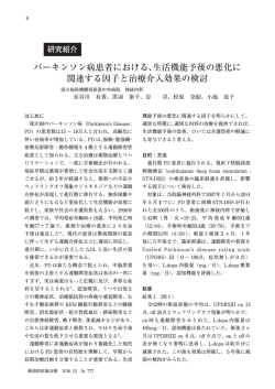
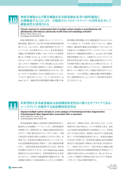
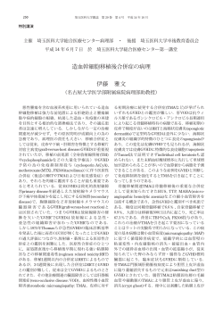
![Microsoft PowerPoint - \217\254\224]\216\270\222\26201302008.ppt](http://s3.paperzz.com/store/data/005626364_1-4cb72c366e12971be31e82be3d00cf94-250x500.png)