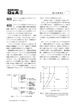
第回臨床解剖研究会記録 .. 腸脛靱帯の構成線維とその機能解剖学的意義について 三浦真弘1 影山幾男2 1大分大学医学部生体分子構造機能制御講座(解剖学 緒 言 腸脛靱帯(iliotibial tract: ITT)は,股関節の屈曲・ 伸展に伴って大転子や Gerdy 結節上部を前後に滑動 する形態学的特徴を有している.そのため両部位には 外側型 snapping hip や腸脛靱帯炎等の疾患がそれぞ れ生じやすい.特に ITT 遠位部では過労性障害が好 発することからこれまで解剖学的・臨床医学的に多く の注意が払われてきた1~3).しかし,その一方で ITT 近位部については構成線維の特徴やその運動制御機構 を含めて未だ不明な点が多く残されている.本研究で は, ITT の構成線維様式,特に ITT 近位部と殿筋と の解剖学的関係を詳細に解析することを目的として肉 眼解剖学的・電顕的検索を行った.また検索結果から 下肢における ITT の運動制御能についても機能解剖 学的検討を試みた. 材料と方法 材料は大分大学医学部に献体された解剖実習用遺体 4 体 7 側 の骨盤 下肢を 用いた. 検索用 試料は 予め ITT を中心に周囲組織を十分温存した状態で摘出し, 35×35 cm 硬質ラバー上に組織深層を上面にしてマッ プピンにて固定することで Whole-mount 伸展標本を 作 製 し た . ITT 構 成 線 維 の 肉 眼 解 剖 学 的 解 析 に は 「裏返し法」を用いた.剖出後の一部試料については 線維構築を走査電子顕微鏡( SEM )にて観察した. また ITT の構成共通線維束については MR 画像にお いても観察した. 結 果 ITT の近位部の構成線維は,合一する線維の走行 の特徴から浅・深 2 層の線維束に区別された( Fig. 1 ).浅層線維束は主に大殿筋表層の腱膜を受けた後 ITT 主部線維・ Kaplan 線維に移行したが,深層線維 大殿筋に は以下に示す線維束から複合形成された.◯ 由来する 4 筋束(上・中・下・最下部)のうちの最 中殿筋の表層筋束ならび 下部を除く 3 筋束から,◯ 大腿筋膜張筋表層・深層部から,な に同筋膜から,◯ ど上記 3 線維束が大転子(GTr)の前後から集束して 形成された.深層領域の線維束解析では,これらの線 6 臨床解剖研究会記録 No. 6 2006. 2 紀 瑞 成1 1) 加藤征治1 2日本歯科大学新潟歯学部解剖学第 1 講座 維束の大部分は GTr 後下方(殿筋粗面上部)におい て指状に噛合うように立体交差(Fig. 1 の*)して集 束したのち大腿骨に付着した.また,一部の線維束は GTr 前方を通り抜けて浅層線維束の形成にも関与し た.各線維束は合一後, ITT 主幹を形成し,それは 外側広筋表層や外側筋間中隔と密接な関係を保ちなが ら膝外側部に向かった.殿筋粗面上部に付着した交差 線 維 束 の 間 には 大 小 発 達 し た 滑 液 包 が 出現 し た . SEM 観察では交差部線維は各々同一方向に並んだ密 性膠原線維束であり,交差線維束間において線維連絡 は認められなかった.また同構造は MR 画像におい ても通常撮像条件内で確認可能であった.大殿筋由来 の 3 線 維 束 の ITT 形 成 線 維 へ の 参 加 形 態 を 含 め た ITT 構成線維様式には,個体間で発達状況に差があ るものの,検索全例で共通の規則性が認められた. 考 察 今回,筆者らは「裏返し剖出法」を用いて ITT 近 位部における詳細な解剖学的特徴(Fig. 1)を明らか にすることで, ITT は静的安定機構に働くばかりで はなく,起立姿勢において GTr の後方に特殊な交差 性線維束(Fig. 1 の*)が移動して,同部を前方に殿 筋からの動的要素を介して強力に押し付けて,いわゆ る「股関節のロック現象」を演じたり,外側筋間中隔 との連結様式から外側広筋筋房コンパートメント圧を 上昇させて伸展作用を増強させたり,伸展作用におい て膝関節の内反防止に働くことなどの新たな役割が推 測された.また,膝関節の screw home 運動の補助動 源として下腿外旋を引き起こす役割や,歩行遊脚相に おいて大腿骨頭を臼蓋に押し付けて股関節の軸性運動 を安定させる動的機能等も加えて推測された.他方, 特殊な交差性線維束については,その器質的肥厚やそ れと GTr の間に異常滑動が生じた場合には snapping hip や大転子部滑液包炎を引き起こす原因となり得る ことが考えられた. 以上のことから, ITT は静的安定機構の役割の他 に,下肢外側支持機構において多くの重要な動的安定 . 機構に関係することが示唆された (Fig. 2 の太い矢印) Fig. 1 A schematic presentation of the morphological characteristics of ˆber connection in the whole-mount preparation of the proximal portion of iliotibial tract (ITT). Deep aspect of the ITT. The asterisk indicates the speciˆc crossing ˆburosal bundles in the upper region of the gluteal tuberosity (GTu). GTr: greater trochanter, GMa-u: gluteus maximus m. (upper portion), GMam: (middle portion), GMa-l: (lower portion), GMa-ml: (lowermost portion), GMe: gluteus medius m., TFL: tensor fasciae latae m. 文 献 1) Terry GC, Hughston JC, Norwood LA: The anatomy of the iliopatellar band and iliotibial tract. Am J Sports Med 14: 3945, 1986 2) Matsumoto H, Seedhom B: Tension characteristics of the Fig. 2 Drawing of the relationship of the lateral stabilizing structures in the lower limb to the proximal portion of iliotibial tract (ITT). Posterolateral aspect of the gluteal region and the lateral of thigh. The wide arrows indicate the stabilizing dynamic elements in the hip joint, respectively. GTr: greater trochanter, GMa-u: gluteus maximus m. (upper portion), GMam: (middle portion), GMa-l: (lower portion), GMa-ml: (lowermost portion), GMe: gluteus medius m., TFL: tensor fasciae latae m. iliotibial tract and role of its superˆcial layer. Clin Orthop 313: 253255, 1995 3) Orchard JW, Fricker PA, Abud AT, Mason BR: Biomechanics of iliotibial band friction syndrome in runners. Am J Sports Med 24: 375379, 1996 An anatomical characteristics of the component ˆbers of the iliotibial tract and its kinematic signiˆcance Masahiro MIURA1, Ikuo KAGEYAMA2, Rui-Cheng JI1, Seiji KATO1 of Anatomy, Biology and Medicine, Faculty of Medicine, Oita University, 2Department of Anatomy, School of Dentistry at Niigata, The Nippon Dental University 1Department The morphological characteristics of the iliotibial tract (ITT) have not been fully elucidated. To clarify the proximal component ˆbers forming the ITT and its kinematic signiˆcance, the detailed anatomical examination of the seven caderveric ITT was performed in the gluteal region and the lateral of thigh. Whole-mount preparations of the ITT specimens facing removed from the hip region were dissected using a steroscopic microscope. In the 3 specimens after the dissection, the attachment condition of each bundle to the gluteal tuberosity (GTu) was observed on scanning electron microscope (SEM ) as a complex structure which became con‰uent from the tensor fasciae latae (TFL) and two gluteal muscles to the distal iliotibial band. As a result, the stratiˆed structure of the proximal ITT was divided into two components on the basis of our ˆndings: the superˆcial and deep layers (Fig. 1). The superˆcial layer of this structure was a wide tendinous-like band formed from the gluteal and TFL muscles and also combined with main portion of the deep layer. In contrast, the deep layer was made up of several separate distinct longitudinal bundles to the GTu of the femur, lateral intermuscular septum (LIMS) and the main portion of ITT including the Kaplan's ˆbers. These bundles crossed just over the greater trochanter (GTr) and often attached to the GTu after fusing with each other at the posterior border of GTr. Such a structure provides stabilization of the hip joint against medially and/or antero-posteriorly directed forces and is dynamically in‰uenced by the vastus lateralis muscle via the LIMS. Incidentally, the proximal ˆbers of ITT were neither ˆxed nor fused at the GTr. In the SEM observation, the crossing band was recognized as a dense ˆne bundle with fully developed blood vessels and bursa in the narrow space between the bundles. Thus, our anatomical data showed the static and/or dynamic tensional band eŠect of the ITT on the femur via the attachment to the GTu mediated by the several tendinous bundles which originated from the two gluteal and TFL muscles. In the kinematics, the proximal ˆber complex of ITT may play an important role in the antero-posterior and rotational stabilizers of the hip joint, as an essential structure in the lateral stabilizing structures of the lower extremity (Fig. 2). Key words: iliotibial tract, component ˆbers, gluteal muscles, tensor fasciae latae, functional anatomy 腸脛靱帯の構成線維とその機能解剖学的意義について 7
© Copyright 2026 Paperzz