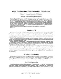
課題番号
研究課題名
主任研究者名
分担研究者名
:24指111
:糖尿病網膜症の新規治療開発に向けた基盤研究
:奥村彰規
:なし
キーワード :糖尿病網膜症、網膜周皮細胞、プロテオーム、ペプチドーム
研究成果
:糖尿病網膜症は年間 4000 人もの患者から光を奪う。さらに、増え続ける糖尿病患者
のうち約 15%は糖尿病網膜症を発症しており、ごく初期から細小血管障害に伴う神経変性が起こって
いる。視神経を不可逆的な神経変性から保護する観点からも、単純網膜症の病期から使用可能である
血管新生阻害剤および血管透過性亢進抑制剤の開発は急務である。特に、網膜周皮細胞が糖尿病の高
血糖により細胞障害を受けたり、網膜毛細血管から脱落したりした結果、周皮細胞由来因子が欠乏し
て引き起こされる血管新生および血管透過性亢進のメカニズムを解明することは重要視されているが、
詳細は明らかにされていない。そこで、本研究では、ヒト網膜由来の周皮細胞を用いて、糖尿病網膜
症の進展抑制や寛解に関連する新規因子の同定を行うことで、糖尿病網膜症の疾患メカニズムの解明
および新規治療法開発ための標的分子の探索を目的としている。
まず、培養細胞レベルで周皮細胞が分泌するペプチド・タンパク質の探索を遂行した。ヒト網膜周
皮細胞の初代培養を行い、培養上清から生理活性ペプチド群の網羅的同定を行った。この結果、培養
上清から 5137 種類のペプチドを同定することに成功した。このうち、質量分析による同定の信用度が
非常に高いペプチドは、129 種類の遺伝子由来よりなる 276 ペプチドであった。これらペプチドのうち、
血管内皮細胞に作用する報告があるペプチドホルモンも含まれており、網膜周皮細胞ではこれまでに
一切報告は無いものであった。また、興味あることとして、血管内皮細胞に対して作用するタンパク
質だけでなく、神経細胞やグリア細胞に作用することが知られているタンパク質についても部分ペプ
チドが多数同定された。この結果は、周皮細胞が網膜毛細血管の外側に存在するため、網膜内の各種
視神経細胞やグリア細胞とも相互作用している可能性を示している。特筆するペプチドとして、これ
まで遺伝子より配列が予測され、検討されてきた血管作動性のペプチドホルモン1種類に注目してい
る。このペプチドは部分的に特異的な切断を受けている配列のみが同定できており、アンジオテンシ
ンが代表例として挙げられるように、部分的な特異的欠損はペプチドホルモンの生理活性を大きく左
右するものであると考えられるため、詳細な機能を検討中である。
並行して、分画操作により生理活性ペプチド候補を段階的に精製することとした。網膜周皮細胞の
培養上清から、上述の通りにペプチド粗画分を調製し、ゲルろ過カラムクロマトグラフィーにて分画
した。得られた各画分を濃縮して、ヒト網膜毛細血管内皮細胞に対する生理活性を測定した。測定手
法は、細胞増殖・管腔形成・血管透過性・血管新生について検討を行ったが、微細な変化をとらえる
ことができるリアルタイム細胞解析装置(ECIS)を使った細胞間バリア機能の解析がより効果的であ
ることが判明し、以降、ECIS を使って活性測定を行うこととした。逆相カラムクロマトグラフィーに
より細胞間バリア機能を強める作用のある生理活性ペプチドを含む画分の調製に成功したが、アミノ
酸配列の決定までは年度内には至らなかった。一方でタンパク質の探索については、分画分子量 3000
のメンブレンフィルターにて濃縮した培養上清をイオン交換カラムクロマトグラフィーにより分画操
作を行った後に、ECIS でヒト網膜毛細血管内皮細胞に対するタンパク質試料の影響を観察した。その
結果、細胞間バリア機能を弱める働きのあるタンパク質画分の調製に成功した。当ラボの質量分析装
置では同定に至らなかったため、共同研究として外部研究機関に解析を依頼している。
以上のとおり、糖尿病網膜症の治療法に向けた研究成果としてペプチドは投与、タンパク質は抗体
療法で、網膜周皮細胞が障害を受けた際に効果的である可能性を見出しているため、合成ペプチドや
組換えタンパク質を使った検証を行いたい。
Subject No. :24 指 111
Title
:Basic studies on the novel treatment of diabetic retinopathy
Researchers :Akinori Okumura
Key word
:Diabetic retinopathy, Retinal pericyte, Proteome, Peptidome
Abstract
:Diabetic retinopathy (DR) is a leading cause of blindness. The number of diabetes
patients continues to increase and approximately 15% of diabetes patients suffer from DR, in
which nerve degeneration associated with microvascular disease begins from a very early stage.
From the standpoint of protecting the optic nerve from irreversible degeneration, there is an
urgent need to develop angiogenesis and vascular hyperpermeability inhibitors that can be used
from the simple DR stage. Retinal pericytes are susceptible to damage by hyperglycemia in
diabetes. There has been particular focus on elucidating the mechanism by which pericyte-derived
factor deficiency, caused by the detachment of retinal pericytes from retinal capillaries, results in
angiogenesis and microvascular hyperpermeability. However, this mechanism remains poorly
understood. Therefore, this study aimed to use the primary culture of human retinal pericytes to
identify new factors relating to the progression of DR in order to elucidate the mechanism of DR
and investigate target molecules for the development of novel treatments.
We first investigated the identification of peptides secreted by retinal pericytes. We performed
shotgun peptidomics experiments to investigate the bioactive peptides in the conditioned medium
of the primary human retinal pericytes. We employed nano-HPLC-MS/MS for comprehensive
peptide identification. Using this technique, we successfully identified 5,137 peptides in the
conditioned medium. Of these, the 276 peptides originating from 129 genes could be identified
with a confidence level at least 95%. These peptides included peptide hormones that have been
reported to act on vascular endothelial cells; it is noteworthy that these have never before been
reported in retinal pericytes. Currently, we focused an oligopeptide that known as the vasoactive
hormone. The oligopeptides lacks 10 residues at the N-terminus and 2 residues at the C-terminus
of the corresponding the full-length oligopeptide hormone. In addition to some of proteins that act
on vascular endothelial cells, it is interesting that we also identified many partial peptides of
proteins that have been reported to act on neurons and glial cells. This finding indicates that since
retinal pericytes are located outside retinal capillaries, they may interact with optic nerve cells
and glial cells. Despite successfully identifying many peptides, the large quantity meant that it
would have been difficult to verify ALL of these peptides by synthesis. Therefore, we opted to use
fractionation to incrementally purify candidate bioactive peptides. We then prepared the crude
peptide fraction from the conditioned medium as described above and used gel filtration column
chromatography to obtain the fractions. After concentrating each of the fractions, we measured
bioactivity on human retinal microvascular endothelial cells (HRMVECs). We measured cell
proliferation, tube formation, vascular permeability and angiogenesis, and established that
intracellular barrier function analysis using electric cell-substrate impedance sensing (ECIS) is
more effective, and therefore, used ECIS to measure activity. We also used reverse-phase column
chromatography to successfully prepare a fraction comprising bioactive peptides that strengthen
intracellular barrier function, but were unable to complete amino acid sequence determination
before the end of the year. To investigate proteins, we used a membrane filter with a molecular
weight cut off of 3,000 to concentrate the conditioned medium before employing ECIS to observe
the effects of sample proteins on HRMVECs. As a result, we successfully obtained a protein
fraction that acts to weaken intracellular barrier function.
Within the context of DR therapy, the above findings suggest that administering peptides or
using antibodies against proteins may be efficacious for the treatment of damaged retinal
pericytes. Therefore, we will conduct identification and verification using synthetic peptides and
recombinant proteins.
Researchers には、分担研究者を記載する。
24指111 糖尿病網膜症の新規治療開発に向けた基盤研究
糖尿病網膜症の新規治療薬開発に向けて、⾎管新⽣または⾎管透過性に
関与する網膜周⽪細胞由来の⽣理活性分⼦群の探索
網膜周⽪細胞の分泌ペプチドミクス解析
分泌プロテオミクス解析に⽐べて分泌ペプチドミクス解析は、転写レベ
ルだけでなく、⽣体内ストレスをより反映した結果となりうる。また、
機能性ペプチドの発⾒にもつながる利点がある。
ヒト由来初代網膜周⽪細胞の培養上清
質量分析器(nanoLC-MS/MS)による網羅的解析
⾎管内⽪細胞に作⽤すると思われるペプチドについて機能解析を継続し
ている。
加えて、視神経やアストロサイトに作⽤しそうなペプチドも同定できて
おり、機能解析を引き続き⾏っていく。
現在検討中のペプチドホルモンの全長
XXXXXXXXXXXXXXXXXXXXXXXXXXXXXXXXX
この3種類のみ検出される
XXXXXXXXXXXXXXXXXXXXX
XXXXXXXXXXXXXXXXXXXX
XXXXXXXXXXXXXXXXXXX
アンジオポエチン様に
特異的な欠落が見られる
⾎管新⽣または⾎管透過性亢進に関与する網膜周⽪細胞由来の
⽣理活性分⼦群(ペプチド、タンパク質)の探索
網膜周⽪細胞の培養上清よりペプチド画分またはタンパク質画分を調製
カラムクロマトグラフィーによる分画
ヒト網膜⽑細⾎管内⽪細胞に対する⽣理活性測定
リアルタイム細胞解析装置による
①創傷治癒アッセイ
②細胞バリア機能アッセイ
リアルタイム細胞解析装置で
確認された⽣理活性画分
(細胞バリア機能アッセイ)
活性画分の再精製
⾼感度質量分析装置(MALDI-TOF、Orbitrap LC-MS)
によるペプチド・タンパク質同定
現在、共同研究として理化学研究所にて、⽣理活性作⽤のあるペプチド
ならびにタンパク質のさらなる⾼純度化と⾼感度質量分析装置で同定を
⾏っている。
研究発表及び特許取得報告について
課題番号:
24指111
研究課題名:
糖尿病網膜症の新規治療開発に向けた基盤研究
主任研究者名:
奥村彰規
論文発表
論文タイトル
著者
掲載誌
掲載号
年
タイトル
発表者
学会名
場所
年月
発表者
発表先
場所
年月日
該当なし
学会発表
該当なし
その他発表(雑誌、テレビ、ラジオ等)
タイトル
該当なし
特許取得状況について ※出願申請中のものは( )記載のこと。
発明名称
登録番号
該当なし
※該当がない項目の欄には「該当なし」と記載のこと。
※主任研究者が班全員分の内容を記載のこと。
特許権者(申請者)
登録日(申請日)
(共願は全記載)
出願国
© Copyright 2026 Paperzz