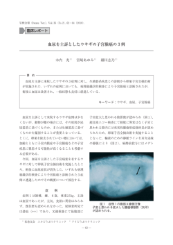
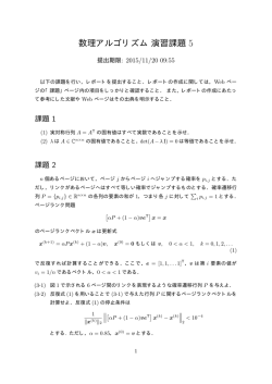
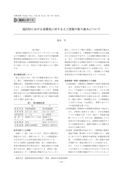
「日本獣医寄生虫学会誌」投稿規程(平成 27 年 7 月 1 日一部改正)
(掲載論文の内容および区分)
察、謝辞、引用文献、連絡責任者の順に記述する。
1 .獣医寄生虫学および獣医寄生虫病学領域に関するも
材料と方法、結果、考察については、小見出しを用
ので、原著、短報、総説、解説、技術情報、施設紹
いることができる。連絡責任者は、和文および英文
介、野外調査・学会参加記などとする。
で、氏名、所属、住所、および E-mail アドレスを
(投稿資格)
記述する。短報では、謝辞、引用文献、連絡責任者
2 .投稿原稿は未公刊のものとし、著者および共著者に
等の項目を除いて、上記の項目分けをしない。
は本会会員が 1 人以上参加していること。但し、編
9 .原著、短報以外の原稿の項目分けは特に設けないが、
内容の理解に繫がる合理的な区分けとする。
集委員長が必要と認めた際には、会員外からの投稿
10.図は JPG もしくは TIFF ファイルとして、個別に
を認めることがある。
3 .投稿原稿は、人権の保護ならびに法令等の遵守に対
提出する。表は EXCEL ファイルとして個別に提出
応していること。また広く、研究に関する倫理を包
する。カラー写真は 4 頁以内まで均一料金(税抜き
3 万円程度)で、著者負担である。
括的に遵守していること。
11.用語は、学名はイタリック体で、度量衡の単位と略
(動物の取り扱い)
語は CGS 単位または SI 単位を用いる。数字および
4 .投稿原稿における症例および実験動物の取り扱いは、
英語は半角文字を用いる。
「動物の愛護及び管理に関する法律」(平成 11 年法
律第 221 号改正)に基づき、動物愛護の精神に則っ
12.引用文献の記載方法を含む、その他の詳細について
は、The Journal of Veterinary Medical Science の
て行われていること。
投稿規定に準ずる。
(採否の決定)
5 .原著論文ならびに短報論文は、編集委員会(以下、
13.総説、解説、技術情報等において、他の文献から図
「委員会」)の委嘱した審査委員の意見をもとに、委
表を引用する場合には、その転載許可は著者の責任
において行う。
員会の議を経て編集委員長(以下、「委員長」)が決
14.カラー印刷料、別刷り作成料、PDF ファイル版作
定する。
成料などは著者の負担とする。
(執筆要領)
6 .原稿の長さは、原則的に、図表などすべてを含み、
15.投稿原稿の送付先および照会先は編集委員長とし
原著、総説では刷り上り枚数 10 頁以内、その他の
て、基本的に電子メール等を介して行う。本会誌は
区分の原稿では 5 頁以内とするが、委員会が長さの
年 2 回(6 月および 12 月)発刊されるので、査読
調整(増・減)を求める場合がある。
および編集委員会での審議を考慮して、時間的余裕
をもって投稿する。
7 .原稿は和文または英文で記述する。和文原稿には英
文要約を附し、英文原稿には和文要約を附す。英文
は十分に推敲し、英文習熟者の校閲を受けたものを
[投稿原稿送付および編集に関する照会先]
投稿する。原稿は A4 版に横書きとし、MS Word
〒 753-8515 文書(1 段組)として作成する。原稿の用紙設定
山口県山口市吉田 1677-1
では上下左右に 30mm ずつ余白をもたせて記述す
山口大学共同獣医学部寄生虫学教室内
る。原稿は、和文は MS 明朝か平成明朝、英文は
日本獣医寄生虫学会編集委員会
Century か Times と し、1 行 38 字、 1 頁 36 行、 文
編集委員長 佐藤 宏
字サイズは 10.5P で記述する。
Phone:083-933-5902
8 .原稿の第 1 頁は、論文題目、著者名、所属機関およ
E-mail:[email protected]
び所在地(または自宅住所)を記述する。第 2 頁以
降は、英文原稿においても和文原稿においても、英
(附則)
文要約(250 語)と Key words(5 語)および和文
この規定は、平成 14 年 4 月 1 日から施行する。
要約(600 字)とキーワード(5 語)をそれぞれ記
本改訂規定は、平成 27 年 7 月 1 日から施行する。
述する。続けて、緒言、材料および方法、結果、考
― i ―
− 編 集 委 員 会 −
編 集 委 員 長 佐藤 宏(山 口 大 学)
編 集 委 員 加藤 大智(北 海 道 大 学)
編 集 委 員 西川 義文(帯広畜産大学)
編 集 委 員 松林 誠(大阪府立大学)
日本獣医寄生虫学会誌(ISSN 1347-961X)
第 14 巻第 2 号(平成 27 年 12 月 25 日発行)
発行者 日本獣医寄生虫学会 理事長 板垣 匡(岩手大学)
印刷者 杜陵高速印刷株式会社
岩手県盛岡市川目町 23 − 2 盛岡中央工業団地
TEL:019−651−2110 ㈹ FAX:019−654−1084
― ii ―
The Japanese Journal of Veterinary Parasitology
(Publication by the Japanese Society of Veterinary Parasitologists)
− Guide for Authors −
1. Unpublished Full papers, Notes, Review articles,
enough margin spaces(30mm)on the four sides.
and Essays, related in principle to veterinary
9. The proper noun should begin with a capital
parasitology science are accepted. Texts must
letter. The scientific terms of animals, plants and
be written in proper Japanese or English. The
microorganisms must be italicized.
manuscript of Full papers and Notes are reviewed
10. Abbreviations except those listed in Item 11
by the referees. Manuscripts are selected for
should be written in parentheses after the full
publication according to editorial assessment
term at their first appearance in the manuscript.
of their suitability and reports from individual
11. In the manuscript Arabic numerals should be
referees. Papers will be rejected if the ethics in the
used for quantum. As a rule, the units and abb-
care and use of animals as well as that in research
reviations should conform to the following exam-
itself have not been followed.
ples:M, mM, μM, N, % , m, cm, mm, μm, nm, pm,
2. Submission of digital files of manuscripts should be
2
cm , l, ml, kg, g, mg, μg, ng, pg, hr, min, sec, msec,
done by e-mail or other network services to the
rpm, Hz, Bq, mBq, μBq, kBq, cpm, dpm, ppm, ℃ , J,
KJ, lux, CPE, LD.
Editor-in-Chief of the journal.
3. The upper half of the first page should notify an
12. References should be arranged in the alphabetical
indication of either Full paper, Note, Review article,
order of the authors’ surname and numbered
or Essay, title, authors’ names and affiliation where
consecutively. In the text they should be cited
the work was done, and the corresponding author
by numbers, e.g.[1, 3-5, 7]. The following are
with his postal address, phone number and e-mail
examples of Reference.
1.Bloom, W. and Faweett, D. W. 1969. A Text Book
address. The lower half of the page should be left
of Histology, 9th ed., B. W. Saunders, Philadelphia.
blank.
4. The second page of each Full paper, Note and
2.Canpen, C. C. 1978. Tumors of the endocrine
Review article should contain an abstract(250
glands. pp.372-429. In:Tumors in Domestic
.
words or less)and Key words(5 or less)
, Univ.
Animals, 2 nd ed. (Moulton, J. E. ed.)
California Press, Berkley.
5. The description on and after the third page of
Full paper should be made in principle in the
3.Nakayama, A. 1978. Rhythm of the body tempe-
following order:INTRODUCTION, MATERIALS
rature. pp.140-145. In:Biorhythm and its Mecha-
AND METHODS, RESULTS, DISCUSSION,
nisms(Suda, M., Hayashi, O. and Nakagawa, H.
ACKNOWLEDGMENTS and REFERENCES. For
, Kodansha. Tokyo(in Japanese)
.
eds.)
Note, such captions as Abstract and Introduction
4.Pedersen, N. C., Ho, E. W., Brown, M. L. and
should not be given and References should follow
Yamamoto, J. K. 1987. Isolation of a T-lymphotr
the text.
opic virus from domestic cats with an immunodeficiency-like syndrome. Science 235:790-793.
6. The length of papers, including tables and figures,
should not exceed 10 printed pages for Full paper
13. On Table, no vertical line is used and each term
and Review article, and 5 for Note and other
or phrase should begin with a capital letter. Any
articles. One printed page containing no title,
explanation essential to the understanding of the
tables or figures, may contain around 870 words in
Table should be given as a footnote at the bottom
all articles.
of the Table. Reference to the footnote should be
designated by symbols in the order a)
, b),c).
7. A summary in Japanese(600 letters for all articles)
should be attached, if the authors include Japanese
14. Figures-Data should be drawn on a white paper
co-author(s). It should include the title, authors
by using a line less than 1 mm wide. Figuresphotographs are preferred to be printed in the
and the affiliation where the work was done.
8. The manuscript should be typed at double spaces
original size and should be 7.7 cm or 16 cm in
(ca. 24-26 lines per page and approx. 70 spaces per
horizontal width by less than 9 cm length. For
line)on A4(215 x 280mm)typewriting paper with
printing any signs(scales, arrows, etc.)on the
― iii ―
photographs should be directly marked. All the
Dr. Hiroshi Sato
Figures should be numbered through those of data
Editor-in-Chief
and photographs in consecutive order, and their
Laboratory of Parasitology, Joint Faculty of
title and legends must be typewritten following
Veterinary Medicine, Yamaguchi University,
the Reference section in the text.
1677-1 Yoshida, 753-8515 Yamaguchi, Japan
15. The author will be charged the reprints, color printing
Tel&Fax:+81-83-933-5902,
and requested PDF file.
E-mail:[email protected]
16. The journal is to be issued every six months(June
18. The copyright for articles which appeared in this
and December).
Journal is held by the Japanese Society of Vete-
17. All papers and inquiries only in writing should be
rinary Parasitologists.
sent to the following address:
− EDITORIAL BOARD −
Editor-in-chief:Hiroshi Sato(Yamaguchi University)
Editor:Hirotomo Kato(Hokkaido University)
Editor:Yoshifumi Nishikawa(Obihiro University of Agriculture and Veterinary Medicine)
Editor:Makoto Matsubayashi(Osaka Prefecture University)
The Japanese Journal of Veterinary Parasitology(ISSN 1347-961X)is published semiannually
by the Japanese Society of Veterinary Parasitologists
(President Prof. Tadashi Itagaki, Iwate University)
.
― iv ―
目 次
投稿規定 ………………………………………………… i
総 説 Guide for authors ……………………………………… iii
馬ピロプラズマ病
五十嵐郁男、横山直明 …………………………… 83
原 著
Phylogenetic relationship of the pinworms of the genus
解 説
Syphacia from murines of Indonesia and some other
生食ブームに潜むトキソプラズマ症のリスク:
regions inferred by molecular analysis
食肉におけるトキソプラズマ汚染の現状
Kartika DEWI, Hideo HASEGAWA,
松尾加代子、上津ひろな、高島康弘 …………… 93
Akiko SATO, and Mitsuhiko ASAKAWA …… 67
調査・学会参加記
ウマの肝臓エキノコックス(多包虫)結節における多房
Molecular and Cellular Biology of Helminth
化の特徴
Parasites Ⅸ 参加記
作井睦子、結城恵美、大西綾衣、中野由佳子、
豊岡大輔、清水俊彦、瀬沼洋二、迫 陽子、
森千惠子、孝口裕一、奥祐三郎、八木欣平
……………………………………… 76
松本 淳 …………………………………………… 97
Original Paper
Phylogenetic relationship of the pinworms of the genus
Syphacia from murines of Indonesia and some other
regions inferred by molecular analysis
Kartika DEWI 1, Hideo HASEGAWA 2, Akiko SATO 2, and Mitsuhiko ASAKAWA 3
1
Zoology Division, Museum Zoologicum Bogoriense, RC. Biology—LIPI, West Java, Indonesia
2
3
Department of Biology, Faculty of Medicine, Oita University
Department of Pathobiology, School of Veterinary Medicine, Rakuno Gakuen University
ABSTRACT
Sequencing of partial cytochrome c oxidase subunit 1 gene(Cox-1)of mitochondrial DNA and 28S ribosomal
RNA gene(rDNA)of nuclear DNA was attempted for pinworms of the genus Syphacia from murines of Indonesia
in order to compare with those from other regions. Cox-1 sequence of Syphacia rifaii from Bunomys penitus
of Sulawesi was characteristic by having deletions of three consecutive nucleotides at two loci in addition to
numerous substitutions, resulting in very long branch in phylogenetic trees. Nevertheless, both in Cox-1 and 28S
rDNA trees, S. rifaii formed a clade with Syphacia muris. Three Syphacia species parasitic in Apodemus, Syphacia
emileromani, Syphacia stroma and Syphacia agraria, formed one clade in 28S rDNA phylograms, suggesting coevolutionary relationship with their hosts. Indonesian examples of S. muris were rather diverged from those in the
laboratory rats, Rattus norvegicus, both in Cox-1 and 28S rDNA, presumably reflecting the process of geographical
dispersal of host Rattus spp. with this pinworm over the world.
Keywords:Syphacia, nematodes, Cox-1, 28S rDNA, co-evolution.
period in external environment or in intermediate host
1.INTRODUCTION
to become infective. Based on the morphological evidence,
The pinworm of genus Syphacia Seurat, 1916(Nematoda:
Syphacia species have been considered to have co-
Oxyuridae: Syphaciinae)has cosmopolitan distribution
evolutionary relationship with their hosts generally
occuring in rodents of the families Cricetidae and
[18, 19]. Meanwhile, analyzed partial sequences of
Muridae[18]. In Indonesia, where murine fauna shows
cytochrome c oxidase subunit 1 gene of mitochondrial
high diversity and endemicity, 10 species of the genus
DNA(Cox-1)and 28S ribosomal RNA gene(rDNA)
Syphacia belonging to three subgenera were hitherto
of Syphacia species of Japanese murines could not
recorded[1-6, 12, 13, 26]. Eight of them belong to the
demonstrate such a co-evolutionary relationship[22,
subgenus Syphacia, while the remaining two have
23]. It was suggested that co-evolutionary relationship
specialized characteristics, having been assigned to two
might not be so strict in Syphacia and host switching
new subgenera[5]
. Because they seem to have rather
probably occurred during the course of evolution. In
strict host specificity, it is of special interest to know
this study, we determined partial sequences of Cox-
their evolutionary relationship with their hosts.
1 and 28S rDNA of Syphacia species collected from
Pinworms of mammals have direct life cycle with
Indonesia and some other countries to know phylogenetic
anal-oral route of transmission. The simplicity of the
relationship of Syphacia species among Indonesia and
life cycle is likely to provide less opportunity to acquire
other areas.
a new host than for other parasites that require a long
― 67 ―
Jpn. J. Vet. Parasitol. Vol. 14. No. 2 2015
Phylogenetic relationship of the pinworms of the genus Syphacia from murines of Indonesia and some other regions inferred by molecular analysis
and sequencing were those used previously[9, 16, 17,
2.MATERIALS AND METHODS
23]or newly designed(Table 2).
2-1.Species examined
The PCR conditions were as follows: initial denaturation
Two species of Syphacia from murines of Indonesia,
at 94 ˚C for 2 min, followed by 30 cycles of 98˚C for 10
i.e. S. muris from Rattus tanezumi collected on Java and
sec, 50 ˚C for 1 min, 68 ˚C for 1 min, 30 cycles of 98 ˚C
Sumatra Islands and S. rifaii from Bunomys penitus
for 10 sec, 55 ˚C for 1 min, 68 ˚C for 1 min, and a post-
captured on Sulawesi Island, were subjected for DNA
amplification extension at 68 ˚C for 7 min for Cox-1.
extraction and amplification(Table 1). In addition, two
PCR products were mixed with Ez-VisionTM Three
species of Syphacia, i.e. S. obvelata from a laboratory
DNA Dye(Amresco, Solon, Ohio, USA)
, electrophoresed
mouse in Japan and S. stroma from a laboratory
in a 1.5% agarose gel plate and visualized using a UV
golden hamster in Czech Republic, were also analyzed.
illuminator. Positive bands were dissected and processed
Besides the Syphacia representatives, Syphatineria sp.
using NucleospinTM column(Machery-Nagel, Düren,
from an Indonesian sciurid and Aspiculuris tetraptera
Germany), and then ethanol precipitated for further
from a laboratory mouse in Japan were also tested for
purification. Proper amount of the DNA was subjected
sequencing(Table 1). All samples were fixed in pure
to direct sequencing using the BigDyeTM Terminator
ethanol.
Cycle Sequencing Kit Version 3.1(Applied Biosystems,
Foster City, California, USA), and purified using
2-2.DNA extraction and amplification
CentriSepTM spin column(Princeton Separations Inc.,
Individual worm was rinsed in phosphate buffer(pH
Adelphia, New Jersey, USA). Then sequencing was
6.5), and homogenized in a 1.5 mL Eppendorf tube
made in an automated genetic analyzer ABI-PRISM
containing 100 μL distilled water using a plastic pestle.
3130(Applied Biosystems)
.
Five μL of the homogenized solution was mixed with
50 μL liquid phase of Dexpat™(Takara Bio. Inc., Otsu,
Table 2.Primers used in the present study
Shiga, Japan)in 200 μL tube, heated at 96 °C for 30
Name of primer
min, and then cooled on ice. Subsequently, 5 μL of the
Partial Cox-1 gene:
Direction
Nucleotide sequence
SyphCoxF1
Forward 5’-GGTCAGTTGTATAATGTTRT-3’
StrCoxAfrF
Forward 5’-GTGGTTTTGGTAATTGAATGGTT-3’
JB3
Forward 5’-TTTTTTGGGCATCCTGAGGTTTAT-3’
MH28R
Reverse 5’-CTAACTACATAAT AAGTATCATG-3’
MgSO4(Toyobo Co., Tokyo, Japan)and 0.25 μL each
JB4.5
Reverse 5’-TAAAGAAAGAACATAATGAAAATG-3’
of forward and reverse primers. PCR was performed
Partial 28S rDNA:
using a thermal cycler, PC-801 (ASTEC Co., Ltd.,
C1′
Forward 5’-ACCCGCTGAATTTAAGCAT -3’
Fukuoka, Japan). The primers used for amplification
D2
Reverse 5’-TCCGTGTTTCAAGACGG-3’
solution was added to the 50 μL PCR mixture, which
contained 0.5 μL of KOD-NeoTM polymerase, 5 μL of
10x PCR buffer, 5 μL of 2 mM dNTP, 5 μL of 2 mM
Table 1.Pinworms examined in this study with the host, locality and accession numbers of DNA sequences in DDBJ
Species
Host
DDBJ accession Nos.
Cox-1
28S rDNA
Muridae: Murinae
Rattus tanezumi
Java, Indonesia
2012
LC038089
LC038096
Syphacia muris
Muridae: Murinae
Rattus tanezumi
Sumatra, Indonesia
2012
LC038090
LC038097
Syphacia rifaii
Muridae: Murinae
Bunomys penitus
Sulawesi, Indonesia
2012
LC038087
LC038094
Syphacia rifaii
Muridae: Murinae
Bunomys penitus
Sulawesi, Indonesia
2012
LC038088
LC038095
Honshu, Japan
2012
LC038086
n.t.**
Muridae: Murinae
Species
Year of
collection
Syphacia muris
Syphacia obvelata
Family: Subfamily
Locality
*
Mus musculus
*
Syphacia stroma
Muridae: Cricetinae
Mesocricetus auratus
Brno, Czech Republic
2013
LC038091
LC038098
Syphatineria sp.
Sciuridae: Sciurinae
Lariscus hosei
Kalimantan, Indonesia
2012
LC038092
LC038099
Honshu, Japan
2012
LC038093
n.t.
Aspiculuris tetraptera
Muridae: Murinae
*
Mus musculus
*
Experimental animal reared in laboratory.
**
n.t.: Not tested.
― 68 ―
Jpn. J. Vet. Parasitol. Vol. 14. No. 2 2015
Kartika DEWI, Hideo HASEGAWA, Akiko SATO, Mitsuhiko ASAKAWA
2-3.Phylogenetic analysis
obtained for each one sample of S. rifaii(LC038087)
Sequences determined in the present study and
and Syphatineria sp.(LC038092). A shorter sequence
those registered in DNA databases were used for
(LC038088)corresponding to 106th to 748th positions of
phylogenetic analysis. They were aligned using Clustal
LC03087 was obtained using primer set StrCoxAfrF-
W, then analyses were made by neighbor-joining(NJ)
JB4.5 from another sample of S. rifaii. This shorter
and maximum likelihood(ML)methods using MEGA5
sequence had one synonymous substitution from C to
(v. 5.2.2) software [25, 27]
. Both nucleotide and
T at 147th position. The sequence of S. rifaii was also
amino acid sequences translated using invertebrate
confirmed by sequencing using primer sets StrCoxAfrF-
mitochondrion code were analyzed for Cox-1. In NJ
MH28R and JB3-JB4.5, which gave 610bp and 366bp,
analysis of nucleotide sequences, the evolutionary
respectively. Meanwhile, S. muris samples responded
distances were computed using the Kimura’s two-
only to the primer set JB3-JB4.5, giving shorter sequences
parameter method[20]
. The bootstrap values were
with 395bp. Because of these differences in length of
. Aspiculuris tetraptera
calculated by 1,000 replicates[8]
the sequences obtained and limitation of the sequences
was used as an outgroup species to root tree of Cox-
in the DNA database, phylogenetic analyses were
1. The nucleotide sequences determined in this study
carried out separately on the two datasets covering
were registered in the DNA Databank of Japan(DDBJ,
618bp(Fig. 1)and 249bp(Fig. 2),respectively.
http://www.ddbj.nig.ac.jp/)with accession numbers
The striking feature is the peculiarity of Cox-1 of
LC038086 to LC038099.
S. rifaii in nucleotide and amino acid sequences. By
Clustal W alignment, it was found that S. rifaii had
deletions of three consecutive nucleotides at two sites
3.RESULTS
causing two amino acid deletions. The genetic distance
In all of the Indonesian Syphacia materials tested,
from other congeners was large, making extraordinarily
partial Cox-1 DNA was successfully amplified and
long branch especially in the NJ tree based on the
sequenced(Table 1). Using primer sets SyphCoxF1-
. If outgroup setting was not
longer sequences(Fig.1)
JB4.5, unambiguous sequence of 749bp of Cox-1 was
done, S. rifaii diverged at the most basal node in the
Fig. 1.NJ reconstruction of phylogeny of Syphacia spp. based on long nucleotide sequences of Cox-1. The
optimal tree with the sum of branch length = 0.81201463 is shown. There were a total of 618 positions
in the final dataset. Nematode taxon is followed by host and locality in parenthesis and accession number
in bracket. Material from laboratory murine is marked with an asterisk.
― 69 ―
Jpn. J. Vet. Parasitol. Vol. 14. No. 2 2015
Phylogenetic relationship of the pinworms of the genus Syphacia from murines of Indonesia and some other regions inferred by molecular analysis
Fig. 2.NJ reconstruction of phylogeny of Syphacia spp. based on short nucleotide sequences of Cox-1. The
optimal tree with the sum of branch length = 0.85148172 is shown. There were a total of 249 positions in
the final dataset. Nematode taxon is followed by host and locality in parenthesis and accession number in
bracket. Material from laboratory murine is marked with an asterisk.
tree, putting Aspiculuris within Syphacia spp.(not
high(Fig. 4). Even when outgroup setting was not
shown here)
. When Aspiculuris was used as an outgroup,
made, A. tetraptera was located most basal. According
S. rifaii and Syphatineria sp. formed a clade clearly
to this tree, Syphacia(Seuratoxyuris)petrusewiczi from
separated from other species of Syphacia. This peculiarity
Japanese Myodes(syn. Clethrionomys)diverged at the
became less prominent but persisted when the analysis
most basal node, and then Syphatineria sp. was separated. was performed based on the short sequences including
Among the species of the subgenus Syphacia, S. rifaii
those of S. muris(Fig. 2)
. Syphacia rifaii shared common
and S. muris formed a clade, sharing a long branch,
ancestor with Syphatineria sp. In the tree based on the
diverging from the common ancestor to the other
short sequences of Cox-1, S. rifaii and S. muris were
Syphacia species. Syphacia agraria, S. stroma and
close to each other, and they shared a common ancestor
S. emileromani, all parasitic in Apodemus spp., are
with Syphatineria sp. though the bootstrap value was
monophyletic. Syphacia vandenbrueli, S. frederici, S.
not high.
obvelata and S. montana formed another monophyletic
Phylogenetic reconstruction using ML method
group. Syphacia muris of R. tanezumi of Sumatra and
was attempted for amino acid sequences translated
Java differed from that of Japan and USA, both were
from the long nucleotide sequences of Cox-1(Fig. 3).
collected from laboratory rats, Rattus norvegicus. The
Again, S. rifaii showed very curious position by having
same groupings were also found in ML tree based
extraordinary long branch.
on 28S rDNA(Fig. 5), but Syphatineria sp. diverged
Amplification of 28S rDNA was successful for S. rifaii,
earlier than S.(Seu.)petrusewiczi.
S. muris and Syphatineria sp.(Table 1). In the NJ tree
based on 28S rDNA, bootstrap values were generally
― 70 ―
Jpn. J. Vet. Parasitol. Vol. 14. No. 2 2015
Kartika DEWI, Hideo HASEGAWA, Akiko SATO, Mitsuhiko ASAKAWA
Fig. 3.ML reconstruction of phylogeny of Syphacia spp. based on amino acids translated from long nucleotide
sequences of mtDNA Cox-1 using the General Reverse Transcriptional model. The tree with the highest
log likelihood(-1382.5027)is shown. There were a total of 206 positions in the final dataset. Nematode
taxon is followed by host and locality in parenthesis and accession number in bracket. Material from
laboratory murine is marked with an asterisk.
Fig. 4.NJ reconstruction of phylogeny of Syphacia spp. based on sequences of partial 28S rDNA. The optimal
tree with the sum of branch length = 1.35815348 is shown. There were a total of 689 positions in the
final dataset. Nematode taxon is followed by host and locality in parenthesis and accession number in
bracket. Material from laboratory murine is marked with an asterisk.
― 71 ―
Jpn. J. Vet. Parasitol. Vol. 14. No. 2 2015
Phylogenetic relationship of the pinworms of the genus Syphacia from murines of Indonesia and some other regions inferred by molecular analysis
Fig. 5.ML reconstruction of phylogeny of Syphacia spp. based on sequences of partial 28S rDNA. The tree with
the highest log likelihood (-4716.7483)is shown. There were a total of 689 positions in the final dataset.
Nematode taxon is followed by host and locality in parenthesis and accession number in bracket. Material
from laboratory murine is marked with an asterisk.
to have diverged in early Pliocene while ancestors of
4.DISCUSSION
Micromys, Mus, Apodemus and Microtines established
Syphaciinae arose with glires as hosts in Paleocene,
much earlier, in Miocene[7]. It is also noticeable that
and evolved as Syphaciini in Muroidea in early Eocene
individuals of S. muris of Java and Sumatra were rather
[18]. Subsequently, syphaciins were divided into those
diverged from those in the laboratory rats both in Cox-1
in murids and sciurids during Eocene, and establishment
and 28S rDNA. Ancestor of S. muris might be adapted
of the subgenera of Syphacia(i.e., Syphacia, Seuratoxyuris
to ancestral Rattus probably in Southeast Asia, and then
and Cricetoxyuris)and genera of syphaciins(including
made dispersal to the surrounding areas. Some Rattus,
Syphatineria and Syphabulea)of sciurids occurred
i.e., R. norvegicus and R. rattus, widened distribution
in Oligocene. However, the phylogenetic trees based
over the world. The laboratory rat was domesticated
on 28S rDNA may suggest that diversification of
from feral R. norvegicus in Europe or North America in
murid- and scuirid-parasitic lineages of Syphaciini and
middle of 19th century, and then distributed to various
diversification of subgenera Seuratoxyuris and Syphacia
laboratories all over the world. Syphacia muris of them
occurred in a relatively short geological period.
also has been maintained in the laboratory conditions.
As shown above, S. rifaii and S. muris are located
Thus, genetic divergence of S. muris in 28S rDNA of
close together both in the phylogenetic trees on Cox-1
U.S. and Japan materials was negligible. Also, Cox-1 of S.
and 28S rDNA. This is not unexpected because ancestors
muris in rats in Chinese laboratories lacked variations
of the host genera, Rattus and Bunomys, are considered
(Fig. 2). However, it is apparent that feral Rattus
― 72 ―
Jpn. J. Vet. Parasitol. Vol. 14. No. 2 2015
Kartika DEWI, Hideo HASEGAWA, Akiko SATO, Mitsuhiko ASAKAWA
spp. harbored S. muris with genetic diversification as
to be related close together[24]. In the phylogenetic
suggested by the present study.
trees based on 28S rDNA, S. agraria, S. stroma and S.
The numerous nucleotide and amino acid substitutions
emileromani formed one clade, suggesting that these
found in Cox-1 of S. rifaii are very curious. At first, the
species have co-evolved with Apodemus. However, host-
sequence obtained was suspected to be a pseudogene.
specificity of Syphacia may not be so strict and host-
However, repeated amplification and sequencing resulted
switching may occur especially when hosts become
in the same sequence except for one synonymous
sympatric. Actually, S. frederici, S. emileromani and
substitution, while no other sequence referable to Cox-1
S. agraria are shared by three Apodemus species
was obtained. Moreover, the sequence was translatable
distributed in Hokkaido, Japan[11]. Moreover, under
to amino acid sequence using invertebrate mitochondrial
an artificial condition such as breeding facilities of
code. Hence, this sequence is regarded as partial Cox-
experimental or pet rodents, Syphacia species could be
1 gene. Presumably, the nucleotide substitutions in
easily transmitted to unfamiliar hosts. It was reported
Cox-1 of this pinworm have been accumulated during
that three species of Syphacia infected concomitantly
isolation on Sulawesi with host genus Bunomys, which
the golden hamsters reared[15]. The present material
.
is also Sulawesi endemic with seven extant species[21]
of S. stroma was such worm collected from the golden
Although S. rifaii has been known from B. prolatus and
hamster.
B. chrycosomus besides B. penitus[1],only two worms
from one host species were subjected to analysis.
Further analyses of DNA sequences of this and other
ACKNOWLEDGMENT
endemic species of Syphacia are indispensable to have
We would like to express our sincere thanks to
comprehensive understanding of pinworm evolution in
the ICBG Project Mekongga team for collecting
Indonesia. the host of Syphacia rifaii, Prof. Gono Semiadii, Y.
Previous studies suggested that co-evolutionary
S. Fitriana and N. Supriyatna(MZB)for collecting
relationship in Syphacia might not be so strict[22,
the host of Syphatineria sp. We are also indebted to
23]. Their conclusion was based on the fact that
Dr. Barbora Kalousova, University of Veterinary and
both Mus-parasitic species and Apodemus-parasitic
Pharmaceutical Sciences Brno, Czech Republic, and Dr.
species did not form their own clades but scattered in
Hideto Kino, Hamamatsu Medical Unversity, for their
different clades. It was also known from morphological
kindness in providing nematode material for analysis.
viewpoint that plural Syphacia species parasitic in a
This study was supported by the RONPAKU Project
murine genus often composed of different lineages.
(ID No. LIPI-11317, year 2013 to 2015),by the Strategic
For example, among the Syphacia species parasitic
Research Foundation at Private Universities(2013-
in Mus, S. obvelata was considered to be close to S.
2017)and by a Grant-in-Aid for Scientific Research
montana, but clearly differed from S. ohtaorum in the
(C-23570120, C-26460513)from the Ministry of the
cephalic morphology, lateral alae shape and egg surface
Education, Science and Culture of Japan.
markings[10, 24].Among those parasitic in Apodemus,
S. frederici has well-developed and pointed alae in the
cervical portion of female and a short tail in male[14,
24], while S. emileromani, S. agraria and S. stroma
REFERENCES
1. Dewi, K. and Hasegawa, H. 2010. Syphacia(Syphacia)
lack such alae in female and have a long tail in male[10,
rifaii n. sp.(Nematoda: Oxyuridae)collected from
24]. The phylogenetic trees presented by Okamoto et
Bunomys spp.(Rodentia: Muridae). J. Parasitol.
al.[22, 23]seemed to congruent with the morphology-
96:125-28.
2. Dewi, K. and Hasegawa, H. 2014. Two new species
based groupings of Syphacia.
Syphacia emileromani and S. stroma, both were
of Syphacia(Nematoda: Oxyuridae)in endemic murid
described originally from Apodemus, were considered
rodents from Sulawesi, Indonesia. J. Helminthol.
― 73 ―
Jpn. J. Vet. Parasitol. Vol. 14. No. 2 2015
Phylogenetic relationship of the pinworms of the genus Syphacia from murines of Indonesia and some other regions inferred by molecular analysis
88:41-49.
Japan, Vol. 7(Kamegai, S., Hayashi, S. and Otsuru,
3. Dewi, K. and Purwaningsih, E. 2013. A checklist
of nematode parasites from Indonesian murids.
M. eds.),Meguro Parasitological Museum, Tokyo.
12. Hasegawa, H. and Syafrudin. 1995. Nematode
Zootaxa 3608:531-546.
fauna of the two sympatric rats, Rattus rattus
4. Dewi, K., Asakawa, M. and Fitriana, Y. S. 2014.
and R. exulans, in Kao district, Halmahera Island,
Syphacia(Syphacia)semiadii n. sp.(Nematoda:
Indonesia. J. Helminthol. Soc. Wash. 62:27-31.
Oxyuridae)from Halmaheramys bokimekot Fabre
13. Hasegawa, H. and Tarore, D. 1996. Syphacia
et al., 2013(Rodentia: Muridae)on Halmahera
(Syphacia)sulawesiensis n. sp. and S.(S.)muris
Island, Indonesia and a key to the species present
(Yamaguti, 1933)
(Nematoda: Oxyuridae)collected
in Sulawesi and the Australian bioregion. Trans.
from Rattus xanthurus(Gray 1867)(Rodentia:
Royal Soc. South Aust. 138:98-104.
Muridae)in North Sulawesi, Indonesia. Trop. Zool.
5. Dewi, K., Hasegawa, H. and Asakawa, M. 2014.
9:165-175.
Description of two new species of Syphacia
14. Hasegawa, H., Asakawa, M., Yagi, K. and Takao,
(Nematoda: Oxyuridae)collected from Eropeplus
Y. 1994. Descriptions of three species of the genus
canus (Rodentia: Muridae), an endemic rat
Syphacia(Nematoda: Oxyuridae)from the murids
of Sulawesi, Indonesia, with proposal of new
of Japan. Biol. Mag. Okinawa 32:7-15.
subgenera. Raffles Bull. Zool. 62:647-654.
15. Hasegawa, H., Sato, H., Iwakiri, E., Ikeda, Y. and
6. Dewi, K., Hasegawa, H., Fitriana, Y. S. and
Une, Y. 2008. Helminths collected from imported
Asakawa, M. 2015. Syphacia(Syphacia)maxomyos
pet murids, with special reference to concomitant
sp. n.(Nematoda: Oxyuridae)from Maxomys spp. infection of the golden hamster with three
(Rodentia: Muridae)from Sulawesi and Sumatra,
pinworm species of the genus Syphacia(Nematoda:
Indonesia. J. Vet. Med. Sci. 77:1217-1222.
Oxyuridae).J. Parasitol. 94:752-754.
7. Fabre, P–H., Pagès, M., Musser, G. G., Fitriana, Y. S.,
16. Hasegawa, H., Sato, H., Fujita, S., Nguema, P. P. M.,
Fjeldså, J., Jennings, A., Jønsson, K. A., Kennedy, J,
Nobusue, K., Miyagi, K., Kooriyama, T., Takenoshita,
Michaux, J., Semiadi, G., Supriatna, N. and Helgen,
Y., Noda, S., Sato, A., Morimoto, A., Ikeda, Y. and
K. M. 2013. A new genus of rodent from Wallacea
Nishida, T. 2010. Molecular identification of the
(Rodentia: Muridae: Murinae: Rattini) and its
causative agent of human strongyloidiasis acquired
implication for biogeography and Indo–Pacific
in Tanzania: dispersal and diversity of Strongyloides
Rattini systematics. Biol. J. Linnean Soc. 169:408-
spp. and their hosts. Parasitol. Int. 59:407-413.
447.
17. Hu, M., Chilton, N. B., Zhu, X. and Gasser, R. B.
8. Felsenstein, J. 1985. Confidence limits on phylogenies:
2002. Single-strand conformation polymorphism-
An approach using the bootstrap. Evolution 39:
based analysis of mitochondrial cytochrome c
783-791.
oxidase subunit 1 reveals significant substructuring
9. Gouÿ de Bellocq, J., Ferté, H., Depaquit, J., Justine,
in hookworm populations. Electrophoresis 23:27-
J. L., Tillier, A. and Durette-Desset, M. C. 2001.
Phylogeny of the Trichostrongylina(Nematoda)
34.
18. Hugot, J. P. 1988. Les nématodes Syphaciinae,
inferred from 28S rDNA sequences. Mol. Phylogenet.
parasites de rongeurs et de lagomorphes. Mém.
Evol. 19:430-442.
Mus. Nat. Hist. Nat., Sér. A 141:1-148.
10. Hasegawa, H. 1991. Syphacia(Syphacia)ohtaorum
19. Hugot, J. P. 1990. The Syphaciinae(Oxyuridae,
n. sp.(Nematoda: Oxyuridae)from Mus caroli on
Nematoda)parasitic in rodents and lagomorpha.
Okinawa Island, Japan. Syst. Parasitol. 18:221-226.
Numerical taxonomy. Cladistic analysis of evolution.
11. Hasegawa, H. and Asakawa, M. 2003. Parasitic
Ann. Parasitol. Hum. Comp. 65
(Suppl. 1):27-29.
helminth fauna of terrestrial vertebrates in Japan.
20. Kimura, M. 1980. A simple method to estimating
pp.129-145. In: Progress of Medical Parasitology in
evolutionary rate of base substitutions through
― 74 ―
Jpn. J. Vet. Parasitol. Vol. 14. No. 2 2015
Kartika DEWI, Hideo HASEGAWA, Akiko SATO, Mitsuhiko ASAKAWA
comparative studies of nucleotide sequences. J.
Mol. Evol. 16:111-120.
15-60.
25. Saitou, N. and Nei, M. 1987. The neighbor-joining
21. Musser, G. G. and Carleton, M. D. 2005. Genus
method: A new method for reconstructing
Bunomys. pp. 1298-1301. In: Mammal Species of the
phylogenetic trees. Mol. Biol. Evol. 4:406-425.
World. 3rd ed.(Wilson, D. E. and Reeder, D. M.
26. Smales, L. R. 2001. Syphacia longaecauda n. sp.
eds.), Johns Hopkins University Press, Baltimore,
(Nematoda: Oxyuridae)Syphacinea from Melomys
Maryland, USA.
spp.(Muridae: Hydromyinae)from Papua New
22. Okamoto, M., Urushima, H., Iwasa, M. and
Guinea and Irian Jaya, Indonesia. Parasite 8:39-43.
Hasegawa, H. 2007. Phylogenetic relationships
27. Tamura, K., Peterson, D., Peterson, N., Stecher, G.,
of rodent pinworms(genus Syphacia)in Japan
Nei, M. and Kumar, S. 2011. MEGA5: Molecular
inferred from mitochondrial CO1 gene sequences. J.
evolutionary genetics analysis using maximum
Vet. Med. Sci. 69:545-547.
likelihood, evolutionary distance, and maximum
23. Okamoto, M., Urushima, H. and Hasegawa, H. 2009.
parsimony methods. Mol. Biol. Evol. 28:2731-
Phylogenetic relationships of rodent pinworms
2739.
(genus Syphacia) in Japan inferred from 28S
rDNA sequences. Parasitol. Int. 58:330-333.
24. Quentin, J. C. 1971. Morphologie comparée des
structures céphaliques et génitales des Oxyures du
genre Syphacia. Ann. Parasitol. Hum. Comp. 46:
Correspondence:Mitsuhiko ASAKAWA, Department of
Pathobiology, School of Veterinary Medicine, Rakuno Gakuen
University, Ebetsu, Hokkaido 069-8501, Japan.
E-mail:[email protected]
インドネシアおよび他地域に産するネズミ亜科動物寄生
Syphacia 属蟯虫類の分子系統解析
カルティカ・デヴィ 1、長谷川 英男 2、佐藤 晶子 2、浅川 満彦 3
1
インドネシア国立生物科学研究所 LIPI 動物学部門、2 大分大学 医学部 生物学教室、
3
酪農学園大学 獣医学類 感染・病理学分野
要 約
ネズミ亜科動物寄生の Syphacia 属蟯虫類をインドネシア産と他地域産で比較するために、ミトコンドリア Cox-1
領域および 28S rDNA の塩基配列解析を試みた。インドネシア産材料としてスラウェシ島産 Bunomys penitus 寄生
の Syphacia rifaii とジャワ島・スマトラ島産 Rattus tanezumi 寄生の S. muris を用いた。S. rifaii の Cox-1 塩基配
列は多くの塩基置換に加えて 3 塩基連続の欠失が 2 ヶ所あって特徴的であり、他の種群からの遺伝的距離が非常に
かけ離れていた。しかし、ミトコンドリア Cox-1 領域および 28SrDNA 双方の系統樹内では S. rifaii は S. muris と
同一クレードに包含された。アカネズミ属各種に寄生する Syphacia emileromani、S. stroma および S. agraria は
28S rDNA 系統樹内で同一クレードに包含され、アカネズミ属との共進化が示唆された。インドネシア産の S. muris
は Cox-1 と 28S rDNA の双方で実験動物のラット R. norvegicus から得られるものとはかなり異なっており、Rattus
spp. がこの蟯虫を伴って世界的に分散した過程が反映していると示唆された。
Keywords:Syphacia、線虫類、Cox-1、28S rDNA、共進化
― 75 ―
Jpn. J. Vet. Parasitol. Vol. 14. No. 2 2015
原 著
ウマの肝臓エキノコックス(多包虫)結節における多房化の特徴
作井 睦子 1、結城 恵美 2、大西 綾衣 3、中野 由佳子 4
豊岡 大輔 5、清水 俊彦 6、瀬沼 洋二 7、迫 陽子 8
森 千惠子 1、孝口 裕一 9、奥 祐三郎 10、八木 欣平 9
1
北海道 帯広食肉衛生検査所、2 北海道 八雲食肉衛生検査所、3 北海道 東藻琴食肉衛生検査所
4
北海道 早来食肉衛生検査所、5 北海道 釧路総合振興局 保健環境部 保健行政室
6
7
北海道 オホーツク総合振興局 保健環境部 北見地域保健室
函館市 食肉検査所、8 旭川市 食肉衛生検査所、9 北海道立衛生研究所
10
鳥取大学 農学部 共同獣医学科 寄生虫病学教育分野
要 約
2011 年度に、北海道の食肉検査機関が協力してウマの多包虫症の全道調査を実施した。多包虫がみられた 11 症例
について肉眼的・病理組織学的検査所見を検討した。多包虫の結節は肝臓にのみ局在し、被膜面、被膜直下あるいは
実質深部にみられ、その個数は単発から 30 個以上と幅があった。大きさは径 5mm 前後の類円形、境界明瞭で、灰
黄白色あるいは灰白色の結節であった。病理組織学的検査では、病巣中心部への好酸球浸潤とその変性もしくは壊死
巣内に大小種々のシスト集合体からなる多房化した多包虫が様々な大きさで認められた。壊死巣の周囲は、類上皮細
胞、リンパ球、好酸球などが集簇し、最外層は結合織で囲まれる肉芽腫性炎を呈していた。病巣内の多包虫は、シス
トの構造や大きさ等の違いにより 5 型に分けられた。微細及び小シスト集合体を呈する多包虫ではクチクラ層を認め
ないシストの混在がみられた。胚細胞や胚層の形成と同時に、クチクラ層の変性など多包虫の退行性変化を認めるも
のもあった。今回検討した症例ではいずれも、繁殖胞ならびに原頭節は全く認められなかったが、ウマでの多包虫の
発育については引き続き十分な検査が必要と考えられた。
Keywords:多包虫、ウマ、多房化、組織学的検査、北海道
包虫症が報告され、それらの多くのものが北海道で感染
1.はじめに
し本州に持ち込まれたと考えられている[1, 3]。ブタと
多包条虫(エキノコックス)はヒトに感染した場合に
異なりウマは寿命も長く、エキノコックスの流行地域と
は重篤な疾病(多包虫症)を引き起こす寄生虫で、自然
なっていない本州への移動も頻繁に行われるため、ウマ
界ではキツネ(成虫:多包条虫)と野ネズミ(幼虫:多
の多包虫で終宿主の感染源となる原頭節の形成が行われ
包虫)でその生活環が維持されている。本邦では北海道
るかどうか、すなわちウマがエキノコックス症流行の拡
でのみ、動物間の流行が確認されているが、近年、北海
大要因となりうるかを判断するためにも、その多包虫の
道以外の地域への流行拡大が懸念されている。家畜のエ
発育状態の詳細な検討は重要である。
キノコックス(多包虫)の自然感染例は 1982 年に初め
2011 年に北海道でウマを処理する屠畜場 8 施設を管
てブタで発見され[11]、翌 1983 年にウマでも自然感染
轄する食肉検査機関の協力の下、病理組織標本の提供が
例が確認された[8]。以後、北海道では多包条虫の流行
得られた 11 例について、肝臓におけるエキノコックス
状況の把握のため、野生動物の調査に加えて食肉検査
結節病巣の多房化に注目して肉眼的・病理組織学的検討
でも調査がなされ、2013 年までの 30 年間に食肉検査さ
を行った。
れたブタ 32,661,207 頭中 41,218 頭(検出率 0.1%)、ウマ
20,732 頭中 50 頭(検出率 0.2%)の陽性例が報告されて
いる[2]。近年、本州で食肉用に検査されるウマでの多
― 76 ―
Jpn. J. Vet. Parasitol. Vol. 14. No. 2 2015
作井睦子、結城恵美、大西綾衣、中野由佳子、豊岡大輔、清水俊彦、瀬沼洋二、迫 陽子、森 千惠子、孝口裕一、奥 祐三郎、八木欣平
シェトランド・ポニー種 4 頭、北海道和種 2 頭であり、
2.材料と方法
最終飼育地は北海道内であった。また、月齢は 24 か月
2011 年度に北海道の 8 屠畜場に搬入されたウマは 158
から 120 か月であった(表 1)。
頭で、肉眼的に肝臓に結節病巣を認めた 49 症例のうち
病理組織学的検査により多包虫が 16 例検出された。病
3.結 果
理組織標本等が提供された 11 頭の肝臓から 1 つずつ、
計 11 個の結節性病巣を検討材料とした。肝臓の結節病
3-1.肉眼的所見
巣を含む組織片は採材後、10%ホルマリン液で固定し、
結節は肝臓にのみ局在し、右葉と外側左葉にほぼ半数
石灰化病巣についてはギ酸などにより脱灰した。薄切部
ずつみられた。発生個数は単発から密発まで、大きさは
位は結節病巣の中心部とし、4μm 厚で薄切切片を作製
径 0.5 ~ 8mm の範囲で、5mm 前後のものが多くみられ
してヘマトキシリン・エオジン染色(HE)あるいは過
た。結節は肝被膜面からやや隆起または被膜直下にみら
ヨウ素酸シッフ反応(PAS)を行い観察した。
れ、単発例以外では肝臓の実質深部にも認めた(図 1)。
今回検討した 11 頭の品種は、サラブレット種 5 頭、
色調は灰黄白色または灰白色で、しばしば結節の外膜は
表1.ウマでの多包虫病巣の確認状況
症例
1
2
3
4
5
6
7
8
9
10
11
a
ウマ品種
シェットランド・ポニー
サラブレッド
サラブレッド
北海道和種
北海道和種
サラブレッド
シェットランド・ポニー
サラブレッド
サラブレッド
シェットランド・ポニー
シェットランド・ポニー
月齢
結節病巣数
発生部位
シスト型 a
120
96
48
24
24
120
40
50
48
42
58
> 30
4
1
> 30
8
2
10
3
2
> 30
4
全葉
右葉・外側左葉
右葉・外側左葉
全葉
右葉・外側左葉
右葉・外側左葉
全葉
右葉・外側左葉
右葉
全葉
右葉・外側左葉・方形葉
Ⅲ型
Ⅲ型
Ⅳ型
Ⅱ型
Ⅴ型
Ⅰ型
Ⅰ型
Ⅱ型
Ⅱ型
Ⅱ型
Ⅰ型
シストサイズと形態により分類した型(本文参照).
図1.ウマでの多包虫の結節病巣肉眼像 .
写真①:症例6(単発例)
.結節は灰黄白色,
類円形で,
半透明の線維性の外膜に包まれる.
挿図は割面像.写真②:症例1(多発例)
.
黄色結節病巣を呈す.挿図は割面像.写真③:
症例4(密発例)
.
― 77 ―
Jpn. J. Vet. Parasitol. Vol. 14. No. 2 2015
ウマの肝臓エキノコックス(多包虫)結節における多房化の特徴
透明感を有した。結節の割面は中心部が膿瘍状で、とき
の有無で、5 つの型に区別された(図 2–4)。すなわち、
に石灰化を伴っていた。3 例に肝被膜面で蛇行する紐状
①中心部が無数の長径約 50μm までの微細シストで構
の病巣の混在を認めた。肝臓の結節病巣数と発生部位は
成されるものを I 型(3 例)、②中心部が微細シスト及
表 1 のとおりである。
び長径約 100μm までの小シストで構成され、周囲は小
シスト及び長径約 300μm までの中シストで構成される
3-2.病理組織学的所見
ものをⅡ型(4 例)、③中心部が中シストあるいは長径
病巣は様々な大きさで、中心部が好酸球を主体とす
300μm 以上の大シストで構成されるものをⅢ型(2 例)、
る変性・壊死巣であった。多包虫は HE で好酸性、PAS
④同心円状に微細及び小シストの層状構造があり、中心
強陽性のクチクラ層を有する種々の大きさのシスト集合
部が中シストで構成されているものをⅣ型(1 例)、⑤
体として観察された。壊死巣周囲は類上皮細胞、リンパ
同心円状に微細及び小シストの層状構造があり、中心部
球、好酸球などの細胞浸潤と最外層には線維性結合織が
が大シストで構成されているものをⅤ型(1 例)とした。
様々な程度にみられる寄生虫性好酸球性肉芽腫性炎を呈
Ⅰ型、Ⅱ型、Ⅳ型の微細~小シスト集合体は活発な多
していた。周囲の小葉間結合組織、すなわちグリソン鞘
房化を示し、Ⅲ型では胚細胞を 2 例に、クチクラ層内側
には好酸球の浸潤がみられた。全例に、繁殖胞ならびに
の胚細胞の間に薄い胚層を 1 例に認めた。これら 2 例の
原頭節は観察されなかった。多包虫病巣のシスト集合体
クチクラ層は宿主細胞によりつぶされて変形し、1 例は
を構成する個別シストの大きさとそれらが作る層状構造
石灰化も伴う退行性変化がみられた。
図2.ウマでの多包虫の病巣組織像(Ⅰ型,Ⅱ型).
写真①:症例6(Ⅰ型,HE 染色)
,病巣中心部にクチクラ層が不明瞭な微細シスト集合体としての多包虫を認める.写真②:症例
11(Ⅰ型,PAS 染色)
,病巣中心部に菲薄なクチクラ層を有する微細シストと周囲の小シスト集合体としての多包虫を認める.写
真③:症例4(Ⅱ型,PAS 染色)
、病巣中心部の微細シストと周囲の小・中シスト集合体としての多包虫を認める.写真④:症例8(Ⅱ
型,PAS 染色)
,中シスト集合体としての多包虫を認める.
― 78 ―
Jpn. J. Vet. Parasitol. Vol. 14. No. 2 2015
作井睦子、結城恵美、大西綾衣、中野由佳子、豊岡大輔、清水俊彦、瀬沼洋二、迫 陽子、森 千惠子、孝口裕一、奥 祐三郎、八木欣平
図3.ウマでの多包虫の病巣組織像(Ⅲ型).
写真①:症例1(HE 染色)
,
病巣中心部に厚い好酸性のクチクラ層をもつ多包虫の大シストを認める.写真②:①の一部強拡大像(HE
染色)
,均一で無構造の厚いクチクラ層とその内側に薄い胚層を認める.写真③:①の PAS 染色像,強陽性のクチクラ層を認める.
図4.ウマでの多包虫の病巣組織像(Ⅳ型,Ⅴ型).
写真①:症例3(Ⅳ型、PAS 染色)中心部の微細シスト集合体と中間部の大シスト,その周囲の層構造を認める.写真②:①の一
部拡大図(HE 染色)クチクラ層は不明瞭あるいは薄く認める.写真③:症例5( Ⅴ型,PAS 染色 ),中心部の大シストと周囲の微
細シストの同心円状のシスト集合体の層構造を認める.写真④:③の一部拡大図(HE 染色)
,クチクラ層は不明瞭あるいは薄く認
める.
シスト集合体の長径は、微細・小型シストで最大約
クラ層の厚さは微細・小シストで 0μm ~約 4μm で、
3mm、中・大シストで最大約 900μm であった。シスト
中・大型シストで 8μm ~ 16μm であった。
集合体に認められたシスト数は、微細・小シストで 10
Ⅳ型は、壊死巣の中心部に多包虫の無数の微細シスト
数個~多数、中・大シストで 2 個~ 3 個であった。クチ
集合体があり(図 4)、その周囲を大シストが取り囲み、
― 79 ―
Jpn. J. Vet. Parasitol. Vol. 14. No. 2 2015
ウマの肝臓エキノコックス(多包虫)結節における多房化の特徴
さらにその外側に微細シスト集合体の層が同心円状に 2
方向的な退行、つまり宿主の囲い込みは簡単には進まな
~ 3 層認められ、壊死巣のほぼ 1/2 領域に多包虫シスト
いと考えられた。退行性変化に陥った多包虫シスト集合
集合体を認めた。胚層あるいは胚細胞は認められなかっ
体は、クチクラ層は厚くなり、病巣は徐々にリンパ球に
た。Ⅴ型は中心部に大シストがあり、その周囲に薄いク
包囲され、豚の肝臓にみられる肝白斑症(milk spot)
チクラ層を有する無数の微細あるいは小シスト集合体と
のように、リンパ濾胞形成に置き換わって終息すると推
して壊死巣に観察され、微細あるいは小シスト集合体の
測されるが、強い宿主反応に対しても生存し続けるヒト
層構造は 3 ~ 4 層みられ、壊死巣のほぼ 2/3 領域を多包
での感染[7]と類似しているものと思われた。肉芽組
虫が占めた。胚層あるいは胚細胞は観察されなかった。
織の形成は、宿主による異物の被嚢化による終息を意味
これらシスト集合体のクチクラ層は薄いものが多く、
するが、ヒトで多包虫は肉芽組織内にシストが侵入して
HE で好酸性、PAS 強陽性を示した。クチクラ層を確認
いくと考えらえられており[10]、長期間にわたり飼養
できないが、多包虫シストと考えられるシスト構造も観
されるウマも同様の経過をたどる可能性がある。
察された。
Ⅳ型、Ⅴ型は、中心部の構造は異なるものの、それに
連続する周囲に微細シスト集合体の層を有し、壊死領域
と微細シスト集合体の層が交互に 2 または 3 層、同心円
4.考 察
状に層が形成されていた。この層状構造はブタの病巣で
ウマの多包虫感染では、宿主反応が著しく、多房化は
は認められないものであった(未発表)。要因について
軽度であり、多包虫にとってウマはブタよりもさらに生
は不明であるが、ウマ特有の病理像であるかもしれない。
残しにくい環境と推測されている[8, 11, 12]。しかし、
Ⅳ型は宿主によって病巣が被嚢され反応が終息に向かっ
その大きさに影響されることなく、病巣内にはすでに
ていて、感染後の経過が長いことが、Ⅴ型は好酸球の浸
種々の程度に多房化した多包虫が観察された。形態分類
潤が著しく、感染後間もないことが推測されたが、感染
したⅠ型は外方出芽が活発で、微細なシストが無数に形
時期は特定できなかった。
成された状態であり、Ⅱ型は周辺のシストが多房化せず
今回、ウマの多包虫結節の形態を多包虫病巣のシスト
大型化し、クチクラ層の形成が優勢で、また、Ⅲ型は多
集合体の構造を構成するシストの大きさとそれらが作る
房化が進行せずシストが大型化した状態であると考えら
層状構造の有無で 5 つの型に分別を試みたが、いずれも
れた。Ⅳ型は大型の病巣であり、宿主の組織反応により
発育途中の一時的組織像であることから、これらの形態
結合織で被嚢された後も、病巣中心部だけでなく、周辺
型は相互に関連すると思われる。また、診断において多
部においても結合組織で多房化が活発に進行した状態と
包虫シスト集合体のクチクラ層が未形成の場合は PAS
考えられた。Ⅴ型は小型の病巣であり、結合織で被嚢さ
強陽性を示さないので、複数の結節病変を採材し、通常
れた後、病巣周辺でシストが大型化した状態と考えられ
の病理組織学的検査に加えて、エキノコックスに特異的
る。豚においては、Ⅲ型の病巣がほとんどであるが、今
な塩基配列確認のための PCR の併用は診断に有効と思
回の馬の病巣では長径約 50μm 以下の微細シストが多
われる。類症鑑別として単包虫を考慮する必要があると
いことが特徴的であった。
思われるが、単包虫は家畜が好適中間宿主であり、その
Ⅰ型、Ⅱ型、Ⅳ型、Ⅴ型など、多包虫の多房化が活
成育は早く、好発臓器は肺であること、肝臓に病変がで
発と思われる場合は、クチクラ層は薄く、多包虫シス
きたとしても通常、包虫内に大量な包液を含む特徴的な
トの退行性変化がほとんど認められない。この所見は
形態をとることから、鑑別は可能である[5, 13]。
Ohbayashi[9]のハタネズミ、マウス、コットンラット
1998 年以降 、北海道以外の地域の食肉検査でも多包
の実験報告での病変に類似している。微細・小シスト集
虫が確認されるようになった(表 2)。青森県で確認さ
合体ではクチクラ層は薄く、輪郭が不鮮明で、好酸性も
れた感染ブタは、北海道で感染後に本州に移動した症例
弱かった。
である可能性が高いとされている[4, 6]。最近は、ウマ
Ⅱ型、Ⅲ型の胚層が認められた中・大シストは、クチ
で多数の感染事例が報告され、その多くのものは北海道
クラ層は厚く、シストの退行性変化がみられており、外
での飼育歴が確認されているが、飼育地が複数にわたる
方出芽から内方の充実へ進行し、クチクラ層や胚層の構
ことから、正確な感染地は特定されていない[1, 3]。今
築に至ると考えられた。
回の病理検査においても、ウマでの多包虫の発育は原頭
多包虫は外方出芽により、大きさを増すことから、全
節形成を伴わず限定的であり、また、多包虫に感染した
― 80 ―
Jpn. J. Vet. Parasitol. Vol. 14. No. 2 2015
作井睦子、結城恵美、大西綾衣、中野由佳子、豊岡大輔、清水俊彦、瀬沼洋二、迫 陽子、森 千惠子、孝口裕一、奥 祐三郎、八木欣平
表2.本州でのイヌを含む家畜の多包条虫(もしくは多包虫)感染確認例
確認年
都道府県
動物種
症例数
参考文献
1998 年
青森県
ブタ
3 例
神谷ら[4]
2005 年
埼玉県
イヌ
1 例
山本ら[15]
2007-2008 年
山形県
ウマ
41 例
Goto et al.[1]
2008 年
青森県
ブタ
6 例
木村ら[6]
2012 年
福岡県
ウマ
39 例
一二三ら[3]
2014 年
愛知県
イヌ
1例
登丸ら[14]
ウマの肝臓が終宿主であるキツネやイヌに捕食されるこ
3. 一二三達郎,池田加江,江藤良樹,井河和仁,西村
とも考えられないことから、北海道以外で終宿主が家畜
耕一,小川卓司,川口博明,三好宣彰.2015.福岡
の臓器から多包虫に感染し、多包条虫の生活環が成立す
県のと畜場に搬入された馬にみられた肝臓灰白色硬
る可能性はほぼないと現在のところ考えられる。
結節と多包虫感染との関連性.日獣会誌 68:253-
北海道におけるエキノコックス症対策では、食肉検査
257.
機関で検出された多包虫の検出結果は、毎年実施される
4. 神谷晴夫,金澤 保.1999.エキノコックス症:青
森県で感染ブタが検出される.IASR 20:248-249.
キツネ、イヌなどの媒介動物解剖検査結果とともに北海
道エキノコックス対策協議会に報告・協議される。これ
5. 兼丸卓美,兼子樹広,及川正明,吉原豊彦,桐生啓治,
らの情報は、各市町村における衛生教育や住民検診の実
佐藤 博,小野 威,広瀬恒夫.1976.馬における
施に反映される。ウマの検査はブタとは異なり、移動が
肝単包虫の 2 症例について.日競研報 13:8-18.
頻繁に行われることや、長期間飼育されることから、感
6. 木村政明,東海林彰,立崎 元,田中成子,原田邦
染の時期や場所の特定が困難であり、疫学情報としての
弘,新井山潤一郎,山崎 浩,杉山 広,森嶋康之,
有用性は限られている。しかしながら、原頭節の形成の
川中正憲.2009.青森県のと畜場に搬入された豚か
有無については、エキノコックスの 伝播に関与するこ
ら検出されたエキノコックス(多包虫)について.
とから引き続き注意が必要であり、継続的な病巣の病理
IASR 30:243-244.
7. 久保田佳奈子.2010.エキノコックス症の病理診断.
形態学的検査が重要と考える。
pp13-14. In: エキノコックス症(多包条虫)診断と
治療のガイドライン,北海道エキノコック症対策協
謝 辞
議会エキノコックス症患者調査専門委員会編.
本研究の推進にあたり、日本医療研究開発機構(AMED)
8. Miyauchi, T., Sakui, M., Ishige, M., Fukumoto. S.,
科学研究費「新興・再興感染症に対する革新的医薬品等
Ueda. A., Ito, M. and Ohbayashi, M.1984. A case of
開発推進研究事業」の援助をいただいたことを深謝しま
multilocular echinococcosis in a horse. Jpn. J. Vet.
す。
Res. 32:171-173.
9. Ohbayashi, M. 1960. Studies on echinococcosis X.
Histological observation on experimental cases of
引用文献
multilocular echinococcosis. Jap. J. Vet. Res. 8:
1. Goto, Y., Sato, K., Yahagi, K., Komatsu, O., Hoshina,
H., Abiko, C., Yamasaki H. and Kawanaka M. 2010.
134-160.
10. 大林正士.1975.包虫(エキノコックス).北獣会
誌 19:126-135.
Frequent isolation of Echinococcus multilocularis
from the liver of racehorses slaughtered in Yamagata,
11. Sakui, M., Ishige, M., Fukumoto, S., Ueda, A. and
Ohbayashi M. 1984. Spontaneous Echinococcus
Japan. Jpn. J. Infect. Dis. 63:449-451.
multilocularis infection in swine in north-eastern
2. 北海道保健福祉部健康安全局.2013.第 5. エキノ
Hokkaido, Japan. Jpn. J. Parasitol. 33:291-296.
コックス症媒介動物関係.pp. 149-156. In: 平成 25
年度食品・生活衛生行政概要.
12. 作井睦子,宮内武夫,石下真通,福本真一郎,上田 ― 81 ―
Jpn. J. Vet. Parasitol. Vol. 14. No. 2 2015
ウマの肝臓エキノコックス(多包虫)結節における多房化の特徴
晃,大林正士.1991.網走地方におけるブタ肝多包
雅也,前野直弘,東 久,水澤 馨,木村 弘,森
虫症の発生状況とその肉眼的・組織学的類症鑑別.
嶋康之,川中正憲.2005.埼玉県内の犬の糞便から
北獣会誌 35:336-340.
検出されたエキノコックス(多包条虫)の虫卵.
13. 作井睦子,森田謙一,大藤 進,石下真通.1992.
IASR 26:307-308.
オーストラリアからの輸入牛にみられた単包虫症と
その類症鑑別.日獣会誌 45:344-347.
14. 登丸優子,福本真一郎,森嶋康之.2014.本州以南
第 2 例目の届出となった犬のエキノコックス(多包
条虫)症-愛知県.IASR 35:183.
連絡責任者:八木欣平、北海道立衛生研究所 〒 060-0819 札幌市北区北 19 条西 18 丁目
Correspondence:Kinpei YAGI, Hokkaido Institute of Public
Health, North 19 West 12, Sapporo 060-0819, Japan.
E-mail:[email protected]
15. 山本徳栄,近真理奈,山口正則,丹野瑳喜子,小山
Alveolar echinococcosis in the horse showing
multilocular vesiculation in the liver
Mutsuko SAKUI 1, Megumi YUUKI 2, Ayae OONISHI 3, Yukako NAKANO 4,
Daisuke TOYOOKA 5, Toshihiko SHIMIZU 6, Youji SENUMA 7, Youko SAKO 8,
Chieko MORI 1, Hirokazu KOUGUCHI 9, Yuzaburo OKU 10 and Kinpei YAGI 9
1
Hokkaido Obihiro Meat Inspection Center; 2 Hokkaido Yakumo Meat Inspection Center; 3 Hokkaido
Higashimokoto Meat Inspection Center; 4 Hokkaido Hayakita Meat Inspection Center; 5 Office of Health
Administration, Department of Health and Environment, Kushiro General Subprefectural Bureau; 6
Office of Kitami Regional Health Center, Department of Health and Environment, Okhotsk General
Subprefectural Bureau;
9
7
Hakodate Meat Inspection Center; 8 Asahikawa Meat Inspection Center;
Department of Infectious Disease, Center for Insfectious Disease Prevention, Hokkaido Institute
of Public Health;
10
Division of Parasitology, Joint Department of Veterinary Medicine, Faculty of
Agriculture, Tottori University.
ABSTRACT
In 2011, a survey for alveolar echinococcosis of the horse was conducted by meat inspection centers in Hokkaido.
In this paper, we report gross and microscopic observations in the 11 cases diagnosed as alveolar echinococcosis.
Histological examination of the lesions revealed aggregation of various sized cysts localized in the central portion
of the nodules . These consisted mostly of degenerated and/or necrotic eosinophils(eosinophilic abscess). The
necrotic areas were surrounded by epithelioid cells, lymphocytes and eosinophils, with the outer layer being fibrous
connective tissues. The cysts were classified into types according to formation and size. Aggregations of minute
and small cysts were intermingled, with or without a cuticular layer. Formation of germinal cells and embryo
layers was associated with degenerative changes in the multilocular hydatid cyst. Brood capsule and scolex forms
were not observed in this study, as is usual for multilocular vesicles in other host species. Further studies on the
development of alveolar echinococcosis of the horse is necessary to determine the risk this parasite poses to public
health.
Keywords:alveolar echinococcosis, horse, multilocular vesiculation, histopathological examination, Hokkaido.
― 82 ―
Jpn. J. Vet. Parasitol. Vol. 14. No. 2 2015
総 説
馬ピロプラズマ病
五十嵐 郁 男、横 山 直 明
帯広畜産大学 原虫病研究センター
要 約
馬ピロプラズマ病は、マダニによってウマに媒介され、赤血球内に寄生・増殖する Babesia caballi と Theileria
equi によって引き起こされる家畜法定伝染病である。我が国での本病の流行は認められていない。しかし、両ピロ
プラズマ原虫を媒介可能なマダニが日本にも生息しており、かつ毎年多数のウマが我が国に輸入されていることから、
両原虫が日本に侵入しないような検疫体制の強化が求められている。本稿では、馬ピロプラズマ病について概説する
ともに、現在我々が取り組んでいる診断法の開発についても紹介する。
Keywords:馬ピロプラズマ病、Babesia caballi、Theileria equi、診断法、マダニ
であり、微細構造の観察、赤血球への侵入、原虫の増殖・
1.はじめに
分裂の機構解明、遺伝子解析、診断用抗原の作製、新し
馬ピロプラズマ病は、赤血球内寄生原虫である Babesia
い薬剤のスクリーニング等に利用されている。
caballi と Theileria equi が原因となる。両原虫はマダニ
によって媒介され、ウマ、ロバ、ラバ、シマウマに感染
1-1.感染赤血球の形態学的変化
が認められている[7]。本病は我が国での流行は認めら
培養で得られた B. caballi の走査型電子顕微鏡を用い
れていないが、家畜法定伝染病に指定されており、動物
た形態観察により、感染赤血球の膜表面には多数の小孔
検疫所により厳重に監視されている。両原虫は、アピコ
があることが発見された[21]。更に透過型電子顕微鏡
ンプレクス門、胞子虫綱、ピロプラズマ目、バベシア科
を用いた形態解析から、赤血球表面から中央に向かって
に属し、それぞれバベシア属とタイレリア属に分類され
切れ込み(管状構造)が存在することが明らかとなった。
ている。B. caballi は、大型で直径が 2.0-5.0μm であり、
また、B. caballi 感染赤血球の厚切り切片を用いた超微
赤血球内に 1 あるいは 2 個の洋梨状の虫体として認めら
観察により、その管状構造は、感染赤血球膜の表面から
れる(図 1 左)。一方、T. equi は、小型で直径が 1.3-3.0
原虫の細胞膜を貫通し、原虫の細胞質まで入り込んでい
μm であり、円形やコンマ状で、分裂時には 4 個の虫体
ることも明らかとなった(図 1 右)。一方、T. equi 感
が十字状に認められる「マルタクロス」が形態的特徴で
染赤血球では、赤血球表面から原虫に連結している 1 本
ある(図 2 左)。また、両原虫とも試験管内培養が可能
の太い管状構造が認められている(図 2 右)。これらの
図1.Babesia caballi の光顕像(左)と電顕像(右).
図2.Theileria equi の光顕像(左)と電顕像(右).
管状構造は感染赤血球膜の表面から細胞質を貫通し,原虫内部
まで内部まで入り込んでいる.
[電顕像は生理学研究所村田和
義博士より提供]
1本の太い管状構造(T)が原虫内部まで入り込んでいる.
[電
顕像は獨協医大川合覚博士より提供]
― 83 ―
Jpn. J. Vet. Parasitol. Vol. 14. No. 2 2015
馬ピロプラズマ病
管状構造はウマ赤血球内でのピロプラズマ原虫の生存に
重要な意味をもつと考えられており、管状構造の起源や
2.生活史
両ピロプラズマ原虫は共にマダニによってウマに媒介
機能について現在検討が行われている。
されるが、B. caballi は経卵伝播、T. equi は経発育期伝
1-2.遺伝子情報
播を行なう[7, 38]
(図 3)。B. caballi は、マダニが吸
1970 年に分離された T. equi フロリダ株のゲノム解析
血した際にウマの血管内に注入されたスポロゾイトが直
により、原虫遺伝子の全体構造が明らかになった[20]。
接赤血球内に侵入して、後に 2 個の原虫へと分裂する。
その後の野外株を用いたゲノム解析から、異なる地域
分裂した虫体(メロゾイト)は赤血球を破壊し赤血球外
から分離された T. equi は、遺伝的にも病原性において
に遊離した後、再び新らたな赤血球に侵入して増殖を繰
も、それぞれ大きく異なることが確認されている。南ア
り返す。このように感染を受けたウマに雌の成ダニが吸
フリカでは、T. equi 感染馬は重篤な症状を示すため集
血すると、無性生殖期(メロゾイト期)の原虫はマダニ
中治療室で管理されるが、アメリカでは軽微な症状しか
の中腸内で破壊されるものの、生殖母体(ガメートサイ
報告されていない[34, 36]。南テキサスでメキシコ原産
ト)は生き残って、赤血球から出て雄と雌の生殖体(ガ
のウマから分離された T. equi は、最近アメリカの流行
メート)に分化・接合して有性生殖を行い、ザイゴート
で分離された株とは相同性が低く、むしろ南アフリカの
からキネートへと発育する。キネートはマダニ腸管壁を
ウマやシマウマで見つかった同原虫の野外株に対して高
貫通して血リンパ内を通って卵巣に到達し、卵に侵入す
い相同性を示すことが明らかになっている[11]。また、
る。経卵感染した卵から孵化した幼ダニや次の若ダニの
EMA(equi merozoite antigen)遺伝子による系統樹解
唾液腺でスポロゾイトが発育し、これらのマダニがウマ
析により、T. equi は、バベシア属とタイレリア属の中
を吸血することにより新たな感染が起こる[38]。一方 T.
間に位置する新たな属に分類すべきであると提唱されて
equi は、感染馬を吸血した幼ダニや若ダニがそれぞれ
いる[20]。B. caballi の全ゲノム解析は、現在アメリカ
若ダニおよび成ダニに発育する間に、体内で有性生殖を
で進められている。また、B. caballi の RAP-1 遺伝子の
行いスポロゾイトが唾液腺で形成される。これらの若ダ
系統樹解析により、南アフリカやイスラエルで分離され
ニおよび成ダニがウマを吸血した際に新たな感染が成立
た原虫株は、アメリカ / カリブ海で分離された原虫株の
する。T. equi は Babesia equi と命名されていたが、そ
配列と約 80% の相同性しか認められず、地域により大
のメロゾイトは血液中のリンパ球内でミクロシゾゴニー
きな違いが報告されている[27, 38]。これら野外株間の
およびマクロシゾゴニーにより増殖した後に、赤血球内
遺伝子情報や病原性の違いは、我々が現在進めている診
に侵入・増殖することが明らかとなり(図 3)、現在は
断法、治療法、ワクチンの開発研究に重要な情報となっ
Theileria 属に再分類されている[23]。更に最近の研究
ている。
により、単球とマクロファージ内でもそのシゾゴニー増
図3.B. caballi と T. equi のマダニ内(左)と馬体内(右)における生活史の違い.
[Vet. Clin. North Am. Equine Pract. 30:677-693 より引用・改変]
― 84 ―
Jpn. J. Vet. Parasitol. Vol. 14. No. 2 2015
五十嵐 郁 男、横 山 直 明
殖が認められている[25]。
の牧場で飼育されたか一時的に滞在したウマが陽性を示
したことから、マダニによる感染が介在していることが
推定された[34]。このため、245,000 頭におよぶウマを
3.流行地域
検査したところ、219 例の T. equi、9 例の B. caballi の
馬ピロプラズマ病は、1888 年に Dupuy により初めて
感染例が摘発された[37]。これらの陽性馬は、最初に
報告され、1901 年に Laveran が T. equi を、1910 年に
摘発された牧場との関連性は認められなかったので、補
は Nuttal が B. caballi を発見している[35]。それ以来、
体結合反応(CFT)が公式検査法として使用されてい
本病の流行地域は、南ヨーロッパ、アジア、中近東、
た 2005 年以前に合法的に輸入された集団と、非合法で
アフリカ、中南米など全世界に及んでおり、T. equi の
輸入されたクォーターホースの集団が発生の原因と考え
分布域は B. caballi より広いとされているものの、ほと
られている。アメリカでは感染馬が発見された場合、本
んどの地域で両原虫が共在して認められている[7, 38]
病の感染を伝播する危険を除くために感染馬を隔離し、
(図 4)。北欧、北米、オーストラリア、日本等の少数の
1)認定された計画により治療する、2)生涯にわたり検
国では本病の流行が認められていない。ただし、アメリ
疫監視下におく、3)淘汰する、のいずれかの対策がと
カでは、1961 年に南フロリダで輸入馬による馬ピロプ
られると規定されている[38]。
ラズマ病が大流行した[37]。これを撲滅するために米
日本は、馬ピロプラズマ病の非汚染国とされ、国内で
国政府は大規模な監視計画を実施し、1988 年には清浄
の流行は認められていないが、1989 年に中国からの輸
化に至っている。また、この状態を維持するために、農
入馬に T. equi 感染例が多数摘発されている[7]。その
務省は流行地域からのウマの輸入に関する厳しい規則を
後も、輸入馬から散発的に陽性馬が摘発され、返送ある
定めた。その結果、非合法な輸入による散発的な発生例
いは殺処分されている[7]。日本では、B. caballi と T.
が認められるのみとなった。しかし、2008 年にはフロ
equi を媒介できる Rhipicephalus sanguineous(クリイロ
リダで再び 7 例の T. equi 感染例が認められた[31]
。
コイタマダニ)が存在しているが、近年その分布が拡大
これはメキシコから非合法的に導入されたウマに本病が
している[7, 19, 30]。また、日本で優勢なマダニ種であ
認められたもので、ベクターとなるマダニが見つからな
る Haemaphysalis longicornis(フタトゲチマダニ)も、
かったことから、注射針や血液ドーピング等の不適切な
2 種類の馬ピロプラズマ原虫を実験的に媒介可能なこと
管理が原因とされている。さらに翌 2009 年には、テキ
が報告されている[18, 28]。そのため、一旦馬ピロプラ
サスで 400 頭もの T. equi 感染馬の発生が判明し、特定
ズマ感染馬が日本に侵入すれば、日本のウマに感染が拡
図4.B. caballi と T. equi の分布図.
[Vet. Clin. North Am. Equine Pract. 30:677-693 より引用]
― 85 ―
Jpn. J. Vet. Parasitol. Vol. 14. No. 2 2015
馬ピロプラズマ病
大することが危惧されている。現在日本には、肉用、競
泡沫を含む滲出液を排出し、窒息により斃死する例もあ
馬、競技のために年間 4,000 〜 5,000 頭のウマが輸入さ
る[7]。B. caballi の死亡率は通常約 10%であるが、時
れている。従って、今後流行国からのウマの輸入には厳
として 40%に達することもある[7]。しかし、通常 0.5%
重な監視が必要で、本邦への侵入阻止のための多数のウ
以下のパラシテミアを示した後に末梢血液から消失して
マに対する正確で速やかな診断法に基づく輸入検疫体制
しまう場合や、症状を示さない慢性感染例が大多数とな
の強化が望まれている。
る[37, 38]。
一方、T. equi の病原性は、
B. caballi よりも高い[38]。
T. equi の潜伏期は 10 〜 30 日間で、メロゾイトの実験
4.感染病理
的静脈内接種後の潜伏期は 7 〜 15 日間となる。急性感
B. caballi あるいは T. equi の感染病理は、赤血球の
染では、30 〜 90%と高いパラシテミアを示し、40℃以
破壊や排除に伴う種々の溶血性貧血による[7, 39]。血
上の発熱に加えて、B. caballi よりも顕著な貧血、黄疸、
色素尿や黄疸の原因となる血管内溶血は、メロゾイトの
血色素尿が見られる。また、T. equi では胎盤感染例も
放出時に赤血球が生理的に破壊されて引き起こされる。
報告されている[3, 8]
。T. equi の死亡率は約 10%であ
一方の血管外溶血は、脾臓内マクロファージによる感染
る[7]。
赤血球や非感染赤血球の除去により起こり、貧血は悪化
感染馬が 3 週間以上生存すると、末梢血液から原虫は
する。非感染赤血球の排除は自己抗体の産生と結合によ
認められなくなる。しかし、B. caballi 感染では 1 〜 4
るⅡ型アレルギー応答に起因すると考えられるが、その
年間、一方の T. equi 感染では生涯にわたって原虫の保
機序は不明である。また、血小板減少や凝固時間の延長
有馬となり、妊娠、負荷の強い運動、免疫抑制剤やステ
など、原虫の感染は血液凝固にも影響を与えている[7,
ロイドの投与、輸送ストレスなどが原因で再び症状を現
37]。
すことがある[37, 38]。流行地や非流行地に関わらず最
もよく見られる感染状態は、症状を示さない不顕性感染
であり、様々な感染伝播のキャリアー(リザーバー)と
5.臨床症状
なる。これらのウマは、馬ピロプラズマ病の清浄化を維
ウマは通常、B. caballi あるいは T. equi が感染した
持しようとする非流行国にとって最も厄介な問題となっ
マダニの刺咬により感染するが、流行地では注射針の使
ている。
い回しやキャリアーとなったウマからのドーピング輸血
によっても感染が成立する[38]。ほとんどの感染馬は、
ヘマトクリット値(Ht 値)、ヘモグロビン濃度、及び赤
6.診 断
血球数の減少を伴う溶血性貧血を示す。急性感染馬の
発熱、貧血、血色素尿や色の濃い尿などの特徴的な
Ht 値は、稀に 10%以下を示すことがあるが、通常 20%
臨床症状や疫学的見地から馬ピロプラズマ病が疑われ
以下になることはない[37]。血小板の減少は普通に見
た場合、血液塗抹染色標本を用いた原虫の形態学的検
られる。末梢血液中の各白血球の割合やフィブリノーゲ
出、血清中に存在する特異抗体を検出する補体結合反応
ンの濃度は、感染ステージや貧血の重篤程度により異な
(CFT)、間接蛍光抗体法(IFAT)、酵素抗体法(ELISA)
る。また、高ビリルビン血症がしばしば認められる[38]。
などの血清診断法、原虫の遺伝子を特異的に検出する
B. caballi の感染から発症までの潜伏期は 7 〜 30 日間
PCR などの遺伝子診断法を実施して、馬ピロプラズマ
である。実験的なメロゾイトの静脈内接種では、潜伏期
の感染を直接的あるいは間接的に診断する[7, 37, 38]。
は 5 〜 16 日間に短縮される[7, 38]。急性感染の場合、
最大 3 〜 7%の赤血球内寄生率(パラシテミア)を示
6-1.鏡検による原虫検出
し、40℃前後の発熱が 2 週間ぐらい続き、粘膜の点状出
血液塗沫スライドをライトギムザ染色あるいはギムザ
血、貧血、黄疸、血色素尿、下腹部や四肢の浮腫、後躯
染色した標本を作製し、赤血球内に寄生している原虫を
麻痺などの臨床症状を示す[7]。また、小さな血管が広
顕微鏡で直接検出するのが、最も確実な方法である[7,
範囲に侵されて、肺、肝臓、腎臓などに播種性血管内凝
39]。ピロプラズマ原虫の鏡検診断の場合、皮膚近傍の
固症候群(DIC)を起こす例や、パラシテミアが 1%以
末梢血液を用いた方が、通常の頸部静脈血液を用いるよ
下にも関わらず肺水腫や腎機能不全等で鼻孔から多量の
りも検出率が高いと言われている。しかし、感染初期や
― 86 ―
Jpn. J. Vet. Parasitol. Vol. 14. No. 2 2015
五十嵐 郁 男、横 山 直 明
慢性感染のように 0.1% 以下のパラシテミアを呈する場
6-3.補体結合反応(CFT)による抗体検出
合、抗原虫薬を投与した場合、他の住血性病原体との混
CFT は、抗原虫抗体を検出するための馬ピロプラズ
合感染が認められる場合など、原虫の検出(判定)に困
マ病の公式検査法として、多くの国で長年にわたり使用
難を伴うことが多い[38]。
されてきた[7, 39]。本法は IgM の検出に優れており、
感染後 8 〜 10 日後に抗体が検出可能であり、感染初期
6-2.培養による原虫検出
の診断には適している。しかし、IgG(T)が趨勢とな
慢性感染(キャリアー状態)のウマは、血清診断法で
る感染 2 〜 3 ヶ月後には検出感度が低下する。これは、
抗体陽性と判定されても、末梢血液の赤血球内に原虫を
ウマの免疫グロブリンの一つである IgG(T)は補体に
鏡検で検出できないことが多い。この場合、試験管内培
対する結合力がないためである。従って、慢性感染や
養によりピロプラズマ原虫を増殖させて検出することが
キャリアー馬の抗体検出には不適当である[37]。また、
可能である[12-14]。我々は、2 種類のピロプラズマ原
CFT 用の原虫抗原を調整するために、実験的に感染さ
虫を 1 種類の培養液で増殖させる方法など、培養法の改
せたウマから大量の感染赤血球を準備する必要がある
良を重ねて、その培養法が馬ピロプラズマ病の確定診断
[39]。しかしながら、動物福祉の観点から原虫を用いた
の一方法として使用可能であることを、モンゴル国にお
ウマ実験感染の実施は今後さらに困難となるものと予想
いて実証した。モンゴル国で抗体陽性馬の赤血球を回収
される。更に、薬剤による治療によってウマの IgM 濃
し 2 週間培養を行った結果、1 週間以内に T. equi が検
度が著しく低下することが知られている[39]。悪質な
出された(表 1)。しかし、ピロプラズマ原虫の培養は、
馬主が、このことを利用して薬剤投与によって IgM の
熟達した研究者が培養設備の整っている特定の研究機関
CFT 抗体価を下げて、検疫体制をかいくぐる例が認め
や大学でのみ実施可能である。今後、動物検疫所におい
られており、違法な感染馬の輸出入の原因となっている
ても設備の充実や人材育成を図り、培養法を用いた確定
[37]。
診断が実施可能な検疫体制が望まれる。
表1.培養によるピロプラズマ原虫の検出
馬番号
原虫の検出
(塗抹標本)
抗体(ELISA)
T. equi
B. caballi
培養(原虫が検出された日数)
T. equi
B. caballi
1
−
−
−
−
−
2
−
+
−
+(6)
−
3
−
+
−
+(7)
−
4
−
+
+
+(7)
−
5
−
+
−
+(7)
−
6
−
+
+
+(2)
−
7
−
+
−
+(7)
−
8
−
+
−
+(6)
−
9
−
+
−
+(7)
−
10
−
+
−
+(7)
−
11
−
+
+
+(2)
−
12
−
+
−
+(2)
−
13
−
+
−
+(3)
−
14
−
+
−
+(7)
−
15
−
+
−
+(2)
−
16
−
+
−
+(3)
−
17
−
+
+
+(7)
−
18
−
+
−
+(2)
−
19
−
+
−
+(7)
−
20
−
+
−
+(6)
−
― 87 ―
Jpn. J. Vet. Parasitol. Vol. 14. No. 2 2015
馬ピロプラズマ病
6-4.間接蛍光抗体法(IFAT)による抗体検出
異的なマウスモノクローナル抗体とその組換え抗原との
IFAT による馬ピロプラズマ病の血清診断は、特異性
結合の阻止率で判定する方法である[39]。しかし、T.
や感度が高いとされている[38]。T. equi と B. caballi
equi 実験感染馬では、cELISA による抗体検出が CFT
の実験感染馬では、感染後 3 〜 20 日後に原虫抗体の検出
や IFAT と比較して 3 週間以上も遅くなることが報告
が可能となる[36]
。IFAT は、CFT や ELISA(Enzyme-
されており、cELISA は感染初期の診断には不適当であ
linked immunosorbent assay)の補完的な診断法として
ると考えられている[10]。また、B. caballi の cELISA
使用されている[38]。しかしながら IFAT は、判定が
では、地域の異なる野外株の抗原性の違いから、南ア
観察者の主観に左右されやすく、多数の検体を用いた判
フリカやイスラエルの感染馬の抗体検出には本 cELISA
定処理には向かない等の欠点がある[39]。また、スラ
キットは使用できないと報告されている[27, 37]。更に、
イド抗原を作製するための馬の実験感染はますます困難
ベネズエラの野外馬から採取された血清を用いた疫学調
になっており、そのため英国では自国での実験感染を断
査では、本 cELISA による陽性群と陰性群の阻止率が接
念し南アフリカから抗原スライドを輸入している現状に
近しており、特異性が低いことが示唆されている[29]。
ある。また、カナダも自国でのスライド作製が困難なた
また、本法では、血清中の抗体濃度と結合阻止率には相
め、馬ピロプラズマ病の世界獣疫事務局(OIE)
・レファ
関が見られないことを我々は経験しており、特異性や感
レンスラボラトリーに認定されている帯広畜産大学・原
度に更なる改善が求められている。
虫病研究センターよりその抗原スライドの提供を受けて
いる。今後、スライド抗原の安定的供給および各国で用
6-6.イムノクロマト法(ICT)による抗体検出
いているピロプラズマ原虫の抗原性の違いに注意する必
イムノクロマト法(ICT)による血清診断は、従来の
要がある。
血清反応と比較して特別な試薬や機器を必要とせず、血
清を ICT ストリップに滴下して 10 〜 15 分後に目視で
6-5.酵素抗体法(ELISA)による抗体検出
簡単に結果を判定できる新しい手技である。そのため、
ELISA の開発当初は、馬ピロプラズマ原虫の感染赤
野外での簡易臨床診断や疫学診断に適している。我々は、
血球から原虫抗原を調整して ELISA に利用していたた
馬ピロプラズマ病に特異的な高感度でかつ短時間で判定
め、非特異反応が高く、その特異性に大きな問題があっ
可能な簡易血清診断法である ICT をすでに開発してい
た。そのため、特異性と感度の高い ELISA の開発には
る[15, 16]
(図 5)。今後、多数の検体を用いた野外で
診断用の原虫抗原の選択と組換え抗原の作製が必要であ
の応用を評価し、実用化に向けた本法活用の普及を図る
ると考えられた。我々はこの問題を解決するため、培養
ことが必要となる。
原虫から構築した cDNA 発現ファージライブラリーと
陽性血清を用いたイムノスクリーニングを行い、血清診
6-7.遺伝子診断法による原虫検出
断法に有用な原虫抗原をコードする遺伝子の検索と同
最近の遺伝子解析技術の普及により、ピロプラズマ原
定を行ってきた。特に T. equi の equi merozite antigen
虫の遺伝子解析の成果も飛躍的に向上している[20]。
(EMA)
[15, 32, 40]と B. caballi の rhoptory associate
これらの遺伝子情報に基づいて、原虫の特異遺伝子を増
protein 1(Bc48)[1, 17, 33]を基にした組換え抗原を
幅させて感染の有無を遺伝子レベルで判定できる様々な
用いた間接 ELISA は、これまでの上述した血清診断法
遺伝子診断法が開発されている。T. equi の 18S RNA や
と同等かそれ以上の特異性と感度を有し、今後、動物検
EMA 遺伝子、B. caballi の rap-1 遺伝子などを標的にし
疫所や国際疫学調査に有用な手法となることが期待され
た PCR 法や nested PCR(nPCR)法が確立され、高い
た。
感度で馬ピロプラズマの感染を検出できるようになって
T. equi の EMA-2 と B. caballi の BC48 の組換え抗原
きた[5]。我々は 2 種類の馬ピロプラズマ原虫を同時に
を用いた競合 ELISA(cELISA)法は、CFT と比較して、
検出可能な PCR 法を確立している[1, 22]。また、簡便
不顕性感染や慢性感染の検出にも有効で、最も感度が高
で迅速な新たな遺伝子増幅法として、近年注目されてい
い方法とされている[37]。その cELISA による診断キッ
る LAMP(Loop-mediated isothermal amplification)法
トがアメリカで市販されており、米国農務省は本法を馬
も開発している[2]
(図 6)。一方で、遺伝子診断の標
ピロプラズマ病の公式検査法として採用している[37]。
的となる原虫遺伝子には地域的な変異の存在が報告され
本法は、ウマ血清中の抗体の有無を、上記原虫抗原に特
ており、遺伝子診断法として活用する際は、それぞれの
― 88 ―
Jpn. J. Vet. Parasitol. Vol. 14. No. 2 2015
五十嵐 郁 男、横 山 直 明
脹がみられ、治療中の副作用の発生には十分注意する必
要がある[37]。
ジミナゼンも、第 2 選択薬として馬ピロプラズマ病の
治療に使用されている[37]
。両ピロプラズマ原虫に対し
て 3.5 mg/kg、
1 日間隔で 2 回の筋肉内注射に用いられる。
Diminazene aceturate の方が Diminazene diaceturate よ
り効果的であるが、両薬剤とも完全に原虫を排除するこ
とはできず、また注射部位の筋肉の損傷や呼吸困難や昏
睡状態といった副作用を引き起こすことがある[37, 38]
。
急性感染の初期に原虫感染を診断し、ただちに適切な
薬剤治療を実施できたなら、感染馬の血液から原虫を消
図5.イムノクロマト法.
1, 血清添加前;2, 陽性;
3, 陰性.
図6.LAMP 法.
1, マーカー;2〜4, 増幅像.
失させることは可能である。しかし、ウマ体内の原虫を
容易にかつ完全に殺滅でき、副作用の少ない抗原虫剤が
ないのが現状である。したがって、一つの薬剤を単独で
用いるよりも、オキシテトラサイクリンなど他の薬剤と
地域の流行株の遺伝子情報に基づいたプライマーの設計
併用したり、症状に応じて輸血や輸液等の対症療法を併
と再評価が重要となる[4, 6]。
用したりして、臨床症状の効率的な改善や副作用の回避
を図ることが重要となる[37]。一方で、原虫の殺滅効
果が高く副作用の少ない新規薬剤の開発も極めて重要で
7.治 療
ある。我々は、培養原虫を用いて種々の薬剤の増殖抑制
馬ピロプラズマ病に対する主な治療薬(抗原虫薬)
効果を検討している[24]。最近我々は多数の化合物の
は、イミドカーブ(Imidocarb dipropionate)とジミナ
効果を同時にかつ簡便にスクリーニングできる新たな方
ゼン(Diminazene aceturate)である。これらの治療薬
法を開発し、馬ピロプラズマ病に対する新規薬剤候補の
は強い副作用があり、また使用する動物の種類によって
探索を進めている[26]。
感受性が異なり、用量や注射部位などの治療方法は十分
に定まっておらず、厳重な注意が必要となる[7, 37]。
一般的に、T. equi は B. caballi よりも完全治療が困難
8.おわりに
であり、たとえ治癒しても感染馬は生涯にわたってキャ
海外から日本へのウマの移動は、国際的競技や食用馬
リアーとなることが多い。また、非流行地における治療
肉の供給・生産のため、今後もますます増加することが
の目的は原虫を完全に排除することであるが、流行地で
予想される。そのため、海外からのウマの輸入には厳重
は急性症状からの回復や軽減をはかることが馬ピロプラ
な監視が必要で、特に本邦への馬ピロプラズマ原虫の侵
ズマ病の治療戦略となる[38]。
入阻止のため、特異性と感度の高い有益な診断法を開発
イミドカーブが、馬ピロプラズマ原虫に対する第 1 選
する必要がある。特に、2020 年の東京オリンピックに
択薬として使用されている[37]。B. caballi には 2 〜 3
おいて開催予定の馬術競技には、約 200 頭のウマが日本
mg/kg、1 日間隔で 2 回の筋肉内注射により、100%の
に同時期に移動してくることが予想され、多数のウマに
治癒率が得られている。T. equi にも、同じ用量で症状
対する正確で速やかな輸入検疫体制の強化が望まれてい
を回復させることがきるが、4 mg/kg、3 日間隔で 4 回
る。したがって、馬ピロプラズマ病の診断法には、簡便
の注射治療でも完全に原虫を体内から排除することはで
で正確な血清診断法や遺伝子診断法を組み合わせて、総
きなかった[9]。また、ロバはイミドカーブに対する感
合的に正確な診断を行なうことができる体制を構築して
受性が高く、T. equi には最小用量の 2.2 mg/kg、1 日
おくことが重要である。帯広畜産大学の原虫病研究セン
間隔で 2 回の筋肉内注射が推奨されている。一方で、イ
ターは、世界で最初に馬ピロプラズマ病に関する OIE
ミドカーブのウマに対する致死量は 16 mg/kg と有効用
レファレンスラボラトリーに認定されている。我々は、
量と極めて近い値を示している。また、イミドカーブは
日本ばかりでなく国際的にも馬ピロプラズマ病の検疫体
抗コリンエステラーゼ作用があり、通常注射部位では腫
制の強化のために貢献したいと願っている。
― 89 ―
Jpn. J. Vet. Parasitol. Vol. 14. No. 2 2015
馬ピロプラズマ病
Theileria equi in a foal in Trinidad. Vet. Parasitol.
引用文献
175:363-366.
1. Alhassan, A., Pumidonming, W., Okamura, M.,
9. Grause, J. F., Bunn, T. O., Schwartz, A., Traub-
Hirata, H., Battsetseg, B., Fujisaki, K., Yokoyama, N.
Dargatz, J. L., Hendrickson, A., Espy, B., Guthrie,
and Igarashi, I. 2005. Development of a single-round
A. J., Fowler, W. K. and Knowles, D. P. 2012. Re-
and multiplex PCR method for the simultaneous
emergence of the apicomplexan Theileria equi in
detection of Babesia caballi and Babesia equi in horse
the United States: elimination of persistent infection
blood. Vet. Parasitol. 129:43-49.
and transmission risk. PLoS One. 7:e44713.
2. Alhassan, A., Thekisoe, O. M., Yokoyama, N.,
10. Grause, J. F., Ueti, M. W., Nelson, J. T., Knowles, D.
Inoue, N., Motloang, M. Y., Mbati, P. A., Yin, H.,
P., Kappmeyer, L. S. and Bunn, T. O. 2013. Efficacy
Katayama, Y., Anzai, T., Sugimoto, C. and Igarashi,
of imidocarb dipropionate in eliminating Theileria
I. 2007. Development of loop-mediated isothermal
equi from experimentally infected horses. Vet. J.
amplification(LAMP)method for diagnosis of
196:541-546.
equine piroplasmosis. Vet. Parasitol. 143:155-160.
11. Hall, C. M., Busch, J. D., Scoles, G. A., Palma-Cagle,
3. Allsopp, M. T., Lewis, B. D. and Penzhorn, B.
K. A, Ueti, M. W., Kappmeyer, L. S. and Wagner,
L. 2007. Molecular evidence for transplacental
D. M. 2013. Genetic characterization of Theileria
transmission of Theileria equi from carrier mares
equi infecting horses in North America: evidence
to their apparently healthy foals. Vet. Parasitol.
for a limited source of U.S. introductions. Parasit.
148:130-136.
Vectors 6:35.
4. Bhoora, R., Franssen, L., Oosthuizen, M. C., Guthrie,
12. Holman, P. J., Frerichs, W. M., Chieves, L. and
A. J., Zweygarth, E., Penzhorn, B. L., Jongejan, F.
Wagner, G. G. 1993. Culture confirmation of the
and Collins, N. E. 2009. Sequence heterogeneity
carrier status of Babesia caballi-infected horses. J.
in the 18S rRNA gene within Theileria equi and
Clin. Microbiol. 31:698-701.
Babesia caballi from horses in South Africa. Vet.
13. Holman, P. J., Hietala, S. K., Kayashima, L. R.,
Parasitol. 159:112-120.
Olson, D., Waghela, S. D. and Wagner, G. G. 1997.
5. Bhoora, R., Quan, M., Franssen, L., Butler, C. M.,
Case report: field-acquired subclinical Babesia equi
van der Kolk, J. H., Guthrie, A. J., Zweygarth, E.,
infection confirmed by in vitro culture. J. Clin.
Jongejan, F. and Collins, N. E. 2010. Development
Microbiol. 35:474-476.
and evaluation of real-time PCR assays for the
14. Holman, P. J., Becu, T., Bakos, E., Polledo, G., Cruz,
quantitative detection of Babesia caballi and Theileria
D. and Wagner, G. G. 1998. Babesia equi field isolates
equi infections in horses from South Africa. Vet.
cultured from horse blood using a microcentrifuge
Parasitol. 168:201-211.
method. J. Parasitol. 84:696-999.
6. Bhoora, R., Quan, M., Zweygarth, E., Guthrie, A.
15. Huang, X., Xuan, X., Yokoyama, N., Xu, L., Suzuki,
J., Prinsloo, S. A. and Collins, N. E. 2011. Sequence
H., Sugimoto, C., Nagasawa, H., Fujisaki, K.
heterogeneity in the gene encoding the rhoptry-
and Igarashi, I. 2003. High-level expression and
associated protein-1(RAP-1)of Babesia caballi
purification of a truncated merozoite antigen-2 of
isolates from South Africa. Vet. Parasitol. 169:
Babesia equi in Escherichia coli and its potential for
279-288.
immunodiagnosis. J. Clin. Microbiol. 41:1147-1151.
7. 中央畜産会.2012.馬プロプラズマ病(第 3 版)
16. Huang, X., Xuan, X., Verdida, R. A., Zhang, S.,
[http://keibokyo.com/wp-content/themes/keibokyo/
Yokoyama, N., Xu, L. and Igarashi, I. 2006. Immuno-
images/learning/pdf/48.pdf]
chromatographic test for simultaneous serodiagnosis
8. Georges, K. C., Ezeokoli, C. D., Sparagano, O.,
of Babesia caballi and B. equi infections in horses.
Pargass, I., Campbell, M., D'Abadie, R. and Yabsley,
M. J. 2011. A case of transplacental transmission of
Clin. Vaccine Immunol. 3:553-555.
17. Ikadai, H., Xuan, X., Igarashi, I., Tanaka, S.,
― 90 ―
Jpn. J. Vet. Parasitol. Vol. 14. No. 2 2015
五十嵐 郁 男、横 山 直 明
Kanemaru, T., Nagasawa, N., Fujisaki, K., Suzuki,
26. Rizk, M. A., El-Sayed, S. A., Terkawi, M. A., Youssef,
N. and Mikami, T. 1999. Cloning and expression of
M. A, El Said el Sel, S., Elsayed, G., El-Khodery,
a 48-kDa Babesia caballi merozoite rhoptry protein
S., El-Ashker, M., Elsify, A., Omar, M., Salama, A.,
and potential use of the recombinant antigen in
Yokoyama, N. and Igarashi, I. 2015. Optimization
an enzyme-linked immunosorbent assay. J. Clin.
of a fluorescence-based assay for large-scale drug
Micorbiol. 37:3475-3480.
screening against Babesia and Theileria parasites.
18. Ikadai, H., Sasaki, M., Ishida, H., Matsuu, A.,
PLoS One. 10:e0125276.
Igarashi, I., Fujisaki, K. and Oyamada, T. 2007.
27. Rapoport, A., Aharonson-Raz, K., Berlin, D., Tal,
Molecular evidence of Babesia equi transmission
S., Gottlieb, Y., Klement, E. and Steinman, A. 2014.
in Haemaphysalis longicornis. Am. J. Trop. Med.
Molecular characterization of the Babesia caballi
Hyg. 76:694-697.
rap-1 gene and epidemiological survey in horses in
19. Inokuma, H., Tamura, K. and Onishi, T. 1995.
Israel. Infect. Genet. Evol. 23:115-120.
Incidence of brown dog ticks, Rhipicephalus
28. Rodríguez Bautista, J. L., Ikadai, H., You, M.,
sanguineus, at a kennel in Okayama Prefecture. J.
Battsetseg, B., Igarashi, I., Nagasawa, H. and
Vet. Med. Sci. 57:567-568.
Fujisaki, K. 2001. Molecular evidence of Babesia
20. Kappmeyer, L. S., Thiagarajan, M., Herndon, D. R.,
caballi(Nuttall and Strickland, 1910)parasite
Ramsay, J. D., Caler, E., Djikeng, A., Gillespie, J. J.,
transmission from experimentally-infected SCID
Lau, A. O., Roalson, E. H., Silva, J. C., Silva, M. G.,
mice to the ixodid tick, Haemaphysalis longicornis
Suarez, C. E., Ueti, M. W., Nene, V. M., Mealey, R. H.,
(Neuman, 1901).Vet. Parasitol. 102:185-191.
Knowles, D. P. and Brayton, K. A. 2012. Comparative
29. Rosales, R., Rangel-Rivas, A., Escalona, A., Jordan, L.
genomic analysis and phylogenetic position of
S., Gonzatti, M., Aso, P. M., Perrone, T., Silva-Iturriza,
Theileria equi. BMC Genomics 13:603.
A. and Mijares, A. 2013. Detection of Theileria equi
21. Kawai, S., Igarashi, I., Abgaandorjiin, A., Ikadai,
and Babesia caballi infections in Venezuelan horses
H., Omata, Y., Saito, A., Nagasawa, H., Toyoda,
using competitive-Inhibition ELISA and PCR. Vet.
Y., Suzuki, N. and Matsuda, H. 1999. Tubular
Parasitol. 196:37-43.
structures associated with Babesia caballi in equine
30. Shimada, Y., Beppu, T., Inokuma, H., Okuda, M. and
erythrocytes in vitro. Parasitol. Res. 33:39-201.
Onishi, T. 2003. Ixodid tick species recovered from
22. Kim, C. M., Blanco, L. B., Alhassan, A., Iseki, H.,
domestic dogs in Japan. Med. Vet. Entomol. 17:
Yokoyama, N., Xuan, X. and Igarashi, I. 2008.
38-45.
Diagnostic real-time PCR assay for the quantitative
31. Short, M. A., Clark, C. K., Harvey, J. W., Wenzlow,
detection of Theileria equi from equine blood
N., Hawkins, I. K., Allred, D. R., Knowles, D. P.,
samples. Vet. Parasitol. 151:158-163.
Corn, J. L., Grause, J. F., Hennager, S. G., Kitchen,
23. Mehlhorn, H. and Schein, E. 1998. Redescription
D. L. and Traub-Dargatz, J. L. 2012. Outbreak of
of Babesia equi Laveran, 1901 as Theileria equi
equine piroplasmosis in Florida. J. Am. Vet. Med.
Mehlhorn, Schein 1998. Parasitol. Res. 84:467-475.
Assoc. 240:588-595.
24. Mosqueda, J., Olvera-Ramirez, A., Aguilar-Tipacamu,
32. Tanaka, T., Xuan, X., Ikadai, H., Igarashi, I.,
G. and Canto, G. J. 2012. Current advances in
Nagasawa, N., Fujisaki, K., Mikami, T. and Suzuki,
detection and treatment of babesiosis. Curr. Med.
N. 1999. Expression of Babesia equi merozoite
Chem. 19:1504-1518.
antigen-2 by recombinant baculovirus and its use
25. Ramsay, J. D., Ueti, M. W., Johnson, W. C., Scoles, G.
in the ELISA. Int. J. Parasitol. 26:803-808.
A., Knowles, D. P. and Mealey R. H. 2013. Lymphocytes
33. Terkawi, M. A., Alhasan, H., Ueno, A., Ratthanophart,
and macrophages are infected by Theileria equi,
J., Luo, Y., Cao, S., Kamyingkird, K., Aboulaila, M.,
but T cells and B cells are not required to establish
Youn-Kyoung, G., Nishikawa, Y., Yokoyama, N.,
infection in vivo. PLoS One. 8:e76996.
Xuan, X. and Igarashi, I. 2012. C-terminal region of
― 91 ―
Jpn. J. Vet. Parasitol. Vol. 14. No. 2 2015
馬ピロプラズマ病
48-kDa rhoptry protein for serological detection of
Knowles, D. P. 2013. Review of equine piroplasmosis.
Babesia caballi antibodies in horses. Parasitol. Int.
J Vet. Intern. Med. 27:1334-1346.
61:493-496.
38. Wise, L. N., Pelzel-McCluskey, A. M., Mealey, R. H.
34. Ueti, M. W., Mealey, R. H., Kappmeyer, L. S., White,
and Knowles, D. P. 2014. Equine piroplasmosis. Vet.
S. N., Kumpula-McWhirter, N., Pelzel, A. M., Grause,
Clin. North Am. Equine Pract. 30:677-693.
J. F., Bunn, T. O., Schwartz, A., Traub-Dargatz, J. L.,
39. World Organisation for Animal Health(OIE),
Hendrickson, A., Espy, B., Guthrie, A. J., Fowler, W.
Equine Piroplasmosis, in Chapter 2.5.8., Manual
K. and Knowles, D. P. 2012. Re-emergence of the
of DiagnosticTests and Vaccines for Terrestrial
apicomplexan Theileria equi in the United States:
Animals. 2016.(in press)[http://www.oie.int/
elimination of persistent infection and transmission
fileadmin/Home/eng/Health_standards/tahm/
risk. PLoS One, 9:e44713.
2.05.08_EQUINE_PIROPLASMOSIS.pdf]
35. Ueti, M. W., Mealey, R. H., Kappmeyer, L. S., White,
40. Xuan, X., Larsen A., Ikadai, H., Tanaka T., Igarashi,
S. N., Kumpula-McWhirter, N., Pelzel, A. M., Hall,
I., Nagasawa, H., Fujisaki, K., Toyoda, Y., Suzuki,
C. M., Busch, J. D., Scoles, G. A., Palma-Cagle, K.
N. and Mikami, T. 2001. Expression of Babesia equi
A., Ueti, M. W., Kappmeyer, L. S. and Wagner,
merozoite antigen 1 in insect cells by recombinant
D. M. 2013. Genetic characterization of Theileria
baculovirus and evaluation of its diagnostic potential
equi infecting horses in North America: evidence
in an enzyme-linked immunosorbent asssay. J.
for a limited source of U.S. introductions. Parasit.
Clin. Microbiol. 39:705-709.
Vectors 6:35.
36. Viljoen, A., Saulez, M. N., Donnellan, C. M., Bester,
L. and Gummow, B. 2009. After-hours equine
emergency admissions at a university referral
hospital(1998-2007): causes and interventions. J. S.
Afr. Vet Assoc. 80:169-173.
37. Wise, L. N., Kappmeyer, L. S., Mealey, R. H. and
連絡責任者:五十嵐郁男、帯広畜産大学 原虫病研究センター
高度診断学分野、〒 080-8555 北海道帯広市稲田町西 2 線 13
E-mail:[email protected]
Correspondence:Ikuo Igarashi, Laboratory of Molecular
Diagnostics, National Research Center for Protozoan Diseases,
Obihiro University of Agriculture and Veterinary Medicine,
Inada-cho, Obihiro, Hokkaido 080-8555, Japan.
Equine piroplasmosis
Ikuo Igarashi, and Naoaki Yokoyama
National Research Center for Protozoan Diseases, Obihiro University of Agriculture and Veterinary Medicine
ABSTRACT
Equine piroplasmosis is caused by Babesia caballi and Theileria equi that are transmitted by ticks to the horse.
The disease is designated as domestic animal infectious disease by law, but outbreak of the disease has not been
reported in Japan. Rhipicephalus sanguineous, that can transmit two protozoan parasites, is distributed in Japan
and a large number of horses are imported to Japan every year. Therefore, reinforcement of a quarantine system
is desired to prevent the invasion of the disease to Japan. In this review, the outline of the disease including
biology, life cycle, morphology of parasites and clinical symptoms was described. Furthermore, we also described
our development of serological and molecular diagnostic methods against equine piroplasmosis.
Keywords:equine piroplasmosis, Babesia caballi, Theileria equi, diagnostics, ticks.
― 92 ―
Jpn. J. Vet. Parasitol. Vol. 14. No. 2 2015
解 説
生食ブームに潜むトキソプラズマ症のリスク:
食肉におけるトキソプラズマ汚染の現状
松 尾 加代子 1,2、上 津 ひろな 1、高 島 康 弘 2
1
岐阜県 食肉衛生検査所、2 岐阜大学 応用生物科学部
要 約
全国の自治体に設置されている食肉衛生検査機関では、食肉衛生について様々な調査研究が行われている。その中
には、屠畜される家畜のトキソプラズマ抗体調査も含まれている。本稿では、これら食肉衛生検査機関の最近の調査
研究結果を我が国の食肉におけるトキソプラズマ汚染の現状として紹介する。
Keywords:トキソプラズマ,家畜,抗体陽性率
1.はじめに
2.調査研究の材料と方法
トキソプラズマは食肉衛生上重要な人獣共通寄生虫の
各食肉衛生検査機関の事業概要、全国及び各ブロック
ひとつであるが、我が国ではそのリスクは軽視される傾
食肉衛生検査所協議会調査研究報告会抄録、全国公衆衛
向にある。感染に対し最も注意が必要な妊婦における抗
生獣医師協議会調査研究発表会抄録等に記載されている
体検査さえも自費診療であるため、国内での感染状況の
2000 年代以降に行われた調査研究報告を対象とした。
把握は困難となっている[3]。小児感染症学会の調査に
抽出された抄録等は、北海道[5]、東京都[6]、静岡県
より 2006 − 2008 年の 3 年間に 5 例の先天性トキソプラ
[17]、岐阜県[11]、神戸市[7]、広島市[13]、愛媛県[8]、
ズマ症が確認され、うち 1 例は妊娠時の加熱不十分な肉
宮崎県[4]、沖縄県[19]からの報告であった。各自治
の喫食による感染が疑われている[12]。また、宮崎県
体からは発表データ使用の許諾を得た。
における妊婦約 4 千人を対象にした疫学調査でもトキソ
いずれの食肉衛生検査機関も所管の屠畜場で食肉検査
プラズマ抗体陽性と生肉喫食の関連が強く示唆されてい
時に採取した血液より分離した血清を用い、ラテックス
る[15]。ネコから排出されるオーシストと食肉に由来
凝集反応キット(トキソチェック-MT:栄研)によるトキ
する感染のどちらが多いのかは地域や条件によって異な
ソプラズマ抗体陽性率を調べていた。対象家畜はブタで、
ると思われるが、近年の生食ブームに伴い、食品由来感
一部の機関でウシ、ウマ、ヤギについても調査が行われ
染症(Food-borne diseases)としてのトキソプラズマ感
ていた。キットにはウシ、ウマ、ヤギでの陽性抗体価の
染のリスクが上昇している可能性が考えられる。しかし、
設定はないが、ブタ及びネコに準じ抗体価 64 倍以上で
最近の我が国での家畜のトキソプラズマ感染状況につい
凝集を示したものを陽性と判定していた。また、いくつ
てのデータは十分とは言えない。全国各地の自治体に設
かの自治体では養豚農家に対しトキソプラズマの認知度
置されている食肉衛生検査機関では、日々屠畜検査を行
やネコの出入りについてアンケート調査も行っていた。
う中で食肉衛生に関する調査研究が業務として行われて
いる。その中で、家畜のトキソプラズマ抗体調査につい
ての報告も散見されるものの、調査研究報告会やその抄
3.調査研究結果について
録は広く公開されておらず、引用は難しい。本稿では、
ブタについてのトキソプラズマ抗体陽性率は、表 1 に
これら食肉衛生検査機関の最近の調査研究結果を、我が
示した。神戸市を除き、いずれの自治体でも陽性率は数
国の食肉におけるトキソプラズマ汚染の現状として紹介
% − 10 数 % で、肥育豚よりも繁殖豚で高い傾向が見ら
する。
れた。北海道の調査では、農家の規模と抗体陽性率につ
いて検討しており、飼育頭数 1,000 頭以下の農家で 6.5%
― 93 ―
Jpn. J. Vet. Parasitol. Vol. 14. No. 2 2015
生食ブームに潜むトキソプラズマ症のリスク:食肉におけるトキソプラズマ汚染の現状
(3/46 戸)、1,000 − 3,000 頭未満で 18.7%(3/16 戸)、3,000
では肥育個体全体で 9.3%(28/301)、うち黒毛和種 9.7%
頭以上では 0%(0/8 戸)であった。また、豚舎周辺の
(19/195)、交雑種 8.5%(9/106)、岐阜県ではウシ全体
ネコの存在についてのアンケート調査では、56%の農家
で 7.3%(31/422)、うち黒毛和種 13.7%(14/102)、交雑
が飼育環境中にネコがいると回答し、ネコの平均頭数は、
種 4.8%(4/105)、ホルスタイン種去勢肥育 3.5%(4/114)、
野良ネコが 5.5 頭、飼いネコ 2.1 頭であった。ネコがト
乳廃用牛 7.9%(8/101)、広島市では全体で 2.5%(11/444)、
キソプラズマの感染源になり得ることは 86%の農家で
うち月齢 48 ヶ月以下のもの 1.8%(6/329)、48 ヶ月超で
理解されていたが、ネコの排泄物の処理はほとんどが屋
4.3%(5/115)であった。沖縄県のウシでは、2.5%(2/80)
外で行われ、出入りするネコに健康診断を受けさせるな
の抗体陽性率であった。
ど具体的な対策をしている農家はなかった。東京都の調
その他の家畜の抗体陽性率は、岐阜県でのウマ 0%
査では、トキソプラズマについて知っていると回答した
(0/100)
、
沖縄のヤギ 56.2%(114/203)と報告されていた。
農家が 24/28 戸(86%)であった。周辺にネコがいると
回答した農家 19 戸のうち 9 戸がトキソプラズマ抗体陽
性であるのに対し、ネコがいないと回答した農家 9 戸で
4.考 察
は陽性農家は 1 戸のみであった。トキソプラズマ症の予
今回調査した各自治体で報告のあったブタのトキソプ
防対策をとっていると回答した農家は 10 戸で、その内
ラズマ陽性率は、神戸市を除き、数 % - 10 数 % であっ
サルファ剤の投与が 7 戸、ネコを寄せ付けないと回答し
た。このことは、かつてのような集団発生[16]は起こ
た農家が 1 戸であった。陽性率が 0%であった神戸市の
さないものの、全国的に一定の割合でトキソプラズマ感
報告では、その考察に 1984 年度にも同様の調査を行なっ
染ブタが存在することを示唆している。陽性率が 0% で
ており、その際の陽性率も 0.55% と低いものだったこと
あった神戸市は、全国的にブタのトキソプラズマが問題
が記されていた。広島市の調査では、養豚農家によって
となっていた 1980 年代の調査でも極めて低い陽性率で
0 - 10.8% と陽性率がかなり異なることが示され、陽性
あったことから、以前より清浄な農家からのブタの搬入
率 8.6% の農家における繁殖個体と肥育個体の比較では、
が多いことが推察される。また、各報告中の農家ごとの
前者で陽性率が明らかに高かった。アンケートにより陽
比較では清浄な農家もあれば、繁殖個体、肥育個体とも
性率 5 - 10% の 4 農家ではネコの出入りがあり、その
に抗体陽性率が高く、トキソプラズマ感染が持続して起
うち、陽性率 10% の 1 農家においては、豚舎内にネコ
こっていると考えられる農家もあった。そして、このよ
が侵入するとの回答を得ていた。愛媛県の調査では、表
うな農家では終宿主であるネコが侵入していることがア
1 に示した繁殖個体の検査結果から陽性率 0% の農家と
ンケートによる調査結果から示されている。やはり、家
18.8% 及び 15.6% の 2 農家を選定し、それぞれの農家か
畜の飼育環境中にはネコを入れないことが、豚肉のトキ
ら出荷される肥育豚 50 頭の抗体陽性率を比べている。
ソプラズマ汚染防止には必須な条件であろう。トキソプ
その結果、繁殖個体の陽性率が 0% の農家では、肥育個
ラズマ感染について清浄農家と汚染農家が明らかに分か
体も 0% であったが、陽性率の高い農家では肥育個体の
れることは半世紀近く前から我が国でも報告されており
陽性率(8.0% 及び 14.0%)も呼応して高いことが示され
[14]、改めてトキソプラズマ症は過去の病気ではないこ
ていた。
とを感じさせる。全体で見ると繁殖個体の方が肥育個体
ウシでの調査報告は少ないが、抗体陽性率は、東京都
に比べ陽性率が高く、これは年齢とともにトキソプラズ
表1.各自治体のブタでのトキソプラズマ抗体陽性率
自治体
北海道
東京都
静岡県
岐阜県
神戸市
広島市
愛媛県
宮崎県
沖縄県
調査年
2003-2004
2012-2014
2008
2012
2003
2013-2014
2013
2010-2011
2006
肥育個体
繁殖個体
全体
0.6%
(2/343)
4.5%
15.0%
(13/290) (26/173)
*
5.2%
0%
6.8%
(8/155)
(0/122)
(5/74)
7.0%
3.3%
14.3%
28.6%*
(6/86)
(5/150)
(6/42)
(2/7)
4.1%
14.9%
1.9%
(8/429)
(18/440) (32/215)
5.2%
0%
(8/155)
(0/122)
*
陽性率 8.6%の 1 農家における結果を示す.
― 94 ―
Jpn. J. Vet. Parasitol. Vol. 14. No. 2 2015
7.1%
9.2%
1.4%
2.4%
(3/218)
(9/374)
2.5%
(34/369) (4/159)
9.2%
1.9%
(39/550) (34/369) (7/377)
17.0%
(17/100)
5.5%
(26/474)
松 尾 加代子、上 津 ひろな、高 島 康 弘
マへの暴露機会が増えることが挙げられる。愛媛県の陽
陽性率を示し、抗体陽性・擬陽性ヤギの 7 割から虫体が
性率の異なる 3 農家や広島市の 1 農家での繁殖個体と肥
分離されたとの報告もある[9]。ヤギはトキソプラズマ
育個体を比較したデータは、この傾向を如実に示してい
に高い感受性を示すことから[1]、加熱不十分なヤギ肉
る(カイ二乗テスト、P<0.01)。しかし、東京都や静岡
の喫食には注意が必要である。
県の結果においては肥育個体と繁殖個体に統計的に有意
食肉の生食は細菌性食中毒だけでなく、トキソプラズ
な差は認められず、調査対象とした複数の農家の汚染度
マ感染を引き起こすリスクがあることを再認識し、特に
や飼育規模などが関係する可能性が推測された。
妊婦への啓発を行っていく必要性があると考えられる。
トキソプラズマ症は家畜伝染病予防法に基づく届出伝
今回紹介したトキソプラズマに限らず、屠畜現場では
染病に指定されているが、この場合の届出対象家畜はヒ
様々な調査研究が行われているにも関わらず、そのデー
ツジ、ヤギ、ブタ、イノシシのみである。屠畜場でのブ
タは広く活用されてはこなかった。風評被害等の問題も
タのトキソプラズマの検出数は、1975 年には全国の屠
含め、事実と対策をしっかりと組み合わせて真摯に向き
畜数 14,406,880 頭に対し 4,426 頭(0.0307%)であるが、
合い、発信することが食の安心安全へつながっていくは
2013 年 に は 16,943,135 頭 中 40 頭(0.0003%) と 激 減 し
ずである。各自治体の調査研究報告が、我が国における
ている。そのほとんどが沖縄県での検出である。しかし、
食肉の生食リスク評価への一助となれば幸いである。
抗体調査結果は全国的に感染ブタが存在することを示し
ている。この理由として、ブタのトキソプラズマ症は、
急性期であれば、屠畜検査においてリンパ節の腫脹や肝
謝 辞
臓の巣状壊死などで発見され、上記のように数字として
調査研究データの提供をご快諾いただいた北海道、東
残るが、慢性期のものでは肉眼検査での摘発は難しいこ
京都、静岡県、岐阜県、神戸市、広島市、愛媛県、宮崎
とが挙げられる[14]。このため、トキソプラズマのシ
県、沖縄県の食肉衛生検査所関係者に謝意を表します。
ストを有する抗体陽性ブタが、屠畜検査に合格し食肉と
して市場に流通している可能性が考えられる。実際、過
去には市販の豚肉から虫体が分離されている[2]。
引用文献
ウシでもブタと同様にトキソプラズマ抗体が検出され
1. Dubey, J. P. 2010. Toxoplasmosis in animals and
ている。このことから、我が国のウシにおいてもトキソ
humans. 2nd edition. CRC Press. Boca Raton, Florida.
プラズマに感染する機会があり、ヒトへの感染源となる
2. 林 滋生,石井俊雄,大塩行夫,小山 力,近藤末
男.1983.本邦における人獣共通寄生虫症.文永堂.
可能性が示唆された。ウシはブタよりも飼育期間が長く、
繁殖農家から哺育農家、そして肥育農家あるいは酪農家
3. 石山聡子,足高善彦.2009.妊婦におけるトキソプ
ラズマ症検査の意義.神戸常磐大学紀要 1:31-39.
などに市場を介して幾度も取引され、国内を長距離にわ
たり移動するため、感染地の特定は困難である。また、
4. 亀井真帆,長尾 暢,岐本博紀,金丸和博,杉田貢
屠畜検査時にどのような所見が得られるのかも定かでは
英,野町太郎,岩切 章.2011.と畜場搬入豚にお
ない。筆者らはこれまでにトキソプラズマ抗体陽性の黒
けるトキソプラズマ抗体調査.平成 23 年度全国食
毛和種 59 頭について、頭部の筋肉を用い、マウスへの
検協微生物部会 :21-23.
経口摂取試験を行なったが、1 匹で抗体が陽転したもの
5. 金子麻理,大内 敏,小笠原徹.2004.と畜場搬入
の虫体の分離には至らなかった。ウシの場合、抗体陽性
豚におけるトキソプラズマ抗体調査.北海道オホー
であったとしても、可食部である大量の筋肉中から虫体
ツク総合振興局平成 16 年度事業概要 .
を探し出すのは極めて難しいと思われる。また、文献的
6. 小林甲斐,大橋比奈子,佐田則臣,永井保守,木村
にも、牛肉はヒトへの伝播にそれほど主要ではないと考
信生,吉川聡一,浦野奈緒子,惠内幸子,松田麻里.
えられるが[18]、抗体陰性の妊婦や免疫的に不安を抱
2015.牛及び豚におけるトキソプラズマ抗体保有状
えた状態では、生食ではなく十分に加熱して喫食するよ
況調査.獣医公衆衛生研究 18:63-66.
7. 神戸市食肉衛生検査所.2003.ブタのトキソプラズ
うに注意が必要である。
国内のウマでのトキソプラズマ抗体陽性例も報告され
マ抗体保有調査について.平成 15 年神戸市食肉衛
ているが[10]、岐阜県の屠畜場におけるウマの調査で
生検査所業務年報.
は陽性例は確認されなかった。沖縄のヤギでは高い抗体
8. 小池正充,池澤紅輔,中村栄久,谷口 宏,岩崎 ― 95 ―
Jpn. J. Vet. Parasitol. Vol. 14. No. 2 2015
生食ブームに潜むトキソプラズマ症のリスク:食肉におけるトキソプラズマ汚染の現状
靖.2013.愛媛県内豚トキソプラズマ抗体保有率に
Hojo, S., Kakinoki, S., Nakata, M., Yasuda, T., Ikenoue,
ついて.平成 25 年全国食肉衛生検査所協議会中国・
T. and Kojima, T. 2012. Anti-Toxoplasma antibody
四国ブロック会議技術研修会.
prevalence, primary infection rate, and risk factors
9. Kyan, H., Taira, M., Yamamoto, A., Inaba, C. and
in a study of toxoplasmosis in 4,466 pregnant women
Zakimi, S. 2012. Isolation and characterization of
in Japan. Clin. Vaccine Immunol. 19:365-367.
Toxoplasma gondii genotypes from goats at an
16. 佐々木栄英,飯田辰夫,土屋好文,木村康治,臼井
abattoir in Okinawa. Jpn. J. Infect. Dis. 65:167-170.
勝夫,辻岡 孝,鈴木 守,河原崎昇.1976.豚ト
キソプラズマ病の集団発生例について.日獣会誌
10. Masatani, T., Takashima, Y., Takasu, M., Matsuu,
29:77-82.
A. and Amaya, T. 2015. Prevalence of antiToxoplasma gondii antibody in domestic horses in
17. 杉本成子,今西 保,竹ヶ原陽一,長岡宏美,神田
政宏,長倉光保,滝戸 守,林 道明,野田哲郎.
Japan. Parasitol. Int. 65:146-150.
2008.豚のトキソプラズマ抗体保有状況について.
11. Matsuo, K., Kamai, R., Uetsu, H., Goto H., Takashima,
静岡県公衆衛生研究発表 2008 :119-120.
Y. and Nagamune, K. 2014. Seroprevalence of
Toxoplasma gondii infection in cattle, horses, pigs
18. 高島康弘.2014.反芻家畜におけるトキソプラズマ
感染率と感染経路.獣医寄生虫誌 13:80-85.
and chickens in Japan. Parasitol. Int. 63:638-639.
12. 森内浩幸.2011.TORCH09 先天性・周産期感染症
19. 津田彩子,喜屋武向子,平良勝也,中村正治.2007.
の実態調査:先天性トキソプラズマ感染.第 43 回
沖縄県の家畜・野生動物等におけるトキソプラズマ
日本小児感染症学会学術集会 .
抗体保有調査.沖縄県獣医師会年報 2007 :15-16.
13. 村田清香,河田みなみ.2015.広島市と畜場におけ
る牛・豚のトキソプラズマ抗体保有状況.獣医公衆
衛生研究 18:67-70.
14. 中村良三郎.1969.ブタの Toxoplasma 症と屠畜検
査.生活衛生 13:111-119.
15. Sakikawa, M., Noda, S., Hanaoka, M., Nakayama, H.,
連絡責任者:松尾加代子、岐阜県食肉衛生検査所、
〒 503-0015 岐阜県大垣市林町 3-167-1
Correspondence:Kayoko MATSUO, Gifu Prefectural Meat
Inspection Office, 3-167-1 Hayashimachi, Oogaki, Gifu 503-0015,
Japan.
E-mail:[email protected]
Raw meat consumption is a potential risk for human
toxoplasmosis: current situation of Toxoplasma gondii
contamination of meat in Japan
Kayoko MATSUO 1,2, Hirona UETSU 1, and Yasuhiro TAKASHIMA 2
1
2
Gifu Prefectual Meat Inspection Office
Faculty of Applied Biological Sciences, Gifu University
ABSTRACT
Meat inspection offices in each local government have been carrying out varieties of screenings and researches
regarding meat hygiene. Seroprevalence of Toxoplasma from slaughtered livestock are often included in those
surveys, however the results are not always published and openly accessible. Recent survey results from the
meat inspection offices are here summarized in this paper to reflect the current status of Toxoplasma gondii
contamination of meat in Japan.
Keywords:Toxoplasma, livestock, seroprevalence.
― 96 ―
Jpn. J. Vet. Parasitol. Vol. 14. No. 2 2015
調査・学会参加記
Molecular and Cellular Biology of Helminth Parasites Ⅸ 参加記
松 本 淳
日本大学 生物資源科学部 獣医学科
要 約
2015 年 8 月 31 日〜 9 月 5 日にイドラ島(ギリシャ)で開催された Molecular and Cellular Biology of Helminth
Parasites Ⅸに参加した。欧米を中心に世界各地から 92 名の参加があり、蠕虫の分子細胞生物学や免疫学に関して計
82 題が発表された。発表の内容は多様であったが、特に線虫寄生に対する宿主免疫応答や、蠕虫由来 small RNA に
関して、興味深い最新の知見が多数発表された。
Keywords:蠕虫、分子細胞生物学、免疫学、イドラ島、寄生虫
この度、Molecular and Cellular Biology of Helminth
れ、日常から離れて何かを深く考えるには絶好の環境で
Parasites Ⅸに参加する機会を得た。日本ではこのミー
ある。
ティングについてはあまり知られていないと思われるの
2015 年の MCBHP Ⅸは、Kleoniki Gounaris(Imperial
で、その概要を紹介したい。
、Rick Maizels(University of Edinburgh,
College, UK)
Molecular and Cellular Biology of Helminth Parasites
UK)、Murray Selkirk(Imperial College, UK)の 3 名を
(以下、MCBHP)は、1997 年に初めて開催された。そ
オーガナイザーとして、8 月 31 日〜 9 月 5 日に開催さ
の後 2 〜 3 年に 1 回のペースで回を重ね、今年(2015 年)
れた。その 2 ヶ月ほど前にはギリシャ経済危機が世界に
で 9 回目となる。最初の 2 回はエジンバラ(スコットラ
緊張をもたらしたばかりだったが、ミーティング開催へ
ンド)で開かれたが、3 回目以降はギリシャのイドラ島
の影響はほとんどなく、6 日間にわたる MCBHP Ⅸは予
(Hydra)が開催地となっている。イドラ島は地中海に
定通り幕を開けた。参加者数は 92 名で、欧米地域から
浮かぶ小島である(図 1)。傾斜地の多い島内には自家
の参加が多かった。他にはアジア、南米、オーストラリ
用車はなく、人や物資の輸送にはもっぱらロバや馬が使
アなどの各地域からそれぞれ数名ほどが参加した。年齢
われている。そのせいか時間の流れもゆるやかに感じら
層は幅広く、大学院生からシニアな研究者にいたる老若
図1.イドラ島の玄関口となっている港.ホテルや商店も港の周りに集中している.
― 97 ―
Jpn. J. Vet. Parasitol. Vol. 14. No. 2 2015
Molecular and Cellular Biology of Helminth Parasites Ⅸ 参加記
図2.一般口演の様子.会場は一室のみ.
図3.ポスター発表の様子.
男女が、普段着のリラックスした雰囲気の中で蠕虫研究
た(図 3)。
について議論を交わした(図 2)。
発表演題を、セッション別(表 1)および寄生虫種別
発表演題総数は 82 題で、その内訳は、基調講演 1 題・
(表 2)にまとめた。発表の内容は多様だったが、ミー
招待講演 5 題・一般口演 42 題・ポスター発表 34 題だっ
ティングの名称にも示されているように、基礎的・実験
た。これらに加えて Genomics Workshop が開催され、
的な研究の話題が大半を占めた。中でも、線虫寄生に対
Welcome Trust Sanger Institute が公開している蠕虫ゲ
ノムデータベースの概要および利用法について解説が
表2.MCBHP Ⅸ における発表演題数(寄生虫種別)
あった。一般口演の持ち時間は 20 分間(招待講演は 40
寄生虫種(各寄生虫に関連する演題数)
分間)で、15 分間の発表とその後の質疑応答がおこな
線虫
Acanthocheilonema vitae(1)
われた。この時間配分からもわかるように、このミー
Ascaris suum(3)
ティングでは一貫してディスカッションが重視された。
Brugia malayi(4)
(うち 2 題は Wolbachia 関連)
Caenorhabditis elegans および近縁種(6)
一方、ポスター発表では、はじめに各発表者がスライド
Cylicocyclus elongatus(1)
1 枚を使って 2 分間、質疑応答なしで自分の研究内容を
Dioctophyma renale(1)
順番に発表した(poster pitch)
。その後、会場の中庭に
Globodera spp.(potato cyst nematodes)
(1)
セットされたポスターを囲んで、ドリンク片手にフリー
Haemonchus contortus(3)
のディスカッションが日暮れまで 2 時間にわたって続い
Heligmosomoides polygyrus(9)
hookworms(1)
Litomosoides sigmodontis(4)
表1.MCBHP Ⅸ における発表演題数(セッション別)
Onchocerca ochengi(1)
セッション名(各セッションの演題数)
Nippostrongylus brasiliensis(7)
Keynote lecture(1)
Srongyloides stercoralis(1)
Development and behavior(4)
Trichinella spiralis(1)
Chemotherapy and drug development(5)
Trichostrongylus colubriformis(1)
Cellular immunology(3)
Trichuris muris(2)
Immunology of helminths(4)
吸虫
Clonorchis sinensis(1)
Molecular interactions with the host(5)
Fasciola hepatica(5)
Immunomodulation(4)
Fascioloides magna(1)
Mini symposium: small RNAs(9)
Opisthorchis viverrini(1)
Molecular biology of helminths(2)
Schistosoma spp.(2)
Molecular immunology(4)
Schistosoma japonicum(2)
Schistosoma mansoni(13)
Genomes and systems biology(2)
Immunity and repair(5)
条虫
Hymenolepis microstoma(1)
Poster sessions(34)
Genomics Workshop(1)
その他
― 98 ―
Jpn. J. Vet. Parasitol. Vol. 14. No. 2 2015
Echinococcus multilocularis(1)
planaria(2)
松 本 淳
する宿主免疫応答に関する演題と、蠕虫が産生・分泌す
luminy, France) に よ る 'How C. elegans fights fungal
る small RNA に関する発表が目立った。蠕虫由来 small
infection' の 5 題が発表された。一般口演・ポスター発
RNA については、虫体発育や宿主との相互作用におけ
表にも興味深いものが多くあったが、紙面の都合によ
る重要性を示す最新の知見が多く報告された。今後この
り割愛する。本ミーティングの HP(http://hydra.bio.
分野の研究が進展することにより、蠕虫による寄生適応
ed.ac.uk/)からプログラム(PDF 版)が入手可能なので、
についての理解がさらに深まるものと期待される。発表
そちらを参照されたい。
演題を虫種別にみると、線虫(Caenorhabditis elegans、
筆者自身は、エキノコックスのグルコース摂取に関わ
Heligmosomoides polygyrus、Nippostrongylus brasiliensis
るトランスポーターについてポスター発表した。poster
ほ か ) お よ び 吸 虫(Fasciola hepatica、Schistosoma
pitch では納得のいく発表ができたものの、2 時間にお
mansoni ほか)を扱った発表が多かったのに対して、条
よぶポスター発表は、英語に不慣れな自分には正直きつ
虫に関する発表はポスターの 2 題のみ(筆者の発表を含
かった。緊張と熱気で大汗をかきながらのポスター発表
む)とさびしい状況だった。
だったが、参加者との情報交換による収穫もあり、今後
基調講演では、
Marie-Anne Félix(CNRS-ENS-INSERM,
につながる発表ができた。
France)が、
「C. elegans in an ecological and evolutionary
開催地が小さな島であること、プログラムが時間的に
context」と題して講演した。現在では実験室内におけ
ゆとりをもって組まれていること、ディナーが 3 回設け
る代表的なモデル生物の 1 つとなった C. elegans だが、
られていることなどから、参加者どうしがいろいろな場
野外で分離される個体群は多様性に富んでおり、まだま
面で顔をあわせられるのもこのミーティングの特徴だと
だ謎に満ちた生物であることについて、病原ウイルスと
感じた。会話の内容は、お互いの発表内容のことはもち
の相互作用や生殖器の形態形成を切り口にして紹介され
ろん、各国の習慣や食文化の違い、研究環境や研究者の
た。「C. elegans は土壌線虫と言われているが、土の中
待遇、学位取得の苦労話といったことにまで及んだ。こ
よりも腐った果物や植物の根っこに多い」といった話な
うした交流も、筆者にとっては新鮮で貴重な経験であっ
ど、現場で汗を流した研究者にしか知り得ない情報も散
た。参加料が若干高額(425GBP)であり、開催地が遠
りばめられており、面白くて示唆に富む素晴らしい講演
いことなどから、日本からの参加は少し敷居が高く感じ
だった。招待講演としては、① James Collins(University
られるかも知れない。しかし、研究者人口が決して多い
of Texas, USA)による 'It's no fluke! Using free-living
とは言えない蠕虫学の分野において、各国の研究者と硬
planarians to guide our understanding of schstosomes'、
軟あわせた交流を持つ機会は限られている。特に若手の
② Graham Le Gros(Malaghan Institute of Medical
研究者には、一度この MCBHP に飛び込んで貴重な数
Research, New Zealand)による 'The biology underpinning
日間を過ごすことをお勧めしたい。2016 年には記念す
helminth parasite induced Type 2 immune responses
べき 10 回目が開催される。その日程も既に決まってお
related to host immunity, tissue repair and pathology'、
り、9 月 4 日から 9 日までの 6 日間である。興味を持た
③ Maria Yazdanbakhsh(Leiden University Medical
れた方は、ぜひ上記 HP をご覧いただきたい。
Center, Netherlands) に よ る 'Helminth infections, the
immune system and metabolism'、④ Richard E. Davis
(University of Colorado School of Medicine, USA) に
よる 'Programmed DNA elimination in nematodes'、⑤
Jonathan Ewbank(Centre d'immunologie de Marseille-
連絡責任者:松本 淳、日本大学生物資源科学部 獣医学科、
〒 252-0880 神奈川県藤沢市亀井野 1866
Correspondence:Jun MATSUMOTO, Department of Veterinary
Medicine, Nihon University College of Bioresource Sciences,
1866 Kameino, Fujisawa, Kanagawa 252-0880, Japan
E-mail:[email protected]
― 99 ―
Jpn. J. Vet. Parasitol. Vol. 14. No. 2 2015
Molecular and Cellular Biology of Helminth Parasites Ⅸ 参加記
A report on Molecular and Cellular Biology of
Helminth Parasites Ⅸ
Jun MATSUMOTO
Nihon University College of Bioresource Sciences
ABSTRACT
I attended Molecular and Cellular Biology of Helminth Parasites Ⅸ held with 92 participants in Hydra, Greece,
during the period of 31st August - 5th September 2015. A total of 82 talks were presented there, focusing on
molecular and cellular biology of helminthic parasites and immunological aspects of helminth infections.
Keywords:helminth, Hydra, immunology, molecular and cellular biology, parasite.
― 100 ―
Jpn. J. Vet. Parasitol. Vol. 14. No. 2 2015
© Copyright 2026 Paperzz