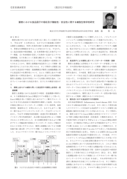
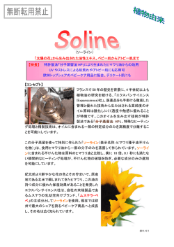
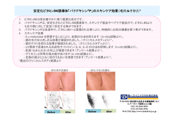
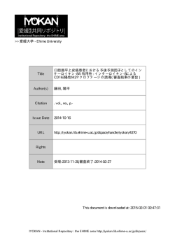
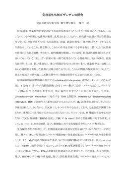
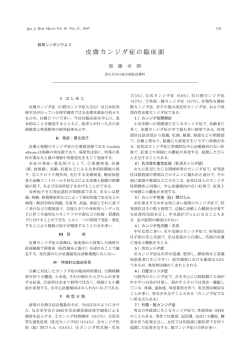
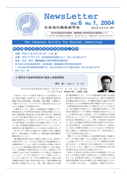
Jpn. J. Med. Mycol.
Vol. 45, 131−136, 2004
ISSN 0916−4804
総 説
サイトカイン産生からみた皮膚真菌症の病態
加 納 塁
日本大学生物資源科学部獣医臨床病理学研究室
要 旨
皮膚は, 直接外界と接し, 多彩な免疫機構によって生体を防御している免疫臓器である. 皮膚の免疫機構の中で貪
食細胞はカンジダや皮膚糸状菌などの感染時に重要な働きをしているが, 貪食細胞が表皮と真皮をとわず皮膚の病巣
に集蔟するためには, 貪食細胞の活性化を誘導する炎症性サイトカインが深く関与しているものと考えられる.
そこで皮膚の真菌感染時におけるケラチノサイトから産生されるサイトカインを検討するため, 培養ケラチノサイ
トにカンジダ, 皮膚糸状菌, マラセチアを感作させ, ケラチノサイトから培養液中に放出された炎症性サイトカイン
(IL-1β, IL-6, IL-8, MCP-1, TNF- α)をELISA法を用いて測定した. その結果, カンジダではIL-8, 皮膚糸状菌で
はIL-8, TNF- α, マラセチアではIL-1β, IL-6, IL-8, TNF- αの産生が確認された.
以上の結果から, これら真菌の刺激が表皮に及ぶと, ケラチノサイトから炎症性サイトカインが分泌され, それに
よって白血球の遊走や, 補体の活性化が惹起され炎症が進行し, 感染真菌が排除されるものと思われる. したがって
皮膚真菌症の病態解明や治療戦略にとって, ケラチノサイトにおける真菌によるサイトカインの誘導や発現を解明す
ることは重要であると考えられる.
key word: サ イ ト カ イ ン(cytokine), ケ ラ チ ノ サ イ ト(keratinocyte), ELISA(enzyme-linked immunosorbent
assay), カンジダ(Candida), マラセチア(Malassezia), 皮膚糸状菌(tinea)
はじめに
皮膚は粘膜とともに直接外界と接する臓器である. と
もすると単純な生体保護膜のように考えられるが. 生体
を防御している免疫臓器であり, その免疫機構は複雑で
ある. 皮膚の厚さは約 20 μm であるが, 角質は有害物質
の浸透や微生物の侵入を物理的に防御している. また各
種化学的物質を分泌して, 微生物の増殖や侵入を防いで
いる. しかしながら角層を通過した異物に対しては, 表
皮を構成するケラチノサイトから, 多種のサイトカイン
が分泌され, それによって周辺の細胞からの抗菌物質の
産生が増強する. また白血球の遊走や, 血管透過性が亢
進して液体成分や補体が浸出し, 炎症が惹起され, さら
に炎症部位周辺の細胞や, 遊走した白血球からもサイト
カインや抗菌物質などの炎症性物質が分泌され, 炎症が
進行することによって侵入物は速やかに排除される.
感染時にステロイド剤が投与されていると, 微生物の
存在を認識しても, 抗菌物質やサイトカイン産生が抑制
されるため, 炎症が起こりにくくなり易感染状態となっ
て感染が拡大する.
そこで皮膚の真菌感染時におけるケラチノサイトから
産生されるサイトカインのうち炎症性サイトカインを検
討するため, 培養ケラチノサイトにカンジダ, 皮膚糸状
別刷請求先:加納 塁
〒252-8510 神奈川県藤沢市亀井野1866
日本大学生物資源科学部獣医臨床病理学研究室
菌, マラセチアを感作させ, ケラチノサイトから培養液
中 に 放 出 さ れ た 炎 症 性 サ イ ト カ イ ン(IL-1β, IL-6,
IL-8, MCP-1, TNF- α)について ELISA 法を用いて測
定した.
1)ケ ラ チ ノ サ イ ト の サ イ ト カ イ ン 産 生 に 及 ぼ す
Candida albicans の影響
皮膚カンジダ症の病理組織像において, 好中球の著し
い浸潤が認められるが, これには, ケラチノサイトから
産生される炎症性サイトカインの関与が考えられる. そ
こでケラチノサイトのサイトカイン産生に対する C.
albicans の影響について検討した.
ヒ ト ケ ラ チ ノ サ イ ト は, Clonetics 社(BioWhittaker
社)の 正 常 ヒ ト 表 皮 細 胞 を NHEK 培 養 液 中 で 37゜
C,
5% CO2 の条件下で培養を行った.
使 用 菌 株 は C. albicans 血 清 型 A の 3 株(TLD-0141,
TLD-0142, TLD-0143)を用い, それぞれサブローブド
ウ糖液体培地で 37゜
C, 3 日間振盪培養後, 菌を回収し
た. 次に NHEK 培養液中で 1×10 3 個の菌数に調整し,
1×10 4 個 の ヒ ト ケ ラ チ ノ サ イ ト と 同 時 に 37゜
C, 5%
CO2 の条件下で培養した. 培養液を経時的に回収し, 培
養液中の炎症性サイトカインを ELISA 法(Cytoscreen
Immunoassay kit; BioSource International 社)によって
測定した. その結果, IL-1β, IL-6, MCP-1, TNF- αは
測定されなかったが, IL-8 は経時的に増加することが確
認された(Fig. 1).
132
真菌誌 第45巻 第 3 号 平成16年
Fig. 2. IL-8 mRNA production in human keratinocytes cocultured with C. albicans.
After 0 h, 1 h, 3 h, and 6 h of co-culture of keratinocytes with
live C. albicans, the IL-8 mRNA levels in the keratinocytes were
determined by RT-RCR. Reaction products were run on 2%
agarose gels. Although IL-8 mRNA in keratinocytes is
undetectable at 0 h of co-culture, IL-8 mRNA was detected in
the keratinocytes after 1 - 6 h of co-culture. Ubiquitin was the
positive transcription control.
Fig. 1. Effect of live, killed and cultured supernatant of C.
albicans on cytokine production by human keratinocytes.
1a, Keratinocytes(1×10 4 cells per well)were co-cultured with
the live C. albicans(1×10 3 or 1×10 4 cells per well). After 1 h,
3 h, 6 h, and 14 h, the IL-8 levels in the supernatants were
measured by the ELISA method.
1b, Keratinocytes (1×10 4 cells)were cultured with the
supernatant of the C. albicans(1×10 7 per ml). After 1 h, 3 h,
and 6 h, the IL-8 levels in the supernatants were measured by
the ELISA method. Results shown are representative of five
experiments.
次 に 1×10 7 個 の C. albicans(TLD-0141)を 10 ml の
NHEK 培養液中に 37゜
C で一晩培養し, その培養液を濾
過した. これをヒトケラチノサイトに加えて培養し, 1×
C, 5% CO2
10 4 個のヒトケラチノサイトに加えて, 37゜
の条件下で培養した. 培養液を経時的に回収し, サイト
カ イ ン を ELISA 法 に よ っ て 同 様 に 測 定 し た. そ の 結
果, IL-8 産生は濾液のみによっても促進されることが認
められた(Fig. 1).
さらにカンジダと共培養することでケラチノサイトか
ら IL-8 mRNA の発現が誘導されることを確認するた
め に, reverse transcription-PCR(RT-PCR)に よ る IL-8
mRNA の発現を検討した.
まず 5×10 5 個のヒトケラチノサイトと 5×10 5 個の
C. albicans(TLD-0141)を同時に 37゜
C, 5% CO2 の条件
下で培養し, 培養 1, 3, 6 時間後に細胞を回収した. 回収
し た 細 胞 か ら, RNeasy total RNA kit(QIAGEN 社)
を 用 い て RNA を 抽 出 し, こ れ か ら OmniscriptTM
Reverse Transcriptase kit(QIAGEN)によって cDNA
を合成した. これを鋳型として IL-8 に特異的なプライ
マーを作成し PCR を行い, IL-8 の mRNA 発現を検出
した.
ヒト IL-8 遺伝子に特異的なプライマーは Gene Bank
に登録されているヒトの IL-8 の遺伝子情報から設計し
た 1). プライマーの塩基配列は下記に示した.
5'-ATGACTTCCAAGCTGGCCGT-3' (primer IL-8
1S; nt. 102-122 in Human mRNA for MDNCF in the
Gene Bank accession nos.-Y00787)
5'- TCCTTGGCAAAACTGCACCT-3' (primer IL-8
1R; nt. 164-183).
PCR の反応条件は 94゜
C 1 分, 55゜
C 1 分, 72゜
C 2 分を
1 サイクルとして 30 サイクル反応を行い, 目的の遺伝子
断片を増幅した.
PCR で増幅した IL-8 の遺伝子断片は, 2%アガロース
ゲル上で泳動後, 臭化エチジウムで染色後, 紫外線照射
下で確認した.
その結果, IL-8 の mRNA は培養 1 時間後から発現す
ることが確認された(Fig. 2).
以上の結果から, C. albicans はヒトケラチノサイト IL-8
の産生を誘導することが示唆された. 一般に皮膚カンジ
ダ症では, 著明な好中球浸潤が認められる 2). 今回得ら
れた結果から, 皮膚カンジダ症においては, ケラチノサ
イトから IL-8 が産生され, それによって好中球浸潤が
起きていることが示唆された. 今後皮膚カンジダ症の病
態解明のためには, IL-8 の産生誘導因子の解析が重要で
あると考えられる 3).
2)ケラチノサイトのサイトカイン産生に及ぼすマラセ
チアの影響
Malassezia 属の 4 菌種(Table 1)をカンジダと同様
Jpn. J. Med. Mycol. Vol. 45(No. 3), 2004
133
Fig. 3. Effect of Malassezia species on cytokine protein release.
Keratinocytes were co-cultured with Malassezia species. After 1 hr, 3 hr, 6 hr 12 hr and 24 hr, the supernatants were
collected and cytokine releases were analyzed by the ELISA method. (a)IL-1β,(b)IL-6,(c)IL-8,(d)MCP-1 and(e)
TNF- α.
真菌誌 第45巻 第 3 号 平成16年
134
Table 1. Species and strains used in this study
Species
Strain
M. furfur
M. furfur
M. furfur
M. pachydermatis
M. pachydermatis
M. slooffiae
M. slooffiae
M. sympodialis
M. sympodialis
VUT-98021
VUT-98022
VUT-98023
VUT-98028
VUT-98010
VUT-98030
VUT-98031
VUT-98032
VUT-98033
(CBS 1878)
(CBS 7019)
(CBS 6000)
(CBS 1879)
(Clinical isolate of cat)
(CBS 7875)
(CBS 7956)
(CBS 7222)
(CBS 7977)
VUT : School of Veterinary Medicine, University of Tokyo
CBS : Centraalbureau voor Schimmelcultures
の条件で, ヒトケラチノサイトと培養し, 経時的に培養
液のサイトカイン量を ELISA 法によって測定した. そ
の 結 果, MCP-1 は 産 生 さ れ な か っ た が,3 菌 種 で IL-1
β, IL-6, IL-8, TNF- αが産生され, 感作に用いた菌種
によってサイトカイン活性が異なっていた(Fig. 3). こ
のことから Malassezia 属の菌種により細胞浸潤や炎症反
応の度合いが異なり, それにともなって皮膚病変が異な
ることが示唆された 4).
3)ケ ラ チ ノ サ イ ト の サ イ ト カ イ ン 産 生 に 及 ぼ す
Trichophyton mentagrophytes の影響
T. mentagrophytes の 6 株を 1/10 サブローブドウ糖寒天
培地上で 25゜
C, 14 日間培養後, 菌体を回収した. 次に菌
体を滅菌水中に懸濁後, 40μmのフィルターに通して,
Fig. 4. Effect of T. mentagrophytes on cytokine protein release.
Keratinocytes were co-cultured with T. mentagrophytes. After 1 hr, 3 hr, 6 hr 12 hr and 24 hr, the supernatants were
collected and cytokine releases were analyzed by the ELISA method. (a)IL-8,(b)TNF- α.
Jpn. J. Med. Mycol. Vol. 45(No. 3), 2004
小分生子と分節分生子を回収した. この懸濁液中の菌体
数計算後, カンジダと同様の条件でヒトケラチノサイト
と培養し, 経時的に培養液のサイトカイン量を ELISA法
によって測定した. その結果, MCP-1 IL-1β, IL-6, は
産 生 さ れ な か っ た が,IL-8, TNF- α が 産 生 さ れ た
(Fig. 4). このことから白癬菌によってもケラチノサイ
トからサイトカインが誘導されることが確認された. ま
た, 白癬の場合も好中球を主体とした炎症細胞浸潤が認
められることから, 白癬菌が角質下の生きているケラチ
ノサイトと接触することによって炎症が惹起されること
が示唆された 5).
ま と め
以上の結果から, これら真菌の刺激が表皮に及ぶと,
ケラチノサイトから炎症性サイトカインが分泌され, そ
れによって白血球の遊走や, 補体の活性化が惹起され炎
症が進行し, 感染真菌が排除されるものと思われる.
生体の細胞が侵入してきた微生物をどのように認識し
ているかについては, 近年侵入微生物の細胞壁や遺伝子
を感知する細胞膜レセプター(Toll-like receptor)が発
見され, その機能について注目されている. このレセプ
ターはヒトやマウスの貪食細胞, 粘膜細胞, 表皮細胞な
どにも存在し, 上記の微生物由来物を認識することで,
炎症性サイトカインの分泌が誘導されることが確認され
ている 6, 7).
このように皮膚真菌症の病態解明や治療戦略にとっ
て, ケラチノサイトにおける真菌によるサイトカインの
誘導や発現を解明することは重要であると考えられる.
135
文 献
1)Matsushima K, Morishita K, Yoshimura T, Lavu S,
Kobayashi Y, Lew W, Appella E, Kung HF, Leonard EJ,
Oppenheim JJ: Molecular cloning of a human monocytederived neutrophil chemotactic factor (MDNCF)and
the induction of MDNCF mRNA by interleukin 1
and tumor necrosis factor. J Exp Med 167: 1883−
1893, 1988.
2)Longley BJ: Fungal disease. In Lever’s Histopathology
of the skin (Elder D ed). 8th ed. 517−551.p.
Philadelphia, Lippincott-Raven Publishers, 1997.
3)Kano R, Watanabe S, Sato H, Nakamura Y,
Hasegawa A: Candida albicans induced interleukin 8
production by human keratinocytes. J Dermatol Sci
31: 233−235, 2003.
4)Nakamura Y, Kano R, Hasegawa A, Watanabe S: IL-8
and TNFα production from human epidermal keratinocytes induced by Trichophyton mentagrophytes. Clin
Diag Laboratory Immun 9: 935−937, 2002.
5)Watanabe S, Kano R, Sato H, Nakamura Y,
Hasegawa A: The effect of Malassezia yeasts on
cytokine production by human keratinocytes. J Invest
Dermatol 116: 769−773, 2001.
6)Kawai K, Shimura H, Minagawa M, Ito A,
Tomiyama K, Ito M: Expression of functional Tolllike receptor 2 on human epidermal keratinocytes. J
Dermatol Sci 30: 185−194, 2002.
7)Pivarcsi A, Bodai L, Rethi B, Kenderessy-Szabo A,
Koreck A, Szell M, Beer Z, Bata-Csorgoo Z, Magocsi
M, Rajnavolgyi E, Dobozy A, Kemeny L: Expression
and function of Toll-like receptors 2 and 4 in human
keratinocytes. Int Immunol 15: 721−730, 2003.
真菌誌 第45巻 第 3 号 平成16年
136
Cytokine Production and Dermatophytosis
Rui Kano
Department of Pathobiology, Nihon University School of Veterinary Medicine,
1866 Kmeino, Fujisawa, Kanagawa 252-8510, Japan
The characteristic pathological feature of dermatomycosis is numerous neutrophilic infiltrates within
the epidermis. However, the precise mechanism of this infiltration remains unknown. In this study,
interleukins 1β, 6, and 8, monocyte chemotactic protein-1(MCP-1), and tumor necrosis factor(TNF)α levels in the medium where keratinocytes were co-cultured with Candida albicans, Malassezia and
Trichophyton mentagrophytes, were determined by enzyme-linked immunosorbent assays(ELISAs)in order
to estimate the effect of these fungi on the cytokine production from human keratinocytes.
The IL-8 level in the supernatants increased with 1 to 14 hours of co-culture in response to live C.
albicans, but the other cytokines were undetectable. Furthermore, the mRNA of IL- 8 in keratinocytes
was also confirmed to increased. This data suggested that C. albicans directly induce interleukin 8
production from human keratinocytes without activated macrophages.
The IL-6, IL-8, and TNF- α levels in the culture supernatants increased with 1 to 24 hours of coculture with keratinocytes and Malassezia species but the MCP-1 level was undetectable.
The IL-8 and TNF- α levels in the culture supernatants increased with 1 to 24 hours of co-culture
with keratinocytes and Trichophyton mentagrophytes but the other cytokine levels were undetectable.
The ELISA analysis of cytokine production by human keratinocytes will provide useful information
in understanding the pathogenesis of dermatomycosis.
この論文は, 第 47 回日本医真菌学会総会の“シンポジウム 1 : 皮膚科領域における抗真菌剤治療のトピックス
−皮膚真菌症の病態と抗真菌薬の新展開−”において発表されたものです.
© Copyright 2026 Paperzz