J. Embryol. exp. Morph. 79, 41-51 (1984)
Printed in Great Britain © The Company of Biologists Limited 1984
Electron microscopic evidence for the presence of an
asialoglycoprotein receptor on isolated foetal rat
hepatocyte surface
By LAURA CONTI DEVIRGILIIS 1 , LUCIANA DINI1 AND
SALVATORE RUSSO-CAIA1
From the Department of Cellular and Developmental Biology, 1st University of
Rome, and the Department of Biology, 2nd University of Rome
SUMMARY
The ontogeny of asialoglycoprotein receptor was investigated by electron microscopic
cytochemistry in hepatocytes isolated from foetal and adult rat. The binding capacity for
asialofetuin coupled to horseradish peroxidase was lacking before the 18th day of intrauterine
life; it arises at this time and increases with developmental age.
The ligand-receptor complexes form small patches. The distribution pattern of positivity is
very similar in pre and postnatal age, covering the entire cell surface.
These results indicate a rather late appearance of the galactose-binding capacity related to
the asialoglycoprotein clearance function, which is typical of adult mammalian liver.
INTRODUCTION
Several carbohydrate recognition systems are present in mammalian liver
involved in the specific binding and internalization of different glycoproteins.
Among them, the asialoglycoprotein (ASG) receptor is the most widely studied,
being located on parenchymal cells, specifically recognizing galactose residues
and thus mediating the removal from the plasma of these modified glycoproteins
(Ashwell & Harford, 1982).
This system, first described by Ashwell & Morell (1974), since then has been
well characterized, the receptor molecule has been isolated and many biochemical and morphological data concerning the binding, internalization and
degradative pathway of ASG have been obtained both in whole liver and in
isolated hepatocytes (Tolleshaug, Berg, Frolich & Norum, 1979; Wall, Wilson
& Hubbard, 1980; Weigel & Oka, 1982; Geuze et al. 1982). The experiments
performed on isolated parenchymal cells have conclusively demonstrated that
the ASG receptor remains present in isolated hepatocytes and is also preserved
Dedicated to the memory of Professor Enrico Urbani, who died on the 9th of June 1983.
1
Authors' address: Dipartimento di Biologia cellulare e dello sviluppo, Facolta di Scienze
M.F.N., I Universita di Roma, p.le Aldo Moro, 00100 Roma, Italia.
42
L. CONTI DEVIRGILIIS, L. DINI AND S. RUSSOCAIA
in cultures of adult rat hepatocytes (Deschuyteneer et al. 1982; Zeitlin & Hubbard, 1982).
All these data have been obtained from adult liver, the only study relating to
foetal life, to our knoweldge, being that of Hickman & Ashwell (1974), who
incidentally reported that foetal liver was essentially devoid of binding activity.
Although several aspects of the ontogeny of protein receptors (mainly for hormones and growth factors) have been carefully investigated (Csaba, 1981), no
information on the time of appearance in the embryonic development of liver
ASG clearance function is available so far.
These considerations, together with the difficulties in administering in vivo any
substances to the foetal liver, have prompted us to utilize dissociated foetal
hepatocytes to investigate the ontogeny of ASG receptor. We have therefore
investigated the presence and distribution of the ASG receptor on foetal rat
hepatocyte surface using electron microscopic cytochemistry to study the
localization of asialofetuin coupled to horseradish peroxidase (ASF-HRP).
MATERIALS AND METHODS
Hepatocytes preparation
Isolated adult hepatocytes were obtained from liver of Wistar rats perfused
with collagenase (Boehringer), according to the method of Moldeus, Hogberg
& Orrenius (1978).
Foetal livers were obtained after rapid hysterectomy from pregnant females
anaesthesized with Farmotal (Farmitalia) 20 mg/100 g.b.w.; embryonal age was
calculated on the basis of the appearance of vaginal plug and checked by foetus
weight and length. The hepatocytes were isolated by immersion of small liver
fragments in a collagenase solution, according to the procedure set up in our
laboratory and detailed elsewhere (Conti-Devirgiliis et al., 1981). This
procedure was sometimes applied to adult livers to test the uniformity of results
obtainable by the two isolation methods.
Cell yeld was 40-60 x 106 hepatocytes/g of fresh liver.
Cell viability was evaluated by the Trypan blue exclusion test and LDH
leakage (Dickson & Pogson, 1977) and further assessed by morphological
examination under electron microscope. In some experiments hepatocytes were
purified by centrifugation in a Percoll (Pharmacia) gradient as described by
Conti-Devirgiliis et al. (1981).
Asialofetuin-horseradish peroxidase (ASF-HRP) preparation and
characterization
Fetuin (type IV, Sigma) was desialated by treatment with insoluble
neuraminidase (Sigma) at 37 °C for 48 h as described by Dunn, Labadie & Aronson (1979).
Asialoglycoprotein receptors on isolated rat hepatocytes
43
Orosomucoid (oti-acid-glycoprotein, Sigma) used for controls, was desialated
in the same way.
Removal of sialic acid was checked by a gas chromatographic assay (Varma
& Varma, 1976).
The conjugate between ASF and HRP (Boehringer) was obtained according
to the method described by Wall etal. (1980) for asialorosomucoid, with minor
modification. The 3H-ASF added to cold ASF (1:20) during conjugation was
prepared by the reductive methylation procedure of Means and Feeney, as
described by Weigel & Oka (1982).
The specific radioactivity obtained was 2 x 10⁷ dpm/mg of protein. The complex ASF-HRP was separated from the monomeric forms of ASF and HRP by
means of a Sephadex G 200 column (cm 3x68) equilibrated in PBS. 2-5ml
fractions were collected at flow rate of 3-85ml/h/cm 2 and were monitored for
absorbance at 280 and 403 nm, and for 3H-radioactivity.
Two peaks were eluted from the column: the first between 150 and 180 ml, and
the second between 185 and 205 ml. Both peaks contained material absorbing at
403 nm (indicating the presence of the peroxidase haem group) but the 3H-ASF
radioactivity was almost entirely detected (97 %) in the first peak.
Analysis of this peak by SDS gel electrophoresis, performed according to
Weber & Osborne (1969) shows only one band, corresponding to a Mr 80000,
which was positive for HRP activity. HRP, ASF and fetuin simultaneously
analysed show RF values clearly different from ASF-HRP complex.
Hepatocyte treatment with conjugate
Freshly isolated hepatocytes, filtered through a nylon gauze (holes 100 //m
dia) and twice rinsed with Krebs-Henseleit solution to remove the collagenase,
were resuspended 1 x 10⁶/ml in Waymouth medium (Gibco) supplemented
with 2 % BSA (Miles), lOi.u./ml of heparin, lOOOi.u./ml of penicillin and
lmg/ml of streptomycin, continuously bubbled with carbogen (95% O2 and
5 % CO2) and maintained for at least 120 min at 37 °C in a rotating bath. This
period was demonstrated to be the minimum essential to recover a good
metabolic capacity after collagenase treatment (Weigel & Oka, 1982; Zeitlin
6 Hubbard, 1982).
After the restoring period, the conjugate (30 to 300/^g ASF-HRP for
5-30min) was added to incubation medium. All the experiments were performed at 37 °C, with oxygenation and rotation, in presence of 25 mM-CaCh. After
incubation, cells were washed with PBS, fixed with 2 % glutaraldehyde in 0-1 Mcacodylate buffer for 15 min at 4°C, rinsed twice in the same buffer before
detection of HRP activity. The hepatocytes were resuspended in 3 mg/ml DAB,
0-02 M-3 amino 2,4 triazole and 0-03M-H 2 O 2 in 0-1 M-Tris HCl buffer, pH 7-5 and
incubated in the dark for 120 min at 24 °C (Wall et al. 1980).
Finally the cells, after rinsing in Tris buffer, were postfixed for l h in 1 %
OSO4 in 0-lM-cacodylate buffer, dehydrated and embedded in Epon 812 for
44
L. CONTI DEVIRGILIIS, L. DINI AND S. RUSSO-CAIA
electron microscopy. Ultrathin sections unstained or stained by lead citrate and
uranyl acetate were observed under a Philips 400 T electron microscope.
Controls
The hepatocytes were treated in the following ways. Before treatment with
conjugate: 15min incubation in Ca++-free medium, containing 20mM-EDTA,
after several rinsing of the cells by Krebs-Henseleit Ca++-free containing EDTA
for complete removal of calcium; preincubation and/or incubation with unconjugated ASF 20-100 fold exceeding the ASF-HRP concentration.
Incubation in presence of 65 mM-N-acetyl galactosamine.
Incubation in presence of a 33-fold excess of asialorosomucoid (ASOR), as
asialoglycoprotein competitor.
RESULTS
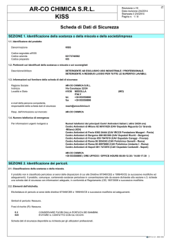
Isolated adult hepatocytes exposed to saturating concentration of ASF-HRP
at 37 °C for 5-15 min, exhibit a quite uniform staining of the entire cell surface;
sometimes the label is particularly concentrated between the microvilli and in the
position of the pits. A few vesicles lying below the plasma membrane are also
stained (Figs 1,2).
By increasing the incubation time (until 30 min) the labelling pattern is practically unchanged. Among the cells of each preparation, all showing a good preserved morphology, some are not at all stained.
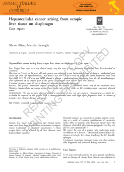
As far as foetal hepatocytes are concerned, the most relevant data deal with the
absence of asialoprotein-binding capacity before the 18th day of intrauterine life;
in fact hepatocytes isolated from foetal rats aging 15,16 or 17 days, treated with
ASF-HRP at the highest concentration employed, are devoid of cell surface reaction product (Figs 4, 5). The hepatocytes of these preparations show always
90 % of viability as evaluated by the Tripan blue exclusion test and LDH leakage.
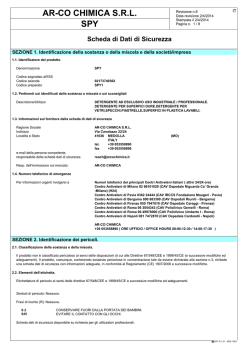
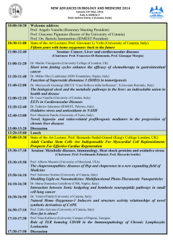
On the contrary, starting from the 18th day of development the positivity
arises and is maintained as the developmental age goes on (Figs 6, 7 and 8).
In spite of the different morphological appearance of foetal hepatocytes which
exhibit a more regular surface, with few microvilli, the distribution pattern of
positivity is quite similar to that observed in the adult hepatocytes, i.e. small
patches cover the entire cell surface, even if the foetal cells are always less
intensely stained (Figs 6, 7 and 8).
Neither specific areas of reaction product accumulation nor engulfed regions
and/or internalization pattern are detectable with the experimental conditions
employed, although some structures similar to coated pits and vesicles are
present since the earliest prenatal stage observed (Figs 4, 5).
Therefore the cytochemical results suggest a rather late appearance of binding
capacity for asialoglycoproteins by the hepatocytes during the ontogenesis.
In the control experiments carried out in presence of excess of ASF (Fig. 3)
Asialoglycoprotein receptors on isolated rat hepatocytes
Fig. 1. Hepatocyte isolated from adult rat (viable cells 95 %) and treated for 5 min
with ASF-HRP. The positivity is quite uniformly distributed along cell surface.
Unstained. Bar = 0-5 ^m.
Fig. 2. Hepatocyte isolated from adult rat and treated for 30min with ASF-HRP.
Near the surface, an internalized vesicle is visible (arrow). Unstained. Bar = 0-5 jum.
Fig. 3. Hepatocyte isolated from adult rat. 3 mg/ml ASF as competitor were added to
30jUg/ml ASF-HRP: reaction product is absent from the cell surface. Bar = 0-5 jum.
45
46
L. CONTI DEVIRGILIIS, L. DINI AND S. RUSSO-CAIA
Figs 4 & 5. Foetal hepatocytes isolated at 16th (4) and 17th (5) day of development
(viable cells 90%) and treated for 15min (4) and 30min (5) with ASF-HRP 300
jug/ml. Cell surfaces appear free of reaction product; coated pits and vesicles are
present (arrows). Bar = 0-
Asialoglycoprotein
receptors on isolated rat hepatocytes
47
Figs 6 & 7. Hepatocytes isolated at 18th (6) and 19th (7) day of intrauterine life
(viable cells 90 %) after 15 min of incubation with ASF-HRP. The positivity is well
visible. Bar = 0-
or ASOR as competitors, or after EDTA treatment, the reaction product is
totally absent, indicating that the binding of ASF-HRP is specific and requires
Ca + + (not shown).
48
L. CONTI DEVIRGILIIS, L. DINI AND S. RUSSO-CAIA
8
Fig. 8. Hepatocyte isolated at 21st day of foetal life (viable cells 95 %) and incubated
for 30min with ASF-HRP. Bar - 0-5 ^m.
When the incubation is performed in presence of N-acetylgalactosamine, the
labelling is not completely abolished and a faint positivity is still evident (not
shown).
DISCUSSION
The data reported here provide another demonstration that isolated rat
hepatocytes from adults and during prenatal life have properties comparable to
those found in the intact liver, making them a suitable in vitro system to study
metabolism.
Among the differences that must be considered the first is the loss of cell
polarity; the sinusoidal and biliary domains are no longer segregated in isolated
hepatocytes (Zeitlin & Hubbard, 1982; Matsuura, Nakada, Sawamura &
Tashiro, 1982): therefore, in many instances receptor-ligand complexes, including ASG receptor complexes, show an uniform distribution on the cell surface,
both in the adult and in the foetus, in contrast to the hepatocytes in situ. A loss
of plasma membrane specialization has also been observed as regards the
Asialoglycoprotein receptors on isolated rat hepatocytes
49
cytochemical localization of several surface enzymes (Groothuis, Hulstaert,
Kalicharan & Hardonk, 1981).
The heterogeneity of ASF-HRP positivity among hepatocytes of some
preparations, observed both in adult and in foetuses, may be explained on the
basis of variability in the number of exposed receptors. This could be related to
a variable degree of loss of receptors during preparation or to a different recovery during incubation at 37°C in Waymouth medium, due to the recycling
mechanism operating on an internal receptor pool (Bridges, Harford, Ashwell
& Klausner, 1982; Weigel & Oka, 1982). A pre-existing heterogeneity among
hepatocytes cannot be ruled out, and may perhaps be linked to their position in
the lobule (Hardonk & Scholtens, 1980).
As far as embryonic development is concerned, in the experiments presented
here the presence of an ASG receptor could be demonstrated by electron
microscopic cytochemistry of ASF-HRP on freshly isolated foetal rat
hepatocytes after the 17th day of intrauterine life; cells isolated from younger
embryos do not show in fact any surface reaction even after incubation in a
medium containing the highest concentration of ASF-HRP and after 30 min of
incubation.
These data seem to indicate a rather late appearance on the hepatocyte
surface of the galactose-binding capacity to which the circulating ASG clearance
function, typical of adult mammalian liver, may be related.
Some general aspects of receptor ontogenesis have been extensively reviewed
in recent papers (Csaba, 1981), in which it has been recalled that receptor
maturation seems to represent one of the key events in cell-membrane differentiation; the ontogenetic patterns of specific receptors may however be quite
different in the same cell type. The glucagon-binding capacity of embryonic rat
liver cells is only 1 % relative to adult liver at 15 days of prenatal life, and still
not more than 23 % at the birth (Blasquez et al. 1976). Insulin-binding capacity
of embryonic hepatocyte membrane, on the other hand, has been found to be
less, similar to, or even greater than, that of adult cells (Blasquez et al. 1976;
Neufeld, Scott & Kaplan, 1980; Autuori et al. 1981; Vinicor & Kiedrowski,
1982).
The absence of a clearance mechanism of circulating ASG in the liver of early
foetuses, which is suggested by our results, may be related either to the absence
in the early embryonic metabolism of extensive desialization processes of plasma
glycoproteins, which become operative only after the 17th day of development,
or to the fact that the removal of these modified proteins could be carried out by
placental tissues.
A point which remains to be investigated is the route of internalization and the
degradative pathway of ASG after the appearance of binding capacity, i. e. in the
last days of intrauterine life, when the lysosomal system of hepatocytes has not
yet reached the functional and morphological development characteristic of the
adult cells (Ciofi-Luzzatto, 1981).
50
L. CONTI DEVIRGILIIS, L. DINI AND S. RUSSO-CAIA
The authors are grateful to Professor F. Autuori for useful discussion through the investigation; to Professor P. Orlando, Director of the Central Radioisotope Service, Faculty of
Medicine, Catholic University of Rome for the preparation of [3H]asialofetuin, and to Mrs V.
Autuori-Pezzoli for her excellent technical assistance. This work was supported by a grant
from the Italian Ministero della Pubblica Istruzione to one of us (S.R.C.).
REFERENCES
ASWELL, G. & MORREL, A. G. (1974). The role of surface carbohydrates in the hepatic
recognition and transport of circulating glycoproteins. Adv. Enzymol. 41, 99-128.
ASHWELL, G. & HARFORD, J. (1982). Carbohydrate-specific receptors of the liver. Ann. Rev.
Biochem. 51, 531-554.
AUTUORI, F., BALDINI, P., CIOFI-LUZZATTO, A., CONTI-DEVIRGILIIS, L., DINI, L., INCERPI, S.
& LULY, P. (1981). Insulin binding and internalization in rat hepatocytes during prenatal
and postnatal life. Biochim. Biophys. Ada, 678, 1-6.
BLASQUEZ, E., RUBALCAVA, B., MONTESANO, R., ORCI, L. & HUNGER, R. H. (1976). Development of insulin and glucagon binding and the adenylate cyclase response in liver membranes
of the prenatal, postnatal and adult rat: evidence of glucagon "resistance". Endocrinology,
98, 1014-1023.
BRIDGES, K., HARFORD, J., ASHWELL, G. & KLAUSNER, R. D. (1982). Fate of receptor and
ligand during endocytosis of asialoglycoproteins by isolated hepatocytes. Proc. natn. Acad.
Sci. U.S.A., 79,350-354.
CIOFI-LUZZATTO, A. (1981). Hepatocyte differentiation during early fetal development in the
rat. Cell Tissue Res. 215, 133-142.
CONTI-DEVIRGILIIS, L., DINI, L., DI PIERRO, A., LEONI, S., SPAGNUOLO, S. & STEFANINI, S.
(1981). An improved non perfusion method for the isolation and purification of rat fetal and
neonatal hepatocytes. Cell, molec. Biol. 27, 687-694.
CSABA, G. (1981). Ontogeny and Phytogeny of Hormone Receptors. Monographs in Developmental Biology, vol. 15, Basel: S. Karger A. G.
DESCHUYTENEER, M., PRIEELS, J. P., MAY, C , PERRAUDIN, J. P. & WANSON, J. C. (1982).
Studies on the liver galactose and fucose recognition systems in cultured and isolated adult
rat hepatocytes. Biol. Cell. 44, 15-24.
DICKSON, A. J. & POGSON, C. J. (1977). The metabolic integrity of hepatocytes in sustained
incubations. FEBS Lett. 83, 27-32.
125
DUNN, W. A., LABADIE, J. H. & ARONSON, N. N. (1979). Inhibition of
I-asialofetuin
catabolism by leupeptin in the perfused rat liver and in vivo. J. biol. Chem. 254,4191-4196.
GEUZE, H. J., SLOT, J. W., STROUS, G. J. A. M., LODISH, H. F. & SCHWARTZ, A. L. (1982).
Immunocytochemical localization of the receptor for ASGP in rat liver cells. J. Cell Biol.
92, 865-870.
GROOTHUIS, G. M. M., HULSTAERT, C. E., KALICHARAN, D. &HARDONK, M. J. (1981). Plasma
membrane specialization and intracellular polarity of freshly isolated rat hepatocytes. Eur.
J. Cell Biol. 26, 43-51.
HARDONK, M. J. & SCHOLTENS, H. B. (1980). A histochemical study about the zonal
distribution of the galactose-binding protein in rat liver. Histochemistry 69, 289-297.
HICKMAN, J. & ASHWELL, G. (1974). Studies on the hepatic binding of asialoglycoproteins by
hepatoma tissue and by isolated hepatocytes. Enzyme Therapy in Lysosomal Storage
Diseases, (eds J. M. Tager, G. J. M. Hooghwinkel & W. T. Daems), pp. 169-172. Amsterdam: North-Holland.
MATSUURA, S., NAKADA, H., SAWAMURA, T. & TASHIRO, Y. (1982). Distribution of an ASG
receptor on rat hepatocyte cell surface. /. Cell Biol. 95, 864-875.
MOLDEUS, P., HOGBERG, J. & ORRENIUS, S. (1978). Isolation and use of liver cells. Methods
in Enzymology. S. Fleischer & L. Packer, vol. 52, part C, 60-71. New York: Academic
Press.
NEUFELD, N. D., SCOTT, M. & KAPLAN, S. A. (1980). Ontogeny of the mammalian insulin
receptor. Studies of human and rat fetal liver plasma membranes. Devi Biol. 78,151-160.
Asialoglycoprotein receptors on isolated rat hepatocytes
51
H., BERG, T., FROLICH, W. & NORUM, K. R. (1979). Intracellular localization
and degradation of asialofetuin in isolated rat hepatocytes. Biochim. Biophys. Ada, 585,
71-84.
VARMA, R. & VARMA, R. (1976). Simultaneous determination of neutral sugars and
hexosamines in glycoproteins and acid mucopolysaccharides (glycosaminoglycans) by gasliquid chromatography. J. Chromat. 128, 45-52.
VINICOR, F. & KIEDROWSKI, L. (1982). Characterization of the hepatic receptor for insulin in
the perinatal rat. Endocrinology, 110, 782-790.
WALL, D. A., WILSON, G. & HUBBARD, A. L. (1980). The galactose-specific recognition
system of mammalian liver: the route of ligand internalization in rat hepatocytes. Cell 21,
79-93.
WEBER, K. & OSBORNE, M. (1969). The reliability of molecular weight determinations by
dodecyl sulfate-polyacrylamide gel electrophoresis. J. biol. Chem. 244, 4406-4412.
WEIGEL, P. H. & OKA, J. A. (1982). Endocytosis and degradation mediated by the asialoglycoprotein receptor in isolated rat hepatocytes. /. biol. Chem. 257, 1201-1207.
ZEITLIN, P. L. & HUBBARD, A. L. (1982). Cell surface distribution and intracellular fate of
ASGP: a morphological and biochemical study of isolated rat hepatocytes and monolayer
cultures. J. Cell Biol. 92, 634-647.
TOLLESHAUG,
{Accepted 12 August 1983)
© Copyright 2026 Paperzz