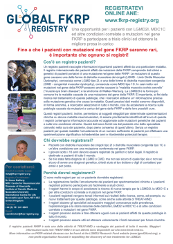
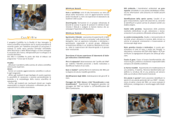
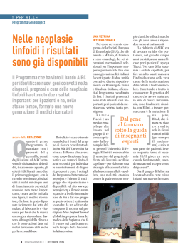
Gene 309 (2003) 71–79
www.elsevier.com/locate/gene
Unusual number and genomic organization of Hox genes in the tunicate
Ciona intestinalisq
Antonietta Spagnuolo1, Filomena Ristoratore1, Anna Di Gregorio1,2, Francesco Aniello1,3,
Margherita Branno*, Roberto Di Lauro*
Laboratory of Biochemistry and Molecular Biology, Stazione Zoologica Anton Dohrn, Villa Comunale, 80121 Naples, Italy
Received 11 December 2002; received in revised form 3 February 2003; accepted 18 February 2003
Received by G. Bernardi
Abstract
Hox genes are organized in genomic clusters. In all organisms where their role has been studied, Hox genes determine developmental fate
along the antero-posterior axis. Hence, these genes represent an ideal system for the understanding of relationships between the number and
expression of genes and body organization. We report in this paper that the ascidian Ciona intestinalis genome appears to contain a single
Hox gene complex which shows absence of some of the members found in all chordates investigated up to now. Furthermore, the complex
appears to be either unusually long or split in different subunits. We speculate that such an arrangement of Hox genes does not correspond to
the chordate primordial cluster but occurred independently in the ascidian lineage.
q 2003 Elsevier Science B.V. All rights reserved.
Keywords: Cluster Hox; Chromosome walking; Ascidian; Spatial and temporal colinearity
1. Introduction
Hox genes encode transcription factors that regulate
developmental fates along the anterior-posterior body axis
of all animals in which they have been examined (Duboule,
1994; Krumlauf, 1994). They usually show a clustered
Abbreviations: PG, paralogous group; lab, labial; pb, proboscipedia;
dfd, deformed; scr, sex comb reduced; ubx, ultrabithorax; ant,
antennapedia, abd-B, abdominal-B; C., Ciona; kb, kilobases; oligo,
oligodeoxyribonucleotide; nt, nucleotide(s).
q
The nucleotide sequence data in this paper have been submitted to
GenBank and the accession numbers are as follows: Cihox1, AJ535671;
Cihox2, AJ535672; Cihox4, AJ535673; Cihox6/7, AJ535674; Cihox10,
AJ535675; Cihox12/13, AJ535676.
* Corresponding authors. Tel.: þ 39-081-583-3278; fax: þ 39-081-5833285.
E-mail addresses: [email protected] (M. Branno), [email protected] (R. Di
Lauro).
1
These authors contributed equally to the work.
Present address: Department of Cell and Developmental Biology,Weill
Medical College of Cornell University, 1300 York Avenue, New York, NY
10021, USA.
3
Present address: Department of Genetics, General and Molecular
Biology, University of Naples ‘Federico II’, via Mezzocannone 8, 80134
Naples, Italy.
2
genomic organization, with an astonishing correlation
between the order of genes in the cluster and both spatial
and temporal order of gene expression (for reviews see
Izpisua-Belmonte et al., 1991; Duboule, 1994; Gehring,
1994).
A single Hox cluster has been described in invertebrates
(Kaufman et al., 1990; Burglin and Ruvkun, 1993; Martinez
et al., 1999) and in the invertebrate chordate amphioxus
(Garcia-Fernandez and Holland, 1994), while four Hox
clusters have been found in tetrapods (McGinnis and
Krumlauf, 1992; Krumlauf, 1994); teleost fish appear to
have at least seven clusters (Amores et al., 1998; Meyer and
Schartl, 1999). The increase in the number of the Hox
clusters in vertebrates could be associated with the increase
in morphological complexity observed in the vertebrate
body plan (Kappen et al., 1989). The vertebrate clusters
probably arose from a single ancestral complex by a twostep duplication event (Kappen et al., 1989); however, none
of the duplicated clusters appears to contain representatives
of all 13 paralogous groups (PG), suggesting that some
genes must have been lost during duplication.
Ascidians are considered the most primitive chordates
(Satoh, 1994; Satoh and Jeffery, 1995; Di Gregorio and
Levine, 1998). During their larval stage they show
characteristic chordate features, like a caudal notochord,
0378-1119/03/$ - see front matter q 2003 Elsevier Science B.V. All rights reserved.
doi:10.1016/S0378-1119(03)00488-8
72
A. Spagnuolo et al. / Gene 309 (2003) 71–79
Table 1
Hox gene clones recovered from the first (lEMBL3) (Di Gregorio et al., 1995) and from next screenings (lGEM11, lEMBL4, cDNA larva, cosmid)
HB1 positives
CiHox1
CiHox2
CiHox3
CiHox4
CiHox5/7
CiHox6/7
CiHox10
CiHox11/12
CiHox12/13
lEMBL3
lGEM11
lEMBL4
cDNA larva
Cosmid
Total isolated
70
200
200
50
3
1
6
4
4
1
9
16
16
4
2
3
1
7
10
15
2
2
6
70
1
4
2
4
4
5
10
8
8
590
4
4
15
12
17
7
38
50
64
6
16
21
6
4
It has to be noted that, in lEMBL3, CiHox4 is not included since CiHbox2, first assigned to PG4, turned out to belong to the Gsx family of homeoboxcontaining genes.
dorsal neural tube and segmental muscles; all these
structures disappear after metamorphosis. Studies carried
out over the last decade have pointed out a certain degree of
conservation, between ascidians and vertebrates, in the
genetic pathways involved in the specification of different
body structures (Di Gregorio and Levine, 1998). Based on
these findings, we started an extensive search for Hox genes
in the ascidian C. intestinalis in order to trace back the
organization of Hox genes in the most primitive chordate
body plan. We have previously carried out a screen for
homeobox-containing genes in a C. intestinalis genomic
library using, as a probe, a degenerate oligonucleotide
(CiHB1) coding for the most conserved region of the Hox
class-homeodomains (Di Gregorio et al., 1995). This
analysis identified, among several other homeobox-containing genes, five putative Hox genes showing sequence
homology with members of mammalian HOX clusters. The
genes were named CiHbox1 (further characterized and
named CiHox3 in Locascio et al., 1999), 2, 3, 4 and 5 and
their encoded homeodomains showed the highest identity to
vertebrate Hox3, Hox4, Hox10, Hox 11/12 and Hox12/13,
respectively. A further screen on a cDNA library at the
larval stage permitted to isolate a gene belonging to the 5/7
paralogy group (Gionti et al., 1998).
In this study, besides the CiHox genes already characterized, we identified new members of this class in the
Ciona genome. By chromosome walking experiments we
confirmed a clustered organization for some of them but,
surprisingly, we found that, differently from what seen in
other chordates, these genes do not form a complete and
contiguous complex. Furthermore, it appears that C.
intestinalis has a smaller complement of Hox genes of all
other chordates investigated thus far.
2. Materials and methods
2.1. Ascidians
Adult C. intestinalis were collected in the Bay of Naples
by fishing service of the Stazione Zoologica. Gametes were
used for in vitro fertilization and embryos were raised in
filtered sea water at 16– 18 8C.
2.2. Isolation of cDNA and genomic clones
Two genomic libraries and a cDNA library from embryos
at larval stage (previously described in Gionti et al., 1998)
were screened using as probe a degenerate oligonucleotide
(CiHB1) coding for the most conserved amino acid
sequence of the Helix III of the Antennapedia type
homeodomain, as previously described (Di Gregorio et al.,
1995). The first genomic library was obtained by partial
digestion of genomic DNA with Sau3AI and insertion into
lGEM11 phage vector (Promega). The second genomic
library was obtained by partial digestion of DNA with
Eco RI and insertion in lEMBL4 phage vector (Stratagene).
The positive clones were analysed by restriction mapping
and hybridization with CiHB1. The positive fragments were
cloned into pBS and sequenced on both strands by
dideoxynucleotide termination procedure (Sanger et al.,
1977). The cDNA clones of interest were also sequenced
entirely on both strands with the same procedure. The
deduced protein sequences were compared with the EMBL
GenBank database.
2.3. Cosmid library construction and screening
A C. intestinalis cosmid library was constructed by the
Reference Library Database (RLDB, MPI for Molecular
Genetic, Berlin-Dahlem, Germany) as described in Burgtorf
et al. (1998). Briefly, high-molecular weight genomic DNA
isolated from sperm of a single animal and included in
agarose blocks, was partially digested with Mbo I and
cloned into the Bam HI site of the cosmid vector Lawrist 7.
High density filter arrays of the library were generated using
a robotic device. Each membrane contained a total of 4 £
104 clones with an average insert size of 35 kb (10-fold
coverage of the Ciona genome). Two filters, containing the
arrayed library, were screened first of all with the
A. Spagnuolo et al. / Gene 309 (2003) 71–79
73
Fig. 1. Alignment of vertebrate, amphioxus, Drosophila and C. intestinalis homeodomain deduced sequences. Dashes indicate amino acid identities. The
percentage of identity and the source organisms are on the right side. Arrows in CiHox1, 2, 3, 4 and 10 mark the position of introns in the coding sequence. The
accession numbers for each sequence are as follows: BAA78620 (Amphihox1), P09022 (mouse Hox-A1), CAB57787 (Drosophila Lab), P31264 (Drosophila
Pb), P31245 (mouse Hox-A2), BAA78621 (Amphihox2), CAA48180 (Amphihox3), P02831 (mouse Hox-A3), Q08624 (mouse Hox-C4), A26638 (Drosophila
Dfd), BAA78622 (Amphihox4), CAA84517 (Amphihox5), P09024 (mouse Hox-B7), P09021 (mouse Hox-A5), P09077 (Drosophila Scr), CAA84519
(Amphihox7), P17509 (Human Hox-B6), A25399 (Drosophila Ant), P02834 (Drosophila Ubx), CAA84518 (Amphihox6), CAA84522 (Amphihox10),
P28359 (mouse Hox-D10), A34220 (Drosophila Abd-B), AAF81909 (Amphihox11), P23812 (mouse Hox-D12), P23813 (mouse Hox-D11), p70321 (mouse
Hox-B13), P70217 (mouse Hox-D13), AAF81903 (Amphihox12), AAF81904 (Amphihox13).
74
A. Spagnuolo et al. / Gene 309 (2003) 71–79
oligonucleotide CiHB1 as previously described (Di Gregorio et al., 1995) in order to identify homeobox-containing
clones. Positive clones were screened again, under high
stringency conditions, using oligonucleotides specific for
each homeobox already identified, in order to classify the
clones. The DNA of the cosmid clones was purified with the
QIAGEN kit (Quiagen Inc., Chatsworth, CA, USA). The
genomic inserts were subjected to digestion with Bam HI
and Eco RI followed by Southern blot hybridization with
CiHB1 oligonucleotide as described. The homeoboxcontaining fragments were cloned and sequenced. For
walking experiments the cosmid/insert ends were sequenced
and specific oligonucleotides were designed for each clone.
These oligonucleotides were used, under high stringency
conditions, in hybridization experiments to isolate adjacent
clones. All genomic regions were hybridized with CiHB1 to
search for new homeobox-containing genes.
3. Results
3.1. Isolation of CiHox genes
The goal of our work was to identify Hox genes in the
ascidian C. intestinalis and to study their genomic
organization. We already reported on the isolation of
some Hox genes belonging to the paralogy groups 3, 4,
5/7, 10, 11/12, 12/13 (Di Gregorio et al., 1995; Gionti et al.,
1998; Locascio et al., 1999) (Table 1).
We then attempted to isolate more Hox genes by looking
at four new sources: two additional C. intestinalis genomic
libraries, a cDNA library prepared from larval mRNAs and
a cosmid library. Each library was screened with the CiHB1
oligonucleotide (Table 1). The positive clones were first rescreened with oligonucleotide probes specific for the CiHox
genes already identified: CiHox3, 4, 5 (renamed in this
paper as CiHox5/7), 10, 11/12 and 12/13 (Di Gregorio et al.,
1995; Gionti et al., 1998; Locascio et al., 1999) to identify
clones containing these genes. From the remaining clones, a
restriction fragment hybridizing with CiHB1 was subcloned
and sequenced from each of them. The sequences thus
obtained were then compared with those present in
GenBank. We identified four new Ciona Hox genes
corresponding to the Hox 1, 2, 4, 6/7 PGs and called them
CiHox1, CiHox2, CiHox4, and CiHox6/7 (Fig. 1). The gene
previously named CiHbox2 (Di Gregorio et al., 1995) and
considered as the ortholog of vertebrate Hox4, turned out to
be a member of the Gsx homeobox family (Hudson and
Lemaire, 2001). A new Hox gene, showing 90% identity
with PG4 members and recovered in these surveys, was
therefore named CiHox4 (Fig. 1). We also obtained the
partial or full cDNA sequences for CiHox1, CiHox4,
CiHox6/7, CiHox10 and CiHox12/13. These results are
shown in Table 1. Names incorporating more than one
paralogy group number, such as CiHox5/7, CiHox6/7,
CiHox11/12 and CiHox12/13, reflect ambiguous assign-
ments, because of the high degree of amino acid similarity
between homeodomains of groups 5, 6, 7 and between
homeodomains of groups 11, 12, 13. Interestingly, the
CiHox1, 2, 3, 4 coding regions are interrupted by introns of
variable length between the sequences coding for the second
and the third helix of their homeodomains (Fig. 1), as is the
case of Drosophila labial and proboscipedia genes
(Burglin, 1994). CiHox10 has an intron between codons
33 and 34 of the homeobox, a rare site for a homeobox
intron.
3.2. Assembly of CiHox genes in genomic contigs
To further analyse the genomic arrangement of the
CiHox genes isolated by these screenings, walking experiments were carried out on a cosmid library (Burgtorf et al.,
1998). Six cosmid clones, Cos1S1 containing CiHox1,
Cos3T4 containing CiHox2 and CiHox3, Cos3S1 containing
CiHox3 and CiHox4, Cos5S1 containing CiHox5/7 and
CiHox6/7, Cos10S1 containing CiHox10 and Cos11S1
containing CiHox11/12 and CiHox12/13 were chosen for
further analysis (Fig. 2). The strategy used was to sequence
the ends of each cosmid clone of interest, to design
oligonucleotide probes and use them to screen the arrayed
library and isolate adjacent genomic contigs. Each presumptive contig was restriction mapped and checked for the
presence of new homeoboxes by Southern hybridization
with the CiHB1 oligo as probe. This analysis allowed us to
discover a linked arrangement for CiHox4, CiHox 3 and
CiHox2; to link CiHox5/7 and CiHox6/7 and to find a paired
homeobox gene (belonging to the SHox family of homeobox-containing gene) downstream from CiHox6/7; finally,
to confirm the linkage between CiHox11/12 and CiHox12/
13 (Di Gregorio et al., 1995) (Fig. 2). PCR amplifications
and restriction digests were used to orient each CiHox
transcript, showing that CiHox2, 3 and 4 as well as CiHox5/
7 and CiHox6/7 are transcribed in the same direction.
CiHox11/12 and CiHox12/13 are divergently transcribed
(Fig. 2, top arrows), differently from vertebrate Hox genes
that are always transcribed colinearly (Kessel and Gruss,
1990; Krumlauf, 1992), but resembling what seen in the
case of the Deformed gene in the Drosophila HOM cluster
(Lewis, 1978). By the end of this round of experiments, we
characterized 40 kb of group 1; walked 50 kb and . 100 kb
from the 50 - and 30 -ends of groups 2 –4; 100 kb and . 120
kb from the 50 - and 30 -ends of groups 5 –7; . 100 kb from
group 10; and 70 kb from groups 11 – 13 (Fig. 2, bottom
arrows). Despite these efforts we were unable to link any of
these groups or to find additional Hox genes in the contigs
isolated with the genome walking experiments. On the other
hand, the genome analysis either has been unable to resolve
the whole organization of Hox genes in C. intestinalis
(Dehal et al., 2002).
A. Spagnuolo et al. / Gene 309 (2003) 71–79
Fig. 2. Contig maps of Ciona Hox genes. Groups are designated by lines with relative positions of homeoboxes shown by boxes differently colored. Constituent clones are shown below each group. Linkage
relationships between individual group is unknown and relative positions of unlinked clones is arbitrary. On the bottom is shown the length of each group (for groups 1 and 10) or the distances between clustered
CiHox genes plus the length of the walk on one end (for groups 11 –13) or on both ends (for groups 2–4 and 5–7). Arrows on top of each Hox gene indicate the direction of transcription. For simplicity shorter
names have been assigned to each cosmid clone isolated. The RLDB original clone’s names are as follows: MPMGc119N0428 (1S1), MPMGc119H2170 (1S2), MPMGc119L0224 (3T2), MPMGc119C0437
(3T4), MPMGc119O1424 (3T7), MPMGc119D1338 (3S1), MPMGc119B114 (4S1), MPMGc119L2130 (4S2), MPMGc119F1185 (5S1), MPMGc119E0321 (5S4), MPMGc119E1725 (10S1),
MPMGc119E1024Q (11S1), MPMGc119B0917Q (3T3), MPMGc119H043 (4S3), MPMGc119C2417Q (4S4), MPMGc119L1835Q (4S5), MPMGc119D044Q (4S6), MPMGc119F1437Q (5S2),
MPMGc119K018Q (5S3), MPMGc119T0248Q (5S5), MPMGc119G2462Q (5S9), MPMGc119B1565 (10S2), MPMGc119E247Q (10S3), MPMGc119F1927Q (10S4), MPMGc119O983Q (10S5),
MPMGc119K0327Q (11S1), MPMGc119M0917Q (11S3), MPMGc119H0948Q (5S6), MPMGc119K1729Q (5S7), MPMGc119K1717Q (5S8).
75
76
A. Spagnuolo et al. / Gene 309 (2003) 71–79
4. Discussion
We have presented the isolation of phage and cosmid
clones containing most of the Ciona Hox genes. Our results
indicate the existence, in the Ciona genome, of representatives of anterior, central and posterior Hox class genes. The
anterior group includes CiHox1, 2, 3 and 4. The central
group includes CiHox5/7 and CiHox6/7. The posterior
group includes CiHox10, CiHox11/12 and CiHox12/13.
Despite our efforts, we did not find representatives of PGs 8
and 9. By genome walking we have found evidence for a
clustered arrangement of some of these Hox genes in the
genome, but we have been unable to link all of them in a
contiguous cluster. Based on the data gathered from C.
intestinalis, we propose that the single, contiguous Hox
cluster present in the common chordate ancestor has
undergone, in ascidians, extensive lineage-specific
rearrangements. These might include the loss of some
genes, such as Hox8 and Hox9, that are found in higher
chordates but appear to be missing in Ciona.
4.1. Hox genes from Ciona intestinalis
4.1.1. Anterior CiHox genes
The anterior Hox genes isolated from C. intestinalis can
be unequivocally assigned to a specific PG, based on the
sequence identities of their deduced homeodomains (Fig. 1).
CiHox1, CiHox2, CiHox3 and CiHox4 show . 80% identity
with PG1, 2, 3 and 4 members.
4.1.2. Central CiHox genes
The central group generally contains three Hox genes
whose orthology relationships are much less clear since
their homeodomain sequences are, in general, very similar
to each other. In Ciona we found two putative medial genes,
CiHox5/7 and CiHox6/7. As previously reported (Gionti
et al., 1998), the deduced homeodomain sequence of
CiHox5/7 shows a very high identity with that of Drosophila
Scr and Antp (88 – 89%) (Burglin, 1994), with Amphihox5
(93%) (Garcia-Fernandez and Holland, 1994) as well as
with vertebrate Hox 7 class (92%) (Krumlauf, 1992) (Fig. 1)
even if this analysis includes the hexapeptide sequence, that
is frequently used to delineate the orthology relationships.
Therefore we renamed CiHox5 as CiHox5/7. CiHox6/7,
which has been found clustered with CiHox5/7, presented
the same problem. In fact, highest identity of the homeodomain deduced sequence was either with PG7 (93%) as
with PG6 (90%) members (Fig. 1), but when the searches
were repeated including the hexapeptide, the identity was
closer to PG7 (87%).
4.1.3. Posterior CiHox genes
The 50 -ends of Hox clusters analysed from different
species contain a number of genes ranging from one
(Drosophila Abd B) (Burglin, 1994) to six (amphioxus)
(Ferrier et al., 2000). As in the case of central genes it is
difficult to identify orthologies between posterior Hox genes
across different phyla. In the Ciona genome we have found
three representatives of this class: CiHox10, CiHox11/12
and CiHox12/13. CiHox10 has been assigned, beyond
doubt, to the PG10 (see Fig. 1). The classification was
instead complicated for CiHox11/12 and CiHox12/13, since
their identity, in the homeodomain deduced sequence, was
63% with PG11 and PG12 members for CiHox11/12 and
60 – 58% with PG13 and PG12 family members for
CiHox12/13 (Fig. 1).
4.2. Organization and expression of the Ciona Hox genes
The putative Ciona Hox cluster seems to contain all
anterior PG members, two representatives of the central
part, and three posterior members. In any case our data
indicate that Ciona genome could lack true members of PG8
and PG9 types (Fig. 3). This is not a novelty, since gain or
loss of Hox genes has been reported in many lineages.
Examples include the absence of PG7 in the pufferfish
(Aparicio et al., 1997), of PG8 (AbdA) in cirripedia
(Mouchel-Vielh et al., 1998), of many Hox genes after
cluster duplication in vertebrates, amplification of posterior
Hox genes in deuterostomes (Izpisua-Belmonte et al., 1991;
Ferrier et al., 2000) and duplication of PG3 in insects
(Stauber et al., 1999) and anellida (Kourakis and Martindale, 2001). The second characteristic of Ciona Hox genes is
that, despite extensive genomic walking, it does not seem
that they are linked in a contiguous cluster as they are in sea
urchin, amphioxus and higher chordates. A detailed
structural analysis of the three larger contigs identified in
our work, indicate that CiHox2, CiHox3 and CiHox4 cover a
genomic region of about 40 kb. CiHox2 and 3 are very close
to each other, while more than 20 kb separate CiHox3 from
CiHox4. This is in accordance with data from vertebrate
(Acampora et al., 1991) and amphioxus (Garcia-Fernandez
and Holland, 1994). CiHox5/7 and CiHox6/7 are also very
close, similarly to what was observed in amphioxus
(Garcia-Fernandez and Holland, 1994). The distances
between CiHox11/12 and CiHox12/13 are comparable to
that found in human and amphioxus Hox clusters (Ferrier
et al., 2000) (Fig. 2).
Therefore, it seems that the intergenic distances within
each subunit remained somewhat conserved, while the
whole cluster split in different subunits. In Drosophila the
cluster is broken into two complexes, Antennapedia and
Bithorax (Burglin, 1994), separated by more than 8 Mb and
located on the same chromosome. Caenorhabditis elegans
also contains a small Hox cluster highly degenerated, split
and located in the center of chromosome III (Burglin and
Ruvkun, 1993). We still do not know whether or not the
CiHox genes are located on the same chromosome. The total
length of the five contigs is approximately 650 kb (Fig. 2).
For comparison, the region containing PG1 to PG10 in
amphioxus spans approximately 270 kb (Garcia-Fernandez
and Holland, 1994), human Hox clusters are 125 kb long on
average (Krumlauf, 1994) and the Hox cluster identified in
A. Spagnuolo et al. / Gene 309 (2003) 71–79
77
Fig. 3. Hox gene organization in Drosophila, amphioxus and Mammals compared to Ciona. Length of each complex is shown on the right side. Code colors are
used to indicate orthologous relationships between Hox genes from different species.
the sea urchin, Strongylocentrotus purpuratus, is . 500 kb
long (Martinez et al., 1999) (Fig. 3).
How to explain the fragmentation of the Hox cluster and
the absence of some PG members in the Ciona genome? In
cirripedia, the loss of AbdA has been correlated with the lack
of pleon (abdomen) in this lineage (Mouchel-Vielh et al.,
1998). Anterior-posterior patterning in C. elegans appears to
require at most three Hox genes, belonging only to the
anterior and posterior PGs, raising the question on how and
when, during evolution, the medial group genes were coopted for embryonic roles (Van Auken et al., 2000). The
ascidian larva contains only , 2500 cells and five
differentiated tissues. One of them is the nervous system
that is divided in an anterior region localized in the head,
containing the sensory vesicle and the visceral ganglion, and
a nerve chord running along the tail. The gut primordium is
a very simple structure since ascidian larvae are nonfeeding. Given these remarks, it could be speculated that C.
intestinalis, a relatively simple specie, might not require a
high number of Hox genes to pattern its larval structures.
Specifically, the absence of central/posterior PG members,
that in higher chordates collaborate with other Hox genes to
pattern gut, axial skeleton and nervous system, could be
related to the simple organization of the axial structures and
gut primordium.
Transgenic approaches have demonstrated that adjacent
vertebrate Hox genes can share enhancer elements or
compete for them (Gould et al., 1997; Zakany et al., 1997;
Duboule, 1998; Sharpe et al., 1998). This is probably due to
the compact structure and to the smaller intergenic distances
observed in vertebrates. In Drosophila it has been shown
that enhancer sharing and competition are prevented by very
high intergenic distances and by the presence of ‘boundary
elements’ (Hagstrom et al., 1996; Zhou et al., 1996).
Furthermore, Drosophila Hox genes do not exhibit the
temporal colinearity as seen in vertebrates. Finally, in
Drosophila the cluster is split into two subunits. This has
suggested that the retention of a clustered organization is
controlled by evolutionary forces that can vary between
phyla. In situ hybridization experiments have shown the
expression of CiHox3 in the visceral ganglion (Locascio
et al., 1999) and of CiHox5/7 in the anterior regions of the
nerve cord (Gionti et al., 1998), suggesting that the CiHox
genes, in ascidians, might be involved in the regionalization
of the larval CNS. Interestingly, the spatial correlation
between the expression territories of CiHox3 and CiHox5/7
genes is conserved, even though they have not been found to
be closely clustered in the genome. The temporal correlation
instead seems to be lost, since it appears that CiHox5/7
(Gionti et al., 1998) is switched on earlier during
embryogenesis than CiHox3 (Locascio et al., 1999),
indicating that Ciona Hox genes, like Drosophila HOM
genes, might not obey a temporal colinearity, therefore
behaving differently from higher chordates. Thus, we
speculate that in Ciona the rapid embryogenesis and the
simple structure of the larva may not require such a
coordinated, colinear and temporal organization. This could
have led to a relaxation of the selective forces controlling
cluster structure allowing the scattering of the Hox cluster.
The presence of repetitive sequence elements in the Ciona
genome (Simmen and Bird, 2000), which has represented a
major technical disadvantage in walking experiments, may
also have facilitated recombination events and hence the
dispersion of the cluster.
78
A. Spagnuolo et al. / Gene 309 (2003) 71–79
4.3. The primitive chordate cluster
The S. purpuratus (sea urchin) genome contains a single
ten-gene Hox complex (Martinez et al., 1999). The cluster
includes genes representing all PGs of chordate Hox
clusters, except that there is a single gene of the PG4-5
types and only 3 genes of the posterior type. The amphioxus
cluster contains homologues of all 13 PGs of vertebrate Hox
genes plus one more posterior Hox14 (Ferrier et al., 2000). It
could, therefore, be speculated that the common ancestor of
echinoderm and chordate may have contained almost all PG
members already before the radiation of these groups. Ciona
is considered the most primitive chordate. Our data strongly
indicate that Ciona Hox cluster show some peculiar
characteristic not shared, so far, by any other chordate. It
is tempting to speculate that Ciona Hox cluster organization
is a derived rather than a primitive condition and that during
chordate evolution, in the ascidian lineage all the rearrangements, including loss of some PGs (PG8 and PG9) members
and splitting of the complex in different subunits, have
occurred independently. It is still food for debate how the
clustered organization of the Hox genes arose in the first
place. The case of Ciona could show that clustered
organization might have been lost at sometime during
chordate evolution.
References
Acampora, D., Simeone, A., Boncinelli, E., 1991. Human HOX homeobox
genes. Oxf. Surv. Eukaryot. Genes 7, 1–28.
Amores, A., Force, A., Yan, Y.L., Joly, L., Amemiya, C., Fritz, A., Ho,
R.K., Langeland, J., Prince, V., Wang, Y.L., Westerfield, M., Ekker, M.,
Postlethwait, J.H., 1998. Zebrafish hox clusters and vertebrate genome
evolution. Science 282, 1711–1714.
Aparicio, S., Hawker, K., Cottage, A., Mikawa, Y., Zuo, L., Venkatesh, B.,
Chen, E., Krumlauf, R., Brenner, S., 1997. Organization of the Fugu
rubripes Hox clusters: evidence for continuing evolution of vertebrate
Hox complexes. Nat. Genet. 16, 79 –83.
Burglin, T.R., 1994. A comprehensive classification of Homeobox genes.
In: Duboule, D., (Ed.), Guidebook to homeobox genes, Oxford
University Press, Oxford, pp. 25 –71.
Burglin, T.R., Ruvkun, G., 1993. The Caenorhabditis elegans homeobox
gene cluster. Curr. Opin. Genet. Dev. 3, 615–620.
Burgtorf, C., Welzel, K., Hasenbank, R., Zehetner, G., Weis, S., Lehrach,
H., 1998. Gridded genomic libraries of different chordate species: a
reference library system for basic and comparative genetic studies of
chordate genomes. Genomics 52, 230–232.
Dehal, P., Satou, Y., Campbell, R.K., Chapman, J., Degnan, B., De
Tomaso, A., Davidson, B., Di Gregorio, A., Gelpke, M., et al., 2002.
The draft genome of Ciona intestinalis: insights into chordate and
vertebrate origins. Science 298, 2157–2167.
Di Gregorio, A., Levine, M., 1998. Ascidian embryogenesis and the origins
of the chordate body plan. Curr. Opin. Genet. Dev. 8, 457–463.
Di Gregorio, A., Spagnuolo, A., Ristoratore, F., Pischetola, M., Aniello, F.,
Branno, M., Cariello, L., Di Lauro, R., 1995. Cloning of ascidian
homeobox genes provides evidence for a primordial chordate cluster.
Gene 156, 253 –257.
Duboule, D., 1994. Temporal colinearity and the phylotypic progression: a
basis for the stability of a vertebrate Bauplan and the evolution of
morphologies through heterochrony. Development Suppl. 1994,
135– 142.
Duboule, D., 1998. Vertebrate hox gene regulation: clustering and/or
colinearity? Curr. Opin. Genet. Dev. 8, 514 –518.
Ferrier, D.E., Minguillon, C., Holland, P.W., Garcia-Fernandez, J., 2000.
The amphioxus Hox cluster: deuterostome posterior flexibility and
Hox14. Evol. Dev. 2, 284–293.
Garcia-Fernandez, J., Holland, P.W., 1994. Archetypal organization of the
amphioxus Hox gene cluster. Nature 370, 563 –566.
Gehring, W.J., 1994. A history of the homeobox. In: Duboule, D., (Ed.),
Guidebook to the homeobox genes, Oxford University Press, Oxford,
pp. 3–10.
Gionti, M., Ristoratore, F., Di Gregorio, A., Aniello, F., Branno, M., Di
Lauro, R., 1998. Cihox5, a new Ciona intestinalis Hox-related gene, is
involved in regionalization of the spinal cord. Dev. Genes Evol. 207,
515 –523.
Gould, A., Morrison, A., Sproat, G., White, R.A., Krumlauf, R., 1997.
Positive cross-regulation and enhancer sharing: two mechanisms for
specifying overlapping Hox expression patterns. Genes Dev. 11,
900 –913.
Hagstrom, K., Muller, M., Schedl, P., 1996. Fab-7 functions as a chromatin
domain boundary to ensure proper segment specification by the
Drosophila bithorax complex. Genes Dev. 10, 3202–3215.
Hudson, C., Lemaire, P., 2001. Induction of anterior neural fates in the
ascidian Ciona intestinalis. Mech. Dev. 100, 189–203.
Izpisua-Belmonte, J.C., Falkenstein, H., Dolle, P., Renucci, A., Duboule,
D., 1991. Murine genes related to the Drosophila AbdB homeotic genes
are sequentially expressed during development of the posterior part of
the body. EMBO J. 10, 2279–2289.
Kappen, C., Schughart, K., Ruddle, F.H., 1989. Two steps in the evolution
of Antennapedia-class vertebrate homeobox genes. Proc. Natl. Acad.
Sci. USA 86, 5459–5463.
Kaufman, T.C., Seeger, M.A., Olsen, G., 1990. Molecular and genetic
organization of the antennapedia gene complex of Drosophila
melanogaster. Adv. Genet. 27, 309–362.
Kessel, M., Gruss, P., 1990. Murine developmental control genes. Science
249, 374–379.
Kourakis, M.J., Martindale, M.Q., 2001. Hox gene duplication and
deployment in the annelid leech Helobdella. Evol. Dev. 3, 145 –153.
Krumlauf, R., 1992. Evolution of the vertebrate Hox homeobox genes.
Bioessays 14, 245–252.
Krumlauf, R., 1994. Hox genes in vertebrate development. Cell 78,
191 –201.
Lewis, E.B., 1978. A gene complex controlling segmentation in
Drosophila. Nature 276, 565–570.
Locascio, A., Aniello, F., Amoroso, A., Manzanares, M., Krumlauf, R.,
Branno, M., 1999. Patterning the ascidian nervous system: structure,
expression and transgenic analysis of the CiHox3 gene. Development
126, 4737–4748.
Martinez, P., Rast, J.P., Arenas-Mena, C., Davidson, E.H., 1999.
Organization of an echinoderm Hox gene cluster. Proc. Natl. Acad.
Sci. USA 96, 1469–1474.
McGinnis, W., Krumlauf, R., 1992. Homeobox genes and axial patterning.
Cell 68, 283–302.
Meyer, A., Schartl, M., 1999. Gene and genome duplications in vertebrates:
the one-to-four (-to-eight in fish) rule and the evolution of novel gene
functions. Curr. Opin. Cell. Biol. 11, 699 –704.
Mouchel-Vielh, E., Rigolot, C., Gibert, J.M., Deutsch, J.S., 1998.
Molecules and the body plan: the Hox genes of Cirripedes (Crustacea).
Mol. Phylogenet. Evol. 9, 382–389.
Sanger, F., Nicklen, S., Coulson, A.R., 1977. DNA sequencing with chainterminating inhibitors. Proc. Natl. Acad. Sci. USA 74, 5463–5467.
Satoh, N., 1994. Developmental Biology of Ascidians, Cambridge
University Press, New York.
Satoh, N., Jeffery, W.R., 1995. Chasing tails in ascidians: developmental
insights into the origin and evolution of chordates. Trends Genet. 11,
354 –359.
Sharpe, J., Nonchev, S., Gould, A., Whiting, J., Krumlauf, R., 1998.
A. Spagnuolo et al. / Gene 309 (2003) 71–79
Selectivity, sharing and competitive interactions in the regulation of
Hoxb genes. EMBO J. 17, 1788–1798.
Simmen, M.W., Bird, A., 2000. Sequence analysis of transposable elements
in the sea squirt, Ciona intestinalis. Mol. Biol. Evol. 17, 1685–1694.
Stauber, M., Jackle, H., Schmidt-Ott, U., 1999. The anterior determinant
bicoid of Drosophila is a derived Hox class 3 gene. Proc. Natl. Acad.
Sci. USA 96, 3786–3789.
Van Auken, K., Weaver, D.C., Edgar, L.G., Wood, W.B., 2000.
Caenorhabditis elegans embryonic axial patterning requires two
79
recently discovered posterior-group Hox genes. Proc. Natl. Acad. Sci.
USA 97, 4499–4503.
Zakany, J., Gerard, M., Favier, B., Duboule, D., 1997. Deletion of a HoxD
enhancer induces transcriptional heterochrony leading to transposition
of the sacrum. EMBO J. 16, 4393– 4402.
Zhou, J., Barolo, S., Szymanski, P., Levine, M., 1996. The Fab-7 element
of the bithorax complex attenuates enhancer-promoter interactions in
the Drosophila embryo. Genes Dev. 10, 3195–3201.
© Copyright 2026 Paperzz