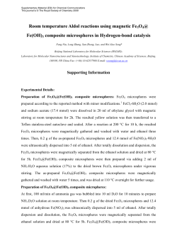
IN VIVO STUDY OF THE EFFECT OF A STRONTIUM-‐RICH INJECTABLE SYSTEM ON BONE REGENERATION, USING A SHEEP MODEL C. Machado1,2, A.H. Lourenço1,2,3 N. Neves1,2,4 , N. Alexandre5,6 M. Lamghari1,2 , A.T Cabral4 M.A. Barbosa1,2,7, C.C. Ribeiro1,2,8 INEB – Ins)tuto de Engenharia Biomédica, Universidade do Porto, Portugal 2 I3S – Ins)tuto de Inves)gação e Inovação em Saúde 3 Faculdade de Engenharia , Universidade do Porto, Portugal 4 Faculdade de Medicina, Universidade do Porto, Portugal 5 Departamento de Zootecnia, Universidade de Évora, Portugal 6 ICAAM – Ins)tuto de Ciências Agro-‐ambientais Mediterrânicas, Universidade de Évora, Portugal 7 ICBAS – In)tuto de Ciências Biomédicas Abel Salazar, Universidade do Porto, Portugal 8 ISEP – Insituto Superior de Engenharia do Porto, Ins)tuto Politécnico do Porto, Portugal. 1 RESULTS & DISCUSSION INTRODUCTION Bone has the capacity to regenerate as part of the repair process, being newly formed bone indis)nguishable from the adjacent uninjured bone. However, there are cases in which bone regenera)on is required in large quan)ty, beyond the normal poten)al for self-‐healing, such as for lesions caused by trauma, infec)on, tumour resec)on or cases in which the regenera)ve process is compromised such as avascular necrosis and osteoporosis. Biomaterials such as alginate are very promising due to its ability to form hydrogels in situ under mild condi)ons in the presence of divalent ca)ons. The combina)on with ceramic microspheres results in a mechanically improved injectable system, adequate for minimally invasive procedures. Moreover, the combina)on with chemical elements such as stron)um, described as promoter of bone forma)on, inhibi)ng bone resorp)on [1], provides ion exchange between the implanted biomaterial and surrounding )ssue, enhancing bone regenera)on. Our goal is to study in an in vivo sheep model, the effect of an injectable system composed of stron)um doped hydroxyapa)te microspheres, delivered in an alginate vehicle, crosslinked with stron)um. Micro-‐CT analysis The degrada)on of the alginate a l l o w e d t h e m i g r a X o n o f inflammatory cells, a reac)on commonly seen in the first phase of bone repair. No signs of degrada)on of the microspheres was observed (lower degrada)on rate than alginate). Fig 1 Orthogonal reconstructed slices of micro-‐computed tomography (micro-‐CT) (A, B, C), of the defects filled with the hybrid system, a_er one week of implanta)on. The hybrid system perfectly filled the defect. During the injec)on, no separa)on of the alginate and the microspheres was observed. Fig 2 Safranin/Light Green stained histological sec)ons of sheep vertebra defect filled with the hybrid system, one week (A, B, C) and eight weeks (D, E, F) post-‐implanta)on. Global view of the defect (A, D, 20x) ; details of the periphery (B, E, 40x, dashed line delimit the defect) and details of the center of the defect (C, F, 100x, microspheres are indicated with the asterisk). New bone formaXon and resorpXon Fig 3 Goldner-‐Masson Trichrome stained histological sec)ons of sheep vertebra defect filled with the hybrid system, eight weeks a_er the implanta)on. Newly formed bone was iden)fied in green (mature bone) and red (immature bone). Lines of newly formed bone surrounding the microspheres are visible in A and B (400x) and between two microspheres in C (400x). Immature bone is shown in the center of the defect in D (100x). Microspheres are iden)fied with *. Images of scanning electron microscopy and energy-‐dispersive X-‐Ray spectra (E) of 8 weeks post implanta)on histological sec)on. Z1, Z2 and Z3 indicate the different analysed areas and correspondent EDS spectra. Bone surrounded the microspheres, both in the periphery and in the center of the defect. EDS analysis confirmed the presence of newly formed bone in the defect (no Sr was detected excluding the possibility of being microspheres fragments). Morphologic evaluaXon of so\ Xssues Fig 4 Tartrate-‐resistant Acid Phosphatase stained histological sec)ons of sheep vertebra defects. Arrows indicate osteoclasts in the empty defect a_er eight weeks of implanta)on (A, 400x), in the filled defect a_er one week (B and C, 400x), and a_er eight weeks (D, 400x). The graphic represents the number of osteoclasts in the empty and in the filled defects at 1 and 8 weeks )me-‐points. A_er eight weeks the stronXum effect at decreasing the number of osteoclasts was evident, possibly by inhibi)ng its recruitment and promo)ng apoptosis. CONCLUSIONS The morphology of k i d n e y s a n d l i v e r The studied system has the ability to promote local bone forma)on as well as inhibi)ng w e r e m a c r o a n d microscopically normal, bone resorp)on, without major systemic effects. Our results suggest that this material sugges)ng the absence may be a promising alterna)ve for bone regenera)on, par)cularly in osteoporo)c of major toxic systemic pa)ents. e ff e c t s c a u s e d b y stronXum. Fig 5 H&E stained histological sec)ons of liver (A) and kidney (B), at 100x magnifica)on. MATERIALS & METHODS In this study, a cri)cal size-‐defect (4.5x3.6 mm), adapted from Lamghari et al [2], was made in the body of 3 lumbar vertebrae of 5 years old Merino Branco sheeps and filled with the injectable hybrid system. An empty defect was used as control. Bone regenera)on was evaluated at one and eight weeks post-‐implanta)on by micro computed tomography and histological analysis a_er methylmethacrylate embedding of bone specimens. So_ )ssues analysis was also performed to evaluate stron)um systemic effects. REFERENCES [1] Marie PJ, Felsenberg D, Brandi ML. How stron)um ranelate, via opposite effect on bone resorp)on and forma)on prevents osteoporosis. Osteoporos Int 2011; 22:1659-‐67. [2] Lamghari M, Huet H, Laurent A, Berland S, Lopez E. A model for evalua)ng injectable bone replacements in the vertebrae of sheep: radiological and histological study. Biomaterials 1999; 20(22): 2107-‐14. ACKNOWLEDGMENTS T h i s w o r k w a s fi n a n c e d b y F E D E R f u n d s , t h r o u g h the Programa Operacional Factores de Compe))vidade — COMPETE and by Portuguese funds through FCT — Fundação para a Ciência e a Tecnologia in the framework of the project PTDC/CTM/103181/2008. Bioimaging 2015. November 2015, Porto . Portugal
© Copyright 2026 Paperzz