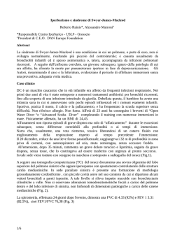
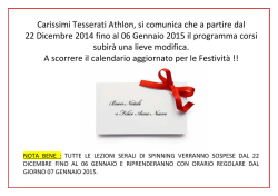
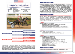
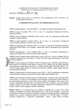
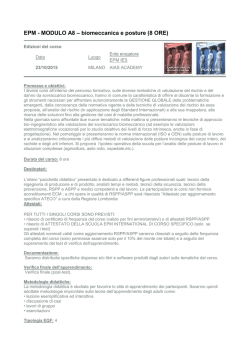
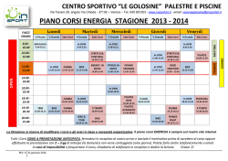
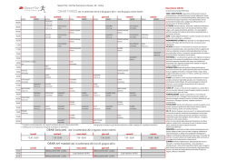
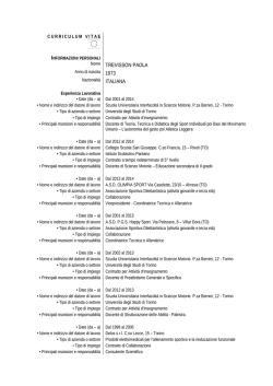
Review
Glosso-postural syndrome
Fabio Scoppa
Scientific and Educational Coordinator, Master’s Degree Course in Posturology
Director: Prof. Giuseppe Amabile
1st Faculty of Medicine and Surgery, University “La
Sapienza”, Rome, Italy
Correspondence to: Prof. Fabio Scoppa
Via Guido Cavalcanti, 2 - 00152 Roma - Italy
E-mail: [email protected]
www.posturologia.biz
Submitted for publication: 31/08/2004
Accepted for publication: 16/11/2004
Summary
Glosso-postural syndrome
Atypical deglutition is correlated not only with posture
and orocraniocervical morphology, but also with the
subject’s general posture.
The tongue is capable of perturbing postural balance
due both to its connections with the key anatomical
structures, and to other neurophysiological reasons.
The hyo-glossus apparatus, owing to its links with the
maxillaries, the skull, the cervicals, the scapula, the
pharynx and the larynx, is the true “trait d’union” between the oral and postural functions of the body.
Due to the prevalently transverse arrangement of its fibres, the tongue may be considered a diaphragm linking the body’s anterior and posterior muscular chains.
In this report, we present a new nosographical entity,
glosso-postural syndrome, which is characterized by
postural imbalance and atypical deglutition.
The most important traits of type I and type II glossopostural syndrome are described.
Key words: atypical deglutition, glosso-postural syndrome, postural imbalance.
Sommario
La sindrome glosso-posturale
La deglutizione atipica è correlata non solo con la postura e la morfologia oro-cranio-cervicale, ma anche
con la postura generale del soggetto.
La lingua è in grado di perturbare l’equilibrio posturale
grazie alle sue connessioni con strutture anatomiche
di capitale importanza, oltre che per ragioni di ordine
neurofisiologico.
L’apparato io-glosso, in virtù dei suoi legami con i mascellari, il cranio, le cervicali, le scapole, la faringe e la
laringe rappresenta il vero “trait d’union” tra funzioni
orali e funzioni posturali corporee.
Annali di Stomatologia 2005; LIV (1): 27-34
Per la disposizione prevalentemente trasversale delle
sue fibre, la lingua può essere considerata un diaframma che mette in comunicazione le catene muscolari
anteriori e posteriori del corpo.
Nel presente lavoro viene presentata una nuova entità
nosografica, la sindrome glosso-posturale, caratterizzata da squilibrio posturale e deglutizione atipica.
Vengono descritti i caratteri salienti delle due varietà
di sindrome glosso-posturale, il tipo I e il tipo II.
Parole chiave: deglutizione atipica, sindrome glosso-posturale, squilibrio posturale.
Introduction
In recent years, specialists from various disciplines
have studied the problem of atypical deglutition: a wide
range of literature exists, also in Italian, describing the
negative effects of deglutition disorders at different levels – orthognathodontic, logopaedic, otorhinolaryngoiatric, paediatric (Balercia, 1993; Caprioglio, 1993 a,
b; Cozza, 1995; Capurso, 1996; Garliner, 1996;
Walther, 1996; Ferrante, 1997; Funt, 1999; Cozza,
2002; Stefanelli, 2003; Ferrante, 2004).
In general, the morphogenetic role of the tongue, capable of modelling the arches and affecting the development of the maxillaries, regards mainly the developmental phase of life, after which it is not rare for the
tongue to fulfil a balancing and a compensatory function, acting as a sort of natural bite. Especially in adulthood, the tongue is capable of compensating for missing teeth, malocclusions, or lack of orthogonality between the occlusal plane and the vertebral sagittal
plane.
In this regard, just as atypical deglutition can be considered a cause of malocclusion, so, in malocclusion, the
tongue can serve an occlusion stabilizing function and
behave in an atypical way.
Such considerations bring us back to the relationship
between structure and function, which, from our point of
view, cannot be regarded as a simple linear relationship
of cause and effect, but rather as a complex, circular
and systemic type relationship (Fig. 1).
The age-old debate over whether it is the orofacial
musculature that affects the teeth or rather the teeth
that affect muscular function can be overcome by performing a differential diagnosis involving muscular
tests, by studying the deglutition pattern, and by examining the occlusion.
It is a topic that continues to be the focus of considerable attention among professionals (Lang, 2004; Peng,
2004; Solomon, 2004).
Many (Balercia, 1993; Caprioglio, 1993 a, b; Bricot,
1996; Ferrante, 1997; Ranaudo, 1997; Cipollari, 2003;
27
F. Scoppa
STRUCTURE
– Teeth position and
intermaxillary
– bone relationships
– Temporomandibular and
craniocervical bone
relationships
FUNCTION
POSTURE AND
MORPHOLOGY
OF THE BODY
AS A WHOLE
– Deglutition
– Suction
– Phonation
– Respiration
Guaglio, 2003; Lentini, 2003; Spinicci, 2003; Stefanelli,
2003; Ferrante, 2004; Zavarella, 2004) also stress the
relationship between lingual dysfunction and postural
problems, these authors describing differently, however,
and in some cases perhaps inaccurately the terms of this
relationship or the types of postural imbalance involved.
Consequently, we feel that there is a need to define a
clear nosographical entity in order to describe the types
of postural imbalance found in subjects with atypical
deglutition. We propose such a nosographical entity,
which we call glosso-postural syndrome.
General considerations on the relationship between
posture and lingual functions
Every individual develops a model of deglutition – an
engram that is based on the phylogenetic, ontogenetic
and environmental information received. The deglutition
pattern can both stimulate and damage stomatognathic
function. In deglutition disorders, the tip of the tongue,
rather than curving upwards in the direction of the
retroincisor spot, may push against the top or bottom
teeth, or interpose itself between them.
This lower and anteriorized position of the tongue, due
to the anomalous thrusts during deglutition, which may
be repeated up to two thousand times a day, can cause
alterations in the spatial arrangement of the teeth and
maxillary bone, and also have negative repercussions
on the general posture of the individual.
Just as occlusion and stomatognathic balance can be
regarded as linked to general postural balance, so lingual functions can be considered part of the global bodily equilibrium.
In our opinion, the tongue constitutes an excellent example of the intimate and reciprocal interrelation between structure and function. The posture and the morphology of the body as a whole are the result of this
reciprocal relationship (Fig. 1).
First of all – and here I take into consideration only the
orocraniocervical region –, the lingual functions and the
intermaxillary, temporomandibular and craniocervical
morphopostural parameters constitute the variables of
a system that is subjected to reciprocal regulation.
Thanks to its peculiar innervation and motility, the tongue
can occupy the space that is put at its disposal, and can
adapt to restricted spaces, and increase in its longitudi-
28
Figure 1 - The tongue constitutes an excellent example of the intimate and reciprocal
interrelation between structure and function.
nal diameter compensating for reduction of its transverse
diameter; it is also capable of filling spaces left by missing teeth, and of twisting about its major axis, adapting to
developmental asymmetries of the palate.
The tongue is thus correlated with the position of the
teeth, with the intermaxillary relations, with the effectiveness, or inadequacy, of the labial seal (nasal or oral
respiration), and with the equilibrium of the occipito-atlantoid joint.
In deglutition disorders, the occipito-atlantoid relationship, influenced by the muscular pull of the tongue, is
altered, giving rise to adaptations and compensations
involving the whole locomotor apparatus.
Atypical deglutition is therefore connected not only with
orocraniocervical posture and morphology, but also
with the general posture of the subject.
The tongue is capable of perturbing postural balance
due to its connections with the key anatomical structures:
– anteriorly: through the hyoid bone, it is part of the
muscular-aponeurotic system that involves the entire
locomotor apparatus;
– posteriorly: through the glossopharyngeal muscles
and the middle constrictor of the pharynx in particular,
it affects the equilibrium of the cervical rachis;
– at cranial level, through the action of the styloglossus
and stylohyoid muscles;
– at mandibular level, both through the direct thrust on
the dental elements and through the action of the genioglossus, the geniohyoid and the mylohyoid.
At cranial level, a function of the tongue that is fundamental for craniosacral mobility is lost in atypical deglutition.
During normal deglutition, the tongue, through contraction of the styloglossus muscle, exerts a traction force
on the base of the skull.
By inserting on the root of the tongue and on the styloid
process of the temporal bone, the styloglossus, during
correct deglutition, pulls the base of the tongue upwards and backwards, simultaneously compressing the
tongue against the palate and exerting a direct action
on the cranial base and on the maxillaries.
During correct deglutition the traction of the styloglossus muscle on the posterior cranial ball favours normal
cranial mobility and harmonizes the craniosacral relations through the dural route.
At the same time, the tip of the tongue presses against
the anterior palate transmitting a force through the vomer
Annali di Stomatologia 2005; LIV (1): 27-34
Glosso-postural syndrome
to the sphenoidal rostrum; this faint pressure allows a
slight mobilization of the sphenoid which is crucial to the
craniosacral system (Perroneaud-Ferré, 1989; Lignon,
1989; Upledger, 1996; Sutherland, 2002 a, b).
The pressure of the tongue on the retroincisor spot during physiological deglutition also has considerable neurophysiological significance, as documented by recent research. Particularly important is the research that has
demonstrated the presence of as many as five types of
exteroceptor in the single square centimetre of the palate
corresponding to the retroincisor spot (Halata, 1999).
Furthermore, other researchers showed that the elevation of the tongue, compared with deglutition, activates a
greater total volume of cerebral cortex, with significantly
increased activation in the cingulate gyrus, supplementary motor area, precentral and postcentral gyrus, premotor cortex, putamen and thalamus (Martin, 2004).
These data give us an idea of just how important, at
neurophysiological level, the elevation movement of the
tongue is in the stimulation of the retroincisor spot, and
of the extent to which the information originating from
this zone may affect the central regulation mechanisms
of posture.
On the other hand, if it is true that deglutition is capable
of affecting posture, the opposite is also true. Correct
postural alignment is important in normal processes of
deglutition and ingestion of food: this aspect is particularly striking in the field of neurological pathologies
(Redstone, 2004).
In short, we do not feel that, to date, adequate consideration has been given to the fundamental nature, in
central regulation mechanisms of posture, of the information originating from this area.
On a functional level, due to the prevalently transverse
arrangement of its fibres, the tongue may be considered a diaphragm linking the body’s anterior and posterior muscular chains. Through the lingual septum and
the hyoglossus membrane, the tongue forms intimate
relationships, in the fascial plane, with the hyoid bone;
the correlation between tongue and general posture is
thus found at aponeurotic as well as at muscular level.
Still on a functional level, the whole muscular-aponeurotic system that links the tongue with the internal organism, might be termed the lingual chain (Clauzade,
1989, 1992, 1998).
Figure 2 - Anteromedial muscular chain (Denis-Struyf, 1982).
Normally, a lingual dysfunction causes a fulcrum of rotation on the hyoid bone leading to rotation and imbalance of the scapular girdle, followed by a succession of
compensations on the whole locomotor apparatus.
The tongue and the hyoid bone, thanks to the superficial cervical aponeurosis, middle cervical aponeurosis
and deep cervical aponeurosis, are able to influence
profoundly the morphopostural organization of the body
as a whole (Fig. 3).
The lingual chain
By lingual chain we mean the ensemble of muscles and
aponeurosis topographically positioned in the anteromedial region of the body, following a longitudinal sequence (Denys-Struyf, 1982; Fig. 2).
On both motor and postural levels, the lingual chain is a
functional unit; anatomically, it is made up of a very rich
network of muscles and aponeurosis, which explains its
importance in posture.
The hyo-glossus apparatus, owing to its links with the
anatomical structures at cranial, caudal, ventral, and
dorsal levels, is the true “trait d’union” between the oral
and postural functions of the body.
In view of its relations with the maxillaries, the skull, the
cervicals, the scapula, the pharynx and the larynx, it is
easy to appreciate the strategic influence of the hyoglossus apparatus on the postural system.
Annali di Stomatologia 2005; LIV (1): 27-34
Figure 3 - The visceral cavity in the inferior zone of the neck, as
described by Testut (1971). 1. superficial cervical fascia; 1’, sternocleidomastoid m.; 1’’ trapezius m.; 2. middle cervical fascia; 3.
deep cervical fascia; 4. prevertebral fascia; 5. common carotid
a.; 5’ arterial vascular fascia; 6. sagittal segment wrapping the
sympathetic; 7. anterior scalenus wrapped in its fascia; 8. internal jugular v.; 8’ venous vascular fascia; 9. sternothyroideus m.
wrapped in its fascia; 10. transverse cervical venous fascia, depending on the external jugular v.; 11. vagus n. included in the
attachment of the vascular laminae; 12. lymph nodes; 13. visceral cavity; 14. vasa fascia of the cephalic intestine; 15. tracheoesophageal sheath where the recurrent n. resides; 16. thyroid
gland sheath or capsule; 17. retrovisceral space; 18. vertebral a.
29
F. Scoppa
Theoretically – and this is because of proprioceptive
control reasons as well as the aponeurotic concatenation mentioned earlier – there is no part of the body that
does not respond in some way to modification of the
hyo-glossus apparatus relations.
On a proprioceptive and postural level, the hyoid bone
has been likened to a gyroscope in a driving system
(Clauzade, 1992; Garliner, 1996).
Not having bone contacts and being suspended rather
like a “hammock”, it can act as a gyroscope supplying
the brain, through the neuromuscular spindles, with information regarding the bodily equilibrium. The position
of the hyoid bone, which should be strictly horizontal,
reflects the tension of the muscles, aponeurosis and ligaments to which it is linked. Anomalous dislocation or
reduction of the mobility of the hyoid bone, active or
passive, are pathognomonic of tension in the individual’s fascial and visceral structure.
Type 1 glosso-postural syndrome
This is the most frequent postural imbalance (85% or
more) in subjects with atypical deglutition (Fig. 4).
The fundamental characteristic of this imbalance is anteriorization of the scapular plane with respect to the
gluteal plane (Fig.s 4, 5, 6). It may be (but is not necessarily) associated with a class II occlusion: indeed, this
anteriorization can also be present in subjects with a
class III functional occlusion (Fig. 5). On a neuromuscular level, this anterior projection of the scapular plane
may be considered the result of predominance of the
anteromedial lingual chain over the other kinetic chains.
Apart from the anterior scapula, which is a major postural problem in itself, type 1 glosso-postural syndrome
is characterized by a tendency to: increased physiological curves, abdominal protrusion with a deficit of the
transverse muscle of the abdomen, anteversion of the
A
30
B
C
Figure 5 - Type 1 glosso-postural syndrome in subject with
class III occlusion.
Figure 4 a, b, c - Type 1 glosso-postural syndrome.
Annali di Stomatologia 2005; LIV (1): 27-34
Glosso-postural syndrome
pelvis, pronation of the foot, and valgism of the rear foot
(Fig. 6).
Asymmetries of the torso may be present, as shown in
figures 4c and 6a.
This forwards projection of the centre of gravity has important effects on posture, resulting in tension at cervicodorsal and lumbosacral joint, trochanter, femur-patella, sural triceps and forefoot level.
In addition to atypical deglutition, which is one of the
most frequent causes, anterior pathological scars (Scoppa, 2004 a, b), and class II occlusions can also be responsible for anterior projection of the scapular plane.
To study this postural imbalance it is helpful to refer to
an anterior body line, located in the sagittal plane, that
links the symphysis menti to the pubic symphysis.
In type 1 glosso-postural syndrome, this anterior line
falls forwards with respect to the pubic symphysis: indeed, the symphysis menti is shifted forwards, while the
pubic symphysis is shifted backwards and downwards.
A
B
In this condition, pressure is exerted on the abdominal
muscle wall and on the anterior ligaments of the pelvis
(especially Poupard’s ligament), rather than on the
bone structure of the pelvis, which is anteverted.
Furthermore, the increased dorsal kyphosis implies an
increase in endoabdominal pressure, with the abdominal muscles tending to yield in order to reduce this
pressure.
These considerations help us to understand why these
patients typically show a protruding and weak abdominal wall (Fig.s 4a, 5, 6b), with a predisposition to abdominal ptosis and inguinal hernia.
In these conditions the synergistic-antagonistic relationship between the diaphragm and the abdominal transverse is altered: upper thoracic hypomobility and a
modification of the diaphragmatic kinetics can easily
develop, predisposing the subject to respiratory dysfunction.
In this postural picture, “upper crossed syndrome” and
“lower crossed syndrome” are frequently present
(Chaitow, 2001; Janda, 1983, 1991, 1993; Lewit, 1991)
and, in time, can result in movement impairment syndromes (Sahrmann, 2002).
These crossed syndromes are due to a neuromuscular
imbalance: the reciprocal relationship between synergistic and antagonistic muscles is permanently altered
at the level of the cervicoscapulothoracic district and
the lumbopelvic district.
There are two possible neurophysiological explanations
for these imbalances:
• according to Sherrington’s law of reciprocal innervation,
a hyperactive and tight muscle inhibits its antagonist;
• at central control level, through alteration of motor and
postural patterns, for every hyperprogrammed muscular chain with massive and early timing of activation,
there is a hypoprogrammed antagonist chain with
poor and delayed timing of activation. Generally, muscles with a prevalently tonic-postural function, predisposed to tension, shortening, and contractions, are in
the first category, and prevalently phasic muscles,
which are prone to weakness and hypotonia, are in
the second.
Upper crossed neuromuscular imbalance
C
D
Figure 6 a, b, c, d - Type I glosso-postural syndrome. Please
note: a – asymmetry of the torso, valgism of the lower limb; b –
anterior scapula, evident abdominal protrusion, anteversion of
the pelvis; c, d – rearfoot valgism and tendency to pronation.
Annali di Stomatologia 2005; LIV (1): 27-34
This type of imbalance is characterized by (Fig. 7):
1) weak lower fixator muscles of the scapular cingulum
(serratus magnus and lower trapezius); strong upper
fixators (scalenus muscles, scapular elevator, upper
trapezius);
2) weak interscapular musculature and strong pectoral
musculature;
3) weak deep flexor muscles of the neck (long flexor of
the neck, long flexor of the head, omohyoid and tyrohyoid); strong neck extensors (paravertebral cervical,
superior trapezius and scapular elevator).
On a neuromuscular level, if the lower fixators of the
shoulder are weak, the upper fixators will become hyperactive and tight. Hyperactivity of the pectoral muscles brings about a forwards projection and rounding of
the shoulders; weakness of the deep flexors of the neck
produces an increase in upper cervical lordosis.
Furthermore, a shortening of the superior portion of the
nuchal ligament may be present, which tends to lock
31
F. Scoppa
TIGHT
Upper trapezius
Scapular elevator
WEAK
Deep flexors
of the neck
TIGHT
Lumbar muscles
WEAK
Lower trapezius
Serratus magnus
WEAK
Abdominal
muscles
TIGHT
Pectorals
WEAK
Gluteal muscles
TIGHT
Iliopsoas
Figure 7 - Upper crossed neuromuscular imbalance.
Figure 8 - Lower crossed neuromuscular imbalance.
the upper cervical rachis in lordosis: overall, the neck
appears sunken and compressed (Fig.s 4a, 6b).
In addition to producing dysfunctional motor and postural schemes, this upper crossed imbalance may influence and alter respiratory function and costodiaphragmatic dynamics.
Type II glosso-postural syndrome
Much less frequent than type I, this type of postural imbalance is characterized by an anomalous relationship
between skull and torso, which, if not treated, persists
in adulthood (Fig. 9).
Lower crossed neuromuscular imbalance
Lower crossed imbalance involves the following pairs of
muscles (Fig. 8):
1) gluteus minimus (weak), and flexor muscles of the
hip (hyperactive and tight);
2) abdominal musculature (weak), and lumbar paravertebral musculature (hyperactive and tight);
3) gluteus medius (weak), and lumbar quadrate muscles and tensor of the fascia lata (hyperactive and
tight).
This neuromuscular imbalance implies actual “substitution” of muscles in the motor patterns, both in static and
dynamic functions.
In order to achieve extension of the hip, weakness of
the gluteal muscles is compensated for by the hyperprogramming of the lumbar and ischiocrural musculature. To guarantee adequate lateral lumbopelvic stabilization, the weakness of the gluteus medius is counteracted by compensatory activity of the tensor of the fascia lata and the lumbar quadrate muscles.
During flexion of the torso, a strong and tight iliopsoas
muscle compensates for the weakness of the abdominal wall.
In postural terms, the result of these neuromuscular imbalances is anteversion of the pelvis with an increase in
lumbar lordosis; in this context, shortened and tight ischiocrural muscles may be considered the expression
of a compensating mechanism that seeks to reduce or
stem the tendency towards anteversion of the pelvis.
32
Figure 9 - Type II glosso-postural syndrome.
Annali di Stomatologia 2005; LIV (1): 27-34
Glosso-postural syndrome
The scapular plane is posteriorized while the head is
markedly anteriorized. This posture appears to be the
crystallization of the protruding movement of the head
that is typically performed during dysfunctional deglutition.
The engram of this forward thrusting movement of the
face (“chicken-like” movement during the act of swallowing) seems, in fact, to be fixed in the posture assumed by these individuals, which is particularly evident in the sagittal plane (Fig. 10).
The tongue is often low on the mandible and the occipito-atlantoid joint tends to be flexed.
On the contrary, in type I glosso-postural syndrome, the
tongue is more frequently interposed anteriorly, with occipito-atlantoid extension.
In both cases, the anomalous behaviour of the tongue
is normally associated with inadequate labial seal, and
a tendency to oral respiration.
As in the type I syndrome, in this case too, the anomalous thrust of the tongue, low and anteriorized,
tends to generate forces that, at postural level, develop prevalently in the sagittal plane. On the other
hand, there is no tendency towards vertebral axial
lengthening, thus the height of the body as a whole is
reduced.
In both the syndrome types, the posture of the patient
with glosso-postural syndrome shows absence of the
righting reflex that occurs through the thrust of the
tongue on the retroincisor spot.
Figure 10 - In type II glosso-postural syndrome, the face is
markedly anteriorized, while the scapular plane is posteriorized.
Conclusion
In conclusion, in considering the two nosographical entities here described, we must not allow ourselves to
forget that the postural system is a non linear, dynamic
Annali di Stomatologia 2005; LIV (1): 27-34
system (Scoppa, 2002, 2003, 2004 a); therefore, in the
presence of other factors, in addition to atypical deglutition, that can perturb the tonic-postural equilibrium, other varieties of postural imbalance, including scoliotic
types, cannot be ruled out.
Aknowledgements
I thank my collaborators, Dr M. Galifi Cerquetti and Dr
R. Sipone for the support given in the preparation of
this paper.
References
11. Balercia L, Balercia P. Fisiopatologia della deglutizione.
Relazioni con occlusione e postura. Dentista moderno
1993; 1:55-83.
12. Bricot B. La reprogrammation posturale globale. Montpellier: Sauramps Medical, 1996.
13. Caprioglio D, Levrini A, Levrini L. Posizione di riposo della
lingua e metodi di indagine. Ortognatodonzia italiana
1993a; 2:543-60.
14. Caproglio D, Levrini A, Corsi C. Aumento della sensibilità
della lingua nella deglutizione atipica. Riv Ital Odontoiatr
Infant 1993b; 2:21-8.
15. Capurso U, Marini I, Alessandri Bonetti G. I disordini
cranio-mandibolari: fisioterapia speciale stomatognatica.
Bologna: Edizioni Martina, 1996.
16. Chaitow L. Muscle energy techniques. London: Churchill
Livingstone, 2001.
17. Cipollari E. La deglutizione. Aspetti posturali e strumenti
diagnostici. Tesi Master in Posturologia, Relatore Prof. F.
Scoppa, Università “La Sapienza” di Roma, a.a. 20022003.
18. Clauzade MA, Daraillans B. Concept osteopathique de
l’occlusion. Perpignan: S.E.O.O. Editeur, 1989.
19. Clauzade MA, Daraillans B. L’homme, le crane, les dents.
Perpignan: S.E.O.O. Editeur, 1992.
10. Clauzade MA, Marty JP. Ortho posturodonthie. Perpignan: S.E.O.O. Editeur, 1998.
11. Cozza P, Di Girolamo R, Pisano L, Celano A, Nofroni I.
Malocclusioni correlate alla deglutizione atipica. Mondo
Ortodontico 1995; 6:521-8.
12. Cozza P, Polimeni A, De Toffol L. Manuale di Terapia
miofunzionale. Milano: Masson, 2002.
13. Denys-Struyf G. Les chaines musculaires e articulaires.
Paris: Maloine, 1982.
14. Ferrante A. Terapia miofunzionale: dalla deglutizione
viziata ai problemi posturali, procedure diagnostiche e terapeutiche. Acquaviva Picena (AP): Futura Publishing Society, 1997.
15. Ferrante A. Manuale pratico di terapia miofunzionale. Roma: Marrapese Editore 2004.
16. Garliner D. Importanza di una corretta deglutizione. Acquaviva Picena (AP): Futura Publishing Society, 1996.
17. Funt LA, Stack B, Gelb S, Pivarnick MJ. Terapia miofunzionale nel trattamento della sindrome craniomandibolare.
In: Gelb H. Trattamento clinico del dolore e della disfunzione dell’ATM, testa collo. Roma: Marrapese Editore,
1999, 609-63.
18. Guaglio G. Ortodonzia dinamica e ripristino delle funzioni.
Perugia: Euroedizioni s.r.l., 2003.
19. Halata Z, Baumann KL. Sensory nerve endings in the
hard palate and papilla incisiva of the rhesus monkey.
Anat Embryol (Berl) 1999; 199:427-37.
33
F. Scoppa
20. Janda V. Muscle Function Testing. London: Butterworth,
1983.
21. Janda V. Muscle spasm - a proposed procedure for differential diagnosis. Journal of Manual Medicine 1991; 6:13641.
22. Janda V. Muscle strength in relation to muscle length,
pain and muscle imbalance, in: Harms-Rindahl K, ed
Muscle Strength. New York: Churchill Livingstone, 1993.
23. Lentini S. Ortodonzia e Postura. Bologna: Edizioni Martina, 2003.
24. Lang IM, Dean C, Medda BK, Aslam M, Shaker R. Differential activation of medullary vagal nuclei during different
phases of swallowing in the cat, Brain Res 2004; 1014:
145-63.
25. Lewit K. Manipulative therapy in rehabilitation of the locomotor system. Oxford: Butterworth, Heineman, 1991.
26. Lignon A. Le puzzle cranien. Aix en Provence: Editions de
Verlaque, 1989.
27. Martin RE, Macintosh BJ, Smith RC, et al. Cerebral areas
processing swallowing and tongue movement are overlapping but distinct: a functional magnetic resonance
imaging (fMRI) study. J Neurophysiol 2004; 92:2428-43.
28. Peng CL, Jost-Brinkmann PG, Yoshida N, Chou HH, Lin
CT. Comparison of tongue functions between mature and
tongue-thrust swallowing – an ultrasound investigation.
Am J Orthod Dentofacial Orthop 2004; 125:562-70.
29. Perronneaud-Ferré R. Ostéopathie cranio-pelvienne. La
technique des blocs. Aix en Provence: Editions de Verlaque, 1989.
30. Ranaudo P, Seyr H. Riflessioni sulla lingua. Roma: Marrapese Editore, 1997.
31. Redstone F, West JF. The importance of postural control
for feeding. Pediatr Nurs 2004; 30:97-100.
32. Sahrmann SA. Diagnosis and treatment of movement impairment syndromes. St. Louis: Mosby Inc., 2002.
34
33. Scoppa F. Posturologia: il modello neurofisiologico, il
modello biomeccanico, il modello psicosomatico. Otoneurologia 2000 2002; 9:3 -13.
34. Scoppa F. Posturologia: dalla dinamica non lineare alla
transdisciplinarietà. Otoneurologia 2000 2003; 15:28-47.
35. Scoppa F. Aggiornamenti scientifici in tema di Posturologia, 2004a; web site: www.chinesis.org
36. Scoppa F, Amabile G. Cicatrici patologiche e squilibri
posturali/Pathological scars and postural disorders.
Résonances Européennes du Rachis 2004b; 38:11922.
37. Solomon NP, Munson B. The effect of jaw position on
measures of tongue strength and endurance. J Speech
Lang Hear Res 2004; 47: 584-94.
38. Spinicci M. Variazioni stabilometriche dopo normalizzazione linguale della deglutizione con metodo Garliner.
Tesi Master in Posturologia, Relatore Prof. F. Scoppa,
Università “La Sapienza” di Roma, a.a. 2002-2003.
39. Stefanelli G. Sistema stomatognatico nel contesto posturale. Milano: Edi Ermes, 2003.
40. Sutherland WG. Texts fondateurs de l’osthéopatie dans le
champ cranien. Vannes, France: Editions Sully, 2002a.
41. Sutherland WG. Enseignements dans la science de l’osteopathie. Fort Worth, Texas: Sutherland Cranial Teaching Foundation. Inc., 2002b.
42. Testut L, Latarjet A. Anatomia Umana. Torino: UTET,
1971.
43. Upledger JE, Vredevoogd JD. Terapia craniosacrale.
Teoria e metodo. Como: Red Edizioni, 1996.
44. Walther DS, Garliner D. Terapia miofunzionale. In:
Walther DS, ed Kinesiologia applicata, Vol. II. Milano:
Castello Editore, 1996.
45. Zavarella P, Asmone C, Zanardi M. Le asimmetrie occluso-posturali: relazione tra ortodonzia e postura. Roma:
Marrapese Editore, 2004.
Annali di Stomatologia 2005; LIV (1): 27-34
© Copyright 2026 Paperzz