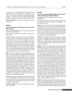
Journal of Pediatric Surgery (2005) 40, E37 – E39
www.elsevier.com/locate/jpedsurg
Congenital hepatic mesenchymal hamartoma associated
with mesenchymal stem villous hyperplasia of the
placenta: case report
Maurizio Cartaa,*, Emiliano Maresib, Mario Giuffrèa, Giuseppe Catalanoa, Ettore Piroa,
Fortunato Siracusaa, Giovanni Corselloa
a
Dipartimento Materno Infantile, Università di Palermo, 90100 Palermo, Italy
Istituto di Anatomia Patologica, Università di Palermo, 90100 Palermo, Italy
b
Index words:
Hepatic mesenchymal
hamartoma;
Mesenchymal stem
villous hyperplasia
of the placenta;
Newborn
Abstract A newborn with an unusual association of hepatic mesenchymal hamartoma and mesenchymal
stem villous hyperplasia of the placenta is presented. At birth, the large hepatic mass caused severe
respiratory distress necessitating early surgical intervention. This report on the association of hepatic
mesenchymal hamartoma and mesenchymal stem villous hyperplasia of the placenta strongly suggests a
common pathogenetic origin of the 2 lesions.
D 2005 Elsevier Inc. All rights reserved.
Hepatic mesenchymal hamartoma (HMH) is an uncommon benign developmental anomaly arising from the
mesenchyma of the portal tract. It is more common in the
first year of life (55% of all cases) [1]; a few cases (15%) have
been reported in the neonatal period [2]. Although histologically benign, large congenital hamartomas of the liver may
present at birth with severe abdominal distension and a
respiratory distress. Careful early fetal ultrasound (US) scans
in the last trimester of pregnancy may allow prenatal
diagnosis, improving clinical evolution of newborns.
Mesenchymal stem villous hyperplasia of the placenta
(MSVHP) is a rare anomaly involving diffuse edema of the
stem villi with relatively normal terminal villi and is
Presented at the 36th Annual Meeting of the Canadian Association of
Pediatric Surgeons, Winnipeg, Manitoba, Canada, September 30- October
3, 2004.
T Corresponding author. Tel.: +39 91 6555454; fax +39 91 6555455.
E-mail address: [email protected] (M. Carta).
0022-3468/$ – see front matter D 2005 Elsevier Inc. All rights reserved.
doi:10.1016/j.jpedsurg.2005.02.018
secondary to vascular malformation of the fetal plate. It has
been described in phenotypically normal fetuses but may be
occasionally associated with severe fetus anomalies [3,4].
We report on a newborn girl with a prenatal history of
placental abnormalities who presented at birth with a huge
HMH causing severe respiratory distress. The diagnosis of
MSVHP led us to investigate a common pathogenetic origin
of HMH and MSVHP.
1. Clinical report
At 18 weeks of gestation of the fifth pregnancy of a
39-year-old woman, US scans revealed an anechoic lesion
occupying more than 50% of the placenta. Amniocentesis
was not performed. Maternal serum a-fetoprotein and hCG
levels at 22 weeks were high. No polyhydramnios or fetal
hydrops was present. At 38 weeks, when the woman referred
to our department, fetal US scans showed multiple cysts in
the fetal liver (107 95 mm) (Fig. 1). A 3150-g infant girl
E38
Fig. 1 Fetal US scan at 38 weeks demonstrating multiple cystic
lesions (F, 107.7 95.4 mm) in the fetal liver.
was delivered through cesarean delivery; the apgar score was
5 at 1V and then the baby required immediate intubation for
respiratory distress. The abdomen was markedly distended
because of a large palpable mass involving almost the entire
abdomen. Abdominal US at birth showed a solid mass
(15 cm) with many cystic lesions (the largest being 30 mm in
diameter) apparently arising from the liver. Renal and cardiac
US scan results were normal. Hematochemical investigations revealed slightly increased levels of liver enzymes and
a high serum level of a-fetoprotein (9592 UI/ml). Cytogenetic analysis of peripheral T lymphocytes showed a normal
female karyotype (46,XX).
The large mass determined a severe impairment to the
respiratory function in spite of intubation and mechanical
ventilation; therefore, the baby underwent surgical resection
at 10 hours of age. At laparotomy, a multicystic and solid
mass (120 70 mm) was totally occupying the right hepatic
lobe and some cysts involved the left hepatic lobe. The
entire right hepatic lobe and the 2 bigger cysts of the left
hepatic lobe were resected. After surgery, mechanical
ventilation was necessary for 6 days. In relation to the large
hepatic resection, the baby developed cholestasis, jaundice,
and generalized edema, which gradually recovered. A
transient hypothyroidism was observed.
M. Carta et al.
Pathological findings demonstrated an enlarged (weight,
970 g; F, 15 cm) and multicystic placenta (Fig. 2A). At
microscopy, the main histological finding was the presence
of hydropic stem villi with normal terminal villi and no
trophoblast proliferation (Fig. 2B). This histological picture
indicated the diagnosis of MSVHP, excluding the first
hypothesis of partial hydatiform mole.
The liver mass (weight, 130 g; F, 10 6 2 cm)
appeared as a multiloculated lesion composed of solid and
cystic areas at macroscopy (Fig. 3A); the cut surface showed
yellow-green gelatinous material. At histology, the lesion
was characterized by many fusiform and star-shaped cells in
a myxomatous connective tissue that compresses the normal
hepatic parenchyma (Fig. 3B). These findings were consistent with the diagnosis of HMH.
Clinical and ultrasonographic follow-up to 8 months
demonstrated good growth and no development of new
abdominal masses; the cysts in the left hepatic lobe do not
tend to enlarge.
2. Discussion
In spite of only a few reports of MSVHP and about 40
reports of neonatal HMH in the literature, there have been
already described 2 newborn patients with the association of
neonatal HMH and MSVHP [5,6]. Only the patient reported
by Kitano et al [5] had a prenatal US detection of both
hepatic and placental mesenchymal lesions as we describe
in the present report. Tsao et al [7] described an infant with
an HMH associated with extensive vascular disease in the
intermediate and stem villi of the placenta. Most of the
reports in the literature lack some clinical (prenatal and
neonatal US, follow-up) or laboratory data (placental
microscopy, a-fetoprotein level, liver function tests), probably because the common pathogenetic origin of the 2
lesions was not suspected. The present patient gives new
and stronger evidence to the hypothesis of a common
pathogenetic origin of HMH and MSVHP.
In the past, an abnormal focal overgrowth of vascular and
epithelial elements isolated from the normal mesenchyma of
Fig. 2 Macroscopic and microscopic studies of the placenta showing mesenchymal stem villous hyperplasia. A, Macroscopic view of the
placenta with severe vesicular degeneration, normal villi are mixed with vesicular ones. B, Histological section showing hydropic and
cavitated stem villi and normal terminal villi (original magnification, 10).
HMH associated with MSVHP
E39
Fig. 3 Macroscopic and microscopic studies of HMH. A, Resected liver mass composed of solid and cystic areas containing yellow-green
gelatinous material. B, Histological section showing many fusiform and star-shaped cells in a myxomatous connective tissue with many
vessels and focal microcystic cavitations; this lesion compresses the adjacent normal hepatic parenchyma where extra medullary
erythropoietic foci and ductal-biliary hyperplasia are visible (original magnification, 40).
the portal tract was suggested [8] for the pathogenesis of
HMH. Therefore, HMH was considered a developmental
anomaly rather than a true tumor and the association of
HMH and MSVHP may represent a synchronous abnormal
development of the mesoderm. The first step of the lesion in
MSVHP may be a continuous inadequate blood supply that
causes both severe aneurysmal dilatations of the chorionic
vessels and hyperplasia of placental stem villi producing
cystic structures. The consequent vascular thrombosis on the
fetal plate of placenta may cause ischemic lesions of the
fetal liver producing, as a reparative response, the proliferation of bile ducts and the obliteration of the central veins,
which may lead to HMH [9]. Placental lesions are not
observed until the second trimester of gestation and precede
the development of hepatic ones, confirming the common
pathogenetic mechanism.
The patient we report on showed an unusual clinical
presentation with severe respiratory distress and compromised hepatic function. The rapid liquid collection in the
hepatic cysts increased pressure over the lungs, causing
respiratory distress. The benign nature of the lesion, the
reported spontaneous regression [10], and the risks of
surgery in newborns could represent elements for choosing
a conservative approach. The severity of symptoms forced us
to conduct a surgery, but the massive involvement of both
lobes did not allow complete excision. In the literature, it has
been described that both hepatic lobes are involved in 3% of
all pediatric hamartoma cases. Recurrence or malignant
transformation has been rarely observed [11]; therefore, a
careful follow-up of any residual lesion has been guaranteed.
When a placental vascular anomaly is observed along
with mesenchymal stem villous hyperplasia, pregnancies
usually result in healthy neonates [4,12]. However, Kitano
et al [5] reported 2 newborns with a mesenchymal
hamartoma of the liver and 1 infant with a hepatic
hemangioma associated with pregnancies with MSVHP.
The present report provides further support of the possible
association of a gestational trophoblastic disease with fetal
hepatic mass. We believe that the association of HMH and
MSVHP has been underdiagnosed; in many patients with
neonatal HMH, MSVHP was probably present especially
when the benign, slowly progressive nature of the hepatic
lesion has led to the diagnosis only several months after
birth. To verify a common pathogenetic origin and address a
specific prognosis of the 2 lesions, a careful histological
investigation of the placenta in any newborn with a hepatic
mass is very important.
References
[1] De Maioribus CA, Lally KP, Sim K, et al. Mesenchymal hamartoma
of the liver. A 35-year review. Arch Surg 1990;125:598 - 600.
[2] Mulrooney DA, Carpenter B, Georgieff M, et al. Hepatic mesenchymal hamartoma in a neonate: a case report and review of the literature.
J Pediatr Hematol Oncol 2001;23:316 - 7.
[3] Lage JM. Placentomegaly with massive hydrops of placental stem
villi, diploid DNA content, and fetal omphaloceles: possible association with Beckwith-Wiedemann syndrome. Hum Pathol 1991;
22:591 - 7.
[4] Chen CP, Chern SR, Wang TY, et al. Pregnancy with concomitant
chorangioma and placental vascular malformation with mesenchymal
hyperplasia. Hum Reprod 1997;12:2553 - 6.
[5] Kitano Y, Ruchelli E, Weiner S, et al. Hepatic mesenchymal
hamartoma associated with mesenchymal stem villous hyperplasia
of the placenta. Fetal Diagn Ther 2000;15:134 - 8.
[6] Alwaidh MH, Woodhall CR, Carty HT. Mesenchymal hamartoma of
the liver: a case report. Pediatr Radiol 1997;27:247 - 9.
[7] Tsao K, Hirose S, Sydorak R, et al. Fetal therapy for giant hepatic
cysts. J Pediatr Surg 2002;37:E31.
[8] Edmonton HA. Differential diagnosis of tumors and tumor-like
lesions of liver in infancy and childhood. Am J Dis Child 1956;91:
168 - 86.
[9] Lennington WJ, Gray GF, Page DL. Mesenchymal hamartoma of the
liver. A regional ischemic lesion of a sequestered lobe. Am J Dis Child
1993;147:193 - 6.
[10] Barnhart DC, Hirschl RB, Garver KA, et al. Conservative management of mesenchymal hamartoma of the liver. J Pediatr Surg 1997;32:
1495 - 8.
[11] Ramanujam TM, Ramesh JC, Goh DW, et al. Malignant transformation of mesenchymal hamartoma of the liver. J Pediatr Surg 1999;34:
1684 - 6.
[12] Moscoso G, Jauniaux E, Hustin J. Placental vascular anomaly with
diffuse mesenchymal stem villous hyperplasia. Pathol Res Pract 1991;
187:324 - 8.
© Copyright 2026 Paperzz