FAU Institutional Repository http://purl.fcla.edu/fau/fauir
This paper was submitted by the faculty of FAU’s Harbor Branch Oceanographic Institute. Notice: ©1981 John Wiley & Sons, Inc. This manuscript is an author version with the final publication available at http://www.wiley.com/WileyCDA/ and may be cited as: Eiseman, N, J., & Moe, R. L. (1981). Maripelta atlantica sp. nov. (Rhodophyta, Rhodymeniales) a new deep‐water alga from Florida. Journal of Phycology, 17(4), 299‐308. doi:10.1111/j.1529‐8817.1981.tb00855.x J. Phycol.
17,299-308 (1981)
MARIPELTA ATLANTICA SP. NOV. (RHODOPHYTA, RHODYMENIALES)
A NEW DEEP-WATER ALGA FROM FLORIDAt
Nathaniel J. Eiseman
Harbor Branch Foundation, Ft. Pierce, Florida 33450
and
Richard L. Moe
Department of Botany, University of California, Berkeley, California 94720
ABSTRACT
A red alga with a cylindrical stipe bearing a single deciduous blade was collected in deep water off the east coast
of Florida. It is described as Maripelta atlantica sp. nov.,
differing from M. rotata (Dawson) Dawson (the type of
the genus, from deep water off California and Baja California) chiefly by having an annular tetrasporangial
nemathecium on the lower surface of the blade rather than
scattered nemathecia on the upper surface. Both species are
vegetatively and reproductively distinct from M. thivyae
Dawson, which is transferred to Halichrysis. New information is given regarding the reproduction and distribution of M. rotata.
Key index words: deep-water algae; Florida; Maripelta
atlantica sp. nov.; Maripelta rotata (Dawson) Dawson;
Halichrysis thivyae (Dawson) comb. nov.; Rhodophyta; Rhodymeniaceae
During an investigation of the deep-water biota
off the east coast of Florida using the JOHNSONSEA-LINK submersibles, an apparently undescribed red alga was found to be common at a depth
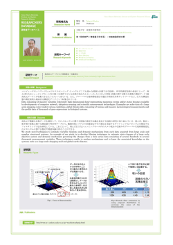
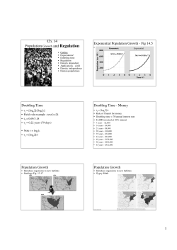
of 65 m. The alga, which has the shape of a delicate
mushroom (Fig. 1), was immediately recognizable as
a member of the Rhodymeniales but could not be
assigned to any Atlantic genus. An examination of
Maripelta rotata (Dawson) Dawson, the type of its
I
Accepted: 7 May 1981.
genus which inhabits the deep water of California
and Baja California revealed a close anatomical and
developmental similarity to the Florida plant. The
two are sufficiently distinct, however, to justify
erecting a new species.
Maripelta atlantica sp. nov.
Thallus ex haptero stipite que laminaque compositus.
Hapteron lobatum. Stipes erectus eramosus. Lamina terminalis 0.25 -0.50 mm crassa 7 cm diametro peltata subcircularis centraliter leviter depressa. Medulla unistrata
cellulis parietibus crassiusculis ecolorata per corticem visibilis, cellulis parvis circumcincta. Cortex distromaticus
pigmentosus, cellulis interioribus 10-25 um diametro
unaquaeque cellula exteria ferentibus, cellulis exteriis ex
parte pilosis, pilis unicellularibus. Lamina supra submarginaliter areis prominentibus procarpia ferentibus. Ramuli
carpogoniferi recti tricellulares. Ramuli auxiliares bicellulares. Cystocarpia superficialia prominentia 1 -2 mm
diametro subsphaerica ostiolata; tela arachnoidea nulla.
Lamina supra submarginaliter soris continuis spermatangia ferentibus, subtus submarginaliter nematheciis continuis tetrasporangia ferentibus. Tetrasporangia cruciatim
divisa, in filamentis nemathecialis intercalata.
A Maripelta rotata (Dawson) Dawson laminis subtus
submarginaliter nematheciis continuis differt.
Holotypus in herb. U.c. (UC 1462532).
Plant with a lobed hold fast and an erect, unbranched stipe bearing a terminal peltate blade (Fig.
300
NAT HAN IE L J. EISE MAN AN D RI CH ARD L.
~I O E
numbers refer to J OH SON-SEA- LI
a d ive num ber):
K I o r II and
J SL 1-307 3 Dec. 1975, 26°47. 0 ' N, 80°00.7' W, 65 .2
m , Ee, lj>
J SL 11-040 5 May 197 6 , 26°57 .4 ' N, 79°59.6 ' W, 59
m, 81,
JSL 11 -1 32 4 O ct. 197 6 , 27°11.7' N, 79°57.3' W, 88 .0
«
In,
F IG.
I.
Marip elta atlantica sp . no v. Habit. Sca le
=
10 mm .
1); stipe irre gularly cylind r ical, with a n n u lar sca rs
in o ld plants, 2 - 5 mm di am ., to 60 mm tall ; blade
nearl y circ u lar, to 70 mm diarn. , often slig h tly d epressed in th e ce nte r , 0. 25 - 0.5 0 mm thi ck ; medulla
with a ce ntral layer o f large , thi ck -wall ed , co lo rless
ce lls (Fig. 2), 200-400 J-tm periclinal di am ., a ngu la r
in su rface view , visibl e through cortex (Fig . 3), su rrounded by sm a ller cells 25-35 J-tm diam ., co rtex o f
two layers of pigmented ce lls, inner ce lls 10-25 J-tm
diam., eac h bearing o ne to se veral smalle r outer ce lls
3-5 J-tm di am. (Fig. 4), whi ch occas io nally bea r hai r
ce lls (Figs. 5, 6).
T etraspo r angia , sper ma ta ngia, a n d ca r pogon ia
borne o n se par a te pl ants. Proca rps in blister-like ,
su b margina l patches o n upper su rface of the blade ,
th e ca r pogon ial branch co m posed of three ce lls in
a straight row a nd a two-celled a ux ilia ry ce ll branch ;
cystoc a r ps protruding , su bs p herica l, os tiola te, to 2
mm diam., without a tela arachnoidea ; spe r mata ng ia
in a co n tin uou s submarginal so r us, bo rne sin gly o n
upper su per ficial ce lls, 2 J-tm di arn ., 5 J-tm long; tetr asporan gia cr uciately di vid ed , produced from intercalary ce lls o f filaments in a co n tin uous, ir r egu lar, su b ma rgina l nemathecium o n lower su rface of
blad e.
Differing fro m M aripelta rotata (Dawso n) Daw son
by ha vin g a co n tin uous, su b margina l tetrasp orang ial nemathecium o n low er su r face o f blade ra ther
than sca tt e r e d n em athe cia o n upp er surface of
blade.
T ype locality: 3.6 km east o f Sin ger Island , Palm
Bea ch Co ., Flo r ida, U.S .A. 26 °46.4 ' N, 79°5 9.5 ' W,
72 m d epth .
Holotyp e : JSL 11-077, 17 June 1976 , 18.3° C; te tra spo r angial , UC 146 2532 .
Isotypes: HBFH , US, US F, D UKE , MI CH .
Specimens exa m in ed (a br id ge d listing; co llec tio n
J SL 1-402 15 Dec. 1976 , 26 °47 .3' N, 79°59.3 ' W,
68 .9 m , EB, lj>
J SL 11-189 2 May 1977 , 27°07 .7' N , 79°50.5' W, 89.2
m,
J SL 11-1 94 5 May 19 77 , 26°48. 0 ' N , 79°59.7' W, 72.3
m , EB, lj>
J SL 1-421 7 July 19 77, 26°4 7.0 ' N, 79°59 .9 ' W, 59
m,
JSL 1-51 6 7 Jun e 19 78 , 26°4 5.4' N, 79°59.5' W, 71. 9
m, Ee, lj>
J SL 1-655 2 1 Ma r. 197 9 , 26 °48 .5' N, 79°59.8 ' W,
69.5 m, EB, lj>
J SL 11 -351 6 A pr. 1978 , 26 °46.2' N , 79°59.5' W,
70 . 1 m , Ee, lj> ,
J SL 1-631 23 Jan . 1979, 27°11. 7' N, 79°57 .2' W, 90 .9
°
m
°
J SL 1-67 0 22 Ma y 19 79, 26 °44. 1' N , 79°58 .6' W,
73.2 m , EB, lj> ,
JSL 1-7 07 12July 19 79, 27°46.5 ' N, 79°59. 2 ' W, 70.1
m, EB,
lj> , o
Gu lf o f Mexi co , Se p t. 1967 26°24' N, 83°43' W, lj> ,
c. J. Dawes we t stac k.
HAB ITAT, DIST R I BUTION AND PHENO LOGY
Maripelta atlantica is present in all seasons o n the
Florida east coast. In the vicin ity of Pal m Beach it
occ urs s parsely a t 60 m , becomes the d omi nan t
mem ber of the flo ra at a bo ut 65 m , a nd is aga in
spa rse at greater d epths. It has been o bse rved fro m
th e su b mersibles to 100 m. It also occ urs fr om 20 60 m in th e Gul f of Mexi co , re po r te d as Fau chea
paltat a Taylo r (Dawes 197 4 , Daw e s a nd van Breedveld 1969 ). A no ther re por t of F. pelt at a by Che ney
a nd Dye r (197 4 ) is p robabl y M . atlan ti ca also, but
th eir specimens co u ld not be locat ed for stu dy . Both
te traspora ngial a nd cystocarpic pl ants are fo u nd at
all seasons . Mal e pl ants are com parative ly rare. T he
sti pes are ofte n e nc r us ted by br yo zoans, sponges, ascid ians , a nd barnacles (Wi ns to n a nd Eise man 1980).
Co mmo nly associate d algae fro m 58-94 m d epths
a re H alymenia sp p ., Peyssonn elia rubra (Greville) J.
Aga r d h, Kallymenia sp p., va r io us Cera miaceae a nd
cr us tose coralline forms.
Vegetative Development
T he stipe of M aripelta atla ntica, whi ch arises fro m
a lobed co nica l h old fast, is persistent a nd a p paren tly
peren n ial. It stores florid ian starch, as ind icated by
a golde n- brown reac tio n with iod in e -KI a n d a
maltese cross pattern under th e polarizin g microsco pe . T he stipe co ntin ues to g ro w in d iamet er a nd
30 1
AIAR IPE LTA ATLANTICA SI'. NOV .
2
3
5
6
8
~
.
,
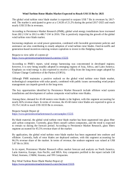
FI Gs . 2- 8. M aripelta atlantica sp . nov. F IG . 2. Section th rough the margin of th e blad e. Scale = 100 /-LIII. F IG . 3. Medulla ry cells see n
through th e co rtex. Scale = 25 /-Lm . FI G . 4 . Surface view o f co rtex . Scale = 10 /-LIII . F IGS . 5, 6. T wo hair ce lls. Scales = 5 /-LIII. FI G. 7.
Longitudinal sectio n o f th e stipe. Arrows indicat e seco nda ry growth of th e cor tex . Scale = I n1l11 . FI G. 8. Cross section of th e stipe
th rough an area of old blad e at tac h me nt . T he da r k rin g is a re mna nt of the old co rtex. Scale = 0.5 m ill.
length throughout th e life o f th e plant. Old co rt ical
layers (Figs . 7, 8) a nd frequently th e calca reous remains of a nimals a re incorporated into th e tissu e of
th e stipe as conce n tr ic rings , particul arly in a reas o f
old blad e attachment.
T he blad es o f M. atlantica a re d eciduou s, but th e
interval be tween successive losses is not kn own . New
blad es a re produced terminally o n the persistent
stipe. Plants brought into th e laborat ory lost th eir
blades within o ne week , p resumabl y as th e result of
302
NATHAN IEL J. EISE M AN AND RI CH ARD L. M O E
9
14
/.
<.
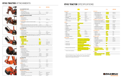
FIGs. 9- 14. M aripelta atlan tica sp. nov . Stages in blad e d evelopmen t. All scales = I m m . F IG. 9. Stipe with a new stipe in itial. FIG. 10.
A mature stipe initi al. FIG. II. A sti pe ini tial with a blad e in itial. The light a rea at the tip ind icat es the cavity. FIG. 12. Near med ian
sec tion th rough an un open ed blade primord ium. FIG. 13. A newly o pe ne d ca p. FIG. 14 . A ca p whi ch has lo st its pigm entat ion in cultu re .
T he abscission zone has form ed but th e ca p ha s not yet been she d: az = abscission zone .
MARIPELTA ATLANTICA SI' . NO V.
shock . Blade regeneration began almost immediately after old blades were shed . ew blade production
begins with an upward growth at th e center of the
stipe forming a cone (Fig s. 9 , 10), which will become
the new stipe in crement. A cylind r ical blade primordium then arises from the apex of the cone (Fig.
11). The blade primordium becomes hollow (Fig .
12), the inner surface lined with small pigmented
cells. It continues to expand , and when it is about
1.5 mm diam . it becomes co nca ve (Fig. 12). At this
stage the hollow primordium apparently ruptures
to give rise to a cup-shaped blade ca. 3 mrn diam .
(Fig. 13), the inner su r face of the primordium beco min g the upper surface of the blade. The conical
stipe primordium continues to grow in diameter
particularly distally, and becomes almost cylindrical.
The margin of the blade becomes meristematic (Fig.
2) and lateral growth results in a Rat, peltate blade.
Production of reprodu ctive organs apparently
terminates vegetative growth. Reproductive structures are always submarginal and have been observed on blades of various sizes. We presume that
blades are abscised after the release of reproductive
bodies since no blades with co nce ntr ic reproductive
areas or co nce n tr ic sca rs have been seen. A definite
abscission zone consisting of a layer of pigmented
cortical cells between the blade and stipe is formed
before a blade is shed (Fig. 14). The origin of thi s
layer, whi ch later gives rise to th e new stipe in crement ha s not been d etermined.
R eproduction
Procarps occur only on the upper surface of the
blade in blister-like submarginal patches of prolife ra tin g cortical cells (Fig . 15). The supporting cell
is indistinguishable in size or stainability from other
cortical cells. It bears a carpogonial branch, a n auxiliary cell branch and sometimes a sterile branch.
The carpogonial branch consists of three cells in a
straight row . The trichogyne is straight and extends
through the superficial mucilage layer. The auxiliary cell branch consists of two cells-an elongate
auxiliary mother cell and a spherical auxiliary cel l.
An auxiliary cell branch alwa ys accompanies a carpogonial branch and is cu r ved so that the auxiliary
cell lies close to the carpogonium (Fig. 15). Cell s of
both the auxiliary cell branch and the carpogonial
branch stain more intensel y with aniline blue than
surrounding cells. The sterile branch, if present,
usuall y consists of more than two cells which do not
stain darkly. If not fertilized, the carpogonium degenerates, but the two lower cells of the branch persist and co ntin ue to stain darkl y as they are buried
by the growth of su r ro u nd ing filaments .
Following presumptive fertilization, the supporting cell fuses with nearby cells whi ch then stain more
darkly with aniline blue. The developmental stages
leading to the fu sion were not seen. As the cortical
cells proliferate to produce the pericarp, there is a
-tr
~ ac
303
15
16
F IGS . 15-17 . M aripelta atlantica sp. no v. Carpos poroph yte de velo pme nt. FI G . 15. A pro ca rpial nem ath ecium . Scale = 10 t.u n .
FI G . 16. A you ng ca rpospo ro p hytc . Scale = 0.1 m m. FI G . 17. A
mature cystoca rp . Section d oes not pass th ro ugh ostiole. Scale =
0.2 mm; tr = tr ichogy ne, cp = car pogonium , ac = auxi liary cell,
a mc = aux iliary mother ce ll, sc = su p porting cell, m = mucilage,
fc = fusion cell, P = placental tissu e.
periclinal rupture through th e filam entous tissue
just above the yo u ng ca r pos poro p hy te. The tissue
a bove the rupture arches outward , crea ting a cavity
in which the carposporoph yte develops (Fig. 16).
304
NAT HAN IE L J. EISE MAN AN D RI CH ARD L. MO E
22
18
~f
nf
/
---t
pc---
FIGs. 18 - 22 . M aripelta atlan tica sp. nov. T etrasporan gial d evelopment. FIG. 18. Section th rough a te tras po ra ng ial nemathecium .
Scale = 20 p.m. FIGs. 19- 21. Stages in tetrasp oran gial d evelopmen t. All scales = 10 p.m . FIG. 22 . Ma ture tet rasp oran gia. Scale = 10 p.m;
n f = nem athecial fila me nt, pc = pit co n nectio n , t = te tras po ra ngi u m , tmc = tetraspo ran gial moth er ce ll.
T he ca vity is traversed by th e mucilaginous rem ain s of ruptured filam ents (the reseau muqueux o f
Hu ve a nd Huve 1976) a network whi ch persists durin g th e early e n lar ge me nt o f th e ca r pos po ro p hy te
and which ma y in part accou nt for th e mucilaginous
co n te nts o f the mature cystoca r p (Fig. 17). A ce llu lar
tela arach noidea is absent. At th e base of th e ca r po spo ro p hy te is a large, su bs p he r ical ce ll whi ch a pparently resul ts from the fu sion of th e auxiliary ce ll
branch, th e primary gonimoblast ce ll, a nd possibly
th e su p porting ce ll (Fig. 17). All th e gonimoblast
filaments arise directl y fr om th e fu sion ce ll. Ben eath
th e fu sion ce ll is a group o f d ense ly sta in ing ce lls
which ha ve been referred to in H alichrysis depressa
as pla cental tissu e (H uve a nd Hu ve 197 6).
T he m ature ca r pos po ro p hy te co n ta ins m an y
lobes of uninucl e a te ca r pos po r a ngia (Fig. 17).
T he re is no evide nce o f th e production of multiple
cro ps of spo ra ngia, as has been reported in so me
Rh od ym eniaceae (Spa rl ing 1957 ). The sp orangia
a re o f abo u t th e sa me size (to 20 j.tm), a p pa re ntly
maturin g sim u lta neous ly. T he cystoc a r p is a nearl y
sp he r ica l protrusion with a broad co n nec tio n to th e
blad e. At th e hi ghest point is a n os tiole, often with
a sligh tly raised co lla r.
T etrasporan gia are produced in a continuous
MARIPELTA ATLANTICA SP. NOV .
23
305
24
nf _ _ _
FIGS. 23, 24. Ma ripelta rotata. FIG. 23 . Section through a tetrasporangial nemathecium . FIG. 24 . A Ietrasporangi al mother cell. Scales =
25 /L m ; nf = nem athecial filam ent, pc = pit co n nec tio n , t = tetrasporangium , tm c = tetrasp orangial moth er cell.
nematheci um-a ring about 2 mm wide j ust inside
the margin of the lo we r surface of the blade. The
nemathecium is formed by filaments cut off from
su pe r ficia l ce lls (Fig. 18). T he nemathecial filame nts
are composed of 3-6 elongate ce lls of 2-3 /Lm diam.
occasionally interconnected by secondary pit connections. T hose filaments which will bear sporangia
are not d ifferentiated at an ea rl y stage from tho se
wh ich will remain sterile. Tetrasporangium mother
ce lls d iffere ntiate from in tercalary ce lls near the
base of the fila me nts (Fig. 19). T he mot her ce ll expa nds asymmetrically (Fig. 20) so that the apica l pit
con nection is di splace d in a basal di r ectio n . U ltimately, a bo u t hal f of th e mother ce ll is above the
a pical pit co n nectio n a nd hal f bel o w. T he mothe r
ce ll di vid es whe n it is abo u t 30 /Lm long (Fig. 2 1).
T he cleavage is se q uen tial-first a pe r iclinal division, th e n anticlina l div isions of th e der iva tives . T he
maximum size of sporangia is 45 /Lm lo ng x 23 /Lm
d iam . T he spores are cruc iately arranged (Fig. 22).
Spermatangia ar e bo r ne in a conti n uous submarginal ri ng like t hat of the te traspora ngia, b u t on the
u p per , ra th e r t han the lo we r surface of the blade.
Spermatangia, 2 /Lm dia m . x 5 /Lm long are cut off
singly from the o uter cortical cel ls. Ma le pl a n ts are
vegetative ly identical to fema le a nd tetrasporic
plants.
REVIEW OF THE GEN US MARIPEL TA
Unti l now, Maripelta has comprised two species,
M. rotata (Dawson) Dawson and M . thivyae Da wso n ,
t he la tte r kno wn o n ly from t he ty pe . Examination
of ma te rial of bot h species has provided n ew in fo rmation pe r tin ent to the taxonomy of the gen us. We
incl ude the results of o ur exa mi natio n of a third
entity-the unna m ed plant from H awa ii considered
by Euba nk (in Dawson 194 9) to be congeneric with
Drouetia, t he genus to which Dawson originally assig ned M . rotata.
Maripelta rotata (Dawson) Dawson
Material exam ined (abridged listin g):
Whi te Cove, Sta. Cata lin a 1., Ca lif. , 66-69 m., leg .
E. Y. Da wso n . Iso type . AHF36404
Pal os Verdes Pt. , Los Angeles Co., Ca lif. , 32°40.0'
N, 119° 10.0 ' W, 20 m. , leg. R. Moe. A HF 79040
Tanner Ba nk , 37 °59.5' N, 123°24. 5 ' W, 69-73 m .,
leg . E. Y. Da wso n , A HF26782
Cordell Bank , 38° 1.5' N, 123°25. 5 ' W, 43 m ., leg . R.
Sch m ieder, UC 1465052; 38 ° 1.5 ' N, 123°25 .5 ' W,
62 m. , leg. R. Sc hm ieder, UC 1446 147.
O ur observations agree in ge ne ral with those of
Dawson ( 1949, 1963). Tetrasporangi um developme n t, whic h was not described by Da wso n , is in itiated by an intercalary ce ll as in Maripelta atlantica
(Fig. 23) . The e nlargement of the tetrasporangium
mother ce ll is somewhat more as ymmetrical than in
M . atlantica, so t hat the apical pit con nection tends
to be d isplaced more basa lly (Fig. 24 ). As mentioned
by Dawson, the sporangia are borne in small pustular nemathecia o n the upper surface of the blade.
The nemathecia tended to occur submarginally in
the p lants we exami ned. Procarps occur in p ust ula r
th icke ni ngs of the cortex of t he u p pe r surface near
the margin. The carpogonial bra nc h is composed of
three or fo ur cells (Figs. 25 , 26) . ·Most bra nches are
straigh t, b u t some ha ve a bend at th e lo we r mo st ce ll.
As in M . atlan tica, when th e procar p aborts on ly the
two basal cells of th e branch re main (Fig. 27) . T he
306
NAT HAN IEL J . EISEMAN AND RI CH ARD L. MOE
25
FI Gs. 25-28 . Maripelta rotata. F IG . 25. A four-celled ca r pogo n ial branch . FI G . 26. A three-ce lled carpogonial branch . FIG. 27. An
unferti lized carpogo nia l branch with a degenerating carpogo nium. All sca les = 20 J-lom . FI G . 28. A d eve lop in g ca r posporo p hyte . Scale =
0.5 m m ; cp = ca rpogo nium, tr = tri chogyn e .
a uxiliary ce ll bran ch is composed of two cells. Carpo spo roph yte d evelop me n t (Fig. 28) is simi lar to
th at of M . atlantica, wit h the forma tion of a subs p~erical fusion cell fro m wh ich gonimoblast in itials
anse.
Bowen (197 1) d escr ibed the process of blade abscission and regene ra tion in cu lture wh ich is ide ntical to that in M . atlantica. H owe ve r , she observed
branchin g of the stipe resu lting from the in itiat ion
by the absc ission laye r of two stipe primordia rather
tha n o ne . We have observed the development of the
fir st blad e in ge r m lings derived from carpos pores
of a pla nt collected from Cordell Ba nk (UC 1446147).
It agrees com p letely with the acco unt given by Bowe n.
M aripelta rotata grows in d ee p wa te r along the
coast of Ca lifo rn ia a nd Baj a Ca lifo rn ia. It has been
obtained fro m Punta Euge nia (2 7°50' N) in the
so u th (Dawson et al. 1960) to Cordell Bank (38° N)
off Pt. Reyes, Ma ri n Co . in the no r th , the latte r record (based on co llect ions by R. Sch m ieder) ex te nd ing the range fro m Carmel Ba y. T he maxim um
depth of collec tion has bee n 79 m (by dredge) from
E mpire La nd ing, Sta . Catalina 1. fide P. C. Silva.
(Metric de pths sup plied in Da wson , 1949, ha ve been
exaggerated through th e use of 2.0 ra ther th an 1.83
in co nverting fat homs to me ters.) It has been found
in the shade of M acrocystis beds as sha llow as 13 m
(Dawson 1963).
MARIPELTA ATLANTICA SP. NOV .
Maripelta thivyae Dawson
Material examined:
Krusadi Island, Gulf of Manaar, Tamil Nadu,
India; leg. F. Thivy, cast ashore; US60368 (Holotype) .
Dawson's (1963) description of M. thivyae is based
on a single cystocarpic plant consisting of a discoid
hold fast and contorted, branched stipe bearing numerous irregularly rotate blades. Blades consist of
a medulla composed of 2-4 layers of thin-walled
compressed cells and a cortex composed of two layers of small pigmented cells. Dawson described the
growth of the stipe as sympodial. The stipe comprises several segments, each of which arises laterally from near the base of the previous segment.
Occasional adventitious branches, not mentioned by
Dawson, are also produced along the stipe. The
stipe has a medulla of large floridian starch-containing cells surrounded by an inner cortex of anticlinally elongated vacuolate cells and an outer cortex
of small pigmented cells. Secondary growth of the
cortex is irregular and does not result in concentric
rings such as those seen in sections of M. atlantica
and M. rotata. The blades attach to one another secondarily through discoid haptera produced marginall y. An abscission layer is formed at the base of the
blades, but in contrast to M. atlantica and M. rotata
new blades are not produced from this layer.
To Dawson's account of the reproduction we can
add only that cystocarps of various sizes ' are scattered at random over the upper surface of the
blade. We have confirmed Dawson's observations
that the carposporophyte is pedicellate (i.e. with a
columnar gonimoblast initial) and that it lacks a filamentous envelope (tela arachnoidea) .
Marip elta thivyae, because of its numerous, coalescent blades and polystromatic medulla, seems
more closely related to Halichrysis than to Maripelta.
The mature carposporophyte could not be distinguished from that of H. depressa as described by
Huve and Huve (1976). We accordingly propose the
following new combination: Halichrysis thivyae (Dawson) comb. nov. = Maripelta thivyae Dawson, 1963,
p. 448, pI. 81: fig. 5.
Th e Hawaiian Plant
Material examined:
Hanauma Bay, Oahu, leg. E. Y. Dawson # 11852,
UC wet stack.
Ewa Beach, Oahu; leg. I. A. Abbott # 1030, UC wet
stack.
Nanakuli , Oahu, leg. G . F. Papenfuss # 10554, UC
wet stack.
This plant, mentioned but not described by Dawson (1949), resembles M. thivyae in having a contorted, apparently sympodial stipe and numerous
irregularly rotate blades which are here and there
united by marginal attachment discs. The blades are
307
up to 600 J..tm thick . The medulla consists of 4-6
irregular layers of large thin-walled cells becoming
smaller toward the surface. The cortex is composed
of much smaller cells in 3-4 layers, with the innermost cells 10-20 J..tm diam. and the surface ce lls 25 J..tm diam . Tetrasporangia are borne primaril y on
the lower surface of the blade . They are grouped in
sori which are at first discrete but which later become confluent and cover the entire surface. The
sporangia are intercalary and cru ciately divided. At
maturity they project slightly above the outer cortical cells. The largest sporangia measure 35 J..tm X
15 J..tm.
Because we have not seen comparable reproductive stages in M . thivyae and the Hawaiian plant, we
hestitate to assert that they are congeneric. However, the latter is probably not congeneric with
Drouetia as postulated by Eubank (in Dawson 1949)
because the tetrasporangia are intercalary rather
than terminal (see below).
TAXONOMI C AFFINITIES OF MARII'EL TA A TLANTICA
Four genera in the Rhodymeniales include species
with peltate blades-Halichrysis U. Agardh) Schmitz,
Sciadophycus Dawson, Drouetia DeToni fil., and Maripelta Dawson . We have rejected Drouetia from consideration because the type and only species, D. coalescens (Farlow) DeToni fil. , has a polystromatic medulla (Kylin 1956) and terminal tetrasporangia
(original observation of material from Academ y
Bay, Isla Sta. Cruz, Galapagos Is., leg. P. C. Silva).
Until sexual plants are found, the placement of
Drouetia in the Rhodymeniales is tentative (Denizot
1968) for there are no vegetative or reproductive
characters demanding its placement in that order.
We have eliminated Sciadophycus (only species: S.
stellatus Dawson) because branching in that taxon
takes place through proliferation from the stellate
margins, the cystocarps possess a tela arachnoidea,
and the tetrasporangia are apparentl y terminal
(Dawson 1945, pl. 21). We have also rejected Halichrysis, including W eberella Schmitz (Huve and Huve
1977), because it has a polystromatic medulla , a carposporophyte with a columnar gonimoblast initial
(Huve and Huve 1976) and irregular, anastomosing
blades. Halichrysis peltata (Taylor) P. and H . Huve,
with which the new species has been confused can
be distinguished by its characteristic patches of
translucent cortical cells (Schneider 1975, as Weberella peltata (Taylor) Schneider) .
We assign the new species to Maripelta on the basis
of close reproductive, vegetative, and developmental similarities with the type species, M. rotata.
In both species the persistent stipe grows by sequential increments interrupted by the loss of the
entire blade. Dawson referred to this pattern in M .
rotata as sympodial. Each new segment (including a
stipe increment and a blade) arises de novo from the
remainder of the previous increment. In both
species the symmetrically rotate mature blade,
308
NATHANIELJ. E[SEMAN AND RICHARD L. MOE
which has a monostromatic medulla, originates
through the rupture of a vesicular blade initial and
abscises following formation of an abscission layer
at the distal end of the stipe. In understanding the
growth as sympodial, one must bear in mind that
the margin of the blade, not the distal portion of
the stipe, is the meristematic region of the plant.
Since the entire blade is lost upon abscission, a new
meristematic region must be generated from the
stipe. No persistent apex is present.
Tetrasporangial development in both species involves the asymmetrical enlargement of an intercalary cell of a nemathecial filament. This differs from
the development usually described for the Rhodymeniaceae-the transformation of an apical cellbut is not without precedent in the family. Lee
(1978) reported intercalary sporangia m Rhodymenia
intricata (Okamura) Okamura, R. pertusa (Pastels
and Ruprecht) J. Agardh, and Chrysymenia wrightii
(Harvey) Yamada. We have also seen sporangia that
appear to be intercalary in Rhodymenia californica
Kylin, Botryocladia pseudodichotoma (Farlow) Kylin,
and Epymenia obtusa (Greville) Kiitzing.
Maripelta atlantica can be distinguished from M.
rotata on the basis of the arrangement of the reprod uctive structures-tetras porangia grou ped in a
continuous, submarginal nemathecium on the lower
surface of the blade in the former, but in scattered
patches on the upper surface in the latter, and spermatangia in a similar ring on the upper surface of
M. atlantica, but in scattered groups in M. rotata. In
both species, cystocarps and tetrasporangia develop
submarginally but those in M. atlantica are produced closer to the margin than those in M. rotata.
We are grateful to the staff of the Harbor Branch Foundation
for technical assistance throughout this study and to the Allan
Hancock Foundation Herbarium and the United States National
Museum for the loan of specimens of Maripelta rotata and M.
thivyae. We thank P. C. Silva and G. F. Papenfuss for guidance
and criticism. M. C. Johnston provided the Latin description and
diagnosis. This is contribution No. 206 from the Harbor Branch
Foundation, Inc.
Bowen, K. Y. 1971. The Growth and Development of the Deep
Growing Alga Maripelta rotata (Dawson) Dawson. Ph.D. Dissertation, University of California, San Diego. University
Microfilms International, Ann Arbor, Michigan.
Cheney, D. P. & Dyer, J. P., III. [974. Deep water benthic algae
of the Florida Middle Ground. Mar. Bioi. 27: 185-90.
Dawes, C. J. [974. Marine Algae ofthe West Coast ofFlorida. University of Miami Press, Miami, Florida, 20 I pp.
Dawes, C. .I. & van Breedveld, J. F. 1969. Benthic marine algae.
Mem. Hourglass Cruises 1:1-47.
Dawson, E. Y. 1945. Some new and unreported sublittoral algae
from Cerros Island, Mexico. Bull. South. Cali]. Acari. Sci.
43:102-12.
- - - 1949. Contributions toward a marine tlora of the Southern California Channel Islands, I-III. Allan Hancock Found.
Occas. Pap. (New Ser.) 8: I-57.
- - - 1963. Marine red algae of Pacific Mexico. Part 6. Rhodymeniales. Nova Hedwigia 5:437-7{j, pis. 77-95.
Dawson, E. Y., Neushul, M. & Wildman, R. D. 1960. New records of sublittoral marine plants from Pacific Baja California.
Pac. Nat. 1:1-30.
Denizot, M. 1968. Les Algues Floridecs Encroutant.cs (a
l'Exclusion des Corallinacees). These, Laboratoire de Cryptogamie du Museum de Paris, Paris. 310 pp.
Huve, P. & Huve, H. 1976. Contribution la connaissance de
I'algue Halichrysi.1 depressa (Montagne 1838 in.J. Ag. 1851)
Bornet 1892 (Rhodophvcces, Rhodymeniales). Phycologia
15:377-92.
- - - 1977. Le genre Halichrvsis (J. Agardh 1851 emend. J.
Agardh 1876) Schousboe mscr. in Bornet 1892 (Rhodvmeniales, Rhodymeniacces). Bull. Soc. Phscol. Fr. 22:99-107.
Kylin, H. 1956. Die Gattungen del' Rhodophyceen. Gleerups Foerlag, Lund, xv + 673 pp.
Lee, I. K. 1978. Studies on Rhodymeniales from Hokkaido. J.
Fae. Sci., Hokkaido Univ. Ser. V Bot. 11:1-194, pis. I-V.
Sparling, S. R. 1957. The structure and reproduction of some
members of the Rhodymeniaceae. Univ. Calij: Publ. Bot.
29:319-96.
Schneider, C. W. 1975. North Carolina Marine Algae. V. Additions to tlora of Onslow Bay, including the reassignment
of Fauchea peltata Taylor to Weberella Schmitz. Br. Phycol. J.
10: 129-38.
Winston, .I. E. & Eiseman, N. J. 1980. Bryozoan-algal associations in coastal and continental shelf waters of eastern Florida. Fla. Sci. 43:65-74.
a
© Copyright 2026 Paperzz