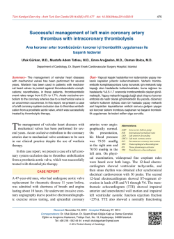
Türk Kardiyol Dern Arş - Arch Turk Soc Cardiol 2014;42(4):412 doi: 10.5543/tkda.2014.21736
412
Stabilization of a slipping balloon during the treatment of in-stent
severe restenosis - anchoring the balloon shaft in the guiding catheter
Stent içi daralmanın tedavisinde kayan balonu sabitleme yöntemi Kılavuz kateter içinde balon şaftını sıkıştırmak
A 68-year-old man
with the diagnosis of acute coronary syndrome unDepartment of Cardiology,
derwent
coronary
Akçaabat Hackali Baba State
angiography
and
Hospital, Trabzon;
subsequently per#
Department of Cardiology,
cutaneous coronary
Ahi Evren Thorasic and
intervention (PCI).
Cardiovascular Surgery Training
He had undergone
and Research Hospital, Trabzon
coronary
artery
bypass
surgery
(CABG) with two vessels two years ago and PCI
four months after CABG. Coronary angiography
demonstrated occluded left anterior descending
artery (LAD) in the osteal region, severe narrowing of the circumflex artery (Cx) proximal stent
(Figure A), non-dominant right coronary artery
(RCA) with 60% narrowing, and open left internal mammary artery (LIMA) and CX-OM1 grafts
(Figure B, C). PCI for the Cx proximal in-stent
lesion was decided. At first, a JL4 guiding catheter was introduced to the left main coronary arAli Rıza Akyüz
Levent Korkmaz#
A
B
D
E
tery (LMCA). Then, repeated balloon
dilatations with 1.5x15 mm semicompliant and 3x15 mm non- compliant catheters were performed. Because the balloon slipped each time the lesion
was dilated with a 3x15 mm balloon (Video 1*),
the JL4 catheter was changed to EBU4, but this
maneuver failed to stabilize the balloon in the lesion. Thus, it was decided to send a second wire
through the lesion, but this also failed to stabilize
the balloon. We then deployed another balloon
(2.5x15 mm) into the second wire, and advanced
it by the distal tip of the guiding catheter. This balloon was then inflated in order to trap the shaft of
the balloon in the guiding catheter, in an effort to
prevent the balloon from slipping. This maneuver
was successful (Figures D, E, Video 2*). Balloon
slipping is not rare, especially in severe calcific
or in-stent lesions. To the best of our knowledge,
this is the first case in which this technique was
utilized. It may help physicians to treat this kind
of lesion securely.
C
Figures– (A) Right caudal
views demonstrates severe instent lesion of the proximal Cx
(white arrow). (B, C) Images of
the Cx and LAD grafts. (D) Anchoring the balloon shaft in the
guiding catheter (balloon in the
catheter [white arrow] and lesion [black arrow]). (E) Cx after
successful balloon dilatation.
*Supplementary video files associated with this presentation
can be found in the online version of the journal.
© Copyright 2026 Paperzz