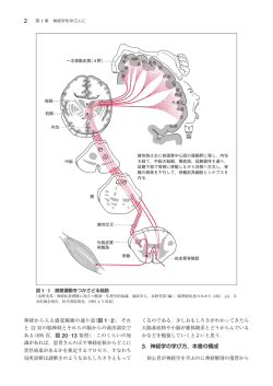
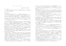
48:945
<シンポジウム 5―2>大脳白質の脳機能画像―オーバービュー―
拡散テンソル画像
森
墾
(臨床神経,48:945―946, 2008)
Key words:磁気共鳴画像,拡散テンソル,異方性,トラクトグラフィー,白質
拡散強調像は超急性期脳梗塞が直接描出可能という,他の
画像検査法にない特徴により広く臨床応用されている.脳梗
塞の検出においては,異方性による白質の信号が脳梗塞の検
出に妨げになることから,等方性拡散強調像が通常もちいら
れてきた.しかし,脳における拡散は白質線維による異方性が
あるので,条件を満たした MRI 撮像をおこないテンソル解析
すると,白質路の画像化
(color map:線維方向の違いの表示,
tractography:特定の白質路の抽出)や白質線維路による拡
散異方性の程度が定量化(FA:異方性比率)できる1).つま
り,従来の等方性拡散強調像は,拡散テンソル近似モデルの固
λ2 および λ3)の平均値に注目した画像であり,trac有値(λ1,
tography は第一固有ベクトル(e1)に注目した画像である.
また,FA 画像(異方性比率画像)は固有値の標準偏差に注目
した画像といえる.
最近,脳白質線維の方向性を加味した拡散テンソル画像と,
その表示法の進歩があり,東京大学放射線科の青木茂樹准教
授,阿部修講師と画像情報処理・解析研究室の増谷佳孝講師
が開発し た dTV(MR 拡 散 テ ン ソ ル 可 視 化 ソ フ ト ウ ェ ア
http:!!www. ut-radiology. umin. jp !people !masutani !dTV !
Fi
g.1 “放線冠”を通る横断像(c
o
l
o
rma
p)
各ボクセルの第一固有ベクトルは緑=前後,赤=左右,青=
頭尾方向を示す.
dTV_frame-j.htm)
により,臨床応用が可能となった.たとえ
ば,放線冠を通るレベルの横断像では放線冠とその他の線維
を区別するのは困難であるが,拡散テンソル画像では脳梁,上
縦束,上前頭後頭束や帯状束との鑑別が容易にできる(Fig.
1)
.また,放線冠内の皮質脊髄路の抽出も可能である.
この拡散テンソル画像は,①白質路の画像化(Fig. 2)と②
白質路の定量的評価に臨床応用される.白質路の画像化では,
拡散テンソルトラクトグラフィーによる主要白質路の描出2),
その白質路と病変(脳梗塞,血管奇形や脳腫瘍)との 3 次元的
位置関係の解析3)による術中ナビゲーションや放射線治療へ
の応用4)や,fMRI との組み合わせ5)などがおこなわれている.
白質路の定量的評価としては,manual trace 法の欠点である
再現性の問題や煩雑さを克服した半自動的な tract-specific
analysis6)や, 必ずしも仮説を必要とせずに全脳解析ができ,
かつ,検定者間のバイアスが低い voxel-based analysis7)8)をお
こなうことができる.また,健常人でのデータベースを構築し
ておけば,群間比較9)ではなく個人の検定もおこなえるように
なる.このように,臨床から研究まで広く応用が期待される分
野である.
東京大学医学部附属病院放射線科〔〒113―8655
(受付日:2008 年 5 月 16 日)
東京都文京区本郷 7―3―1〕
Fi
g.2 弓状線維
48:946
臨床神経学 48巻11号(2008:11)
拡散テンソル画像(とくに tractography)の限界としては
① FA の閾値10),②交叉線維の問題がある.前者は,FA の閾
4)Maruyama K, Kamada K, Ota T, et al: Tolerance of pyramidal tract to gamma knife radiosurgery based on
値設定によって恣意的に線維路を描出できてしまう問題であ
diffusion-tensor tractography. Int J Radiat Oncol Biol
る.生体内の解析のため,描かれた線維に対する validation
がえられにくいのも欠点である.また,後者は,皮質脊髄路,
脳梁と上縦束など神経線維交叉部で描出能が低下する問題で
Phys 2008; 70: 1330―1335
5)Kamada K, Todo T, Masutani Y, et al: Visualization of the
frontotemporal language fibers by tractography com-
ある.一つのボクセルを一つの拡散テンソルで代表させるた
bined with functional magnetic resonance imaging and
め,ボクセル内で交叉していたり,屈曲接触している神経線維
は線維間を乗り越えるように描出されてしまう.そもそも,異
方性の組織学的背景が不明である点も問題である.
magnetoencephalography. J Neurosurg 2007; 106: 90―98
6)Aoki S, Iwata NK, Masutani Y, et al: Quantitative evaluation of the pyramidal tract segmented by diffusion ten-
将来的には,MR angiography と同様に通常の MRI 撮像の
sor tractography : feasibility study in patients with
一環として,病変と近傍白質路との関係を示した拡散テンソ
amyotrophic lateral sclerosis. Radiat Med 2005; 23: 195―
ル画像を提供し,血液生化学などの臨床検査値と同様に,全脳
あるいは特定の白質路に対応した拡散テンソルパラメータを
臨床に提供できるようになることを一つの目標と考えてい
199
7)Abe O, Yamada H, Masutani Y, et al: Amyotrophic lateral
sclerosis: diffusion tensor tractography and voxel-based
る.
analysis. NMR Biomed 2004; 17: 411―416
文
8)Abe O, Yamasue H, Kasai K, et al: Voxel-based diffusion
献
tensor analysis reveals aberrant anterior cingulum integ-
1)Masutani Y, Aoki S, Abe O, et al: MR diffusion tensor im-
rity in posttraumatic stress disorder due to terrorism.
aging: recent advance and new techniques for diffusion
Psychiatry Res 2006; 146: 231―242
tensor visualization. Eur J Radiol 2003; 46: 53―66
9)Yasmin H, Nakata Y, Aoki S, et al: Diffusion abnormalities
2)Mori H, Fujishiro T, Hayashi N, et al: Partially uncrossed
of the uncinate fasciculus in Alzheimer s disease: diffusion
pyramidal tracts shown by tractography in horizontal
tensor tract-specific analysis using a new method to
gaze palsy and scoliosis. Am J Roentgenol 2005; 184: S4―
measure the core of the tract. Neuroradiology 2008; 50:
6
293―299
3)Itoh D, Aoki S, Maruyama K, et al: Corticospinal tracts
10)Kunimatsu A, Aoki S, Masutani Y, et al: The optimal
by diffusion tensor tractography in patients with arte-
trackability threshold of fractional anisotropy for diffu-
riovenous malformations. J Comput Assist Tomogr 2006;
sion tensor tractography of the corticospinal tract. Magn
30: 618―623
Reson Med Sci 2004; 3: 11―17
Abstract
Diffusion tensor imaging
Harushi Mori
Department of Radiology, University of Tokyo Hospital
Diffusion tensor imaging of magnetic resonance imaging, including diffusion tensor tractography, is a unique
tool to visualize and segment the white matter pathways in vivo and one can evaluate the segmented trace quantitatively. Three dimensional visualization of the white matter fibers, such as corticospinal (pyramidal) tracts, with
relationship to brain lesions (infarcts, vascular malformations and brain tumors) is extremely helpful for stereotactic radiosurgery, preoperative evaluation and intraoperative navigation. Quantitative measurement of the tract is
a very sensitive method to detect differences in the tract in neurodegenerative!neurocognitive!psychiatric patients such as amyotrophic lateral sclerosis, schizophrenia and Alzheimer diseases. Importance of this tool will become more significant in clinical and neuroscience fields in the future.
(Clin Neurol, 48: 945―946, 2008)
Key words: magnetic resonance imaging, diffusion tensor, anisotropy, tractography, white matter
© Copyright 2026 Paperzz