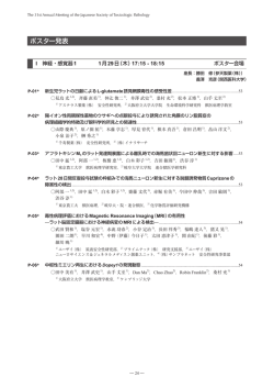
第 31 回日本毒性病理学会総会及び学術集会 目 次 第 31 回日本毒性病理学会総会及び学術集会概要 ………………………… 3 年会長挨拶 …………………………………………………………………… 5 会場へのアクセス 会場案内 …………………………………………………………… 7 ……………………………………………………………………… 8 ポスター・商業展示 参加者へのご案内 ……………………………………………………… 10 ………………………………………………………… 11 座長の先生方へ……………………………………………………………… 13 発表者の先生方へ…………………………………………………………… 14 協賛法人・企業一覧 ……………………………………………………… 16 日本毒性病理学会のあゆみ 日程表 ……………………………………………… 17 ……………………………………………………………………… 18 司会・座長一覧 …………………………………………………………… 20 プログラム 特別講演 ……………………………………………………………… 22 教育講演 ……………………………………………………………… 22 シンポジウム ………………………………………………………… 22 STP セミナー ………………………………………………………… 23 ポスター ……………………………………………………………… 24 講演要旨 特別講演 ……………………………………………………………… 39 教育講演 ……………………………………………………………… 40 シンポジウム ………………………………………………………… 42 STP セミナー ………………………………………………………… 50 ポスター ……………………………………………………………… 51 IATP レクチャー …………………………………………………………… 77 発表者索引 ………………………………………………………………… 150 The 31st Annual Meeting of the Japanese Society of Toxicologic Pathology 第 31 回日本毒性病理学会総会及び学術集会 The 31st Annual Meeting of the Japanese Society of Toxicologic Pathology 1. 期 日 2015 年 1 月 29 日 ( 木 )・30 日 ( 金 ) 2. 会 場 タワーホール船堀(東京) 〒 134-0091 東京都江戸川区船堀 4-1-1 Tel: 03-5676-2211 3. 年会長 中山 裕之 (東京大学 大学院農学生命科学研究科 獣医病理学研究室) 4. プログラム委員会 委員長 鈴木 雅実 (中外製薬株式会社) 委 員 小川 久美子 (国立医薬品食品衛生研究所) 渋谷 淳 (東京農工大学大学院) 寺西 宗広 (第一三共株式会社) 中江 大 (東京都健康安全研究センター) 福田 良 (武田薬品工業株式会社) 吉田 緑 (国立医薬品食品衛生研究所) (50 音順) 5. 事務局 【会期前】 東京大学大学院 農学生命科学研究科 獣医病理学研究室 第 31 回日本毒性病理学会総会及び学術集会事務局 担当:内田 和幸 〒 113-8657 東京都文京区弥生 1-1-1 Tel: 03-5841-5401 Fax: 03-5841-8185 E-mail: [email protected] 【会期中 2015 年 1 月 29・30 日】 タワーホール船堀(東京) Tel: 03-5676-2211 Fax: 03-5676-2501 6. 事務局補佐 株式会社アイペック 〒 170-0002 東京都豊島区巣鴨 1-24-12 Tel: 03-5978-4067 Fax: 03-5978-4068 E-mail: [email protected] 7. ホームページ http://www.ipec-pub.co.jp/31jstp/ ̶3̶ The 31st Annual Meeting of the Japanese Society of Toxicologic Pathology 年会長挨拶 今回の学術集会のテーマは「毒性病理学と比較病理学」です。このテーマに関する教育講演 を 2 題、シンポジウムを 2 題企画しております。また、特別講演として東京大学の難波成任先 生に植物病理学についてお話いただきます。動物の病理学を専門とする私たちにとって植物病 理学は未知の世界ですが、きっと共通する病態があるはずです。 現在、ヒトの疾患研究に利用されている動物はほとんどがげっ歯類ですが、そこで得られた 成果をヒトに直接外挿するには両者の進化的距離が大きすぎます。ヒトとげっ歯類との間に存 在する牛、羊、馬、豚などの有蹄類、犬、猫などの肉食類の生理・病理についても考察するこ とで、進化を軸とした比較学問体系が確立すれば、こうした missing link も解消できると考えら れます。すなわち、「比較病理学」分野における成果を「毒性病理学」分野で利用していくこと が、今後の「毒性病理学」を大きく発展させる原動力になるものと期待しております。今回の 学術集会への参加が、1)「比較病理学」と「毒性病理学」との異同、2) 「比較病理学」の「毒 性病理学」への応用、および 3) 「比較病理学」と「毒性病理学」のトランスレーショナル・リサー チにおける位置づけを考えるきっかけになれば望外の喜びです。 最後になりましたが、学術集会の準備を円滑に行うにあたり、プログラム委員各位のご尽力 と日本毒性病理学会理事会のご支援をいただいております。また、多くの企業よりご賛助を賜 りました。心より感謝いたします。 第 31 回日本毒性病理学会総会及び学術集会 年会長 中山 裕之 東京大学 大学院農学生命科学研究科 獣医学専攻獣医病理学研究室 ̶5̶ The 31st Annual Meeting of the Japanese Society of Toxicologic Pathology 会場へのアクセス タワーホール船堀は都営新宿線「船堀」駅前、徒歩 1 分です。 会場へは、公共の交通機関をご利用ください。 電車をご利用の場合 都営新宿線 新宿駅 JR 総武線快速 約5分 徒歩 馬喰町駅 JR 山手線 徒歩 秋葉原駅 約 15 分 ( 急行 10 分 ) 都営新宿線 岩本町駅 約5分 約 17 分 空港から電車をご利用の場合 船 堀 駅 約4分 都営新宿線 馬喰横山駅 約2分 羽田空港 東京モノレール 徒歩 浜松町駅 約4分 大門駅 約4分 京成本線 成田空港 都営大江戸線 京成八幡駅 約 60 分(特急) 森下駅 約 15 分 徒歩 約4分 本八幡駅 都営新宿線 約 11 分 徒歩 約1分 都営新宿線 約 11 分 空港からバスをご利用の場合 リムジンバス 羽田空港 約 40 分 一之江駅 京成バス 成田空港 都営新宿線 約2分 約 60 分 タワーホール船堀周辺地図 船堀橋東詰 信号 新大橋通り コンビニ コンビニ 船堀街道 勤労福祉会館 P 船堀駅 信号 東 駐車場入口 正面入口 銀行 大島 江 一之 船 堀 タワーホール船堀 駅 宿線 新 都営 ̶7̶ タワーホール船堀 東京駅 約 30 分 (急行 約 21 分) The 31st Annual Meeting of the Japanese Society of Toxicologic Pathology 会場案内 駐車場出口 1階 サービスヤード 駐車場入口 つきじ植むら (和食) 書籍店 サービスヤード 警備室 (ドリンクサービス・休憩) 1 展示ホール 2 サービスヤード 展示ホール ポスター・商業展示会場 精養軒 (洋食) インフォメーション エントランスホール 正面入口 2階 ランチョンセミナー 懇親会場 (イベントホール 瑞雲・平安) ロビー ̶8̶ The 31st Annual Meeting of the Japanese Society of Toxicologic Pathology 会場案内 4階 楽屋 1 楽屋 2 楽屋3 楽屋4 楽屋5 講師 控室 特別 会議室 講演会場 (大ホール) 研修室 401 会議室 スタッフ控室 (会議室 407) 会 議 室 (会議室 402) クローク 会 議 室 大会本部 (会議室 403) 会 議 室 (会議室 404) (会議室 406) (会議室 405) 5階 楽屋1 (ロビー) ̶9̶ 楽屋4 総合受付 楽屋3 楽屋2 講演会場 (大ホール) The 31st Annual Meeting of the Japanese Society of Toxicologic Pathology ポスター・商業展示 ブース 番号 企業名 1 株式会社ボゾリサーチセンター 2 PDS パソロジー・データシステムズ・リミテッド 3 日本エスエルシー株式会社 4 株式会社スリーエス・ジャパン 5 株式会社 DIMS 医科学研究所 6 アジア器材株式会社 7 株式会社新組織科学研究所 8 株式会社エイチ・アンド・ティー 9 ライカマイクロシステムズ株式会社 10 クラボウ/株式会社イナリサーチ 11 富士通株式会社 12 白井松器械株式会社 13 サクラファインテックジャパン株式会社 14 株式会社新日本科学 15, 16 1階 展示ホール ポスター・商業展示会場 (ドリンクサービス ・2 休憩) 1 展示ホール AnaPath GmbH/Safety Alliance P-053 P-052 P-051 P-050 P-049 P-048 P-047 P-046 P-045 P-044 P-043 P-042 P-041 P-040 P-039 P-038 P-037 P-036 P-035 P-034 P-033 P-032 P-031 P-030 P-029 P-028 P-027 P-026 P-025 P-024 P-023 P-022 P-021 P-020 P-088 P-089 P-019 P-087 P-090 P-018 P-086 P-091 P-017 P-085 P-092 P-016 P-015 P-014 P-013 4 5 P-084 P-093 3 6 P-082 P-095 P-054 P-055 P-056 12 13 P-083 P-094 P-058 11 14 P-081 P-096 P-010 P-009 P-008 P-059 P-060 P-012 P-011 P-057 P-061 2 7 1 8 10 15 ドリンクサービス P-062 P-063 9 16 P-007 P-006 休憩スペース P-005 P-004 P-003 P-064 P-065 P-066 P-067 P-068 P-069 ̶ 10 ̶ P-070 P-071 P-072 P-073 P-074 P-075 P-076 P-077 P-078 P-079 P-001 ポスター受付 P-080 P-002 The 31st Annual Meeting of the Japanese Society of Toxicologic Pathology 参加者へのご案内 ●参加者の方へ 1. 総合受付は 5 階ロビーにて行います。受付時間は下記のとおりです。 1 月 29 日(木)9:00 ∼ 18:00 1 月 30 日(金)8:30 ∼ 15:00 2. 事前登録者はあらかじめ講演要旨集とともに送付された参加証(ネームカード)を持参し、 会期中は必ず着用ください。ネームホルダーは総合受付にご用意いたしますので、ご利用 ください。なお、ネームホルダーは大会終了後に総合受付までご返却くださいますよう、 お願いします。 3. 当日参加者は「当日参加申込書」に必要事項を記入の上、 「当日参加受付」にて下記参加費 を納入して参加証を受け取り、氏名・所属を記入の上、会期中は必ず身につけてください。 学生の場合は学生証の提示が必要です。 一般会員:12,000 円 学生会員: 6,000 円 非 会 員:17,000 円 ※講演要旨集 5,000 円含む 4. 講演要旨集は事前に郵送いたしますので、忘れずにご持参ください。 お忘れの場合は一般会員・学生会員:2,000 円、非会員:5,000 円での販売となります。 5. 質問 ・ 討論される方は、マイクの前に並んだ上、座長の指示に従って所属と氏名を述べて から発言してください。 6. 館内はすべて禁煙です。 7. 場内では携帯電話の電源を OFF またはマナーモードにしてください。 8. ドリンクコーナー・休憩所(1 階 ポスター・商業展示会場)でドリンクを無料配布します。 休憩の際、ご利用ください。また、無線 LAN ・ 有線 LAN によるインターネット接続をご提 供しますのでご利用ください。 9. 昼食はランチョンセミナー、あるいは会場 1 階・7 階のレストランなどをご利用ください。 10. クロークは 4 階 大会本部の隣(会議室 406 )にございます。 受付時間は下記のとおりです。 1 月 29 日(木)9:00 ∼ 18:30 1 月 30 日(金)8:30 ∼ 18:30 懇親会の際には懇親会会場(瑞雲・平安)のクロークをご利用ください。学術集会のクロー クは 18 : 30 で終了とさせて頂きますので、お預けになったお荷物は必ず 18 : 30 までにお 受け取りください。 11. 会場内での呼び出しは緊急時以外にはいたしませんのでご了承ください。 5 階 総合受 付前にメッセージボードを設置いたしますのでご利用ください。 12. 会場内での写真撮影・録画・録音は禁止させていただきます。 ̶ 11 ̶ The 31st Annual Meeting of the Japanese Society of Toxicologic Pathology ●評議員会及び総会のご案内 評議員会及び総会は学会第 2 日目(1 月 30 日(金))16:00 から 5 階 大ホールにて行います。 ●ランチョンセミナーのご案内 1 月 29 日(木) ・30 日(金)両日ともに、2 階 イベントホール(瑞雲・平安)にてランチョンセミナー を行います。チケットは、5 階 総合受付の前にて配布します。配布時間は下記のとおりです。 1 月 29 日(木)9:00 1 月 30 日(金)8:30 ランチョンセミナー I 1 月 29 日(木) 12:15 ∼ 13:15 2 階 イベントホール(平安) 「Jic:CB6F1-Tg rasH2@Jcl マウスを用いた短期(26 週間)がん原性試験の背景データ」 演 者:山口 裕子(株式会社ボゾリサーチセンター) 座 長:土井 邦雄(東京大学名誉教授) 協 賛:株式会社ボゾリサーチセンター ランチョンセミナー II 1 月 29 日(木) 12:15 ∼ 13:15 2 階 イベントホール(瑞雲) 「Endocrine Tumors in Carcinogenicity Studies: Mechanisms and Relevance to Man」 演 者:Dianne Creasy(Huntingdon Life Sciences Inc.) 座 長:高橋 道人(病理ピアレビューセンター) 協 賛:ハンティンドンライフサイエンス株式会社 ランチョンセミナー III 1 月 30 日(金) 12:30 ∼ 13:30 2 階 イベントホール(瑞雲) 「様々な科学技術を応用した病理学的評価手法」 演 者:Klaus Weber (AnaPath GmbH) 座 長:高橋 道人(病理ピアレビューセンター) 協 賛:Safety Alliance (AnaPath GmbH, Accelera S.r.l., Vivotecnia Research S.L., BIOSERVICE Scientific Laboratories GmbH, Dr. Knoell Consult GmbH and AnaPath Toxicology Consulting Japan) ●懇親会のご案内 1. 懇親会は 1 月 29 日(木)18:30 から 2 階 イベントホール(瑞雲・平安)にて開催します。 2. 事前登録制ですが、総合受付にて若干名を懇親会費 9,000 円(学生 7,000 円)で申し受けます。 定員に達した場合は締め切る場合がございます。予めご了承ください。 ̶ 12 ̶ The 31st Annual Meeting of the Japanese Society of Toxicologic Pathology ●座長の先生方へ ■ 特別講演・教育講演・シンポジウム座長の先生へ 1. 来館されましたら、5 階 総合受付の口演座長受付にお越しください。会長賞選考のための 評価票をお渡しします。 2. ご担当セッション開始時間の遅くとも 20 分前までには、口演座長受付にお越しください。 3. セッション開始 15 分前には、会場内の次座長席にご着席ください。 ■ ポスター発表の座長の先生へ 1. 来館されましたら、1 階 ポスター ・ 商業展示会場のポスター受付にお越しください。 会長賞選考のための評価票をお渡しします。 2. ご担当セッション開始時間の遅くとも 15 分前までには、ポスター受付にお越しください。 指示棒、リボンをお渡しします。討論時間には参加証(ネームカード)の他に、リボンを必 ず着用ください。セッション終了後、指示棒は必ずご返却ください。 3. 会長賞は 40 歳未満の筆頭演者(演題番号に*を表示)によるポスター発表を対象に選考 していただきます。それぞれの評価担当演題の抄録とポスター発表を評価していただき、 1 月 30 日(金)13:00 までに評価票を 5 階 総合受付にご提出ください。 ̶ 13 ̶ The 31st Annual Meeting of the Japanese Society of Toxicologic Pathology ●発表者の先生方へ ■ 利益相反について 発表者の皆様は可能な限り COI(利益相反)の開示を お願いいたします。企業に所属の場合は別企業と COI 状態にある場合に開示してください。 口演発表者は発表スライドの 2 枚目(表題の次のスライ ド)に、ポスター発表者は一番最後に記載してください。 ■ 特別講演・教育講演・シンポジウムの発表者の先生方へ 1. PowerPoint での口演発表用スライド作成にあたっては、外国人の参加を考慮して可能な限り 英語で作成ください。 2. 発表時間の遅くとも 30 分前までに、口演発表受付にお越しください。発表用データの動作確 認を行っていただきます。 3. 発表機材は PC 液晶プロジェクターを使用します。スライド ・ ビデオは使用できませんので ご注意ください。 4. 発表は、演壇におかれたパソコン画面を見ながら、ご自身で画面操作をしてください。 5. 前演者の発表が始まりましたら、次演者席にお着きください。 6. 発表データは、会場のパソコンに一時保存いたしますが、これらのデータは本学会終了後、 責任を持って廃棄します。 プレゼンテーションデータ作成時の注意 • 環境の違いにより、画面レイアウトが乱れるなどの不具合が発生する可能性があります。 会場のパソコン使用環境は以下の通りですので、ご留意ください。 パソコン :Windows PC(Macintosh は用意しておりません) OS :Windows 7 解像度 :XGA(1024 × 768) プレゼンテーション用ソフト:PowerPoint 2007 / 2010 / 2013 フォント :OS(Windows7)標準 • 動画は不可といたします。 プレゼンテーションデータの提出方法 • シンポジウムでご発表の先生方は、発表当日の口演発表受付時に USB メモリに保存してご 提出ください。 ̶ 14 ̶ The 31st Annual Meeting of the Japanese Society of Toxicologic Pathology ■ ポスター発表をされる先生方へ 1. ポスター発表・掲示・撤去時間 掲示時間 1 月 29 日(木)9:00 ∼ 10:00 ※ 1 月 29 日(木)10:00 までに、1 階 ポスター・商業展示会場のポスター受付にて受付を 行ってください。 撤去時間 1 月 30 日(金)16:15 ∼ 18:00 ※ 発表者用リボン、ピンは元の位置に返却してください。 時間までに撤去されないポスターは事務局で処分いたします。 発表日時 コアタイム 1 月 29 日(木) 17:15 ∼ 18:15 1 月 30 日(金) 11:15 ∼ 12:15 1 月 30 日(金) 13:45 ∼ 14:45 2. 各パネルの前に掲示用のピンと発表者用のリボンをご用意しますので、ご使用ください。ま た、コアタイムには参加証(ネームカード)のほかに、このリボンを必ず着用してください。 3. パネルのサイズは縦 180 cm ×横 90 cm です。演題番号については主催者側で用意いたしま す。演題名と発表者氏名 ・ 所属の表題を下記の図に従ってご準備ください。 4. 発表は、1 演題あたり発表 5 分、質疑 5 分で行います。発表者は、座長の指示に従って要約発 表 ・ 討論をお願いします。発表時間は厳守してください。 20 cm 20 cm 演題番号 70 cm 演題名・発表者名・所属 180 cm 160 cm 内容発表 90 cm ̶ 15 ̶ The 31st Annual Meeting of the Japanese Society of Toxicologic Pathology 協賛法人・企業一覧 第 31 回 日 本 毒 性 病 理 学 会 総 会 及 び 学 術 集 会 を 開 催 す る に あ た り、 多 く の 企 業・ 団 体 様 よりご支援ご協力を賜りました。ここにお名前を掲載して厚く感謝申し上げます。 第 31 回日本毒性病理学会総会及び学術集会 年会長 中山 裕之 学会後援 日本獣医学会 日本毒性学会 日本獣医病理学専門家協会 協賛 旭化成ファーマ株式会社 味の素株式会社イノベーション研究所 アステラス製薬株式会社 アスビオファーマ株式会社 エーザイ株式会社 株式会社 LSI メディエンス 株式会社大塚製薬工場 小野薬品工業株式会社 協和発酵キリン株式会社 株式会社札幌総合病理研究所 三栄源エフ・エフ・アイ株式会社 参天製薬株式会社 一般財団法人残留農薬研究所 塩野義製薬株式会社 公益財団法人食品農医薬品安全性評価センター ランチョンセミナー共催 AnaPath GmbH/Safety Alliance 一般財団法人食品薬品安全センター 住友化学株式会社 第一三共株式会社 大正製薬株式会社 大鵬薬品工業株式会社 武田薬品工業株式会社 田辺三菱製薬株式会社 中外製薬株式会社 帝人ファーマ株式会社 日産化学工業株式会社 一般財団法人日本生物科学研究所 日本たばこ産業株式会社 日本ベーリンガーインゲルハイム株式会社 バイエル薬品株式会社 ハンティンドンライフサイエンス株式会社 株式会社ボゾリサーチセンター 商業展示 アジア器材株式会社 株式会社エイチ・アンド・ティー クラボウ/株式会社イナリサーチ サクラファインテックジャパン株式会社 白井松器械株式会社 株式会社新組織科学研究所 株式会社新日本科学 株式会社スリーエス・ジャパン 講演要旨集への広告 AnaPath Toxicology Consulting Japan 株式会社 株式会社アマネセル Safety Alliance (AnaPath GmbH, Accelera S.r.l., Vivotecnia Research S.L., BIOSERVICE Scientific Laboratories GmbH, Dr. Knoell Consult GmbH and AnaPath Toxicology Consulting Japan) 株式会社 DIMS 医科学研究所 日本エスエルシー株式会社 PDS パソロジー・データシステムズ・リミテッド 富士通株式会社 株式会社ボゾリサーチセンター ライカマイクロシステムズ株式会社 株式会社バイオ病理研究所 文永堂出版株式会社 キッコーマン株式会社 ネームホルダーの提供 株式会社イナリサーチ (50 音順) ̶ 16 ̶ The 31st Annual Meeting of the Japanese Society of Toxicologic Pathology 日本毒性病理学会のあゆみ シンポジウム 特別講演 ワークショップ その他 一般 回 開催地 会 長 (所 属) 会 期 1 東 京 西山 保一 (北里学園) 1985. 3. 25 2 0 0 2 東 京 藤原 公策 (東京大学) 1986. 2. 7-8 1 5 29 3 名古屋 伊東 信行 (名古屋市立大学) 1987. 2. 6-7 1 8 47 4 浜 松 榎本 眞 (安評センター) 1988. 2. 5-6 1 7 55 5 横 浜 蟹澤 成好 (横浜市立大学) 1989. 1. 27-28 1 6 53 6 札 幌 板倉 智敏 (北海道大学) 1990. 2. 13-14 1 6 80 7 東 京 林 裕造 (国立衛試) 1991. 1. 17-18 1 20 53 8 奈 良 小西 陽一 (奈良医科大学) 1992. 1. 23-24 1 19 71 9 東 京 土井 邦雄 (東京大学) 1993. 1. 21-22 2 4 126 10 広 島 伊藤 明弘 (広島大学) 1994. 1. 27-29 1 18 136 11 大 阪 佐久間貞重 (大阪府立大学) 1995. 1. 26-27 2 10 151 12 東 京 高橋 道人 (国立衛試) 1996. 1. 24-25 0 16 147 13 鳥 取 梅村 孝司 (鳥取大学) 1997. 1. 23-24 1 11 151 14 東 京 前川 昭彦 (佐々木研) 1998. 2. 3-4 1 9 143 15 水 戸 真板 敬三 (残農研) 1999. 1. 28-29 0 7 142 16 岐 阜 森 秀樹 (岐阜大学) 2000. 1. 26-27 1 12 125 17 淡 路 奈良間 功 (摂南大学) 2001. 1. 25-26 1 10 146 18 東 京 津田 洋幸 (国立がんセンター) 2002. 1. 24-25 0 2 119 19 東 京 布谷 鉄夫 (日生研) 2003. 1. 23-24 1 5 102 20 神 戸 * 福島 昭治 (大阪市立大学) 2004. 2. 15-18 2 31 172 21 浜 松 今井 清 (安評センター) 2005. 1. 20-21 1 23 100 22 鹿児島 吉田 浩己 (鹿児島大学) 2006. 1. 26-27 1 15 109 23 東 京 三森 国敏 (東京農工大学) 2007. 1. 30-31 0 15 107 24 名古屋 白井 智之 (名古屋市立大学) 2008. 2. 6-7 1 13 106 25 浜 松 真鍋 淳 (第一三共㈱) 2009. 1. 27-28 2 17 124 26 金 沢 田中 卓二 (金沢医科大学) 2010. 2. 3-4 2 10 125 27 大 阪 大石 裕司 (アステラス製薬㈱) 2011. 1. 27-28 2 13 144 28 東 京 西川 秋佳 (国立衛研) 2012. 2. 2-3 2 21 108 29 つくば 原田 孝則 (残農研) 2013. 1. 31-2. 1 1 8 107 30 徳 島 泉 啓介 (徳島大学) 2014. 1. 30-31 2 15 114 31 東 京 中山 裕之 (東京大学) 2015. 1. 29-30 1 11 96 *Joint international meeting of JSTP/IFSTP ̶ 17 ̶ 演題 The 31st Annual Meeting of the Japanese Society of Toxicologic Pathology 日 程 表 1 月 28 日(水曜日) 講演会場 ポスター・商業 展示会場 1 月 29 日(木曜日) 懇親会・各種 会議室 講演会場 ポスター・商業 展示会場 懇親会・各種 会議室 8:30 9:00 受付開始 9:00 ∼ 開会式 10:00 9:45 ∼ 試験問題解説 10:00 ∼ 11:00 11:00 毒性病理組織学 改訂委員会 10:30 ∼ 12:00 (会議室) 12:00 編集委員会 13:00 14:00 10:00 ∼ 12:00 4 名(30 分) (質疑応答 を含む) ランチョンセミナー 12:15 ∼ 13:15 (イベントホール) 教育委員会 特別講演 13:45 ∼ 14:45 11:15 ∼ 17:05 (会議室) 9:00 ∼ 10:00 シンポジウム 1 12:00 ∼ 13:30 (会議室) 第 27 回 スライド カンファレンス ポスター貼付 資格認定委員会 12:15 ∼ 13:15 ポスター閲覧 13:30 ∼ 14:45 難波先生(75 分) 15:00 理事会 16:00 15:00 ∼ 17:00 (会議室) 教育講演 15:00 ∼ 17:00 ①Dr. Rosol ②山手先生 2名(各 60 分) 17:00 18:00 ポスター発表Ⅰ IATP レクチャー 17:15 ∼ 18:15 17:30 ∼ 18:30 19:00 懇親会 18:30 ∼ 20:30 20:00 21:00 International President’s Meeting 20:45 ∼ 21:45 22:00 23:00 IFSTP EC Meeting 22:00 ∼ 23:00 ̶ 18 ̶ The 31st Annual Meeting of the Japanese Society of Toxicologic Pathology 日 程 表 1 月 30 日(金曜日) 講演会場 8:30 9:00 ポスター・商業 展示会場 懇親会・各種 会議室 受付開始 8:30 ∼ シンポジウム 2 10:00 9:00 ∼ 11:00 4 名(30 分) (質疑応答を含む) ポスター閲覧 11:00 ポスター発表Ⅱ 11:15 ∼ 12:15 12:00 13:00 ランチョンセミナー 12:30 ∼ 13:30 (イベントホール) 14:00 ポスター閲覧 ポスター発表Ⅲ 13:45 ∼ 14:45 15:00 STP セミナー 15:00 ∼ 15:45 Dr. Ryan 30 分+15 分 ポスター閲覧 16:00 評議員会・総会 17:00 表彰式・閉会式 16:00 ∼ 17:30 ポスター撤去 16:15 ∼ 18:00 18:00 19:00 20:00 21:00 22:00 23:00 ̶ 19 ̶ The 31st Annual Meeting of the Japanese Society of Toxicologic Pathology 司会・座長一覧 セッション名 日 時 演題番号 1 月 29 日(木)13:30 ∼ 14:45 教育講演 1 1 月 29 日(木)15:00 ∼ 16:00 教育講演 2 1 月 29 日(木)16:00 ∼ 17:00 シンポジウム 1 1 月 29 日(木)10:00 ∼ 12:00 シンポジウム 2 1 月 30 日(金) 9:00 ∼ 11:00 STP セミナー 1 月 30 日(金)15:00 ∼ 15:45 P-001 ∼ 006 中山 裕之(東京大学) 福田 良(武田薬品工場(株)) 鰐渕 英機(大阪市立大学) 鈴木 雅実(中外製薬(株)) 吉見 直己(琉球大学) 渋谷 淳(東京農工大学) 吉田 緑(国立医薬品食品衛生研究所) 小川久美子(国立医薬品食品衛生研究所) 寺西 宗広(第一三共(株)) 林 新茂(三栄源エフ・エフ・アイ(株)) 中江 大(東京都健康安全研究センター) 勝田 修(参天製薬(株)) 義澤 克彦(関西医科大学) 梅村 隆志(国立医薬品食品衛生研究所) 細川 暁(エーザイ(株)) 澤本 修((株)大塚製薬工場) ポスター発表Ⅰ 1 月 29 日(木) P-034 ∼ 038 (日本ベーリンガーインゲルハイム(株)) 17:15 ∼ 18:15 P-049 ∼ 053 P-064 ∼ 069 P-081 ∼ 085 P-007 ∼ 011 1 月 30 日(金) 11:15 ∼ 12:15 P-039 ∼ 043 P-054 ∼ 058 P-070 ∼ 075 P-086 ∼ 091 塚本 徹哉(藤田保健衛生大学) 深町 勝巳(名古屋市立大学) 小泉 治子((株)イナリサーチ) 高橋 智(名古屋市立大学) 佐藤 洋(岩手大学) 田村 啓(キッセイ薬品工業(株)) 尾崎 清和(摂南大学) 桑村 充(大阪府立大学) 丸山 敏之(塩野義製薬(株)) 横平 政直(香川大学) 鈴木 智(大鵬薬品工業(株)) 古川 賢(日産化学工業(株)) 山口 裕子((株)ボゾリサーチセンター) 渡邊 厚(旭化成ファーマ(株)) 髙場 克己(協和発酵キリン(株)) 仲辻 俊二(アステラス製薬(株)) 小林 欣滋((株)新日本科学) 美谷島克宏(日本たばこ産業(株)) ̶ 20 ̶ ポスター会場︵展示ホール︶ P-023 ∼ 028 ポスター発表Ⅱ 杉元 陽子 ポスター会場︵展示ホール︶ P-018 ∼ 022 会場 講演会場︵大ホール︶ 特別講演 司会・座長(所属) The 31st Annual Meeting of the Japanese Society of Toxicologic Pathology セッション名 日 時 演題番号 司会・座長(所属) P-012 ∼ 017 P-044 ∼ 048 ポスター発表Ⅲ 1 月 30 日(金) 13:45 ∼ 14:45 P-059 ∼ 063 P-076 ∼ 080 宮田かおり(住友化学(株)) 吉田 敏則(東京農工大学) 田中 雅治(田辺三菱製薬(株)) 涌生 ゆみ((株)LSI メディエンス) 金子 英志(帝人ファーマ(株)) 桑原 真紀((一財)残留農薬研究所) 小笠原裕之(アスビオファーマ(株)) 平田 暁大(岐阜大学) 岡崎 欣正(AnaPath GmbH) 石上 紀明(小野薬品工業(株)) 河部 真弓((株)DIMS 医科学研究所) P-092 ∼ 096 志賀 敦史 ((公財)食品農医薬品安全性評価センター) ̶ 21 ̶ ポスター会場︵展示ホール︶ P-029 ∼ 033 会場 The 31st Annual Meeting of the Japanese Society of Toxicologic Pathology プログラム 特別講演 1 月 29 日(木)13:30 - 14:45 講演会場(大ホール) 座長:中山 裕之(東京大学) SL 植物病理学の新展開:基礎から臨床へ ...................................................................................................................39 難波 成任 東京大学大学院 農学生命科学研究科 植物病理学研究室 教育講演 1 1 月 29 日(木)15:00 - 16:00 講演会場(大ホール) 座長:福田 良(武田薬品工業(株)) 鰐渕 英機(大阪市立大学) EL-1 Toxicologic pathology and comparative pathology of the endocrine glands ..............................40 Thomas Rosol Ohio State University, Columbus, Ohio, USA 教育講演 2 1 月 29 日(木)16:00 - 17:00 講演会場(大ホール) 座長: 鈴木 雅実(中外製薬(株)) 吉見 直己(琉球大学) EL-2 マクロファージと毒性病理学 .....................................................................................................................................41 山手 丈至 大阪府立大学 生命環境科学研究科 獣医病理学 シンポジウム 1 1 月 29 日(木)10:00 - 12:00 講演会場(大ホール) 座長: 渋谷 淳(東京農工大) 吉田 緑(国立医薬品食品衛生研究所) 毒性病理と獣医病理学を繋ぐ比較の生物学̶共通性と種差を知ろう SY1-1 アルツハイマー病の病理に関する比較生物学.......................................................................................................42 ○チェンバーズ ジェームズ,内田 和幸,中山 裕之 東京大学 獣医病理学研究室 SY1-2 種を超えて繁殖を制御する中枢メカニズム:70 年来の謎が解けるのか? ..................................................43 ○前多 敬一郎 1),束村 博子 2) 東京大学大学院 農学生命科学研究科 獣医繁殖育種学,2)名古屋大学大学院 生命農学研究科 生殖科学 1) SY1-3 外来化学物質の代謝の種差 ........................................................................................................................................44 ○石塚 真由美,中山 翔太,水川 葉月,池中 良徳 北海道大学大学院 獣医学研究科 SY1-4 実験動物とヒト間における副腎病変の類似性と相違点 : 副腎病変の in vivo 解析の有効性と限界 .........45 ○笹野 公伸 東北大学大学院 医学系研究科 医科学専攻 病理診断学分野 ̶ 22 ̶ The 31st Annual Meeting of the Japanese Society of Toxicologic Pathology シンポジウム 2 1 月 30 日(金)9:00 - 11:00 講演会場(大ホール) 座長:小川 久美子(国立医薬品食品衛生研究所) 寺西 宗広(第一三共(株)) 毒性病理評価と比較病理学 SY2-1 Patient-derived Xenografts (PDXs) の特徴と留意点 ....................................................................................46 ○加藤 淳彦,藤井 悦子 中外製薬(株) 安全性研究部 SY2-2 げっ歯類の肝臓腫瘍のヒトへの外挿性− CARKO マウスを用いた実験結果からの考察− ......................47 ○井上 薫 国立医薬品食品衛生研究所 病理部 SY2-3 安全性評価で遭遇する血管炎について ...................................................................................................................48 ○福田 良,穴山 久志 武田薬品工業(株) 医薬研究本部 薬剤安全性研究所 SY2-4 げっ歯類の系統による病理像の相違 .......................................................................................................................49 ○星谷 達 (株)ボゾリサーチセンター 病理部 STP セミナー 1 月 30 日(金)15:00 - 15:45 講演会場(大ホール) 座長:中江 大(東京都健康安全研究センター) 林 新茂(三栄源エフ・エフ・アイ(株)) STP Nonclinical Aspects of Biosimilar Drug Development ...........................................................................50 ○ Anne M. Ryan Pfizer Inc., Drug Safety Research and Development, Groton CT, USA ̶ 23 ̶ The 31st Annual Meeting of the Japanese Society of Toxicologic Pathology ポスター発表 I 神経・感覚器 1 1 月 29 日(木)17:15 - 18:15 ポスター会場 座長:勝田 修(参天製薬(株)) 義澤 克彦(関西医科大学) P-01* 新生児ラットの日齢による L-glutamate 誘発網膜毒性の感受性差 ................................................................53 ○見鳥 光 1,2),斉藤 直美 1),仲辻 俊二 1),井澤 武史 2),桑村 充 2),松本 正博 1),山手 丈至 2) アステラス製薬(株) 安全性研究所,2)大阪府立大学大学院 生命環境科学研究科 獣医病理学教室 1) P-02* 陽イオン性両親媒性薬物のウサギへの点眼投与により誘発された角膜のリン脂質症の 病理組織学的特徴及び眼科学的所見との関連性 ..................................................................................................53 ○山際 慶典 1),原ノ園 祐 1),木藤 学志 1),厚見 育代 1),根本 真吾 1),倉田 昌明 1),畠山 洋文 2), 小泉 治子 2),榊 秀之 1) 千寿製薬(株) 安全性研究所,2) (株)イナリサーチ 1) P-03* アフラトキシン M1 のラット発達期暴露による離乳時での海馬歯状回ニューロン新生に対する影響 .....53 ○田中 猛 1,2),阿部 一 1,2),板橋 恵 1,2),白木 彩子 1,2),吉田 敏則 1),渋谷 淳 1) 東京農工大学 獣医病理学研究室,2)岐阜大学大学院 連合獣医学研究科 1) P-04* ラット 28 日間反復投与試験の枠組みでの海馬ニューロン新生に対する脱髄誘発物質 Cuprizone の 障害性の検出 .................................................................................................................................................................53 ○阿部 一 1,2),田中 猛 1,2),白木 彩子 1,2),齋藤 文代 3),赤堀 有美 3),今田中 伸哉 3),吉田 敏則 1), 渋谷 淳 1) 東京農工大 獣医病理,2)岐阜大・院・連合獣医,3)化学物質評価研究機構 1) P-05* 毒性病理評価における Magnetic Resonance Imaging(MRI)の有用性 ̶ラット脳固定臓器における神経病変の MRI による検出̶ .............................................................................54 ○武田 賢和 1),塩谷 元宏 1),水流 功春 2),小谷 定治 3),長田 祥秀 3),福嶋 達人 3),猪又 晃 1), 園田 二朗 1),早川 和宏 4),中野(伊藤)今日子 1),太田 恵津子 1),関 由妃 1),後藤 彩 1), 細川 暁 1) 1) エーザイ(株) 筑波安全性研究部,2)プライムテック(株) 研究支援部,3)エーザイ(株) ニューロサイエンス&ジェネラルメディスン創薬ユニット,4) (株)サンプラネット 安全性研究事業部 P-06* 中枢性ミエリン再生における Dopey1 の発現動態 ..............................................................................................54 ○田中 美有 1),井澤 武史 1),山手 丈至 1),Dan Ma2),Chao Zhao2),Robin Franklin2),桑村 充 1) 大阪府立大学 獣医病理学教室,2)ケンブリッジ大学 1) ̶ 24 ̶ The 31st Annual Meeting of the Japanese Society of Toxicologic Pathology II 神経・感覚器 2 1 月 30 日(金)11:15 - 12:15 ポスター会場 座長:尾崎 清和(摂南大学) 桑村 充(大阪府立大学) P-07* ウサギの網膜におけるニューロフィラメントの蓄積に関する病理組織学的検索 .........................................54 ○川迫 一史,押方 孝文,菅野 剛,隈部 志野,満石 三喜男,浜村 政夫 (株)LSI メディエンス 試験研究センター 病理研究部 P-08* 眼球腫大が認められたスナネズミの眼組織及び視覚中枢の病理学的変化 ...................................................54 ○福永 八千代 1),鈴木 穂高 2),岡田 由美子 2),野口 真紀 1),春木 美那都 1),鶴本 和子 1), 伊倉 佐織 1),小川 竜也 1),久世 博 1),花見 正幸 1) (株)ボゾリサーチセンター,2)国立医薬品食品衛生研究所 1) P-09* ネコのメルケル細胞癌の病理学的研究 ...................................................................................................................55 ○堂畑 厚志,チェンバーズ ジェームズ,内田 和幸,中山 裕之 東京大学 獣医病理学研究室 P-10* カニクイザルの三叉神経節に発生した神経節神経芽腫の 1 例 .........................................................................55 ○森山 亜紀子 1),井手 鉄哉 2),仲辻 俊二 2),松本 正博 2),内田 和幸 2), チェンバーズ ジェームズ 2),岡崎 孝信 1),小林 欣滋 1) (株)新日本科学 安全性研究所,2)アステラス製薬(株),3)東大院 農 獣医病理 1) P-11* 摂水不良状態のミニブタで認められた好酸球性髄膜脳炎(食塩中毒)...........................................................55 ○伊藤 格,長瀬 孝彦,遠藤 克己,松下 久美,小池 恒雄,今井 順,木村 均 (株)日本バイオリサーチセンター III 免疫系・毒性試験 1 月 30 日(金)13:45 - 14:45 ポスター会場 座長:宮田 かおり(住友化学(株)) 吉田 敏則(東京農工大学) P-12* 7 動物種における Serum Amyloid A protein(SAA)N 末端配列のアミロイド原性の比較解析...........55 ○吉本 早慧子,水川 真緒,菅原 豪,上家 潤一,代田 欣二 麻布大学 獣医学部 病理学研究室 P-13* クルクミンの AA アミロイド沈着抑制メカニズムに関する研究 .....................................................................56 ○庄司 亜香音,渡邉 謙一,鄭 明奈,潮 奈々子,チェンバーズ ジェームズ,内田 和幸, 中山 裕之 東京大学 獣医病理学教室 P-14* IL-1raKO マウスにおける AA アミロイドの沈着と分解に関する研究 ..........................................................56 ○渡邉 謙一,内田 和幸,チェンバーズ ジェームズ,潮 奈々子,中山 裕之 東京大学大学院 農学生命科学研究科 獣医病理学研究室 P-15* 新生児低栄養環境による児の免疫組織への形態学的影響 .................................................................................56 ○等々力 舞 1),瀬沼 美華 1),熊谷 文明 1),臼見 憲二 1),千坂 亜希子 1),野口 聡 1),小川 哲郎 2), 斉藤 義明 1),桑形 麻樹子 1) (一財)食品薬品安全センター 秦野研究所 毒性部,2)埼玉医科大学 医学部 生理学 1) ̶ 25 ̶ The 31st Annual Meeting of the Japanese Society of Toxicologic Pathology P-16* gpt delta ラットを用いた短期反復投与毒性・遺伝毒性併合試験 ...................................................................56 ○赤木 純一 1),豊田 武士 1),Young-Man Cho1),水田 保子 1),能美 健彦 2,3),西川 秋佳 2), 小川 久美子 1) 国立衛研・病理,2)国立衛研・安全性生物試験研究センター,3)医薬基盤研 1) P-17* 幼若ラットにおける DXR (doxorubicin hydrochloride) 投与後の経時的変化 ........................................57 ○伊倉 佐織,藤原 壮志,西原 義人,福永 八千代,松島 圭太,霜山 奈津美,金光 弘幸, 花見 正幸 (株)ボゾリサーチセンター IV 試験法・関連技術 1 月 29 日(木)17:15 - 18:15 ポスター会場 座長:梅村 隆志(国立医薬品食品衛生研究所) 細川 暁(エーザイ(株)) P-18* Acorus gramineus のラットの亜慢性毒性試験 .................................................................................................57 ○ Yong-Hoon Lee1), Duyeol Kim1), Mi Ju Lee1), Myoung Jun Kim1), Ho-Song Jang1), Tae-Woo Kim1), Sun Hee Park1), Jung-Min Lee1), Hye-Yeong Lee1), Cheol-Beom Park1), Jin Seok Kang2), Jong-Koo Kang1,3) (株)バイオトクステック,2)南ソウル大学校,3)チュンブク大学 1) P-19* 抗体医薬品の組織交差性試験における免疫染色法の検討̶抗体の予期せぬ結合の検出を 容易にする染色法の検討について̶ .......................................................................................................................57 ○才 貴史,安部 絵美,宮内 泰,上田 耕平,久保 浩子,望月 英典 (株)鎌倉テクノサイエンス 生物試験業務部 P-20 毒性試験病理ピアーレヴューをとりまく最近の動向............................................................................................57 ○岩田 聖 1),安齋 享征 2) ルナパス(合) 毒性病理研究所,2)昭和大学 医学部 1) P-21 Use of Hyperspectral Nanoscale Analysis for Tracking of Test Items in Tissues ......................58 ○ Klaus Weber1),岡崎 欣正 1),Alexander Waletzky2),Anne-Laure Leoni3),Marc Kunze3), Andres Koenig4),Raphaela Schnurbus5),林 大祐 6) 1) AnaPath GmbH, 2)AnaPath Services GmbH, 3)BSL BIOSERVICE Scientific Laboratories GmbH, Vivotecnia Research S.L., 5)Accelera S.r.l., 6)AnaPath Toxicology Consulting Japan 4) P-22 Use of Laser Scanning Microscopy as a Tool for Pathology Evaluation .......................................58 ○岡崎 欣正 1),Klaus Weber1),Alexander Waletzky2),Susanne Paepke2),Diana Fendl3), Patricia Ordonez4),Marc Kunze2),Andres Koenig4),Enrico Pesenti5),林 大祐 6), Ivano Romano7) 1) AnaPath GmbH, 2)AnaPath Services GmbH, 3)BSL BIOSERVICE Scientific Laboratories GmbH, Vivotecnia Research S.L., 5)Accelera S.r.l., 6)AnaPath Toxicology Consulting Japan, 7)Olympus Schweiz AG 4) ̶ 26 ̶ The 31st Annual Meeting of the Japanese Society of Toxicologic Pathology V 呼吸器系 1 1 月 30 日(金)11:15 - 12:15 ポスター会場 座長:丸山 敏之(塩野義製薬(株)) 横平 政直(香川大学) P-23 ナノ材料の気管内投与試験における投与器具及び投与液量の影響 ................................................................58 ○小林 俊夫,大嶋 浩,坪倉 靖祐,菊池 純一,橋爪 直樹,井上 義之,中井 誠,安心院 祥三, 古川 浩太郎,今田中 伸哉 (一財)化学物質評価研究機構 P-24 ニホンザルにみられたアスベスト肺の 1 例 ...........................................................................................................58 ○津郷 孝輔,樫村 茜,宇根 有美 麻布大 獣医 病理 P-25 多層カーボンナノチュ−ブによるラット中皮及び肺増殖性病変誘発に対する phenyl N-tert-butyl nitrone(PBN) の影響..................................................................................................................................................59 ○坂本 義光 1),小縣 昭夫 1),広瀬 明彦 2),猪又 明子 1),中江 大 1,3) 東京都健安研センター,2)国立衛研,3)東京農業大 1) P-26 多層カーボンナノチューブの腫瘍発生プロファイル ............................................................................................59 ○酒々井 眞澄 1),沼野 琢旬 1),深町 勝巳 1),二口 充 1),津田 洋幸 2) 名市大 院 分子毒性,2)名市大 津田特任研 1) P-27 NNK でイニシエートされた A/J マウスにおける磁性ナノ粒子マグネタイト 気管内スプレー投与の影響 ........................................................................................................................................59 ○多田 幸恵 1),高橋 博 1),湯澤 勝廣 1),安藤 弘 1),久保 喜一 1),長澤 明道 1),北條 幹 1), 海鉾 藤文 1),長谷川 悠子 1),猪又 明子 1),中江 大 1,2) 東京都健康安全研究センター,2)東京農業大学 1) P-28* F344 ラットの Quartz 粒子単回気管内投与による肺腫瘍発生の検討 ............................................................59 ○中野 裕子 1),横平 政直 1),山川 けいこ 1),岸 宗佑 1),蟹江 尚平 1),竿尾 光祐 1,2),今井田 克己 1) 香川大学 医学部 腫瘍病理学,2)苫小牧市立病院 1) VI 呼吸器・循環器 1 月 30 日(金)13:45 - 14:45 ポスター会場 座長:田中 雅治(田辺三菱製薬(株)) 涌生 ゆみ((株)LSI メディエンス) P-29 マウス肺腫瘍における CD44v6 の発現に関する検討 .........................................................................................60 ○山川 けいこ 1),横平 政直 1),岸 宗佑 1),中野 裕子 1),蟹江 尚平 1),竿尾 光祐 2),今井田 克己 1) 香川大学 医学部 腫瘍病理学,2)苫小牧市立病院 1) P-30* A/J マウスを用いた Urethane,NNK もしくは B[a]P による肺腫瘍誘発能の用量反応性の検討 ..........60 ○大波 冴子,飯田 一,本橋 友里恵,鈴木 啓明 日本たばこ産業(株) R&D グループ 製品科学部 ̶ 27 ̶ The 31st Annual Meeting of the Japanese Society of Toxicologic Pathology P-31 高脂肪食で誘発した ApoE-KO マウスの動脈硬化病変の病理・分子生物学的解析 ...................................60 ○柴田 雅朗 1),柴田 映子 2),斯波 真理子 2) 大阪保健医療大学大学院 保健医療学研究科 解剖病理学研究室,2)国立循環器病研究センター 研究所 病態代謝部 1) P-32 下肢の異なる部位の血管へのステント留置により生じる組織変化の比較 ...................................................60 ○早場 純子 1),坂岡 篤 1),田崎 雅子 1),磯部 厚志 1),寺尾 壽子 1),岩谷 綱一 1),井上 知紀 1), 長野 嘉介 2),萩原 仁美 1) テルモ(株),2)長野毒性病理コンサルティング 1) P-33* ステントを留置したブタ膝窩動脈に認められた水腫様変化 .............................................................................61 ○磯部 厚志 1),坂岡 篤 1),田崎 雅子 1),早場 純子 1),岩谷 綱一 1),臼見 憲司 2),熊谷 文明 2), 斉藤 義明 2),寺尾 壽子 1),井上 知紀 1),長野 嘉介 3),萩原 仁美 1) テルモ(株),2) (一財)食品薬品安全センター 秦野研究所,3)長野毒性病理コンサルティング 1) VII 上部消化器 1 月 29 日(木)17:15 - 18:15 ポスター会場 座長:澤本 修((株)大塚製薬工場) 杉元 陽子(日本ベーリンガーインゲルハイム(株)) P-34* ラットの臼歯脱灰標本を用いた自然発生性の齲蝕の検討 ..................................................................................61 ○中澤 朋美,永谷 真理子,山川 誠己,大平 東子,枝元 洋,霜山 奈津美,田村 一利, 星谷 達 (株)ボゾリサーチセンター P-35 雄の SD ラットに自発発生した周辺性歯牙エナメル上皮腫の組織化学的及び免疫組織化学的解析 .......61 ○李 英花 1),Han-Ik Bae2),Hak-Soo Kim1),Min-Soo Kang1),Bo-Ho Gong1), Won-Hee Jung1),Sranna Lee1),Jin-Sook Bae1),Kap-Ho Kim1),Si-Whan Song1), Boo-Hyon Kang1) 1) P-36* Chemon Co. Ltd., 2)Department of Pathology, Kyungpook National University Medical Center イヌの消化管間質腫瘍の病理組織学的および免疫組織化学的検索 ................................................................61 ○村田 洋介,二瓶 和美,チェンバーズ ジェームズ,内田 和幸,中山 裕之 東大獣医病理 P-37* 糖尿病ラットの C.albicans 誘発慢性炎症および扁平上皮癌における TLR4 シグナルの解析 ..................62 ○寺山 由依,松浦 哲郎,奈良間 功,尾崎 清和 摂南大学 薬学部 病理学研究室 P-38* Wnt シグナル活性化による胃上皮細胞の分化・増殖制御機構の破綻: 胃癌の発生・進展機構の解析....................................................................................................................................62 ○平田 暁大 1),山田 泰広 2),富田 弘之 3),塚本 徹哉 4),原 明 3) 岐阜大学 生命化学総合研究支援センター 動物実験分野,2)京都大学 iPS 細胞研究所, 岐阜大学大学院 医学研究科 腫瘍病理学分野,4)藤田保健衛生大学 医学部 病理診断科 1) 3) ̶ 28 ̶ The 31st Annual Meeting of the Japanese Society of Toxicologic Pathology VIII 下部消化器 1 月 30 日(金)11:15 - 12:15 ポスター会場 座長:鈴木 智(大鵬薬品工業(株) ) 古川 賢(日産化学工業(株) ) P-39* マウス大腸における遺伝毒性非発がん物質 と Dextran sulfate sodium (DSS) の併用による 大腸腫瘍の誘発 .............................................................................................................................................................62 ○関 由妃 1),園田 二朗 1),羽倉 昌志 1),小山 直己 1),後藤 彩 1),武田 賢和 1),太田 恵津子 1), 中野(伊藤)今日子 1),猪又 晃 1),早川 和宏 2),細川 暁 1) エーザイ(株) 筑波安全性研究部,2) (株)サンプラネット 安全性研究事業部 1) P-40 デオキシコール酸誘発マウス大腸病変の病理及び分子生物学的検討 .............................................................62 ○久野 壽也 1),加藤 寛之 1),内木 綾 1),佐藤 慎哉 1),鈴木 周五 1),田中 卓二 2),高橋 智 1) 名古屋市立大学 大学院医学研究科 実験病態病理学,2)岐阜市民病院 1) P-41* 抗酸化剤と血小板凝集抑制剤のマウス dextran sulfate sodium (DSS) 誘発性大腸炎抑制効果 .........63 ○寒川 祐見 1,2),吉田 敏則 2),阿部 一 2,3),渡辺 朝子 1),入山 昌美 1),丸山 潔 1),林 新茂 4), 渋谷 淳 2) 科研製薬(株),2)東京農工大学 獣医病理学研究室,3)岐阜大学大学院 連合獣医学研究科, 三栄源エフ・エフ・アイ(株) 1) 4) P-42 マウス炎症関連大腸発がんモデルにおける Alkannin の修飾作用 .................................................................63 ○杉江 茂幸 1),田中 卓二 1,2) 朝日大・歯・村上記念病院・病理,2)岐阜市民病院・病理 1) P-43* 大腸粘膜修復過程で生じる再生陰窩における大腸上皮幹細胞,増殖細胞,分化細胞の動態..................63 ○村井 厚子 1,2),平田 暁大 3),柳井 徳磨 1),原 明 4) 岐阜大学 応用生物科学部 獣医病理,2)小野薬品工業(株) 創薬研究統括部 安全性研究部, 岐阜大学 生命科学総合研究支援センター 動物実験,4)岐阜大学大学院 医学研究科 腫瘍病理 1) 3) IX 肝臓・膵臓 1 1 月 30 日(金)13:45 - 14:45 ポスター会場 座長:金子 英志(帝人ファーマ(株) ) 桑原 真紀((一財)残留農薬研究所) P-44* Protox 阻害剤による肝肥大と CAR の関与 ..........................................................................................................63 ○桑田 和倫 1,2,3),井上 薫 3),高橋 美和 3),市村 亮平 3,4),森川 朋美 3),児玉 幸夫 5),吉田 緑 3) 田辺三菱製薬(株) 研究本部,2)東京農工大学 農学部,3)国立衛研 病理部,4)持田製薬(株) 開発研究所,5)国立衛研 毒性部 1) P-45* ピペロニルブトキシドによる肝薬物代謝酵素誘導のマウス系統間差について ...........................................64 ○横尾 諭,石井 雄二,高須 伸二,木島 綾希,土屋 卓磨,吉田 緑,梅村 隆志 国立医薬品食品衛生研究所 病理部 ̶ 29 ̶ The 31st Annual Meeting of the Japanese Society of Toxicologic Pathology P-46* gpt delta ラットにおける 1,2-dichloropropane 及び dichloromethane の強制経口投与による in vivo 変異原性試験 ..................................................................................................................................................64 ○平田 直 1,3,4),Cho Young-Man1),豊田 武士 1),赤木 純一 1),鈴木 勇 1),西川 秋佳 2), 小川 久美子 1) 国立衛研 病理部,2)国立衛研 安全性生物試験研究センター,3)日本たばこ産業(株) R&D グループ 製品科学部,4)昭和大学大学院 薬学部 1) P-47 肝細胞癌における新規分子マーカーとして CNPY2 および CACHD1 の同定 ...............................................64 ○梯 アンナ,石井 真美,魏 民,藤岡 正喜,鰐渕 英機 大阪市立大学 大学院医学研究科 分子病理学 P-48 非遺伝毒性肝発がん物質ダンマル樹脂の発がんメカニズムの検討 ................................................................64 ○藤岡 正喜,魏 民,山野 荘太郎,下村 衣里,三島 胡桃,鰐渕 英機 大阪市大 院 医 分子病理学 X 肝臓・膵臓 2 1 月 29 日(木)17:15 - 18:15 ポスター会場 座長:塚本 徹哉(藤田保健衛生大学) 深町 勝巳(名古屋市立大学) P-49* connexin 32 機能低下状態におけるアルコールのラット肝発がん促進作用 ................................................65 ○加藤 寛之,内木 綾,久野 壽也,高橋 智 名古屋市立大学大学院 医学研究科 実験病態病理学 P-50* ラット肝発がんにおける Valerian の予防効果 ......................................................................................................65 ○石井 真美 1),梯 アンナ 1),魏 民 1),福島 昭治 2),鰐渕 英機 1) 大阪市立大学大学院 医学研究科 分子病理学,2)中央労働災害防止協会 日本バイオアッセイ 研究センター 1) P-51* フェニルプロペノイド系化合物の遺伝子突然変異誘発と細胞増殖シグナル ................................................65 ○石井 雄二 1),高須 伸二 1),横尾 諭 1),土屋 卓磨 1),木島 綾希 1),能美 健彦 2),小川 久美子 1), 梅村 隆志 1) 国立衛研 病理部,2)国立衛研 変異遺伝部 1) P-52* Diethylnitrosamine 及び furan の肝発がん早期過程における sulforaphane の影響 .............................65 ○高須 伸二 1),石井 雄二 1),木島 綾希 1),横尾 諭 1),土屋 卓磨 1),西川 秋佳 2),梅村 隆志 1) 国立衛研 病理部,2)国立衛研 安全性生物試験研究センター 1) P-53 N,N- ジメチルアセトアミドの 104 週間吸入暴露によるラットとマウスの肝臓における発がん性 .........66 ○片桐 卓,高信 健司,妹尾 英樹,梅田 ゆみ,相磯 成敏,福島 昭治 中央労働災害防止協会 日本バイオアッセイ研究センター ̶ 30 ̶ The 31st Annual Meeting of the Japanese Society of Toxicologic Pathology XI 肝臓膵臓 3 1 月 30 日(金)11:15 - 12:15 ポスター会場 座長:山口 裕子((株)ボゾリサーチセンター) 渡邊 厚(旭化成ファーマ(株)) P-54* ペンタクロロフェノール誘発マウス肝内胆管腫瘍進展への Nrf2 の関与 ........................................................66 ○田崎 雅子 1),黒岩 有一 1),井上 知紀 1),日比 大介 1),松下 幸平 1),木島 綾希 1),西川 秋佳 2), 梅村 隆志 1) 国立衛研 病理部,2)国立衛研 安全性生物試験研究センター 1) P-55 マウス胆嚢における Hyalinosis ..............................................................................................................................66 ○志賀 敦史,太田 泰史,高見 成昭,細井 理代,宮島 留美子,長谷川 和成,奈良間 功 (公財)食品農医薬品安全性評価センター 試験部 病理検査室 P-56* 肝中期発がん性試験における Wistar Han ラットのブリーダーの比較検討 ...................................................66 ○土井 悠子,河部 真弓,今井 則夫,勝呂 繭子,沼野 琢旬,古川 文夫,玉野 静光 (株)DIMS 医科学研究所 P-57* ラット 28 日間反復投与における発がん予測指標分子の肝発がん物質・プロモーターに対する 90 日間反復投与での反応性 ......................................................................................................................................67 ○木村 真之 1,2),水上 さやか 1,2),寒川 祐見 1),吉田 敏則 1),渋谷 淳 1) 東京農工大学 獣医病理学研究室,2)岐阜大学大学院 連合獣医学研究科 1) P-58 ハムスター BOP 二段階膵胆管発がんモデルを用いた 1,2-dichloropropane (1,2-DCP) の 発がん修飾作用の検討 ................................................................................................................................................67 ○下村 衣里,魏 民,藤岡 正喜,山野 荘太郎,梯 アンナ,鰐渕 英機 大阪市立大学大学院 医学研究科 分子病理学 XII 肝臓・膵臓 4 1 月 30 日(金)13:45 - 14:45 ポスター会場 座長:小笠原 裕之(アスビオファーマ(株)) 平田 暁大(岐阜大学) P-59 雌性 SDT fatty rats に認められた NASH 様病変の解析 ...................................................................................67 ○美谷島 克宏,太田 毅,勝田 佳朋,石井 幸仁,柿本 恒知,安井 雄三,剣持 佑介,中村 明子, 豊田 薫,谷合 枝里子,高橋 明美,宮川 義史,正田 俊之 日本たばこ産業(株) 医薬総合研究所 安全性研究所 P-60* 非アルコール性脂肪性肝炎モデルラットにおける鉄過剰の病理学的役割の検討 ........................................67 ○井澤 武史,新 真智,宮城 伶奈,桑村 充,山手 丈至 大阪府立大学 獣医病理学教室 P-61 Effect of HSG4113 on Nonalcoholic Fatty Liver Disease in a Mouse Model of Type II Diabetes .............................................................................................................68 ○ Duyeol Kim1), Yong-Hoon Lee1), Mi Ju Lee1), Myoung-Jun Kim1), Sun Hee Park1), Ho-Song Jang1), Mi-Suk Jeong1), Jong-Koo Kang2), In Geun Jo3), Sang Ku Yoo3) 1) Biotoxtech Co., Ltd., 2)Department of Laboratory Animal medicine, College of Veterinary Medicine, Chungbuk National University, 3)ErumMedichem Incorporation ̶ 31 ̶ The 31st Annual Meeting of the Japanese Society of Toxicologic Pathology P-62 血清診断マーカー N-ERC/mesothelin による抗癌剤の治療効果の判定 .......................................................68 ○深町 勝巳 1),二口 充 1),津田 洋幸 2),酒々井 眞澄 1) 名古屋市立大学大学院 医学研究科 分子毒性学,2)名古屋市立大学 津田特任教授研究室 1) P-63 DEN 及び cisplatin 反復投与マウスの肝臓における DNA 二重鎖切断マーカー(γH2AX)の 免疫組織化学的解析.....................................................................................................................................................68 ○阿部 正義,萩尾 宗一郎,辻 菜穂,黒田 雄介,林 清吾,古川 賢 日産化学工業(株) 生物科学研究所 安全性研究部 XIII 泌尿器 1 1 月 29 日(木)17:15 - 18:15 ポスター会場 座長: 小泉 治子((株)イナリサーチ) 高橋 智(名古屋市立大学) P-64 Nrf2 欠損 gpt delta マウスを用いたニトロフラントインの in vivo 変異原性機序の解析 ..........................68 ○木島 綾希,石井 雄二,高須 伸二,横尾 諭,土屋 卓磨,梅村 隆志 国立医薬品食品衛生研究所 病理部 P-65* 酸化ストレス産生系を有する腎発がん剤が誘発する酸化的 DNA 損傷及び遺伝子突然変異への Nrf2 の役割 ....................................................................................................................................................................69 ○土屋 卓磨,石井 雄二,高須 伸二,横尾 諭,木島 綾希,小川 久美子,梅村 隆志 国立医薬品食品衛生研究所 病理部 P-66 3- アミノフェノールの経口投与によるラットとマウスに対する発がん性評価 .............................................69 ○妹尾 英樹,高信 健司,片桐 卓,梅田 ゆみ,相磯 成敏,福島 昭治 中央労働災害防止協会 日本バイオアッセイ研究センター P-67* ラット同所同種移植による新規腎癌肺転移モデルの確立及び評価 .................................................................69 ○三島 胡桃,山野 荘太郎,魏 民,鰐渕 英機 大阪市立大学大学院 医学研究科 分子病理学 P-68* ウラン誘発マウス腎障害における kidney injury molecule-1(Kim-1) 発現に関する基礎検討 .............69 ○今村 朋美,大町 康,池田 瑞代,宍倉 恵理子,金 ウンジュ,栗原 治 (独)放射線医学総合研究所 緊急被ばく医療研究センター 被ばく線量評価研究プログラム P-69 SD ラットの腎臓 Fornix における加齢性病変 .......................................................................................................70 ○友成 由紀 1),黒滝 哲郎 1),爰島 洋子 1),佐藤 順子 1),土居 卓也 1),菅野 剛 1),土谷 稔 1), John Curtis Seely2) (株)LSI メディエンス 試験研究センター 病理研究部,2)Experimental Pathology Laboratories, Inc. 1) ̶ 32 ̶ The 31st Annual Meeting of the Japanese Society of Toxicologic Pathology XIV 泌尿器 2 1 月 30 日(金)11:15 - 12:15 ポスター会場 座長:髙場 克己(協和発酵キリン(株)) 仲辻 俊二(アステラス製薬(株)) P-70 シスプラチン(CDDP)誘発ラット腎線維化における骨形成因子(BMP-6)とオステオポンチンの 関与 ..................................................................................................................................................................................70 ○山手 丈至,ゴルバー ホサイン エム,矢野 遼,井澤 武史,桑村 充 大阪府立大学 獣医病理 P-71* シスプラチン投与によりカニクイザルでは遠位尿細管が傷害される ............................................................70 ○小林 亮介 1),小林 大礎 2),平嶋 昂 2),三木 篤子 2),原田 英樹 2),大西 康之 2),山田 直明 1), 土居 卓也 1),菅野 剛 1),涌生 ゆみ 1),土谷 稔 1) 1) (株)LSI メディエンス 試験研究センター 病理研究部,2) (株)LSI メディエンス 試験研究センター 安全性研究部 P-72 Chronic Ingestion of High Dosed Phikud Navakot Extraction Induces Mesangiolysis in Rats with AQP-1 Downregulation ................................................................................................................70 ○ Sumate Ampawong1), Kanchana Kengkoom2) 1) P-73 FTM-MU, Bangkok, Thailand, 2)NLAC-MU, Nakorn-Pathom, Thailand Gentamicin 誘発イヌ急性腎障害における各種 biomarker の比較...............................................................71 ○佐藤 洋 1),矢部 光一 2),岩間 亮祐 1),一條 俊浩 1),高砂 浄 2),古濱 和久 1) 1) 岩手大学 農学部 共同獣医,2)第一三共(株) P-74 ゲンタマイシンによる幼若ラットの糸球体硬化性病変 ......................................................................................71 ○甲斐 清徳,大澤 徹也,本多 久美,佐藤 里子,新野 訓代,寺西 宗広,高崎 渉 第一三共(株) 安全性研究所 P-75* Crl:WI(Han) 雌ラットにみられた自然発生性糸球体病変 ..................................................................................71 ○後藤 彩 1),関 由妃 1),武田 賢和 1),太田 恵津子 1),中野伊藤 今日子 1),猪又 晃 1), 早川 和宏 2),園田 二朗 1),細川 暁 1) 1) エーザイ(株) 筑波安全性研究部,2)サンプラネット(株) 安全性研究事業部 XV 泌尿器・副生殖器 1 月 30 日(金)13:45 - 14:45 ポスター会場 座長:岡崎 欣正(AnaPath GmbH) 石上 紀明(小野薬品工業(株)) P-76* MTBITC の F344 ラット膀胱への高用量投与の影響 ...........................................................................................71 ○鈴木 勇 1,3,4),Young-Man Cho1),豊田 武士 1),赤木 純一 1),西川 秋佳 2),中村 考志 5), 小川 久美子 1) 1) 国立医薬品食品衛生研究所 病理部,2)国立医薬品食品衛生研究所 安全性生物試験研究センター , (株)ボゾリサーチセンター,4)岐阜大学大学院 連合獣医学研究科,5)京都府立大学大学院 食品化学研究室 3) ̶ 33 ̶ The 31st Annual Meeting of the Japanese Society of Toxicologic Pathology P-77* ラット膀胱に対する遺伝毒性および発がん性評価指標としての ȖH2AX ......................................................72 ○豊田 武士 1),Young-Man Cho1),赤木 純一 1,2),水田 保子 1),小川 久美子 1) 1) 国立医薬品食品衛生研究所 病理部,2)国立医薬品食品衛生研究所 薬理部 P-78* Luteolin によるラット前立腺発がん抑制効果 ......................................................................................................72 ○内木 綾,加藤 寛之,鈴木 周五,佐藤 慎哉,久野 壽也,高橋 智 名古屋市立大学 大学院 医学研究科 実験病態病理学 P-79* HDAC 阻害剤 OBP-801 によるラット前立腺浸潤癌発生に対する抑制効果 .................................................72 ○佐藤 慎哉,鈴木 周五,内木 綾,加藤 寛之,久野 壽也,高橋 智 名古屋市立大学 大学院 医学研究科 実験病態病理 P-80 Pioglitazone によるラット前立腺発癌抑制効果 .................................................................................................72 ○鈴木 周五 1,2),佐藤 慎哉 1),加藤 寛之 1),内木 綾 1),久野 壽也 1),高橋 智 1) 1) 名市大・院 医 実験病態病理,2)名古屋市立東部医療センター 病理診断科 XVI 生殖器・乳腺 1 月 29 日(木)17:15 - 18:15 ポスター会場 座長:佐藤 洋(岩手大学) 田村 啓(キッセイ薬品工業(株)) P-81* Ethynylestradiol の新生児期曝露による遅発影響の感受期の検索 ...............................................................73 ○市村 亮平 1,2,3),高橋 美和 1),森川 朋美 1),井上 薫 1),臼田 賢人 2),渡辺 元 2),吉田 緑 1) 1) 国立医薬品食品衛生研究所 病理部,2)東京農工大学 獣医生理学研究室,3)持田製薬(株) 開発 研究所 P-82 B6C3F1 マウス雌生殖器の性周期別組織像 ..........................................................................................................73 ○山口 裕子,工藤 佳代子,池崎 信一郎,安藤 亮,斉藤 翼,田村 一利,星谷 達 (株)ボゾリサーチセンター P-83* ヒト肝細胞キメラマウス(PXB マウス)における卵胞発育不全 ......................................................................73 ○高橋 美和 1),立野 知世 2,3),石田 雄二 2,3),井上 薫 1),吉田 緑 1) 1) 国立衛研 病理,2) (株)フェニックスバイオ,3)広島大学 肝臓プロジェクト研究センター P-84* CD4 陽性 T 細胞を SCID マウスに移入すると卵巣炎を発症する .....................................................................73 ○高田 志織,中村 隆一,西村 友成,落合 忍仁,渡邉 諒,吉岡 亮介,小笠原 裕之 アスビオファーマ(株) P-85 C3H-BALB/c 背景 p53 ヘテロノックアウトマウスの自然発生腫瘍................................................................74 ○今井 俊夫,小松 輝夫,打屋 尚章 国立がん研究センター 研究所 動物実験支援施設 ̶ 34 ̶ The 31st Annual Meeting of the Japanese Society of Toxicologic Pathology XVII 生殖器・内分泌 1 月 30 日(金)11:15 - 12:15 ポスター会場 座長:小林 欣滋((株)新日本科学) 美谷島 克宏(日本たばこ産業(株)) P-86* SD 系ラットを用いたがん原性試験における精巣腫瘍の背景データ ...............................................................74 ○太田 泰史,志賀 敦史,長谷川 和成,細井 理代,宮島 留美子,高見 成昭,奈良間 功 (公財)食品農医薬品安全性評価センター 試験部 病理検査室 P-87* 動物実験施設におけるハムスターの精巣・精巣上体への季節的影響についての探索 ...............................74 ○中村 明子,柿本 恒知,美谷島 克宏,安井 雄三,剣持 佑介,豊田 薫,谷合 枝里子,高橋 明美, 正田 俊之 日本たばこ産業 医薬総合研究所 安全性研究所 P-88 Sprague-Dawley ラット精管前立腺部 Ampullary Glands に認められた腫瘍性病変 .............................74 ○本橋 昌也 1),木村 啓成 1),鷹橋 浩幸 2),武藤 朋子 3),猪股 智夫 1),浅利 昌夫 1),池上 雅博 2), 和久井 信 1) 麻布大学 獣医学部 比較毒性学・実験動物学,2)東京慈恵会医科大学 病理学,3) (株)薬物安全 性試験センター 1) P-89* 雄ラットにおける Streptozotocin 誘発性膵島腫瘍の病理学的及び網羅的遺伝子解析 ............................75 ○加藤 祐樹 1,2),近藤 千晶 1),三好 貴子 1),藤澤 可恵 1),宮内 秀之 1),兼藤 雅子 1),高須 伸夫 1), 上野 元伸 1),真砂 有作 1),奥田 智彦 1),善本 亮 1),雪岡 日出男 1),上原 健城 1),井澤 武史 2), 桑村 充 2),山手 丈至 2) 塩野義製薬(株),2)大阪府立大学 1) P-90* マウス副腎に及ぼす aminoglutethimide の影響に関する病理組織学的検討 .............................................75 ○六鹿 麻由,浅岡 由次,井村 奈緒子,三好 智也,冨樫 裕子,林 亮司 東レ(株) 医薬研究所 安全性研究室 P-91* ケトコナゾール投与ラットにおけるコルチコステロン及びその前駆物質の血漿中濃度の変化, 並びにその副腎の病理学的変化との関連性 ..........................................................................................................75 ○栃谷 智秋,山下 晃人,河内 眞美,藤井 雄太,松本 泉美,宮脇 出,山田 徹,船橋 斉 大日本住友製薬(株) ̶ 35 ̶ The 31st Annual Meeting of the Japanese Society of Toxicologic Pathology XVIII 皮膚・運動器 1 月 30 日(金)13:45 - 14:45 ポスター会場 座長:河部 真弓((株)DIMS 医科学研究所) 志賀 敦史((公財)食品農医薬品安全性評価センター) P-92* 皮膚バリア破綻とナノ銀粒子皮膚透過性の検討 ..................................................................................................75 ○熊谷 文明 1),関 剛幸 1),松本 亜紀 1),古谷 真美 1),福永 裕基 1),等々力 舞 1),千坂 亜希子 1), 臼見 憲司 1),野口 聡 1),丸茂 秀樹 1),斉藤 義明 1),吉岡 靖雄 2),堤 康央 2),桑形 麻樹子 1) 食品薬品安全センター 秦野研究所,2)大阪大学大学院 薬学研究科 1) P-93* 悪性黒色腫 RMM ラットモデルにおける腫瘍関連マクロファージ (TAMs) の特徴 .....................................76 ○ Alexandra Bondoc,加藤(市川)智彩,ホサインエムー ゴルバル,田中 美有,井澤 武史, 桑村 充,山手 丈至 大阪府立大学 獣医病理 P-94* F344 ラットにみられた多臓器への転移を伴う脂肪肉腫の 1 例 ........................................................................76 ○加藤 由隆,伊藤 強,嶋田 悠子,小山 彩,高橋 尚史,桑原 真紀,原田 孝則 (一財)残留農薬研究所 毒性部 病理研究室 P-95* Ishibashi rat(ISR)にみられた脊椎病変の病理組織像 ....................................................................................76 ○高木 みづほ,山口 裕子,笠原 健一郎,鷹野 正生,浅野 裕三,田村 一利,星谷 達 (株)ボゾリサーチセンター P-96 若齢 SD ラットの大腿骨骨幹端にみられた focal chondrocyte dysplasia ................................................76 ○石上 紀明,日比 大介,村井 厚子,中村 賢志,橋本 愛,下内 孝司 小野薬品工業(株) 創薬研究部 ̶ 36 ̶ 講演要旨 特別講演 教育講演 シンポジウム STP セミナー 特別講演 The 31st Annual Meeting of the Japanese Society of Toxicologic Pathology 特別講演 SL 植物病理学の新展開:基礎から臨床へ ○難波 成任 東京大学大学院 農学生命科学研究科 植物病理学研究室 動物は移動して病原体の攻撃から逃げることができるが,植物は移動出来ないため,病原体による攻撃は空間的に モデル化できる。例えば,土壌病原微生物はキノコが芝地に同心円状に発生するフェアリーリングのように,地中を 拡散しその発生は同心円状になる。植物病に感染すると,菌はマイコトキシンという毒性を持った物質を産生し,植 物はファイトアレキシンを産生するため,動物の健康を害することがある。 病原体はいったん植物に感染すると全身に広がるが,動物と違い,植物は細胞壁を持っているので,細胞壁や原形 質連絡を通じて細胞を貫通するシステムが必要である。ウイルスは自身のゲノムを運ぶために,原形質連絡を通過で きる特殊なタンパク質(細胞間移行タンパク質)をゲノムにコードしている。細菌や糸状菌は細胞壁を溶かす酵素を 持っている。 植物はウイルスの攻撃に対して,その進化の過程で,動物とは異なる強力な防御応答システムを獲得したにもかか わらず,人間による野生植物の作物化(育種)により品質を向上させる遺伝子を導入する代償として,防御に関わる 遺伝子を捨て去るという皮肉な結果となった。一方で,究極的な抵抗性機構「RNA サイレンシング」を発揮するよう になった。 最近,アメーバに感染する巨大ウイルス「パンドラウイルス」が発見され,さらに巨大ウイルスに感染するスプー トニクウイルスも見つかり,生物と無生物の境界が曖昧になってきた。遺伝子数で比較すると,大腸菌>巨大ウイル ス>マイコプラズマ>ファイトプラズマ>植物ウイルスと,生物が無生物に挟まれてしまうと言うこれまでの常識を 覆す事態となった。 ファイトプラズマは地球上で最小の生命体である。ファイトプラズマは植物に葉化,天狗巣,黄化,枯死などの症 状を引き起こす植物病原細菌の一種で,世界中で 1,000 種類以上の植物に感染し,農業生産上甚大な被害をもたらし ている。一方で,葉化した花は,1,000 年以上前から花葉化品種として珍重され,いまでも,アジサイをはじめとし て葉化した園芸植物は高値で取引される。また,クリスマスになると高値で取引されるポインセチアは,小ぶりで小 枝が沢山出ているが,これは全てファイトプラズマに感染させ,天狗巣症状にさせて出荷されている。このように, 植物病原体に感染させ,高付加価値の園芸品種として国境を越え出荷される農産物は,新たな感染源として農業生産 に脅威を与える存在となる。ポインセチアは本来 2 m にもなる低木植物である。我々はファイトプラズマの葉化誘導 因子「ファイロジェン(Phyllo 植物の+ gen 遺伝子)」,天狗巣誘導因子「TENGU」をそれぞれ突き止めた。 萎縮や枯死は植物を衰弱させる病原体の病理学的性質であるが,ウイルス,ファイトプラズマ,ウイロイドなど, 生物・無生物の境界を越え,植物側の共通した防御応答反応である。我々は,これらの病原体をナノ病原体として統 合的に研究し,その普遍的な病原性誘導メカニズムを明らかにするべく研究している。 これらの植物病原体は農業生産に甚大な被害をもたらしている。世界の食糧生産可能量の 1/3 が植物病によって失 われている。我々は植物病を臨床的に研究する研究分野「植物医科学」を確立し,植物病院を開設し,園芸愛好家や企業, 農業法人などを対象に植物病を診断し,先端臨床技術を開発し,植物病の抑止を目指している。 ̶ 39 ̶ The 31st Annual Meeting of the Japanese Society of Toxicologic Pathology 教育講演 EL-1 Toxicologic Pathology and Comparative Pathology of the Endocrine Glands ○ Thomas Rosol 教育講演 Ohio State University, Columbus, Ohio, USA The pathophysiology of endocrine diseases and toxicologic changes in the endocrine glands can vary between the different species of animals and humans. However, most human endocrine diseases have one or more representative spontaneous animal models that partially or closely mimic the human disease. Toxic changes in the endocrine glands tend to be species specific due to different physiologic mechanisms and genetic predispositions in the glands between the species despite similarity in overall function of the endocrine glands between species. If toxic changes occur in the endocrine glands of animals in preclinical toxicology studies, it is important to determine the mode of action (MOA) to help predict human relevance of the change. Specific diseases and toxicities of the endocrine glands have been chosen to demonstrate mechanisms of disease that are similar or dissimilar between animals and humans. The examples include goiter (diffuse thyroid follicular hyperplasia), nodular goiter, and tumors of C cells, adrenal gland, pituitary, and pancreatic islets. Goiter can be induced by dietary imbalances of iodide, chemical inhibition of thyroid hormone synthesis or increased metabolism and excretion of thyroid hormones. The role of chemicals in the pathogenesis of goiter will be compared in humans, rats, and dogs. Goitrogens are tumorigenic in rats, but not in humans, which is an important consideration in preclinical toxicity studies. Nodular goiter is a spontaneous condition in humans and cats with similar pathology. Nodular goiter can progress to neoplasia. Even though the disease is common in cats, the cause remains unknown. C-cell tumors occur spontaneously in multiple species, including rats, humans, horses, dogs, and cattle. Glucagonlike peptide receptor (GLP-1) agonists are an important new class of antidiabetic drugs. GLP-1 agonists induce C-cell tumors in rats and mice, but do not appear to do so in dogs, primates or humans. However, serum calcitonin is used as a precautionary biomarker for C-cell tumors in humans treated with GLP-1 agonists. Adrenal tumors have various pathogenic mechanisms and clinical syndromes and will be compared between animals and humans. Increases in luteinizing hormone (due to disruption of gonad function) can induce functional adrenal cortical tumors in mice and ferrets and other species. Pheochromocytomas are particularly common in rats. Pituitary tumor incidence and cause vary by species. Pituitary adenomas are common in humans. Common pituitary tumors will be compared between different species and humans. Calcitonin-induced pituitary tumors have been reported to occur in rats. Pancreatic islet tumors will be compared in humans, dogs, rats, cattle, and ferrets. Some chemicals can induce islet cell tumors in rats. Rodent islet beta cells have increased proliferative capacity compared to primates and humans. Regeneration of beta cells is an important area of research for humans. In addition, alpha cell hyperplasia occurs in animals treated with glucagon receptor antagonists. In summary, understanding disease mechanisms and comparative pathology are essential to investigate toxic mechanisms of action of the endocrine glands. ̶ 40 ̶ 教育講演 The 31st Annual Meeting of the Japanese Society of Toxicologic Pathology EL-2 マクロファージと毒性病理学 ○山手 丈至 1892 年 Metchnikoff は貪食活性のある細胞をマクロファージ(大食細胞)と命名した。マクロファージは,高い遊走 能を有するアメーバーと活発な貪食能を有するゾウリムシの双方の特徴を備えた細胞で,多細胞動物に共通して存在 する最も原始的な細胞といえる。本教育講演では,毒性病理学の観点からマクロファージの魅力(重要性)について 紹介したい。 マクロファージの発生と M1/M2 分極化:マクロファージは,単球由来マクロファージ,組織固着マクロファージ, 抗原提示マクロファージ/樹状細胞に分けられる。固着マクロファージと樹状細胞は,胎生期にその前駆細胞が全身 の組織に分布し,未熟な状態で組織に定着し,刺激によりその機能を発揮する。しかし,病変部位では,その由来に 拘わらず多種多様なマクロファージが相互に係わりながら機能している。近年,そのようなマクロファージを M1 と M2に分けて評価する概念が提唱された(M1/M2分極化)。これは,マクロファージの二面性(ジキルとハイド)を現わす。 M1 は,炎症初期に INF- γにより誘導され,高い貪食活性を示すことから古典的活性化マクロファージとも呼ばれる。 M2 は,IL-4 により誘導され,IL-10 や TGF-β を産生し線維化の形成に関わることから修復性マクロファージとも呼ば れる。M2 は,さらに,複雑な因子を産生することから,機能的に M2a,M2b,M2c に細分類され,進行性の慢性病変(肉 芽組織)の形成に関与する。また,血管新生を促進することで腫瘍の増悪に係わるとされる。 化学物質による肝障害とマクロファージ:チオアセトアミド(TAA)投与の肝傷害では,M1 機能に関わる INF-γ, TNF- α,IL-6 と,M2 機能に関わる IL-4 の発現が,組織傷害に先立ちすでに増加していた。これに続いて,CD68 発現 M1 と CD163 発現 M2 が肝小葉中心の傷害部位に誘導され,同時に修復に係わる TGF-β1 や IL-10 が上昇した。CD68 発 現 M1 は,MHC クラス II と Iba1 を,CD163 発現 M2 は,CD204 と Gal-3 を表出することが分かった。クロドロネート前 投与によるマクロファージ枯渇条件下での TAA 病変を解析したところ,初期では肝小葉中心部の凝固壊死の形成が遅 延し,修復期では異栄養性石灰沈着が生じ,治癒が遷延した。マクロファージ枯渇による異物除去作用の欠失が原因 と考えられる。 肝恒常性とクッパー細胞:リポソームを投与すると,それを貪食した CD163 発現クッパー細胞が活性化し,AST と ALT は減少した。一方,クロドロネート投与によるクッパー細胞枯渇条件下では,AST と ALT は増加した。クッパー 細胞は肝逸脱酵素のクリアランスに関わることから,クッパー細胞の機能状態を把握しておくは肝機能の解析におい て重要である。 腎線維化とマクロファージ:シスプラチン投与による尿細管傷害後の腎線維化モデルでは,初期には CD68 発現 M1 と M1 機能因子が増加し,線維化が始まる時期に一致して CD163 発現 M2 が増加しはじめた。線維化にともない MHC クラス II と CD204 発現細胞が増加し,それら細胞は M2 へ分極化する傾向にあった。 まとめ:マクロファージは微小環境因子に依存し多様な機能を現すことを認識しつつ,薬物誘発病変を鏡検する必 要がある。単なる「マクロファージの浸潤」ではない。M1/M2 分極化に基づいた毒性病変の解析手法は,薬物誘発病 変の新たな病理発生機序の解明につながる。 ̶ 41 ̶ 教育講演 大阪府立大学 生命環境科学研究科 獣医病理学 The 31st Annual Meeting of the Japanese Society of Toxicologic Pathology シンポジウム SY1-1 アルツハイマー病の病理に関する比較生物学 ○チェンバーズ ジェームズ,内田 和幸,中山 裕之 東京大学 獣医病理学研究室 ヒトでは加齢性に脳にアミロイドβ(A β)が沈着して老人斑を形成し,次いで高リン酸化タウ蛋白が神経細胞内に 蓄積して神経原線維変化(NFT)を形成して,神経細胞が脱落することでアルツハイマー病(AD)を発症すると考えら れている。このカスケードを動物モデルで再現することができないため,AD の病態に関する研究は主にヒトの死後 シンポジウム 脳を用いて行われてきた。また,AD の 99% 以上は散発性であり,「加齢」以外の決定的な発症原因は未だ分かってい ない。さらに,健常(非認知症)老人の脳にも老人斑が形成されること,AD 患者では Aβ の N 末端側が切断された A β pN3 分子種が老人斑を形成することも知られている。サル類やイヌなど多くの動物種では Aβ のアミノ酸配列がヒト と相同であり,加齢性に多数の老人斑を形成する。また,高齢の鳥類でも脳血管に Aβ が沈着することが知られている。 これらの動物でも A β pN3 は沈着するが,NFT は殆ど形成されず,そのため神経細胞の脱落はみられない。これに対し, マウスやラットの A βは,ヒト型と比較して N 末端側が 3 アミノ酸残基異なっており,これらの動物は NFT ばかりで なく老人斑も形成しない。また,様々な家族性 AD の遺伝子変異を導入したトランスジェニックマウスが作出されて いる。これらのマウスでは加齢性に脳に A βが沈着し,嗜銀性の老人斑を多数形成するが,やはり NFT 形成されない。 すなわち,ヒト以外の動物は寿命が短いため,Aβ が脳に沈着して老人斑を形成しても NFT の形成には至らない可能 性が考えられた。一方,イエネコ,ヤマネコ,チーターなどのネコ亜科の動物は加齢性に脳に Aβ が沈着するが,嗜 銀性老人斑や脳血管アミロイド沈着は殆ど観察されない。これらの動物の Aβ は,ヒト Aβ と比較して N 末端側 7 番目 のアミノ酸1残基が異なる。興味深いことに,これらの動物ではAβ沈着に伴って高リン酸化タウ陽性のNFTを形成され, 海馬の錐体細胞が有意に減少する。また,海馬の錐体細胞の細胞質に Aβ が観察され,その一部はエンドソーム内に 分布することが分かった。これらの知見から,AD のカスケードによる神経細胞脱落には細胞外に蓄積した Aβ(老人斑) ばかりでなく,細胞内の A βとタウの過剰リン酸化が重要ではないかと考えた。ネコ亜科以外で老人斑と NFT を形成 する動物としてデグー(Octodon degu)が報告されている。デグーの Aβ は,ヒト Aβ と比較すると N 末端側 13 番目の アミノ酸が 1 残基異なっている。すなわち,A βのアミノ酸配列で一部の動物でみられる N 末端側の配列の違いが,A βの凝集性の違いや NFT 形成に関与している可能性が考えられる。 ̶ 42 ̶ シンポジウム The 31st Annual Meeting of the Japanese Society of Toxicologic Pathology SY1-2 種を超えて繁殖を制御する中枢メカニズム:70 年来の謎が解けるのか? ○前多 敬一郎 1),束村 博子 2) 東京大学大学院 農学生命科学研究科 獣医繁殖育種学,2)名古屋大学大学院 生命農学研究科 生殖科学 1) 1977 年は,神経内分泌学にとって記念すべき年であった。3 名の科学者が成し遂げた成果が,現代生理学に大きな 一歩をもたらし,さまざまな病態の解明に貢献したのである。Roger Guillemin と Andrew Schally には,脳のペプチド ホルモン生産に関する発見に関する業績で,Rosalyn Sussman Yalow には,ペプチドホルモンのラジオイムノアッセイ っていまだに欠くことのできない方法論と概念を提供したのである。1940 年代,イギリスの解剖学者 Geoffrey Harris は視床下部の神経細胞から分泌される何らかの因子が,下垂体門脈を介して前葉からのさまざまなホルモンの分泌を 制御していることを実験的に証明し,「神経内分泌学の父」と呼ばれるようになった。Guillemin と Andrew はこの予言 に基づき,数々の視床下部因子を単離,構造決定したのである。 1970 年代には,Ernst Knobil がアカゲザルにおいて黄体形成ホルモン(LH)がパルス状に分泌されていることを発見 した。このパルス状 LH 分泌の頻度は,卵胞発育を精緻に制御しており,ホルモンが用量―反応的に末梢器官の機能 を制御しているという概念を覆すこととなった。パルス状 LH 分泌を引き起こしているのが,GnRH 産生神経細胞から パルス状に分泌される GnRH であることは,数々の実験からわかっており,このメカニズムは GnRH pulse generator と 呼ばれるようになった。一方,LH の大量放出(LH サージ)が成熟卵胞から分泌される大量のエストロジェンにより 引き起こされ,排卵を誘起することは古くからわかっていたが,この LH サージが GnRH サージによって誘起される ことが明らかになったのは,1990 年代になってからである。 GnRH パルスとサージ,この 2 つの分泌を制御するメカニズムの解明は,動物やヒトの性と生殖のメカニズムを解 明する上で不可欠であり,永らく謎に包まれていた。2001 年,武田薬品の大瀧らが G タンパク共役型受容体の一つで ある GPR54 の内因性リガンドを発見した。それからほどなく,ヒトでは 54 個のアミノ酸からなるこのペプチド,キ スペプチンが,GnRH の上位に位置する制御因子であることが徐々に解明されはじめた。 キスペプチンニューロンは脳内では 2 つの明瞭な集団を形成する。一つは前腹側室周囲核(AVPV),もう一つは視 床下部弓状核(ARC)である。この 2 つの神経核は以前からそれぞれサージおよびパルスの中枢であろうと考えられて きた。その後,AVPV のキスペプチンニューロンがエストロジェンの正のフィードバック作用の標的であり,GnRH サージを誘起していることは,数々の証拠から明らかになりつつある。ARC のキスペプチンニューロンには,ニュー ロキニン B およびダイノルフィンという 2 つのペプチドが共在することから,KNDy(キャンディ)ニューロンと呼ば れている。KNDy ニューロンが GnRH pulse generator そのものであるかどうかは,いまだ明らかではない。しかし,世 界中の生殖内分泌学者による研究により,そのメカニズムは近い将来解明されるであろう。 ̶ 43 ̶ シンポジウム 法の開発に関する業績で,ノーベル医学生理学賞が授与された。いずれの業績も内分泌学あるいは神経内分泌学にと The 31st Annual Meeting of the Japanese Society of Toxicologic Pathology シンポジウム SY1-3 外来化学物質の代謝の種差 ○石塚 真由美,中山 翔太,水川 葉月,池中 良徳 北海道大学大学院 獣医学研究科 外来性化学物質の代謝は第 I 相と第 II 相反応に分けることができる。第 I 相反応ではシトクロム P450(P450)が主要 な代謝経路であり,第 II 相反応では第 I 相反応の代謝物の極性をさらに増加させるグルクロン酸抱合(UGT)や硫酸抱 合(SULT)などの抱合反応が主要な代謝となっている。化学物質の代謝能力は,生物の化学物質に対する感受性を決 シンポジウム 定する重要なファクターである。例えば,臨床系薬物の副作用の原因の 80%は代謝によることが報告されており,が ん原生物質の多くは P450 などの代謝酵素による代謝的活性化を受けて発がん性を有するようになる。一方で,化学物 質の代謝能力には大きな種差が存在することが報告されてきた。特に,我々はこれまでの研究から,鳥類や哺乳類に おいて,P450 および第 II 相反応のグルクロン酸抱合に大きな動物種差があることを報告してきた。鳥類では,従来, ニワトリが実験動物のモデルとして用いられてきたが,我々のこれまでの研究では,ダイオキシンへの反応性や農薬 である殺鼠剤に対する抵抗性が他の鳥類と大きく異なっていることが分かっている。その主原因は解毒に寄与する代 謝経路や代謝酵素を発現調節する転写因子の違いであり,ニワトリをモデルとして毒性評価を行った場合,他の鳥類 種に対して化学物質の毒性に関して過大及び過小評価となることを報告した。また,哺乳類では,第 II 相反応の主体 となるグルクロン酸抱合酵素について食肉目でその活性が低いことが報告されているが,同じ食肉目でも,低活性の 原因が偽遺伝子や遺伝子重複によるコピー数の違いなど,種によって異なることがわかってきた。また教科書的に特 にブタで活性が欠損しているとされてきた硫酸抱合については,ブタの肝臓ミクロソームの Km/Vmax による酵素効 率の比較では他の動物種と大きな差はないことも報告している。そこで,今回,鳥類と哺乳類が有する化学物質代謝 の種差と毒性発現の違いについて紹介したい。 ̶ 44 ̶ シンポジウム The 31st Annual Meeting of the Japanese Society of Toxicologic Pathology SY1-4 実験動物とヒト間における副腎病変の類似性と相違点 : 副腎病変の in vivo 解析の有効性と限界 ○笹野 公伸 東北大学大学院 医学系研究科 医科学専攻 病理診断学分野 内分泌系の病変は種々の feed back 機構などの全身的な影響を強く受けることから,一般に培養細胞を中心とする in vitro の検索だけでは十分な結果を得ることは困難であり,種々の実験動物を用いた in vivo レベルでの検索が他の臓器 りも大きく,得られた所見のヒトへの外挿は他の臓器系よりも慎重に考えなければならない。そして内分泌系の中で も特に副腎でこの点が重要になる。副腎の中でも視床下部−下垂体−副腎皮質系の制御に見られるように中枢神経系 の影響をかなり受ける副腎皮質は,ヒトと実験動物,中でもげっ歯類どの間で機能的にも形態的にもかなり異なる。 一方,副腎髄質は種々の哺乳類間で副腎皮質程には大きな種差は認められない。 1. 副腎皮質 正常構造と機能:ヒト副腎皮質ではコーチソールが主たる糖質コルチコイドとして働いているが,げっ歯類では C17(17 水酸化酵素)が発現していないことからコルチコステロンが糖質コルチコイドとして作用している。 鉱質コ ルチコイドは両者共にアルトステロンが主体となる。更に c17 が発現していないことから DHEA などの副腎アンドロ ケンはげっ歯類では認められない。更に形態学/機能的にヒト副腎皮質は球状層,束状層,網状層の 3 層に分かれるが, げっ歯類副腎皮質は基本的に外層と内層の 2 つから構成されている。一方,Guinea Pig 以上の哺乳類ではコーチゾー ルがヒト同様に糖質コルチコイドとして作用していて,形態学的にも三層が同定されることが多い。 副腎皮質の病変:ヒト副腎皮質において結節と癌や腺腫を含む腫瘍病変は比較的多く発生するが,げっ歯類副腎で は結節はよく認められるが副腎皮質腫瘍の発生は種々の化学物質投与でも極めて稀である。この傾向は犬,猿など実 験によく用いられる動物種でも認められる。更にヒトでは副腎皮質結節の多くは高血圧他に起因する副腎内血管病変 に伴う皮質細胞の虚血変性による周囲の皮質細胞の代償性肥大であることが多い。しかし実験動物の場合には腫瘍性 病変である事例もありその解釈には注意が必要である。 実験動物とヒト双方でよく認められる副腎皮質病変は副腎嚢包であり,共に拡張した血管から発生する場合がほと んどである。一方実験動物でもっとも頻度が高く認められる病変である“lipid depletion”は一種のストレスに対する副 腎皮質細胞の生体反応であるが,糖質コルチコイド合成,分泌の予備能の低下を示す場合も含まれておりヒトへの外 挿を考えるに際しては慎重さが要求される場合もある。 2. 副腎髄質 副腎髄質は実験動物とヒト間でその機能,形態学的所見に大きな差異はない。しかしヒトでは褐色細胞腫/ paraganglioma はあまり多い病変ではないが,げっ歯類,特にラットでは飼育条件にもよるが自然発症例も極めて多い。 しかしヒトの場合褐色細胞腫/ paraganglioma の 1/4 以上は遺伝性であり悪性であるが,ラットの場合悪性は極めて稀 であり毒性試験で発症した場合は他の種の動物で確認する等慎重な対策が望まれる。 ̶ 45 ̶ シンポジウム 系よりも更に重要であり不可欠な事例が多い。しかし内分泌系臓器の病変はヒトと実験動物間での差異が他の臓器よ The 31st Annual Meeting of the Japanese Society of Toxicologic Pathology シンポジウム SY2-1 Patient-derived Xenografts (PDXs) の特徴と留意点 ○加藤 淳彦,藤井 悦子 中外製薬株式会社 安全性研究部 近年の分子標的薬開発においては,標的分子の疾患組織や正常組織における分布や振る舞いを評価・検討する,ト ランスレーショナル研究の重要性が増しており,種々のバイオバンクが立ちあがるなど基盤整備が進んでいる。一方 で,そのようなヒト組織を用いた検討のみでは達成しえないトランスレーショナル研究上の課題として,組織間の多 シンポジウム 様性(個人差)と組織内の時間的多面性(病態の推移)の問題があると考えられる。例えば,遺伝的・環境的に統御さ れ得ないヒト組織において,単に標的分子の発現解析などをおこなっても,個人差のため当該分子の意味合いにまで 解釈が及ばないケースはしばしば経験される。同様に,ヒト組織が生体中でおかれている環境は日々変化しているこ とから,単純にその一様態を切り取ったスナップショット的標的分子解析では,その実像が明確にならないことも考 えられる。 癌研究においても,同様の問題が指摘されるようになり,これを補う比較病理学的な研究ツールの開発が求められ るようになった。その候補としてまず,長年にわたって腫瘍生物学研究や抗癌剤効果判定に用いられてきた xenograft モデル(in vitro 条件で安定培養可能な株化ヒト腫瘍細胞を移植した免疫不全マウス)の応用が考えられる。しかしな がら近年,同モデルの腫瘍生物学上の限界として,in vitro 化を含む株化の過程で,移植前の腫瘍細胞には無い遺伝子 変異を含む不可逆的な変化が生じたため,その増殖や浸潤といった基本的な腫瘍生物学的特性に変化が生じる場合や, 人工的な環境中で腫瘍細胞が培養継代される過程で,移植前腫瘍組織中に存在していた特定細胞集団が欠落し,一部 の細胞集団のみが選択的に維持されるなどの問題が指摘されつつある。 以上から,今後の癌研究において求められる比較病理学的研究ツールの要件としては,癌組織の複雑さがそれを生 み出す成因(遺伝子変異など)を含めて維持され,またその複雑さが繰り返し再現されることが考えられ,それをあ る程度以上実現できる可能性のある patient-derived tumor xenograft(PDX)モデルへの期待が高まっている。現在のと ころその定義や作出方法について若干の差異はあるものの,多く研究グループで PDX モデルは,外科切除腫瘍組織を 直接免疫不全マウスにて in vivo 継代し,安定的に維持できる in vivo 株化細胞を得ることにより作出されている。また この際に用いる免疫不全マウスについては,NOD/SCID/γcnull(NOG)マウスが,比較的長期生存可能であり,また超 免疫不全状態(リンパ球・NK 細胞の欠損,マクロファージの機能不全)であることより移植組織の生着率向上が期待 できるため好ましいと考えられている。 以上のようなNOGマウスを用いたPDXモデルの作出とその解析に,我々も過去10年以上にわたって取り組んできた。 そこで今回,まずその作出過程で経験した課題を留意点としてまとめ,また作出された PDX モデルの腫瘍生物学的特 性の一端を紹介し,ヒト臨床腫瘍との比較病理学的観点から考察する。さらに上記の取り組みの過程で,いくつか非 腫瘍性組織の移植についても検討を行ってきたので,この生着組織についての生物学的特性の一端も報告し,その毒 性検討への応用の可能性について毒性病理学的観点から考察する。 ̶ 46 ̶ シンポジウム The 31st Annual Meeting of the Japanese Society of Toxicologic Pathology SY2-2 げっ歯類の肝臓腫瘍のヒトへの外挿性 − CARKO マウスを用いた実験結果からの考察− ○井上 薫 国立医薬品食品衛生研究所 病理部 発がん性試験において,げっ歯類,特にマウスでは肝細胞腫瘍は自然発生でも高頻度に発生し,化学物質の投与 によってその発生頻度が増加する例が多数認められる。化学物質の安全性評価において,評価対象物質の投与により 種差を把握することが重要である。化学物質をげっ歯類に投与すると,肝臓において Constitutive androstane receptor (CAR)など核内受容体を介して薬物代謝酵素等が誘導されることが知られる。CAR 欠損(KO)マウスを用いた研究 より,CAR はフェノバルビタール(PB)による肝細胞腫瘍の発生に重要な役割を果たすことが明らかになった。マウ ス型とヒト型 CAR は構造的・機能的に異なることから,マウス型 CAR を介して生じた反応はヒトには外挿されない とされる。つまり,マウス肝細胞腫瘍の Mode of Action としてマウス型 CAR の活性化が確認されたとき,そのマウス 肝発がん性はヒトへは外挿されない。したがって,CAR は化学物質の肝発がん性について種差を知るための重要な 因子の一つである。我々の研究グループは,既知の非遺伝毒性肝発がん物質のマウスにおける肝発がん性にマウス型 CAR が関与するかを明らかにするため,肝腫瘍好発系統の C3H 系を遺伝的背景とする CARKO マウスを用いた二段階 肝発がんモデルにより検討した。その結果,投与した CYP2B 誘導剤(ビペロニルブトキサイド,イチョウ葉エキス), トリアゾール系殺菌剤(シプロコナゾール,フルコナゾールおよびテブコナゾール)および PPARα アゴニスト(クロ フィブラート)全てについて,CARKO マウスでは増殖性病変(変異肝細胞巣および肝細胞腺腫)の発生が Wild-type マ ウスに比し減少した。このことから,これら 6 剤によるマウス肝増殖性病変の発生機序には,主にマウス型 CAR が関 与し,マウスにおける 6 剤の肝発がん性は,ヒトには外挿されない可能性が示された。このように,CARKO マウスは, マウスにおける肝発がんのヒトへの外挿性を検索するための有用なツールとなるが,ヒト型 CAR(および PB 投与に より CAR と共に作用する核内受容体 pregnane X receptor)を発現する遺伝子改変マウスや,マウスの肝組織のほとん どがヒト肝組織に置換されているキメラマウスが開発され,肝発がん性のヒトへの外挿性に関してさらに新しい科学 的知見が期待される。本発表では,CARKO マウスを用いた我々の研究成果を中心に,化学物質によるマウス肝発が ん機序における CAR の役割や種差についてまとめ,マウスでみられた肝発がん性のヒトへの外挿性について考察する。 ̶ 47 ̶ シンポジウム 発生増加した肝細胞腫瘍の意義を議論する場合,その発生機序がげっ歯類特異的か,あるいはヒトへ外挿されるか, The 31st Annual Meeting of the Japanese Society of Toxicologic Pathology シンポジウム SY2-3 安全性評価で遭遇する血管炎について ○福田 良,穴山 久志 武田薬品工業株式会社 医薬研究本部 薬剤安全性研究所 医薬品の前臨床試験で血管(動脈)の病理変化が認められた場合,供試動物あるいはその系統において散発的にみ られる自然発生性血管病変と薬物誘発性変化とを鑑別した適切な毒性病理学的評価・解釈が重要となる。評価時に考 慮されるべき点として,用量依存性及び発生頻度(背景データとの比較)はもとより,解剖学的な血管構造の理解, シンポジウム 変化が主座する血管の種類,その径及び分布,病理組織学的特徴(内膜,中膜あるいは外膜各層)あるいは病期に関 する詳細な観察が必要となり,薬物起因か否かの判断及び病変発現の機序を考える上で,毒性病理の果たす責任は大 きい。また,変化がみられた血管が灌流する臓器・組織への影響,病理組織学的変化と臨床所見あるいは臨床病理デ ータとの関連性,既知の薬物誘発性血管病変との類似性に関する比較検討は,医薬品開発を進める上で重要な要素と なってくる。前臨床試験において血管拡張作用を有する薬物を投与した際,ラットでは腸間膜動脈が,イヌでは心冠 状動脈がしばしば標的となり,その誘発感受性あるいは病変好発部位に動物種差が存在することはよく知られている。 また,薬物誘発性血管病変の機序は一般的には非免疫性と考えられ,過剰な薬理作用で血行動態が変化し,血管内皮 の変化から中膜平滑筋の傷害へと進むものと考えられているが,炎症性変化の原因あるいは病変の進展機序について, その詳細は不明な点も少なくない。一方,ヒトにおける血管の炎症性疾患は大血管を侵襲する高安動脈炎,中型血管 炎(結節性多発性動脈炎),小型血管を優位に侵襲するタイプ,あるいはいずれのサイズの血管にも変化が認められる 病型などに細分類され(The 2nd International Chapel Hill Consensus Conference Nomenclature of Vasculitides, CHCC2012), これらの多彩な疾患には,炎症による閉塞性病変の出現,動脈瘤あるいは心不全に至る重篤かつ難治性疾患が含まれ, 自己抗体産生を含む免疫機能異常との関わりが示唆されている。これらの病因・診断・治療研究のために,血管炎病 態モデル動物の開発,改良の努力が続けられており,その過程においても,病理組織学的な評価は重要となる。前臨 床試験で遭遇する血管病変のヒトにおけるリスクアセスメント及びリスクマネジメントとして,血管炎関連バイオマ ーカーに大きな期待が寄せられており,その有用性,新規バイオマーカー探索に,毒性病理学が果たしていく役割は 大きいといえよう。講演では動物種差を念頭に薬物誘発性血管炎を概述してみたい。 ̶ 48 ̶ シンポジウム The 31st Annual Meeting of the Japanese Society of Toxicologic Pathology SY2-4 げっ歯類の系統による病理像の相違 ○星谷 達 株式会社ボゾリサーチセンター 病理部 実験動物を用いた前臨床試験や薬理試験などでは,系統の異なったラット及びマウスが試験目的によって使い分 けられている。その中で,一般毒性試験や長期がん原性試験では,マウスは CD-1(ICR) や B6C3F1 が繁用さているが, 最近では 6 カ月間の短期がん原性試験を見越して Tg rasH2(Non-Tg を含む)マウスなどの遺伝子改変動物の使用も増 病理学的な所見採取は,これら系統の背景病変を理解した上で,病理組織学的な評価を行う必要があり,それぞれの 系統の自然発生病変の特徴を理解する必要がある。自然発生病変並びに腫瘍の発現頻度に系統差がみられることは多 くの研究者が知るところであるが,同一病変でも系統によっては組織学的に特徴が異なる病変も観察されることがあ る。自然発生腫瘍を例にとってみると,長期のがん原性試験では B6C3F1 マウスでは肝腫瘍が好発し,F344 ラットで は精巣にライディッヒ細胞腫瘍が 100% 近く発生し,LGL 白血病の発生も多いことがよく知られている。非腫瘍性病 変では,CD-1 マウスの胃に腺胃粘膜過形成が好発することを経験している。組織学的な特徴という観点からみると, 近年使用が多くなった Wistar Han ラットでは,SD や F344 ラットでは観察されなかった肝臓の色素沈着などの病変や 特徴的な胸腺腫瘍がみられるなど,系統特有な形態的特徴をもつ所見が観察されるようになった。最近では,自然発 生病変及び腫瘍は,用語の統一化を目的として,所見の特徴,診断根拠を含め INHAND の診断基準に基づき所見名が 決められるようになった。そこで,今回のシンポジウムでは,自然発生性の非腫瘍性並びに腫瘍性病変の発現頻度を 含め,同一診断名でも異なった特徴を示す所見を紹介する。 ̶ 49 ̶ シンポジウム 加している。ラットでは SD,F344,Wistar,最近では Wistar Han が多く用いられてくるようになった。一方で,毒性 The 31st Annual Meeting of the Japanese Society of Toxicologic Pathology STP セミナー STP Nonclinical Aspects of Biosimilar Drug Development ○ Anne M. Ryan Pfizer Inc., Drug Safety Research and Development, Groton CT, USA セミナー S T P Biopharmaceuticals, produced by recombinant DNA technology, are generally more complicated to produce than small molecule drugs because they represent heterogeneous mixtures of protein produced in living cells. As patents around the development and manufacturing of biopharmaceuticals expire, biosimilars of many marketed biopharmaceuticals are being developed to offer comparable and more affordable alternative therapies to improve patient access and market competition. Basic principles of the nonclinical development for monoclonal antibody biosimilars, including extensive analytic characterization, the use of rational, scientifically–based comparative in vivo study designs and available regulatory guidances, will be presented. For global development, different guidelines and requirements may apply in different geographies, and navigating these guidelines requires upfront dialogue with regulators to clarify their expectations or negotiate an acceptable alternative. Comparative nonclinical in vivo studies are challenging due to the number of animals required and the limitations associated with obtaining the reference product (cost, quantity, procurement and expiry). As examples, the nonclinical development program for 2 proposed biosimilar molecules, rituximab and trastuzumab, will be compared and contrasted. For both molecules, the nonclinical in vivo studies were comparative in design, but the choice of the marketed reference product, nonclinical species and study endpoints differed for these 2 monoclonal antibodies. These examples illustrate that it is possible to customize the strategy for biosimilar development to the specific molecule, leveraging publically-available information on the marketed reference product, and taking a weight of evidence approach in designing the nonclinical in vivo studies. ̶ 50 ̶ ポスター 要 旨 P01 ∼ P96 ポスター The 31st Annual Meeting of the Japanese Society of Toxicologic Pathology P-01* P-02* 新生児ラットの日齢による L-glutamate 誘発網膜毒性の 感受性差 ○見鳥 光 1,2),斉藤 直美 1),仲辻 俊二 1),井澤 武史 2),桑村 充 2), 松本 正博 1),山手 丈至 2) アステラス製薬(株) 安全性研究所, 大阪府立大学大学院 生命環 境科学研究科 獣医病理学教室 1) 2) 【目的】新生児ラットに L-glutamate を皮下投与すると投与時の日 齢により異なる網膜障害像を示すことが報告されている。そこ で日齢による L-glutamate 感受性差を明らかにすることを目的と し本実験を実施した。【材料及び方法】1 ∼ 14 日齢の Crl:CD(SD) 系ラットに L-glutamate(24 μ moles/g)を単回皮下投与し,21 日 齢時に眼球を採材し,4%グルタールアルデヒド固定,HE 染色 を施し,病理組織学的検査に供すると共に,視神経近傍部及び 辺縁部の網膜各層の厚さを計測した。なお,1 ∼ 14 日齢及び 21 日齢の無処置ラットの眼球を対照群とした。 【結果】対照群では, 日齢と共に網膜は発達し,21 日齢で成熟ラットとほぼ同様の組 織像を示した。網膜の発達は辺縁部に比べ視神経近傍部で早か った。1 日齢時投与ラットと対照群で差はみられなかったが,2 ∼ 9 日齢時投与ラットでは,対照群に比べ,網膜の全域に亘っ て網膜内層(神経線維層∼内顆粒層)が薄かった。菲薄化の程度 は投与時の日齢が増すにつれ強くなり,8 日齢時投与ラットで 最も顕著で,網膜内層をほとんど確認できなかった。10 ∼ 12 日齢時投与ラットでは程度が軽く,13 日齢以降投与ラットでは 対照群と差がなかった。なお,同一個体内では,辺縁部に比べ 視神経近傍部で菲薄化の程度が強い傾向にあった。【結論】2 ∼ 12 日齢のラットで L-glutamate 誘発網膜毒性がみられ,8 日齢が 最も強い感受性を示すことが明らかになった。 陽イオン性両親媒性薬物のウサギへの点眼投与により誘 発された角膜のリン脂質症の病理組織学的特徴及び眼科 学的所見との関連性 ○山際 慶典 1),原ノ園 祐 1),木藤 学志 1),厚見 育代 1),根本 真吾 1), 倉田 昌明 1),畠山 洋文 2),小泉 治子 2),榊 秀之 1) 千寿製薬株式会社 安全性研究所,2)株式会社イナリサーチ 1) 【目的】薬物誘発性リン脂質症(PLD)は,陽イオン性両親媒性 薬物(CAD)投与によって生じ,リン脂質の細胞内への蓄積を 特徴とする。我々は第 41 回日本毒性学会で CAD のクロロキン (CQ)をウサギに 2 週間反復点眼した眼に角膜混濁が生じ,投与 後 1 週より角膜にリン脂質症(PLD)が発生したことを報告した。 眼局所における PLD の影響を明確にするため,CAD の CQ 及 びアミオダロン(AD)をウサギに点眼し,惹起された所見の眼 科学的・病理組織学的特徴及びその関連性を検討した。【方法】 8週齢Kbs:JW雄ウサギに3%CQを30 ∼ 40日間,3%ADを60日間, 1 日 3 回反復点眼し,細隙灯顕微鏡検査,眼球の病理組織学的検 査及び角膜の超微形態学的解析を行った。【結果】CQ,AD 投与 眼とも病理組織学的検査で角膜上皮細胞,角膜固有質細胞,及 び角膜内皮細胞に細胞質内微小空胞が認められ,透過型電子顕 微鏡観察では PLD の指標である Lysosomal lamellar body が認め られた。細隙灯顕微鏡検査では投与開始後 2 週目より角膜混濁 を認め,CQ では 4 週目に角膜上皮障害を示すフルオレセイン染 色斑が認められた。【結論】一連の結果から,1) CQ の投与期間 が長くなるに従い,角膜の細胞質内微小空胞の蓄積が程度を増 した。2) PLD の誘発後に角膜混濁,角膜上皮障害が誘発された ことから,細胞質内微小空胞の蓄積と眼科学的異常との関連性 が示唆された。 ポスター P-03* P-04* アフラトキシン M1のラット発達期暴露による離乳時で の海馬歯状回ニューロン新生に対する影響 ラット 28 日間反復投与試験の枠組みでの海馬ニューロン 新生に対する脱髄誘発物質 Cuprizone の障害性の検出 ○田中 猛 1,2),阿部 一 1,2),板橋 恵 1,2),白木 彩子 1,2),吉田 敏則 1), 渋谷 淳 1) ○阿部 一 1,2),田中 猛 1,2),白木 彩子 1,2),齋藤 文代 3),赤堀 有美 3), 今田中 伸哉 3),吉田 敏則 1),渋谷 淳 1) 東京農工大学 獣医病理学研究室,2)岐阜大学大学院 連合獣医学研 究科 1) 東京農工大 獣医病理,2)岐阜大・院・連合獣医,3)化学物質評価研究 機構 1) 【背景および目的】アフラトキシン(AF)M1は AFB 1を摂取した動 物の乳汁中に分泌される代謝物であり,牛乳及びその加工品へ の汚染が懸念されている。本研究では AFM1の乳幼児暴露によ る神経発生毒性の病理学的なリスク評価を目的として,ラット を用いて母動物に AFB1を妊娠期・授乳期暴露し,離乳児の海 馬歯状回のニューロン新生への影響を検討した。 【方法】妊娠 SD ラットに,妊娠 6 日目から分娩後 21 日目(離乳時)まで AFB1を 0, 0.1,0.3,1.0 ppm の濃度で混餌投与した。離乳時の児動物脳を 用いて海馬歯状回の顆粒細胞層下帯(subgranular zone: SGZ)にお けるニューロン新生の各段階にある顆粒細胞系譜の細胞数と歯 状回門での GABA 性介在ニューロンの分布を免疫組織化学的に 検討した。【結果】児動物は 1.0 ppm で出生後 11 日目に有意な体 重低値を示した。児動物の SGZ では 0.3 ppm 以上で doublecortin 陽性細胞が減少した。Pax6,Tbr2 及び NeuN 陽性細胞数は変 動しなかった。また,PCNA 陽性細胞数が 1.0 ppm で減少した が,TUNEL 陽性細胞数は変動しなかった。歯状回門では reelin, parvalbumin,calbindin,calretinin 及び NeuN 陽性細胞は変動しな かった。【考察】AFM1のラットに対する妊娠期・授乳期暴露に より,離乳時において SGZ の細胞増殖抑制を伴うニューロン新 生障害が認められ,その標的は type-3 神経前駆細胞であること が示唆された。 【背景及び目的】Cuprizone(CPZ)は,齧歯類への投与により脳に 脱髄を起こす。本研究では一般毒性試験の枠組みでの CPZ の発 達神経毒性の検出を目的とし,ラットを用いた 28 日間強制経口 投与を行い,海馬歯状回(DG) におけるニューロン新生への影響 を検討した。【方法】雄性ラットに CPZ を 0,120 及び 600 mg/kg の投与量で 28 日間経口投与した後,免疫染色により DG の顆粒 細胞層下帯(SGZ)におけるニューロン新生の各段階にある細胞 数の変動及び歯状回門における GABA 性介在ニューロンの分布 を検討した。また,海馬歯状回での real-time RT-PCR 法による ニューロン新生関連遺伝子の発現解析を行った。【結果】Myelin basic protein 染色では,600 mg/kg で帯状束及び小脳髄質に脱髄 が認められた。また SGZ では Tbr2,Dcx 陽性細胞及び PCNA 陽性増殖細胞が> 120 mg/kg で減少し,Sox2 陽性細胞が> 120 mg/kg で増加した。一方,歯状回門における reelin,calbindin, calretinin 陽性介在ニューロン陽性細胞は変動しなかった。また, 海馬歯状回におけるコリン作動性神経受容体 Chrna7 の mRNA 発 現が 600 mg/kg で低下した。【考察】CPZ の 28 日間投与による海 馬ニューロン新生障害は SGZ における分化中∼後期の細胞を標 的とした増殖抑制であり,その障害は投射するコリン作動性神 経の機能障害が原因であると考えられた。 ̶ 53 ̶ The 31st Annual Meeting of the Japanese Society of Toxicologic Pathology P-05* ポスター P-06* 毒性病理評価における Magnetic Resonance Imaging (MRI)の有用性─ラット脳固定臓器における神経病変の MRI による検出─ ○武田 賢和 1),塩谷 元宏 1),水流 功春 2),小谷 定治 3),長田 祥秀 3), 福嶋 達人 3),猪又 晃 1),園田 二朗 1),早川 和宏 4),中野(伊藤) 今日子 1),太田 恵津子 1),関 由妃 1),後藤 彩 1),細川 暁 1) エーザイ(株) 筑波安全性研究部,2)プライムテック(株) 研究支援 部,3)エーザイ(株) ニューロサイエンス&ジェネラルメディスン創薬 (株)サンプラネット 安全性研究事業部 ユニット,4) 中枢性ミエリン再生における Dopey1の発現動態 ○田中 美有 1),井澤 武史 1),山手 丈至 1),Dan Ma2), Chao Zhao2),Robin Franklin2),桑村 充 1) 大阪府立大学 獣医病理学教室,2)ケンブリッジ大学 1) 1) 【目的】Magnetic Resonance Imaging(MRI)は,非侵襲的かつ高 感度に生体内の病巣を捉える方法として,臨床的に幅広く用い られている。本研究では,非臨床安全性試験での毒性評価に おける MRI の有用性を検証するため,高性能コンパクト MRIHistology システムを用いて,中枢神経病変モデルでの固定臓器 における MRI 画像と病理組織学的変化の相関性を検討した。 【方 法 】雄 SD ラ ッ ト に pilocarpine(30 mg/kg)を 単 回 投 与 し, 1 週間後に 4%PFA にて全身灌流固定後に脳を採取した。Aspect Imaging 社製永久磁石を用いた 1T MRI システムによる解析を行 なった後,病理観察を実施した。 【結果・考察】Pilocarpine 投与により,てんかん重積状態が認め られた。MRI 解析により,大脳の梨状皮質,視床外側核および 視床下部後野において,short T2 パターンが認められた。同一 脳組織における病理観察の結果,MRI の short T2 パターン部位 と一致して,神経細胞の変性/壊死が認められた。以上から, 剖検後の固定臓器における MRI は,病変の有無とその分布を予 め把握し,毒性病理評価の有効なツールとして利用できる可能 性が示された。 【背景・目的】我々はこれまでに,ミエリン低形成と軸索周囲の 異常な空胞形成を特徴とする vacuole formation(VF)ラットの原 因遺伝子として,Dopey1を同定した。さらに,正常ラット脊髄 では,DOPEY1 蛋白が,主に神経細胞に加え,オリゴデンドロ サイト前駆細胞(OPC),オリゴデンドロサイト(OL)にも発現 していることを明らかした。今回は,ミエリン再生時における DOPEY1 蛋白の発現動態を解析した。【材料・方法】8 ∼ 10 週齢 の SD ラットの caudal cerebellar peduncles(CCPs)に臭化エチジウ ムを投与し,実験的脱髄・再ミエリン化モデルを作製した。投 与後,5(OPC 動員),10,14(OPC 分化),21(OL 成熟とミエリ ン形成)日において,DOPEY1 と各種 OPC,OL マーカーとの二 重蛍光免疫染色を行った。【結果・考察】投与後 5 日から,多数 のNkx2.2陽性OPCおよび少数のAPC陽性OLが病変部に出現し, APC 陽性 OL は投与後 14,21 日においてさらに増加した。これ らの病変部に出現した OPC,OL は全て DOPEY1 陽性であり, その染色強度は正常部位よりもやや増強していた。以上の結果, ミエリン再生時において,Dopey1が OL の分化・成熟において 重要な役割を担っている可能性が示された。 ポスター P-07* P-08* ウサギの網膜におけるニューロフィラメントの蓄積に関 する病理組織学的検索 眼球腫大が認められたスナネズミの眼組織及び視覚中枢 の病理学的変化 ○川迫 一史,押方 孝文,菅野 剛,隈部 志野,満石 三喜男, 浜村 政夫 ○福永 八千代 1),鈴木 穂高 2),岡田 由美子 2),野口 真紀 1), 春木 美那都 1),鶴本 和子 1),伊倉 佐織 1),小川 竜也 1),久世 博 1), 花見 正幸 1) (株)LSI メディエンス 試験研究センター 病理研究部 1) 【背景】Neurofilaments(NF)の蓄積は多くの神経変性性疾患にお いて神経細胞の機能障害をもたらす重要な因子とされている。 今回,我々はウサギの網膜に NF が蓄積することを明らかにし たので報告する。【材料と方法】ニュージーランドホワイト種 (NZW)87 例,日本白色種(JW)89 例,ダッチ種(Dutch)93 例の 眼球を用いて,HE 染色,抗 NF 抗体を用いた免疫染色,透過型 電子顕微鏡観察を行った。【結果】眼球背側の網膜の内顆粒層か ら外網状層において,球状の好酸性小体(EB)が認められ,EB は外網状層を水平に走行する神経突起と連続していた。EB は 抗 NF 抗体に陽性を示した。また,EB 内には,それと連続する 神経突起内の中間径フィラメントと同様の,太さ約 10 nm の中 間径フィラメントが認められたが,ミトコンドリアなどの細胞 内小器官は認められなかった。なお,視細胞や神経節細胞など に組織学的変化は認められなかった。EB は NZW で 44.8 %,JW で 31.5 %,Dutch で 33.3 % に認められた。【考察】今回認められ たウサギの網膜における EB は,水平細胞の神経突起内におけ る NF の蓄積であることが明らかとなった。近年,蓄積した NF を除去する方法の発見に非常に高い関心が寄せられていること から,ウサギの網膜に自然発生性に蓄積するNFに関する研究は, 多くの神経変性性疾患の有効な治療方法の確立や発展に貢献す るかもしれない。 (株)ボゾリサーチセンター,2)国立医薬品食品衛生研究所 スナネズミ(Meriones unguiculatus) は,脳梗塞 / 虚血・てんかん・ 感染症など様々な分野の研究で用いられているが,自然発生性 の眼球病変に関する知見は乏しい。 【材料及び方法】本症例は,国立医薬品食品衛生研究所にて, 4 ペア(分与元:独立行政法人医薬基盤研究所)をもとに自家繁 殖された MG-W 近交系スナネズミである。約 3 年半の期間に合 計産仔数 849 匹(合計離乳仔数 686 匹)が得られ,6 ∼ 23 週齢の 5 例(雄 4 例,雌 1 例)に片側性の眼球腫大が認められた。そのう ちの 4 例について,眼科学的検査終了後,麻酔下で開腹,放血 致死させ剖検に供した。眼球・視神経は摘出後 3 vol% グルタル アルデヒド・2.5 vol% ホルマリン液にて,脳については 10% リ ン酸緩衝ホルマリン液にて固定し,常法に従って HE 染色標本 を作製し,病理組織学的検査を行った。 【結果】肉眼的に腫大していた眼球の長径は,同週齢正常動物の 約 1.2 ∼ 1.6 倍,重量は約 1.8 ∼ 3.4 倍であった。同側の眼窩は 広がり,視神経は細径であった。病理組織学的には,上下眼瞼 側または何れか側の毛様体,チン小帯,シュレム管が不明瞭で あり,4 例中 3 例では虹彩前癒着を認めた。その他,個体により, 角膜炎,水晶体線維の変性(白内障),網膜の菲薄化,視神経乳 頭の陥凹拡大等の所見も認められ,緑内障を疑う変化であった。 これらの変化に関連した中枢神経の組織学的変化の詳細につい ても報告する。 ̶ 54 ̶ ポスター The 31st Annual Meeting of the Japanese Society of Toxicologic Pathology P-09* P-10* ネコのメルケル細胞癌の病理学的研究 カニクイザルの三叉神経節に発生した神経節神経芽腫の 1例 ○堂畑 厚志,チェンバーズ ジェームズ,内田 和幸,中山 裕之 ○森山 亜紀子 1),井手 鉄哉 2),仲辻 俊二 2),松本 正博 2),内田 和幸 3), チェンバーズ ジェームズ 3),岡崎 孝信 1),小林 欣滋 1) 東京大学 獣医病理学研究室 1) (株)新日本科学 安全性研究所,2)アステラス製薬(株),3)東大院 農 獣医病理 【背景と目的】ネコのメルケル細胞癌(MCC)の病理学的特徴は まだ詳細に検索されていない。これらに加えて,ネコの正常メ ルケル細胞(MC)の形態学的,免疫組織学的特徴を明らかにす ることを目的とした。【材料と方法】ネコの正常皮膚に対して HE 染色および免疫染色を行った。3 例のネコの皮膚腫瘤につ いて FNA を行った後,ホルマリン固定,HE 染色および免疫染 色を行った。【結果】ネコの正常皮膚において,MC は空胞を有 する明調な細胞として認められ,採取されたどの部位の皮膚基 底層にも存在した。免疫組織化学では,MC は Cytokeratin(CK) 20,CK18,p63,NSE,synaptophysin,PGP 9.5 陽性であった。 MCC3 症例の細胞診標本ではリンパ球様の大型独立円形異型細 胞が認められた。組織検査では,線維性間質に区画されて円 形の異型細胞が増殖していた。同細胞は CK20,CK18,p63, NSE,synaptophysin 陽性で,正常 MC と同様であった。【結論】 MCC はリンパ腫と形態的に類似するため,老齢ネコの皮膚腫瘍 症例において細胞診の結果の評価には注意を要する。さらに, CK20,CK18,NSE,synaptophysin はネコの MCC の有用な免疫 染色マーカーと考えられた。 【症例】症例は,毒性試験の対照群に用いた雄カニクイザル(5 歳 11 か月齢)で,剖検時,三叉神経節に連続して灰白色結節状腫 瘤(15 × 12 × 10 mm)が認められた。その他の検査項目に特記す べき異常はみられなかった。腫瘤は 10% 中性緩衝ホルマリン固 定後,HE 染色および免疫染色を行った。 【結果】核小体明瞭な円形核とその周囲に明暈を持つ細胞が線維 成分で区画されながら小葉状に増殖し,小葉辺縁部ではクロマ チンに富む多形核と乏しい細胞質を有する細胞の集簇巣も散 見された。小葉内には豊富な神経網の形成も認められた。前 者の細胞は NeuN,Synaptophysin,beta 3-tubulin に陽性であり, 分化度の高い神経細胞と考えられた。一方で,後者の細胞は Doublecortin に陽性で,一部の細胞は beta 3-tubulin にも陽性であ り,分化度の低い幼若な神経細胞(神経芽細胞)と考えられた。 Ki-67 には後者の細胞のみ陽性であった。なお,いずれの細胞 も GFAP,Nestin には陰性であり,グリア細胞への分化は認めら れなかった。 【まとめ】以上の病理学的特徴より,本例は神経細胞への単一な 分化を示す腫瘍と考えられ,神経節神経芽腫と診断した。カニ クイザルの神経節神経芽腫に関する報告はなく,極めて稀な症 例と考えられた。 ポスター P-11* P-12* 摂水不良状態のミニブタで認められた好酸球性髄膜脳炎 (食塩中毒) ○伊藤 格,長瀬 孝彦,遠藤 克己,松下 久美,小池 恒雄,今井 順, 木村 均 (株)日本バイオリサーチセンター 7 動物種における Serum Amyloid A protein(SAA) N 末端配列のアミロイド原性の比較解析 ○吉本 早慧子,水川 真緒,菅原 豪,上家 潤一,代田 欣二 麻布大学 獣医学部 病理学研究室 【症例】3 ヶ月齢で入荷した雄ミニブタ(NIBS,供給源:日生研 株式会社)のうち,入荷後 3 日の 1 例で認められた所見である。 ミニブタは,おびえが強い個体が存在し,特に若齢でその傾向 がみられる。それらの個体では,輸送のストレスにより,摂食・ 摂水行動が抑制されることがあり,飼育管理上の留意事項とし ている。本症例は,入荷後 2 日に発作が認められ,脱水症状が 疑われたため,皮下に補液するとともに,給水を自動給水ノズ ルから器へ切り替えた。しかしながら,入荷後 3 日に,頻回発 作や遊泳運動がみられ,動物の回復が見込めないため,安楽死 させた。 【組織所見】大脳皮質のウイルヒョウ・ロビン腔に好酸球浸潤が 認められ,好酸球性髄膜脳炎と診断した。 【考察】食塩中毒は食塩摂取と水摂取のバランスが崩れることに より発症し,好酸球性髄膜脳炎が特徴病変である。豚は比較的 食塩濃度の高い餌を与えられているので他の動物に比べて発症 しやすく,通常 1 ∼ 4 ヶ月齢で発症すると言われている。本症 例は,入荷後給水ノズルから飲水が出来なかったため,食塩中 毒を発症したと考えられる。特に若例でミニブタを入荷する場 合には,給水ノズルから確実に飲水出来ることが確認されるま で,器を用いて給水する等の特別なケアが必要である。本症例 の病変は,短時間に発症していることから,毒性試験等での全 身状態の悪化に伴う好酸球性髄膜脳炎は,摂水不良に起因した 二次的影響も考慮する必要がある。 【目的】AA アミロイド症の発生頻度には動物種差が認められる。 近年,AA アミロイドの前駆タンパク質である SAA のアミロイ ド線維形成には N 末端の十数残基が重要であることが報告され た。SAAのN末端の十数残基は動物種に特異的な配列を示すが, 同部位のアミロイド原性について比較生物学的な解析はされて いない。本研究では牛,豚,犬,猫,鶏,ヒトの SAA の N 末端 配列のアミロイド原性を明らかにし,比較解析することを目的 とした。【方法】牛,豚,犬,猫,鶏,ヒト,および豚のアミロ イド症個体から同定された SAA アイソフォーム(aSAA)の N 末 端ペプチド 12 ∼ 19 残基を合成し,10%酢酸溶液中で反応させ た。各ペプチドのアミロイド線維の形成を,電子顕微鏡観察お よびチオフラビン T 蛍光法により評価した。【結果】牛,鶏およ び豚の aSAA 由来のペプチドは,超微形態的にねじれを有する 幅 10nm の長いアミロイド様の線維を形成し,チオフラビンT に陽性を示した。他のペプチドではアミロイド線維様の構造は 形成されなかった。豚 SAA では幅 5nm の短い線維が形成され, ヒトでは短い線維が束状に集まる構造を示した。また,犬と猫 では微細な結晶構造が形成されたが, 線維は形成されなかった。 【総括】SAA の N 末端のアミロイド原性は,動物種間で差がある ことが示された。 ̶ 55 ̶ The 31st Annual Meeting of the Japanese Society of Toxicologic Pathology P-13* ポスター P-14* クルクミンの AA アミロイド沈着抑制メカニズムに関す る研究 IL-1raKO マウスにおける AA アミロイドの沈着と分解 に関する研究 ○庄司 亜香音,渡邉 謙一,鄭 明奈,潮 奈々子, チェンバーズ ジェームズ,内田 和幸,中山 裕之 ○渡邉 謙一,内田 和幸,チェンバーズ ジェームズ,潮 奈々子, 中山 裕之 東京大学 獣医病理学教室 東京大学 大学院農学生命科学研究科 獣医病理学研究室 ポスター 【背景と目的】Interleukin-1 receptor antagonist(IL-1ra)KO マウス の AA アミロイド症モデルでは,クルクミン投与により AA アミ ロイドの沈着が軽減することが明らかにされている。今回は本 現象のメカニズムについて,血中 IL-1 と Serum amyloid A(SAA) の動態およびクルクミンとアミロイドの結合性に着目し検証し た。【材料と方法】IL-1raKO マウスに AA アミロイドが沈着した マウスの臓器乳剤を腹腔内に,2%硝酸銀水溶液を皮下に投与 (炎症惹起)した。クルクミン混餌投与を上記処置前から続けた 群(事前投与群),処置後に始めた群(惹起後投与群)および非投 与群(対照群)を設け,それぞれ処置後 20 日まで経時的に採血し, 血清中の IL-1 および SAA を ELISA で測定した。また各群 3 例を 経時的に剖検し,肝臓と脾臓の凍結切片を用いて,アミロイド とクルクミンの結合を調べた。【結果と考察】事前投与群の血清 IL-1(7.8 pg/ml, n=8)は,処置後 1 日において対照群(20.2 pg/ml, n=6)および惹起後投与群(18.5 pg/ml, n=7)と比較して,有意に 低かった。また,事前投与群の血清 SAA(13 日目 55.8 μg/ml, 20 日目 70.0 μ g/ml)は,処置後 13 と 20 日において対照群 (13 日目 336.4 μ g/ml, 20 日目 267.2 μg/ml, n=5)と比較して,有意に低か った。いずれの群でも AA アミロイド沈着部位にクルクミンの 蛍光は観察されなかった。以上から,クルクミンのアミロイド 沈着抑制は血中 IL-1 の上昇抑制による SAA の産生抑制によるも のと考えられた。 【背景と目的】マウス実験的 AA アミロイド症において,SAA 値 の低下に伴う AA アミロイド(AA)沈着量の減少,この時期の 再刺激による AA の重度の再沈着が報告されている。我々は IL-1raKO マウスを用いて同現象を再現し,生体内における AA アミロイドの動態について考察した。【材料と方法】8 週齢の Interleukin-1 receptor antagonist knockout(IL-1raKO)マウスに対 し,AA 含有肝臓乳剤および 2% 硝酸銀水溶液(AgNO3)を投与 し,AA アミロイド症を誘発した。1)処置後 60 日まで定期的に 血清 SAA 量と臓器 AA 沈着量を測定した。2)処置後 5,10,35, 50 日目に脾臓の一部を採取するとともに再度 AgNO3を投与(再 刺激)した。再刺激の 10 日後に剖検を行い,血清 SAA 量と臓器 AA の沈着量を測定した。血清 SAA 量および SAA オリゴマーの 量は ELISA および Westernblot により評価した。【結果と考察】 1)各臓器の AA 沈着量は処置後 20 日がピーク(スコア 10.7)で, 35 日以降は顕著に減少した。AA の沈着パターンは,び漫性か ら血管周囲性へと変化した。また,沈着減少期の血清には SAA ダイマーが検出された。2)再刺激実験の結果,AA 沈着量の増 加は処置後 35 日で最も顕著であった(スコア 16.3)。以上の結果 より,臓器に沈着した AA は血中の SAA オリゴマーとの平衡状 態を保ちながら徐々に消失すると考えられた。 P-15* P-16* 新生児低栄養環境による児の免疫組織への形態学的影響 gpt delta ラットを用いた短期反復投与毒性・遺伝毒性 併合試験 ○等々力 舞 1),瀬沼 美華 1),熊谷 文明 1),臼見 憲二 1),千坂 亜希子 1), 野口 聡 1),小川 哲郎 2),斉藤 義明 1),桑形 麻樹子 1) ○赤木 純一 1),豊田 武士 1),Young-Man Cho1),水田 保子 1), 能美 健彦 2,3),西川 秋佳 2),小川 久美子 1) (一財)食品薬品安全センター 秦野研究所 毒性部,2)埼玉医科大学医 学部 生理学 1) 国立衛研・病理,2)国立衛研・安全性生物試験研究センター,3)医薬基 盤研 【目的】発達期の栄養環境変化は成人期の生活習慣病発症リスク を高める。新生児期低栄養暴露後の肝臓 DNA マイクロアレイ 解析の結果,免疫関連遺伝子が変化していた。本実験ではマウ ス新生児期の栄養環境変化と免疫系組織の発達について組織学 的に検討した。【方法】母マウス(C57BL/6J)に,分娩後 7 日間 50% 給餌制限(FR)し,対照群は固形飼料 CE-2 を自由摂取させ た。生後 7 日(PD7)及び PD21 の児を解剖し,肝臓,脾臓,胸腺 について HE 染色及び CD3,CD45R,Iba-1 免疫組織化学染色を 行った。【結果】PD7 の対照群の肝臓では造血像がみられクッパ ー細胞が Iba-1 陽性を示した。FR 群は対照群と比較して造血像 の程度は強かった。PD21では両群ともに造血像の程度は弱まり, Iba-1 陽性細胞分布には両群間に差はなかった。PD7 の対照群の 脾臓では Iba-1 陽性細胞は辺縁帯領域に,CD3 陽性細胞は中心動 脈周囲に,CD45R 陽性細胞は辺縁帯周囲に観察されたが,FR 群の陽性細胞数はいずれも少なく局在も不明瞭であった。PD21 ではこれらの局在の違いはより明確になった。PD7 の対照群の 胸腺は被膜直下に CD3 陽性大型リンパ球が認められた。FR 群 ではこの陽性細胞がより多かった。PD21 では差は認められな かった。【結論】遺伝子解析では自然免疫系の遺伝子発現の上昇 が認められ,炎症の誘発が示唆されたが,組織観察像では各免 疫細胞の発達に変化のあることが明らかになった。 【背景】突然変異検出のためのレポーター遺伝子を導入した遺伝 子組換えげっ歯類(TGR)は臓器レベルで in vivo 遺伝毒性を解析 することが可能であるためリスク評価に有用であり,OECD に おいてもそのガイドラインが作成され,将来的には反復投与毒 性試験との統合も期待されている。 【目的】TGR を用いた反復投与毒性・遺伝毒性併合試験を確立す るため,SD 系および F344 系の gpt delta ラットとそれぞれの野 生型ラットの毒性学的同等性を検証する。本年度は病理組織診 断,gpt遺伝子突然変異スペクトラムおよび Spi –変異体頻度につ いて解析を実施した。 【材料と方法】SD 系,F344 系の gpt delta および野生型雄ラット に既知の肝発がん物質であるジエチルニトロソアミン(DEN)ま たはフタル酸ビスエチルヘキシル(DEHP)を 2,4 または 8 週間 投与し,体重,臓器重量,血液生化学,病理組織診断,GST-P+ foci,gpt変異頻度および Spi –変異体頻度を解析した。 【結果】gpt delta ラットは SD 系,F344 系いずれの系統も野生型 ラットと同様の項目において同程度の反復投与毒性を示した。 また,同じ動物サンプルを用いて DEN 遺伝毒性の検出が可能で あった。 【結論】これらの結果から gpt delta ラットを用いた反復投与毒性・ 遺伝毒性併合試験により,一般毒性と遺伝毒性を同時に検出可 能であると考えられる。 1) ̶ 56 ̶ ポスター The 31st Annual Meeting of the Japanese Society of Toxicologic Pathology P-17* P-18* 幼若ラットにおける DXR (doxorubicin hydrochloride) 投与後の経時的変化 Acorus gramineus のラットの亜慢性毒性試験 ○ Yong-Hoon Lee1), Duyeol Kim1), Mi Ju Lee1), Myoung Jun Kim1), Ho-Song Jang1), Tae-Woo Kim1), Sun Hee Park1), Jung-Min Lee1), Hye-Yeong Lee1), Cheol-Beom Park1), Jin Seok Kang2), Jong-Koo Kang1,3) ○伊倉 佐織,藤原 壮志,西原 義人,福永 八千代,松島 圭太, 霜山 奈津美,金光 弘幸,花見 正幸 (株)ボゾリサーチセンター (株)バイオトクステック,2)南ソウル大学校,3)チュンブク大学 1) DXR は骨髄毒性を有することが知られており,成熟動物の毒 性は多数報告があるが幼若動物についての報告は極めて少な い。DXR が幼若ラットに及ぼす経時的変化を検討した。3 週齢 の雌雄Crl:CD(SD)ラットにDXR 5 mg/kgを単回尾静脈内投与後, 3(3 h 群),24(24 h 群),48(48 h 群)と 96 時間(96 h 群)に深麻 酔下で採血し安楽殺した後,組織を採取し血液学,骨髄検査, 病理組織学検査を行った。病理組織学検査で,3 h 群の骨髄と脾 臓に中等度以上の程度で tingible-body macrophage(TBM)がみら れ,両器官の細胞減少が 3 h 群から観察され,96 h 群で最も強い 変化を示した。胸腺と腸間膜リンパ節では 48 h 群で TBM の出 現が最も強く,リンパ球減少は 24 h 群からみられ,96 h 群で最 も強い変化となった。骨髄検査で,24 h 群の赤血球系細胞とリ ンパ球が減少,48 h と 96 h 群で赤血球系細胞とリンパ球に加え 骨髄球系細胞が減少した。骨髄巨核球数に差はなかった。血液 学検査で,24 h,48 h と 96 h 群で白血球数が減少,96 h 群雄で 赤血球数が減少した。骨髄と脾臓の組織変化は 3 h 群より,胸 腺と腸間膜リンパ節の組織変化は 24 h 群よりみられ 96 h 群で最 も強い変化を示し,骨髄検査で白血球系と赤血球系の細胞の変 化は 24 h 群より,血液検査では白血球系の変化は 24 h 群より, 赤血球系は 96h 群で減少が認められた。 [Background]: Acorus gramineus (AG) is a medical herb from the family Araceae that has been used as sedative, analgesic, diuretic, digestive and antifungal agent in Eastern Asia. [Aim]: This study was conducted to evaluate the no-observed-adverse-effect level (NOAEL) and the toxicity of AG, following repeat oral administration to rats for 13 weeks. [Materials and Methods]: AG was administered by oral gavage to groups of rats (n = 10/group, each sex) at dose levels of 0, 25, 74, 222, 667 or 2,000 mg/kg/day 5 times per week for 13 weeks. [Results]: No mortality or remarkable clinical signs were observed during this 13-week study. No adverse effects on body weight, food consumption, urinalysis, hematology, serum chemistry, organ weights, gross lesion, histopathology, vaginal cytology, sperm motility or deformity were observed in any of male or female rats treated with AG. [Conclusion]: On the basis of these results, the NOAEL of AG is determined to be 2,000 mg/kg/day for male and female rats. ポスター P-19* P-20 抗体医薬品の組織交差性試験における免疫染色法の検討 毒性試験病理ピアーレヴューをとりまく最近の動向 ○才 貴史,安部 絵美,宮内 泰,上田 耕平,久保 浩子,望月 英典 ○岩田 聖 1),安齋 享征 2) −抗体の予期せぬ結合の検出を容易にする染色法の検討について− (株)鎌倉テクノサイエンス 生物試験業務部 ルナパス(合) 毒性病理研究所,2)昭和大学 医学部 1) 【目的】抗体医薬の組織交差性(TCR)試験では,被験抗体と組 織の結合を免疫染色で検出することが定められている。しか し,TCR 試験では,目的の組織だけではなく,内在性 IgG およ び Fc gamma 受容体に結合した被験抗体も二次抗体により検出 される。そこで,被験抗体の特異的反応を簡便かつ明瞭に検出 可能な評価系の構築を検討した。【材料および方法】予備検討で はパラフィン包埋マウス組織を,本検討では凍結ヒト組織を用 いた。各組織スライドに,各々の種由来の正常 IgG を添加し免 疫染色を実施し ALP で発色させた。用いた二次抗体あるいは発 色基質を加熱処理し失活させた後,特異抗体(マウス抗マウス Cytokeratin(CK)AE1/AE3 抗体あるいはヒト抗ヒト Desmin 抗体) を用いた追加免疫染色を実施し DAB で発色させた。【結果】正 常 IgG を用いた免疫染色では間質の間葉系細胞に陽性反応が認 められ,CK AE1/AE3 あるいは Desmin 抗体を用いた追加免疫染 色では形態的に本来陽性となるべき部位のみに反応が認められ た。従って,正常 IgG を用いた免疫染色を行った後に被験抗体 を用いた免疫染色を実施し,各反応を異なる色で可視化するこ とにより,被験抗体に特異的な抗体反応を簡便かつ明瞭に見分 けることが可能であることが示唆された。被験抗体に特異的な 反応を検出できる本手法は,抗体の予期せぬ結合の検出を容易 にする点で TCR 試験に利用できると考えられた。 【背景】病理ピアーレヴュー(PR: Peer Review)は GLP 省令で規 制されているものではないが,定性データである病理組織所見 が毒性評価に大きく影響することから,米国 FDA や EPA,欧 州 EMA 等の当局が病理組織所見のデータの品質確保を目的に PR を推奨した。その後一般に毒性試験で前向きに実施される ようになった。2010 年には STP 学会連合推薦案が,2012 年に OECD ガイドライン , さらに 2014 年 9 月には同追補が公表され た。【現状】日本ではデータ最終化前の実施や,スポンサーによ る実施についての見解が行政当局と JSTP 学会とで違いがあっ た。このため PR 導入が躊躇されたり,各機関により PR 実施方 法に違いがあったりした。【今後】公表された OECD ガイドライ ン追補は STP 学会の推薦案と内容がほぼ合致しており,今後 PR 実施法は統一されるものと思われる。データ最終化前に実施す る Prospective PR(事前 PR)では病理担当者とレビュアーの所見 合意の記録が要求されるが,レビュアーが記録した Note は生 データとはされない。【国際標準化】医薬品審査期間短縮のため Standard for Exchange of Nonclinical Data(SEND)導入も計画され ており,国際的に統一された形式/用語での申請が求められて いる。日本でも INHAND 利用による用語の標準化や PR 実施に よる品質確保の伸展が期待される。 発表では今後 GLP 毒性試 験で行われる適切な PR 実施法を提示したい。 ̶ 57 ̶ The 31st Annual Meeting of the Japanese Society of Toxicologic Pathology P-21 ポスター P-22 Use of Hyperspectral Nanoscale Analysis for Tracking of Test Items in Tissues Use of Laser Scanning Microscopy as a Tool for Pathology Evaluation ○ Klaus Weber1),岡崎 欣正 1),Alexander Waletzky2), Anne-Laure Leoni3),Marc Kunze3),Andres Koenig4),Raphaela Schnurbus5),林 大祐 6) ○岡崎 欣正 1),Klaus Weber1),Alexander Waletzky2), Susanne Paepke2),Diana Fendl3),Patricia Ordonez4),Marc Kunze3), Andres Koenig4),Enrico Pesenti5),林 大祐 6),Ivano Romano7) 1) AnaPath GmbH, 2)AnaPath Services GmbH, 3)BSL BIOSERVICE Scientific Laboratories GmbH, 4)Vivotecnia Research S.L., 5)Accelera S.r.l., 6)AnaPath Toxicology Consulting Japan 1) AnaPath GmbH, 2)AnaPath Services GmbH, 3)BSL BIOSERVICE Scientific Laboratories GmbH, 4)Vivotecnia Research S.L., 5)Accelera S.r.l., 6)AnaPath Toxicology Consulting Japan, 7)Olympus Schweiz AG Hyperspectral imaging technology by microscopy was specifically designed to provide quantitative spectral analysis of nanoscale materials by Cytoviva. This includes spectral analysis of both biological and materials-based nanoscale samples, which may be isolated or integrated in cells, tissue or other materials-based matrices. This technology has been successfully been applied by AnaPath GmbH to trace test items in tissue samples including blood smears. Regarding tissues, frozen section were shown to provide best results in the absence of wash-out effects by processing. Meanwhile, small molecules, proteins, nanoparticles etc. underwent hyperspectral analysis at our laboratory. The technology is applied mainly for mechanistical studies but is even used in clinical trials. Laser Scanning Microscopy is a technique using a focused laser for scanning an object. The Olympus LEXT OLS4000 is using Differential Interference Contrast (DIC), a method for visualizing nanometer (nano-sized) micro asperities, which are far beyond the resolving power of a typical laser microscope. The obtained live images are comparable to those of a scanning electron microscope under relatively low power magnifications. The technique was developed to evaluate technical surfaces and materials. So far it has not been used in biology. Attempts were made to use LEXT for the morphological analysis of tissues, test items, erythrocytes, sperm, nanotubes, implant surfaces etc. Recently, this technique was presented as a new technique for sperm evaluation using a focused laser for scanning an object at magnifications up to x17'090 (Weber et al., 2014). A number of further examples is presented. ポスター P-23 P-24 ナノ材料の気管内投与試験における投与器具及び投与液 量の影響 ニホンザルにみられたアスベスト肺の 1 例 ○小林 俊夫,大嶋 浩,坪倉 靖祐,菊池 純一,橋爪 直樹,井上 義之, 中井 誠,安心院 祥三,古川 浩太郎,今田中 伸哉 ○津郷 孝輔,樫村 茜,宇根 有美 (一財)化学物質評価研究機構 麻布大 獣医 病理 【背景及び目的】近年,ナノ材料の簡易的有害性評価法として, 気管内投与試験が広く実施されている。しかし,気管内投与試 験の標準的ガイドラインは整備されておらず,各機関で様々な 条件が採用されており,試験条件の違いが結果に影響している 可能性がある。そこで気管内投与試験の標準化を目的として, 投与器具及び液量の違いによる影響を検討した。【材料と方法】 12 週齢,雄の F344 ラットに,2 mg/mL リン酸二ナトリウム水溶 液に分散したナノ二酸化チタン P25 を複数の濃度で調製し,投 与用量が 3.0 mg/kg となるように液量を変えて気管内投与した。 投与には経口投与に使用される経口ゾンデ及び気管内投与専用 器具であるスプレーゾンデを使用した。投与液量は,経口ゾン デでは0.5 ∼ 3 mL/kg,スプレーゾンデでは0.5 ∼ 2 mL/kgとした。 投与 3 日後に気管支肺胞洗浄液(BALF)検査を実施した。【結果】 同一器具での液量間の比較では,両器具とも 0.5 ∼ 2 mL/kg の 範囲で顕著な差はみられなかったが,経口ゾンデの 3 mL/kg で は炎症反応の程度が弱かった。同一液量での器具間の比較では, スプレーゾンデ使用時に一部の検査項目で有意な低値がみられ た。【結論】P25 の気管内投与試験において,0.5 ∼ 2 mL/kg の範 囲では,投与液量の違いは結果に影響しないと考えられた。一 方,投与器具の違いによって肺の炎症の程度が変化する可能性 が示唆された。発表では炎症の程度と肺内分散性の関係も含め 考察する。 【はじめに】アスベストは,6 種類の繊維状鉱物の総称で,中皮 腫などのアスベスト関連疾患を引き起こす原因物質である。今 回,動物で世界初となるアスベストの自然曝露症例を発見した ので報告する。【材料と方法】ニホンザル,雌,25 歳以上。剖検 後,定法に従って病理組織検査を行った。また,ホルマリン固 定後の肺組織からアスベストを抽出し,位相差顕微鏡,X 線回 析を用いて種類の同定と定量を行った。【結果】肉眼的には肺全 葉に亘って高度な炭粉沈着がみられたが,胸膜癒着や腫瘍はみ られなかった。組織学的には,ほとんどの中,小動脈周囲,一 部の細気管支周囲や肺胞壁が軽度から中程度に肥厚し,その間 質に光輝性のある針状結晶物が沈着していた。沈着した結晶物 の中には,褐色,鉄アレイ状を呈するアスベスト小体がみられ た。この構造物は,肺胞腔,肺胞壁にも観察された。これらの 結晶物は,X 線回析でアスベストの一種であるトレモライトと 同定された。また,乾燥肺 1g 当たりの沈着量は,アスベスト小 体が 16,633,968 個,トレモライト繊維数が 465,334,411 本であっ た。【考察】本例の組織像は,ヒトのアスベスト肺に一致した。 検出されたトレモライトは,日本での使用報告例は少なく,飼 育施設での使用記録もないため,曝露時期や場所については不 明であった。本例のヒトと異なる所見として,著しい曝露量で あったにもかかわらず腫瘍性病変がなかったこと,動脈周囲へ の高度なアスベスト沈着がみられたことが挙げられるが,原因 は不明であった。 ̶ 58 ̶ ポスター The 31st Annual Meeting of the Japanese Society of Toxicologic Pathology P-25 P-26 多層カーボンナノチュ−ブによるラット中皮及び肺増殖 性病変誘発に対する phenyl N-tert-butyl nitrone(PBN) の影響 ○坂本 義光 ,小縣 昭夫 ,広瀬 明彦 ,猪又 明子 ,中江 大 1) 1) 2) 1) 1,3) 多層カーボンナノチューブの腫瘍発生プロファイル ○酒々井 眞澄 1),沼野 琢旬 1),深町 勝巳 1),二口 充 1),津田 洋幸 2) 名市大 院 分子毒性,2)名市大 津田特任研 1) 東京都健安研センター,2)国立衛研,3)東京農業大 1) 【目的】多層カ−ボンナノチュ−ブ(CNT)によるラット中皮腫発 現には,繊維を貪食した大食細胞による炎症反応と酸化ストレ スの関与が提唱されている。一方,PBN は活性酸素捕捉などに より,酸化ストレスによる毒性・発がん性を抑制する。本研究は, CNT によるラット発がんに対する PBN の影響を観察した。【材 料・方法】動物は Han:WIST 系雄ラット 10 週齢を用いた。CNT は,MWNT-7[長さ 2 μ m,径 75 nm,Fe 含有量 0.344%]を懸濁 し,0・0.25 mg/kg を単回腹腔内投与,または 0・0.01・0.05・ 0.25 mg/kg を 1 回 /4 週間・12 回経気管投与した。PBN は,CNT 0・ 0.25 mg 腹腔内及び経気管投与追加群に,0.065% の濃度で飲水 投与した。実験期間は,104 週間とした。【結果・考察】CNT 投 与に関係した中途死亡・瀕死,臨床症状または体重変化なかっ た。CNT 群では,主に腹膜中皮腫と,肺の細気管支末梢・肺胞 の増殖性病変を認めた。PBN 併用は,これらの病変の発現に明 らかな影響を示さなかった。以上より,CNT によるラット発が んにおいては,酸化ストレスとそれによるシグナル異常の関与 が限定的であり,それ以外の要因が介在する可能性が示唆され た。 MWCNT の肺ばく露後の長期経過の影響については明らかでは ない。ふるい板(25 μ m)を使い MWCNT を濾過(FT 分画平均長 2.6 μ m) ,非濾過(R 分画平均長> 2.6 μm)および原液(P 分画平 均長 4.2 μ m)に分画化し,各分画をラット肺内に噴霧した。雄 F344 ラットに 2 週間で計 8 回(計 1.0 mg)肺内噴霧した。分散剤 として PF68(Sigma)を用いた。2 年経過まで観察し途中死亡例 および定期解剖例の腫瘍発生を記録,組織学的に調べた。実験 終了時までに計 20 個体に腫瘍が発生した。65 週目に 1 個体に中 皮腫が発生し以降剖検までに 11 個体に中皮腫が発生した。計 12 個体中 8 個体が心外膜あるいは胸膜原発であり,4 個体が腹膜 中皮腫であった。胸腔での中皮腫発がんまでの平均経過は 92 週 であった。無処置群および vehicle control 群には中皮腫は認めな かった。腫瘍および各臓器の組織学的検索の結果,気管内にス プレーされた MWCNT は上縦隔リンパ節,中皮腫組織,肥厚し た横隔膜中皮などに存在した。少なくとも本実験条件下では, CNT が気管あるいは肺内から胸腔に移動し胸膜や心外膜を標的 に中皮腫発がんに至ったと考えられる。 ポスター P-27 P-28* NNK でイニシエートされた A/J マウスにおける磁性ナ ノ粒子マグネタイト気管内スプレー投与の影響 F344 ラットの Quartz 粒子単回気管内投与による肺腫瘍 発生の検討 ○多田 幸恵 1),高橋 博 1),湯澤 勝廣 1),安藤 弘 1),久保 喜一 1), 長澤 明道 1),北條 幹 1),海鉾 藤文 1),長谷川 悠子 1),猪又 明子 1), 中江 大 1,2) ○中野 裕子 1),横平 政直 1),山川 けいこ 1),岸 宗佑 1),蟹江 尚平 1), 竿尾 光祐 1,2),今井田 克己 1) 香川大学 医学部 腫瘍病理学,2)苫小牧市立病院 1) 東京都健康安全研究センター,2)東京農業大学 1) 【背景・目的】マグネタイトの安全性に関する情報が限られてい るため,我々は,ラットを用いた急性及び慢性毒性・肺発がん性・ 体内動態について報告した。マグネタイトの慢性毒性試験では 肺胞上皮の過形成性変化を観察し,肺に対する発がん性も示唆 されたが,二段階肺発がん試験でイニシエーション・プロモー ション作用を示さず,むしろ肺発がんを抑制する傾向が示され た。本研究は,A/J マウスを用いた二段階肺発がん試験のプロ モーション期に投与したマグネタイトの影響を検討した。 【方法】実験は,雌の A/JJmsSlc マウスを 5 週齢で 4 群に分け, 生 理 食 塩 液 あ る い は NNK を 投 与 後, マ グ ネ タ イ ト を 0 及 び 5.0 mg/kg 体重で 4 週間毎に 1 回反復経気管投与し,実験開始後 18 週間でと殺した。 【結果】試験期間中の体重の推移は,NNK 投与群で投与後 6 週ま で低い値を示したが,その後有意差なく推移した。血液学的性 状には,NNK 及びマグネタイトの影響を認めなかった。肺の重 量は,マグネタイト投与群で有意に増加した。NNK による肺の 重量変化は,認められなかった。解剖時の肉眼観察で,NNK 投 与群には結節 / 白斑が多数認められたが,結節の発現率及び個 体あたりの結節数にマグネタイトの影響は認められなかった。 肺の組織学的検索は,現在遂行中である。 Quartz 粒子の長期的吸入暴露によりラットの肺に腫瘍性病変が 発生することが報告されている。しかし,単回気管内投与によ る肺への腫瘍性病変の発生に関する報告はない。そこで今回 我々は,Quartz 粒子単回気管内投与による肺での炎症反応と腫 瘍性病変の発生に着目して検討を行った。 8 週齢の雄性 F344 ラット 36 匹を用い,Quartz 投与群(Q 群,26 匹) はラット1匹あたり4 mg quartz/0.2 ml salineを,対照群(S群,10匹) は0.2 ml salineを気管内投与した。投与後96週目に生存ラット(Q 群 18 匹,S 群 5 匹)の屠殺剖検を行い,肺,肝臓,腎臓を摘出し, 臓器重量測定後,病理組織学的検討を行った。 Q 群の肺重量は有意に増加し,病理組織学的に肺に強い炎症所 見が観察された。さらに,Q 群では,肺の増殖性病変として, 過形成(頻度 : 100%, 個数 : 18.1),腺腫(85.7%, 2.1),腺癌(81.0%, 2.7),気管支乳頭腫 (66.7%, 2.3)が観察された。一方,S 群では, 過形成(100%, 5.6),腺腫(20.0%, 0.2),腺癌(20.0%, 0.2),気管 支乳頭腫(40.0%, 1.0)であった。よって,Q 群の過形成の個数, 腺腫と腺癌の頻度および個数は S 群に比べて有意に増加してい た。 今回の結果より,Quartz 粒子の単回気管内投与 96 週後の肺にお いて,肺腫瘍発生が確認された。また,Quartz は単回の投与に も関わらず,炎症を長期に持続させることが明らかとなった。 ̶ 59 ̶ The 31st Annual Meeting of the Japanese Society of Toxicologic Pathology P-29 ポスター P-30* マウス肺腫瘍における CD44v6 の発現に関する検討 A/J マウスを用いた Urethane、NNK もしくは B[a]P に よる肺腫瘍誘発能の用量反応性の検討 ○山川 けいこ 1),横平 政直 1),岸 宗佑 1),中野 裕子 1),蟹江 尚平 1), 竿尾 光祐 2),今井田 克己 1) ○大波 冴子,飯田 一,本橋 友里恵,鈴木 啓明 香川大学 医学部 腫瘍病理学,2)苫小牧市立病院 日本たばこ産業株式会社 R&D グループ 製品科学部 1) ポスター 【目的】接着分子である CD44 の variant form である CD44 variant6 (CD44v6)は,多くのヒト腫瘍で発現が認められ,予後との相関 性が報告されている。今回,我々は,マウス肺発がんモデルを 用い,肺発がん過程における CD44v6 の発現を免疫組織学的に 検討した。 【材 料 と 方 法 】7 週 令 の 雌 性 A/J マ ウ ス に 2 mg 4-(methylnitrosamino)-1-(3-pyridyl)-1-butanone(NNK)を 1 週目に 単回腹腔内投与を行い,52 週目に発生した肺腫瘍(12 匹)を対象 として CD44v6 の免疫染色を行った。 【結果】正常気管支上皮細胞では,7/12 例で細胞膜に CD44v6 の 発現を認めたが,同一気管支内または同一切片内において発現 にばらつきがあり,一定の傾向は認められなかった。肺胞上 皮細胞の多くは CD44v6 の発現が認められなかったが,肺胞上 皮細胞の細胞密度が高い箇所では強発現している例も認めら れた。一方,増殖性病変における CD44v6 発現は過形成 16/25 例,腺腫 25/31 例,腺癌 10/10 例で認められた。過形成病変から CD44v6 発現は高頻度に認められ,組織型による発現の差は認 められなかったが,特に乳頭状構造を示す腫瘍細胞で強発現し, 充実性部では陰性または弱陽性であった。 【考察】CD44v6 は腫瘍の発育・増殖環境だけでなく,過病変形 成の段階から関与していることが示唆された。これは CD44 が 細胞接着因子であることに関係するものと思われ,今後,NNK 投与後早期の病変についても検討を行う予定である。 【背景・目的】現在,マウスに肺腫瘍を誘発させる化学物質は複 数報告されている。それらの報告では,一般的に化学物質を高 用量で投与し,高頻度にマウスに肺腫瘍を発生させているが, 化学物質と肺腫瘍発生率の用量反応性について検討した研究は 少ない。そこで,マウスに肺腫瘍を誘発させることが知られて いる既知の化学物質の内,Urethane,NNK および B[a]P について, 低用量域における用量反応性の有無を検討した。【材料と方法】 7 週齢の雌雄 A/J マウスに対し,Urethane(0, 0.2, 1, 5 mg/head)も しくは NNK(0, 0.25, 0.5, 1, 2 mg/head) ,また雌性 A/J マウスに対 し,B[a]P(0, 0.25, 0.5, 1 mg/head)を 100 ul/head で単回腹腔内投 与した。投与 26 週間後に剖検し,病理組織学的に解析した。【結 果】肺腫瘍の incidence は,Urethane では雄が 5 ∼ 93%で雌が 8 ∼ 88%,NNK では雄が 10 ∼ 68%で雌が 10 ∼ 98%,B[a]P では 10 ∼ 45%であった。肺腫瘍の multiplicity は,Urethane では雄が 1.1 ∼ 2.6 個で雌が 1.0 ∼ 2.7 個,NNK では雄が 1.0 ∼ 2.1 個で雌が 1.3 ∼ 4.0 個,B[a]P では 1.0 ∼ 1.6 個であった。【結論】いずれの 化学物質も,マウスの肺腫瘍誘発能において,低用量域におけ る用量反応性が明らかとなった。 P-31 P-32 高脂肪食で誘発した ApoE-KO マウスの動脈硬化病変の 病理・分子生物学的解析 下肢の異なる部位の血管へのステント留置により生じる 組織変化の比較 ○柴田 雅朗 1),柴田 映子 2),斯波 真理子 2) ○早場 純子 1),坂岡 篤 1),田崎 雅子 1),磯部 厚志 1),寺尾 壽子 1), 岩谷 綱一 1),井上 知紀 1),長野 嘉介 2),萩原 仁美 1) 大阪保健医療大学大学院 保健医療学研究科 解剖病理学研究室, 国立循環器病研究センター研究所 病態代謝部 テルモ(株),2)長野毒性病理コンサルティング 1) 1) 2) 【背景】動脈硬化病変に起因した死亡は死因の上位を占め,その 発症を抑制することは極めて社会的に意義がある。【目的】動脈 硬化治療薬の開発において,薬効評価に用いる小動物での解析 法の確立は重要であり,今回,ApoE-KO マウスで発症させた 動脈硬化病変の基礎的解析を行った。【方法】ApoE-KO 雄マウ スに高脂肪食を 16 週間投与し,動脈硬化病変を作製し,病理 学組織学的・分子生物学的に解析した。【結果】病理組織学的に ヒト類似の病変が多数観察され,石灰化を伴う複合病変も認め られた。F4/80 陽性のマクロファージの集簇やテネイシン C 発 現が各病変に認められた。ホルマリン固定パラフィン包埋組織 (FFPE)から RNA 抽出を行い,プライマーアレイを用いた Realtime PCR 解析を行った結果,動脈硬化を含む動脈では幾つかの 炎症性サイトカインが著しく上昇した。また,動脈硬化病変を 含む動脈の凍結組織からタンパク抽出を行い,抗体アレイ解析 を行った結果,TIMP-1 が上昇を示し,病変部に一致してその発 現が免疫組織学的に観察された。【結論】マウスの微小な FFPE 組織を活用することで,網羅的な解析が可能となり,抗体アレ イでは TIMP-1 発現を検出し得た。これらの解析手法を総合的 に導入することにより,動脈硬化モデル動物を用いた治療実験 において,薬剤効果の評価や作用機序を分子レベルで解明する 上で手がかりになるものと期待された。 【目的】下肢の血管は脚の動きに伴い伸展,短縮,圧迫,屈曲お よび捻じれが生じることが報告されているが,解剖学的に異な る下肢の血管にステントを留置した場合の組織変化の報告はな い。そこで,通常ステントが留置される浅大腿動脈(SFA)と, より動きの多い膝窩動脈(POP)にステントを留置し,組織変化 を比較した。 【材料と方法】ブタ(LW 種)の SFA および POP にステントを留置 し,4 週間後に剖検した。ステント留置血管はホルマリンによ る灌流固定後に摘出し,横断面の樹脂包埋研磨標本を作製した。 【結果】SFA および POP ともに,内皮細胞に被覆された新生内 膜の形成が認められた。新生内膜の血管内腔側には成熟した平 滑筋層が認められた。新生内膜の厚みは SFA でほぼ均一であっ たが,POP では片側で厚く形成される傾向にあった(偏心性の 内膜肥厚)。ステントストラット周囲の変化を観察したところ, POP では,出血,フィブリン沈着,血管新生,肉芽形成が SFA と比較して高頻度に観察され,新生内膜が厚く形成された部位 で血管新生が多く認められる傾向があった。 【考察】POP ではステント周囲の組織治癒が血管内腔側と比較し て遅れることが明らかとなった。また,同血管では偏心性の内 膜肥厚が血管新生を伴うことも特徴的な変化であると考えられ た。このような解剖学的部位による違いを考慮して組織変化を 解析することが重要である。 ̶ 60 ̶ ポスター The 31st Annual Meeting of the Japanese Society of Toxicologic Pathology P-33* P-34* ステントを留置したブタ膝窩動脈に認められた水腫様 変化 ラットの臼歯脱灰標本を用いた自然発生性の齲蝕の検討 ○磯部 厚志 1),坂岡 篤 1),田崎 雅子 1),早場 純子 1),岩谷 綱一 1), 臼見 憲司 2),熊谷 文明 2),斉藤 義明 2),寺尾 壽子 1),井上 知紀 1), 長野 嘉介 3),萩原 仁美 1) ○中澤 朋美,永谷 真理子,山川 誠己,大平 東子,枝元 洋, 霜山 奈津美,田村 一利,星谷 達 テルモ(株),2) (一財)食品薬品安全センター 秦野研究所,3)長野毒 性病理コンサルティング (株)ボゾリサーチセンター 1) 【目的】ラットでは臼歯に齲蝕があることは一般に知られてお り,毒性試験においてもときおり齲蝕が原因と思われる臼歯の 欠落に遭遇する。しかし,自然発生性の臼歯の齲蝕に関する報 告は少ない。そこで一般毒性試験に用いる週齢のラットにおい て自然発生性にみられる齲蝕の組織像及び週齢ごとの発生頻度 について検討した。 【材料と方法】10 週齢から 32 週齢の Crl:CD(SD)ラット及び RccHan TM:WIST ラット,合計 151 匹の下顎の臼歯(左右)につい て肉眼観察を行った後,脱灰を行い,ヘマトキシリン・エオジ ン染色標本を作製した。また,一部の例ではグラム染色を実施 した。それぞれの標本について光学顕微鏡下で観察した。 【結果】脱灰標本ではエナメル質に止まる初期の齲蝕は捉えられ ないが,象牙質に及ぶ齲蝕の初期像として象牙質表面への細菌 塊の付着が観察された。さらに程度の進んだものでは,歯髄腔 内に極めて小型の限局性のものから,歯髄腔全体を置換するも のまで様々な程度の第三象牙質の形成がみられた。また,第三 象牙質付近の既存の象牙質では象牙細管内に細菌が観察され た。齲蝕の程度の強いものでは歯冠部が欠失し,歯髄腔内及び 周囲組織に強い炎症がみられた。週齢別の齲蝕の発生率は 15 週 齢以下の雄 57.9%,16 週齢から 24 週齢の雄 67.6%,雌 64.7%, 25 週齢以上の雄 100%,雌 80% であり,15 週齢以下の動物にみ られた齲蝕の多くは,象牙質表面に細菌塊が付着する程度であ った。 P-35 P-36* 雄の SD ラットに自発発生した周辺性歯牙エナメル上皮 腫の組織化学的及び免疫組織化学的解析 イヌの消化管間質腫瘍の病理組織学的および免疫組織化 学的検索 ○李 英花 1), Han-Ik Bae2), Hak-Soo Kim1), Min-Soo Kang1), Bo-Ho Gong1), Won-Hee Jung1), Sranna Lee1), Jin-Sook Bae1), Kap-Ho Kim1), Si-Whan Song1), Boo-Hyon Kang1) ○村田 洋介,二瓶 和美,チェンバーズ ジェームズ,内田 和幸, 中山 裕之 東大獣医病理 1) Chemon Co. Ltd., 2)Department of Pathology, Kyungpook National University Medical Center The present report describes a spontaneous tumor in a male SD rat. The clinically confirmed nodule in the right mandibular region was observed from 42-week-old until 48-week-old terminal sacrifice. At necropsy, a well demarcated nodule (2.5 mg) was present in the ventral area of the right mandible. The nodule was not attached to mandibular bone and was not continuous with the normal teeth. The tumor was characterized by the simultaneous occurrence of an ameloblastomatous component and composite odontoma-like elements within the same tumor. Mitotic figures were rare, and areas of cystic degeneration were present. Immunohistochemically, the epithelial component was positive for cytokeratin AE1/AE3, and the mesenchymal component and odontoblast-like cells were positive for vimentin, in the same manner as in normal teeth. On the basis of these findings, the tumor was diagnosed as a peripheral POA in an extraosseous mandibular region in a SD rat. In the present study, we report the first case of spontaneous POA in the SD rat. 【背景と目的】消化管間質腫瘍 (GIST)はカハール介在細胞 (ICC) が起源とされており,ヒトでは ICC が c-kit や CD34 に陽性である ことから,抗 c-kit,CD34,S-100,desmin 抗体を用いた免疫組織 化学染色 (IHC)により,平滑筋腫瘍,神経鞘腫瘍などと鑑別す ることができる。イヌでは,c-kit 陽性であることが GIST の定義 であり,c-kit 陰性のものは GIST とは診断されない。本研究では, イヌの本腫瘍について病理組織学的および IHC 的検討を行った。 【材料と方法】イヌの消化管壁原発腫瘍 23 例のホルマリン固定 パラフィン包埋切片を用いた。HE 染色標本の観察により,腫 瘍細胞の形態,増殖形態から,紡錘細胞型,上皮様型,多形 型,粘液型,神経節腫様型の 6 型に分類した。また,抗 c-kit, CD34,S-100,SMA,desmin 抗体を用いて,IHC を行った。 【結果と考察】c-kit のみ陽性が 12/23 例(52.2%),CD34 のみ陽 性が 5/23 例(21.7%),c-kit と CD34 両陽性が 1/23 例(4.3%)だっ た。c-kit のみ陽性の細胞増殖巣では,S-100,SMA が陽性で, desmin が陰性(9/12 例)に,CD34 のみ陽性の細胞増殖巣では, S-100,SMA,desmin が陰性(5/5 例)になる傾向があった。組 織型と IHC 的特徴の間に相関は認められなかった。イヌでは, c-kit 陰性の腫瘍は GIST に分類されないが,今回の結果から c-kit 陰性でも CD34 陽性で,SMA と desmin が陰性であれば,GIST に含められると考えられた。以上より,イヌの GIST は c-kit と CD34 の染色性,腫瘍細胞の発生母地,分化の方向がヒトの GIST と異なる可能性がある。 ̶ 61 ̶ ポスター 【背景】ブタ膝窩動脈にステント留置し 4 週間後の組織変化を観 察した所,ステント留置血管遠位側の新生内膜が解離して水腫 様を呈した病変に遭遇した。本血管の中央部に観察されたステ ント破損と本病変の因果関係を検索した。 【方法】血管横断面の樹脂包埋薄切切片を作製し観察した。また, 薄切切片を倒立法で樹脂包埋し,水腫様部位を透過型電子顕微 鏡で観察した。 【結果】本病変部には偏心性内膜肥厚が認められ,内皮細胞下に は平滑筋層が形成されていた。この平滑筋層とストラット部の 間に血管断面積の 1/2 を超える大型の水腫様の腔が存在した。 腔内は赤血球を含む血漿蛋白様物質で満たされ,さらに束状フ ィブリン線維で腔内は区隔されていた。中膜および外膜には炎 症性細胞浸潤が全周性に認められた。本病変部の中枢側切片に, 出血を伴うステントの破損部が存在し,ストラット周囲に大型 の腔と同様の特徴を持つ腔が観察された。 【考察】我々はステントを膝窩動脈に留置すると,ストラット周 囲の組織治癒が遅れ,新生血管が多数生じることを報告した。 本病変の中枢側にステント破損と出血および血漿蛋白様物質で 満たされた腔が存在したことに加えて,本病変部の中膜および 外膜に炎症が認められたことから,ステントの破損により血管 の力学的負荷に対する耐性が低下し,大型の水腫様変化が形成 されたと考えた。 The 31st Annual Meeting of the Japanese Society of Toxicologic Pathology P-37* ポスター P-38* 糖尿病ラットの C.albicans誘発慢性炎症および扁平上 皮癌における TLR4 シグナルの解析 Wnt シグナル活性化による胃上皮細胞の分化・増殖制御 機構の破綻:胃癌の発生・進展機構の解析 ○寺山 由依,松浦 哲郎,奈良間 功,尾崎 清和 ○平田 暁大 1),山田 泰広 2),富田 弘之 3),塚本 徹哉 4),原 明 3) 岐阜大学 生命化学総合研究支援センター 動物実験分野,2)京都大学 iPS 細胞研究所,3)岐阜大学大学院 医学研究科 腫瘍病理学分野, 4) 藤田保健衛生大学 医学部 病理診断科 1) 摂南大学 薬学部 病理学研究室 【背景】アロキサンにより糖尿病を誘発したラットの舌,食道お よび前胃では,Candida albicans感染を伴った慢性炎症と扁平上 皮増殖性病変が形成され,その一部は扁平上皮癌に進展するこ とを我々は明らかにしてきた。本病変の発癌メカニズムを解析 するため,C.albicans認識の中心的役割を果たす TLR4 およびそ の下流シグナルに関して解析を行った。【方法】アロキサン誘発 糖尿病 WBN/Kob ラットの前胃扁平上皮において,TLR4 ならび に下流シグナルである NF- κ B,Akt および MAPK 経路に関する タンパク発現を免疫染色により正常,過形成,癌に隣接する過 形成,癌と連続する過形成および扁平上皮癌の各領域で解析し た。【結果】TLR4 発現は,癌化に伴って基底細胞の細胞質,表 層および基底細胞の細胞質,全層の細胞質へと発現の局在が変 化した。NF- κ B 経路の p-NF- κ B および NF-κB 発現は,正常か ら癌までみられ,過形成表層の炎症部直下で最も強く,癌では 正常よりも減弱した。Akt 経路の p-Akt および p-mTOR も,NFκ B 経路と同様な発現傾向を示したが,癌では正常より軽度に 増強した。MAPK 経路のタンパクは主に炎症部位に発現してい たが,SAPK の発現は一部の癌細胞において認められた。【結論】 C.albicans感染を伴う扁平上皮癌の発癌過程において,TLR4 は 癌化に伴って発現が増強したが,NF- κ B,Akt および MAPK 経 路との明瞭な相関は認められなかった。 【目的】胃癌において Wnt シグナルは高率に活性化しているが, その活性化が胃癌の発生・進展にどのように寄与しているかに ついては,分子機構も含めて,不明な部分が多い。そこで,今回, β -catenin の発現誘導により Wnt シグナルを強制的に活性化し, マウスの胃上皮細胞に及ぼす影響を検討した。【方法】ドキシサ イクリン(Dox)の投与により(Tet-on system)安定型 β-catenin の 発現を誘導可能なβ -catenin inducible マウスを用いた。5 ∼ 7 週 齢のマウスに Dox を 5 日間飲水投与し,摘出した腺胃の組織形 態学的および遺伝子発現変化を解析した。【結果】Dox を投与し たマウスの胃底腺および幽門腺においてβ -catenin の蓄積が認 められ,峡部から胃小窩にかけて Ki-67 陽性の小型細胞が著明 に増殖し,同細胞では粘液産生の減少が認められた。遺伝子発 現解析では,分化細胞マーカーの抑制,胃および腸管上皮幹細 胞マーカーの亢進を認められた。マイクロアレイにより発現量 が 2 倍以上増加したプローブを抽出してパスウェイ解析を行っ たところ,Wnt シグナルに加えて,Hippo シグナル,TGF-β シ グナル等の他のシグナル経路に関連した遺伝子群が有意に変動 していた。【考察】Wnt シグナルの活性化は,他のシグナル経路 と協調して,胃上皮細胞を未分化・増殖状態へと導くことが明 らかとなった。 ポスター P-39* P-40 マウス大腸における遺伝毒性非発がん物質 と Dextran sulfate sodium (DSS) の併用による大腸腫瘍の誘発 デオキシコール酸誘発マウス大腸病変の病理及び分子生 物学的検討 ○関 由妃 1),園田 二朗 1),羽倉 昌志 1),小山 直己 1),後藤 彩 1), 武田 賢和 1),太田 恵津子 1),中野(伊藤)今日子 1),猪又 晃 1), 早川 和宏 2),細川 暁 1) ○久野 壽也 1),加藤 寛之 1),内木 綾 1),佐藤 慎哉 1),鈴木 周五 1), 田中 卓二 2),高橋 智 1) エーザイ(株) 筑波安全性研究部,2) (株) サンプラネット 安全性 研究事業部 名古屋市立大学 大学院医学研究科 実験病態病理学,2)岐阜市民病院 1) 1) 【背景と目的】我々はこれまで,大腸に対して遺伝毒性を有する が発がん性は示さないベンゾ [a] ピレン(BP)をマウスに経口投 与後,大腸炎誘発物質の DSS に処理より,高率に大腸腫瘍が 誘発されることを報告してきた。本研究では,BP 以外の大腸 における遺伝毒性非発がん物質である,o- アミノアゾトルエン (AAT),N- エチル -N- ニトロソ尿素(ENU),7,12- ジメチルベン ツ [a] アントラセン(DMBA),1- メチル -γ- カルボリン -3- アミ ン(TrP-P-2),2- アセチルアミノフルオレン(2-AAF)をマウスに 投与後,DSS 処理により,大腸に腫瘍性病変が発現するかを調 べた。 【材 料 と 方 法 】AAT(125 mg/kg),ENU(2 mg/kg),DMBA (25 mg/kg),TrP-P-2(20 mg/kg),2-AAF(75 mg/kg)を CD2F1雄 マウスに 5 日間経口投与後,4% DSS を 1 週間飲水投与した。 DSS 投与終了から 12 週間後に剖検し,大腸の病理組織学的検索 を行った。 【結果と考察】いずれの化合物も,DSS 処理により,大腸に腫瘍 性病変が誘発され,その発現頻度は AAT,ENU,DMBA,TrPP-2,2-AAF で,それぞれ 8/8 例,7/7 例,4/6 例,1/8 例,4/8 例で あった。それぞれの投与群において,特徴的な組織像を有する 腫瘍性変化が認められた。以上から,BP 以外の大腸における遺 伝毒性非発がん物質においても,DSS の併用により腫瘍性病変 が誘発されることが確かめられた。 【背景】デオキシコール酸(DC)の経口摂取はアゾキシメタン (AOM)誘発マウス大腸腫瘍を促進するのみならず近位部大腸に おいては単独で病変を形成するが,その腫瘍形成メカニズムは 明らかにされていない。近位大腸病変は発生部位が盲腸付近に 限局しており広基性の肉眼形態と過形成的な構造が見られるこ とからヒトにおける鋸歯状病変に相当する可能性がある。【目 的】DC で誘発した近位部大腸病変の病理学的特徴を明らかに し,ヒト sessile serrated adenoma/polyp(SSA/P)との類似性を検 討する。【材料と方法】8 週齢雄性 C57B6J マウスを 8 群に分け, 1–4 群に AOM(10 mg/kg)を週 1 回計 4 回腹腔内投与し,対照群 (5–8 群)には生食を投与した。2–4 及び 5–7 群にはそれぞれ 0.04 %,0.2% および 0.5%デオキシコール酸を混餌投与し,8 群は基 礎食のみを与えた。実験開始 24 週後に近位部,遠位部に生じた 大腸病変を病理組織学的に評価し,Braf,Krasの遺伝子変異を 検索した。【結果】近位大腸病変は多くが陰窩の延長からなる平 坦な病変で DC 摂取群にのみ見られ,一部に浸潤を認めた。い ずれの近位大腸病変にもβカテニンの核内蓄積は見られなかっ たが,Ki-67 陽性細胞は表層付近にまで見られた。DC 摂取群で 発生した近位部病変には一部に Brafの変異を認めたが, Kras変 異は観察されなかった。【結論】DC 誘発マウス近位大腸病変は ヒト SSA/P のモデルとなる可能性があり,現在,詳細な検討を 行っている。 ̶ 62 ̶ ポスター The 31st Annual Meeting of the Japanese Society of Toxicologic Pathology P-41* P-42 抗酸化剤と血小板凝集抑制剤のマウス dextran sulfate sodium (DSS) 誘発性大腸炎抑制効果 マウス炎症関連大腸発がんモデルにおける Alkannin の 修飾作用 ○寒川 祐見 1,2),吉田 敏則 2),阿部 一 2,3),渡辺 朝子 1),入山 昌美 1), 丸山 潔 1),林 新茂 4),渋谷 淳 2) ○杉江 茂幸 1),田中 卓二 1,2) 1) 科研製薬株式会社,2)東京農工大学獣医病理学研究室,3)岐阜大学大学 院連合獣医学研究科,4)三栄源エフ・エフ・アイ株式会社 1) 【目的】大腸がんの背景病変として炎症性腸疾患が知られ,そ の効果的な予防・治療法の開発が健康維持を考える上での重 点課題となっている。今回,抗酸化剤である酵素処理イソク エルシトリン(EMIQ)及び α - リポ酸(ALA)と血小板凝集抑制 剤であるシロスタゾール(CZ)のマウス DSS 誘発性大腸炎の抑 制効果を検証した。【方法】雌性 BALB/c マウスに 1.5%EMIQ, 0.2%ALA 及び 0.3%CZ を混餌投与し,5%DSS 混水投与により 誘発した大腸炎への抑制効果(炎症評価)を DSS 投与終了時,回 復促進効果(回復評価)を DSS 投与終了の 1 週間後に検証した。 【結果】炎症評価では,薬剤併用処置群で糞便の出血スコア,大 腸長短縮あるいは組織傷害が抑制された。さらに各併用処置群 において血漿中 IL-2,IL-6,IL-17 及び TNF-α 濃度の上昇が抑 制され,マイクロアレイ解析では,ケモカインや MAP キナー ゼシグナルに関わるパスウェイの変動がみられた。回復評価で は全ての併用処置群で下痢又は出血スコアの抑制が認められ た。【結論】以上のように EMIQ,ALA 及び CZ の DSS 誘発性大 腸炎抑制効果が認められ,その有効性は抗炎症作用によると考 えられた。 朝日大・歯・村上記念病院・病理,2)岐阜市民病院・病理 Alkannin は,シコニンの光学異性体で,古来,和漢薬や染料に 用いられてきた紫根,むらさきやフランス南部で自生するルリ ジサから抽出される天然色素で,抗酸化性と,抗菌性を有する。 このほか,抗ガン剤,抗血栓剤としての効果も知られている。 前学会で,マウス炎症関連大腸発がんモデルにおいて alkannin の post-initiation 期投与で抑制傾向を報告した。また,以前,ラ ットの大腸発がんモデルであるが,シコニンの initiation 期投与 で抑制作用を報告もしている。今回,炎症関連マウス大腸発が んモデルを用いて,alkannin の initiation 期投与について検討した ので報告する。6 週齢雄 ICR マウスを 5 群に分け,第 1 ∼ 3 群に azoxymethane(AOM)10 mg/kg 体重を i.p. 投与し,第 1, 2, 4 群に 1 週間後より 1 週間 2.0% dextran sodium sulfate(DSS)を飲水投与し た。AOM 投与 1 週間前から 2 週間第 2 群に 200 ppm alkannin を混 餌投与し,実験開始 20 週後に実験終了した。腫瘍発生個体数は, AOM/DSS 処 置 群(陽 性 対 照 群 ):85%,AOM/DSS + 200 ppm alkannin 処置群:70% であった。腫瘍平均個数は,AOM/DSS 処 置群(陽性対照群) :3.55 ± 3.07,AOM/DSS + 200 ppm alkannin 処 置群:2.95 ± 3.28 であった。腫瘍発生個体数,平均腫瘍数に有 意差はなかったが,低下傾向を認めた。alkannin は,炎症関連 大腸発がんモデルにおける initiation 期で大腸発がんを低下させ ることが示唆された。 ポスター P-43* P-44* 大腸粘膜修復過程で生じる再生陰窩における大腸上皮幹 細胞、増殖細胞、分化細胞の動態 Protox 阻害剤による肝肥大と CAR の関与 ○村井 厚子 1,2),平田 暁大 3),柳井 徳磨 1),原 明 4) ○桑田 和倫 1,2,3),井上 薫 3),高橋 美和 3),市村 亮平 2,3,4), 森川 朋美 3),児玉 幸夫 5),吉田 緑 3) 岐阜大学 応用生物科学部 獣医病理,2)小野薬品工業(株)創薬研究統 3) 岐阜大学 生命科学総合研究支援センター 動物実験, 括部 安全性研究部, 4) 岐阜大学大学院 医学研究科 腫瘍病理 1) 1) 【目的】我々は,大腸粘膜修復過程において,陰窩の分岐により 生じる再生陰窩の底部に Lgr5 陽性腸管上皮幹細胞が存在するこ とを報告した(第 31 回本学会)。正常な大腸陰窩では,陰窩下部 において幹細胞から生み出された一過性増殖細胞が活発に増殖 し,上部に移動するとともに細胞は分化し,増殖を停止する。 しかし,再生陰窩における増殖・分化様式は明らかになってい ない。そこで今回,Dextran sulfate sodium(DSS)誘発性大腸炎モ デルを用いて再生陰窩における増殖・分化細胞の動態を解析し た。【材料および方法】C57BL/6J マウスおよび Lgr5 発現細胞を GFP で標識可能な Lgr5-GFP ノックインマウスを用いた。5 週齢 のマウスに,2% DSS を 5 から 7 日間飲水投与し,投与後 3 から 21 日後に解剖し,大腸を組織学的および免疫組織学的に検索し た。【結果および考察】DSS 投与後,大腸上皮細胞が変性脱落し, 陰窩欠損部位に隣接する陰窩では分岐により複数の再生陰窩が 生じていた。それぞれの再生陰窩の先端には GFP 陽性の Lgr5 発 現細胞が,下部には Ki67 および未分化細胞マーカーである Sox9 に陽性を示す細胞が,上部には Ki67 陰性,Carbonic anhydraseII 陽性の分化細胞が認められた。今回の結果から,再生陰窩は, 分岐してから比較的早い段階で,幹細胞,増殖細胞,分化細胞 に階層化されていることが明らかとなった。 田辺三菱製薬(株)研究本部,2)東京農工大学農学部,3)国立衛研病理部, 持田製薬(株)開発研究所,5)国立衛研毒性部 4) 【背景】ポルフィリンの蓄積を誘発する Protox 阻害剤はげっ歯 類に肝障害,肝肥大及び肝腫瘍を誘発し,その腫瘍発生 MOA は ヒ ト に 外 挿 性 あ り と 考 え ら れ て い る。 一 方,Constitutive Androstane Receptor(CAR)はマウスにおいて CYP2B 誘導,肝 肥大及び肝腫瘍発生への関与が注目されている核内受容体だ が,Protox 阻害剤との関与は明らかでない。そのため,Protox 阻害 Oxadiazon を用いて肝肥大への CAR の関与を検索した。 【方 法】C3H(wild)および C3H 由来 Car -/- 雄マウス(KO)に Oxadiazon 1000 ppm を 1, 4, 13 週間混餌投与し,肝重量,病理組織学的変 化,CYP 発現及び細胞増殖を検索した。【結果】全ての投与期間 で wild 及び KO とも肝重量は顕著に増加し,病理組織学的に小 葉中心性/び慢性の肝細胞肥大が認められた。肥大の程度は 4 週間投与で最も著しかった。4,13 週間投与でポルフィリンを 示唆するクッパー細胞褐色色素沈着がwild,KOとも認められた。 免疫組織化学的に Cyp4A の発現が肝細胞肥大に一致して認めら れ Real-time PCR で wild 及び KO の Cyp4a10が全ての投与期間で 顕著に増加した。Cyp2b10発現は wild で増加したが,KO では軽 微であった。また,全ての投与期間で wild 及び KO とも肝細胞 の単細胞壊死,あるいは炎症細胞浸潤が認められ,PCNA 陽性 細胞数は増加していた。【結論】Oxadiazon 誘発肝肥大に CAR の 関与は乏しく,PPAR が主因と考えられた。 ̶ 63 ̶ The 31st Annual Meeting of the Japanese Society of Toxicologic Pathology P-45* ポスター P-46* ピペロニルブトキシドによる肝薬物代謝酵素誘導のマウ ス系統間差について ○横尾 諭,石井 雄二,高須 伸二,木島 綾希,土屋 卓磨,吉田 緑, 梅村 隆志 国立医薬品食品衛生研究所 病理部 gpt delta ラットにおける 1,2-dichloropropane 及び dichloromethane の強制経口投与による in vivo 変異 原性試験 ○平田 直 1,3,4), Cho Young-Man1),豊田 武士 1),赤木 純一 1), 鈴木 勇 1),西川 秋佳 2),小川 久美子 1) 国立衛研 病理部,2)国立衛研 安全性生物試験研究センター,3)日本た ばこ産業株式会社 R&D グループ 製品科学部,4)昭和大学大学院 薬学部 1) ポスター 【目的】化学物質により誘発される肝肥大の毒性学的意義につい て明らかにすることを目的とした研究の一端として,毒性試験 で広く使用されている C57BL/6 系および B6C3F1系マウスに様々 な肝 CYP 分子種を誘導することが報告されているピペロニルブ トキシド (PBO)を投与して,肝重量変化及び病理組織学な肝肥 大の程度,一般毒性試験で検索される血清生化学的検査項目並 びに誘導される薬物代謝酵素レベルについて系統間で比較した。 【方法】9 週齢の雄性 C57BL/6 系および B6C3F1系 gpt delta マウス のそれぞれに 0,60,600 あるいは 6000 ppm の PBO を 4 週間混 餌投与し,体重・摂餌量測定,肝重量測定,病理組織学的検査, 血液生化学的検査,肝 CYP 発現解析を実施した。 【結果】いずれの系統においても 6000 ppm 投与群で肝重量の有 意な上昇,病理組織学的な肝細胞肥大並びに Cyps2b10,3a11 及び 1a2 mRNA 発現レベルの有意な上昇が認められた。一方, B6C3F1系の 6000 ppm 投与群でトリグリセリドの有意な上昇, Cyp4a10 mRNA 発現レベルの有意な上昇,600 ppm 以上の投与 群で CYP4A 蛋白発現レベルの上昇が認められたが,これらの 変化は C57BL/6 系マウスでは認められなかった。 【考察】PBO による血清トリグリセリドの上昇並びに CYP4A の 発現誘導に系統間差が認められた。CYP4A の発現誘導に関与す る PPAR αの発現には種差が存在することが報告されているこ とから,今後,PPAR αの発現の系統間差について検討し,考 察する予定である。 【背景・目的】大阪の印刷事業場で頻発した胆管がんの原因候 補物質に 1,2-dichloropropane(1,2-DCP),dichloromethane(DCM) が挙げられている。しかし齧歯類を用いたがん原性試験にお いてこれらの物質と胆管がんとの関連を示す報告はない。本 研究では 1,2-DCP 及び DCM のラット肝臓における遺伝毒性影 響を解析すべく,gpt delta ラットを用いて in vivo 変異原性試験 を実施した。【材料と方法】6 週齢の雄の F344 gpt delta ラット に 1,2-DCP(100 及び 200 mg/kg BW),DCM(250 及び 500 mg/kg BW),また 1,2-DCP と DCM の混合物について 4 週間強制経口 投与を実施した。投与終了時に肝臓を摘出し,病理組織学的検 査,in vivo 変異原性試験としてレポーター遺伝子変異解析,こ れらの代謝酵素である CYP2E1,GSTT1 のウエスタンブロッ ト及び qPCR を用いて発現解析を実施した。【結果】全ての群に おいて in vivo 変異原性に顕著な変化は認められなかった。ま た,CYP2E1 及び GSTT1 の発現も顕著な変化は確認されなかっ た。【結論】以上より,4 週間の強制経口投与による 1,2-DCP 及 び DCM がラット肝臓において in vivo 変異原性を示さないこと が明らかとなった。今後は解析を深耕し,これらの物質につい て総合評価をする予定である。 P-47 P-48 肝細胞癌における新規分子マーカーとして CNPY2 およ び CACHD1 の同定 非遺伝毒性肝発がん物質ダンマル樹脂の発がんメカニズ ムの検討 ○梯 アンナ,石井 真美,魏 民,藤岡 正喜,鰐渕 英機 ○藤岡 正喜,魏 民,山野 荘太郎,下村 衣里,三島 胡桃, 鰐渕 英機 大阪市立大学 大学院医学研究科 分子病理学 大阪市大 院 医 分子病理学 本研究では肝臓癌の有用な新規分子ターゲットの解明を目指 した。HCV 陽性及び non-alcoholic steatohepatitis(NASH)由来の 肝細胞癌部及び非癌部の LC-MS/MS,Ingenuity Pathway 解析, 臨床病理組織学的解析ならびに肝癌細胞株(HepG2, Huh7)を 用いた in vitro機能解析を行った。プロテオーム解析結果によ り,両方の肝臓癌において canopy 2 homolog(CNPY2)や cache domain containing 1(CACHD1)の有意な過剰発現が認められ た。HCV 陽性の肝細胞癌において,CNPY2 の発現が強い癌を 持つ患者は弱い発現の癌を持つ患者より予後が悪いことが認め られ,CACHD1 の過剰発現の NASH 有来の肝細胞癌患者の生 存率の減少が認められた。CNPY2 及び CACHD1 をノックダウ ンした細胞(CNPY2kn,CACHD1kn)の細胞増殖が有意な抑制が 認めれ,CNPY2kn の有意な浸潤活性の抑制が見られた。さら に,CNPY2kn 及び CACHD1kn 細胞では,TGF-beta の活性化及 び NFE2L2,CEBPA,HNF1A,FOXA2(CNPY2kn,CACHD1kn),ま たは c-myc と N-Myc(CACHD1kn のみ)の活性の抑制が認められ た。今回の結果より,CNPY2及びCACHD1は肝細胞癌において, 大事な役割を持つ新規分子マーカーになりうると考えられた。 【背景】ダンマル樹脂はフタバガキ科又はナンヨウスギ科の分泌 液より得られる多糖類であり,多くの飲食物に増粘安定剤とし て使用されている。これまでに我々はダンマル樹脂がラット肝 発がん性を有すること,非遺伝毒性肝発がん物質であることを 明らかにした。しかし,その肝発がん過程に非遺伝毒性分子 機序がどのように関与するかについては未だ不明である。 【目 的】本研究ではダンマル樹脂の発がんメカニズムを解明するこ とを目的とし,酸化的ストレスおよび遺伝子発現変動について 検討した。【方法】動物は 6 週齢の F344 ラットを用いて,無処置 群および 2% ダンマル樹脂投与群の 2 群を設定し,4 週間飼育し た。剖検後,肝臓について酸化的ストレスの指標である 8OHdG 量の検討およびマイクロアレイを用いた網羅的遺伝子発現量解 析を行った。【結果】8OHdG 量にダンマル樹脂投与による有意 な変化はみられなかった。またマイクロアレイの結果について Ingenuity Pathway Analysis software より canonical pathway 解析を 行った結果,異物代謝酵素誘導系や芳香族炭化水素受容体誘導 系の関与が疑われ,さらにヒストン脱アセチル化酵素,DNA メ チル基転移酵素がダンマル樹脂投与によって発現減少がみられ た。以上より,ダンマル樹脂がシトクロム P450 による水酸化の 亢進およびエピジェネティック修飾機構の異常を誘導し,発が んに寄与している可能性が示唆された。 ̶ 64 ̶ ポスター The 31st Annual Meeting of the Japanese Society of Toxicologic Pathology P-49* P-50* connexin 32 機能低下状態におけるアルコールのラット 肝発がん促進作用 ラット肝発がんにおける Valerian の予防効果 ○加藤 寛之,内木 綾,久野 壽也,高橋 智 ○石井 真美 1),梯 アンナ 1),魏 民 1),福島 昭治 2),鰐渕 英機 1) 名古屋市立大学大学院医学研究科実験病態病理学 1) 大阪市立大学大学院 医学研究科 分子病理学,2)中央労働災害防止 協会,日本バイオアッセイ研究センター 【目的】西欧で精神安定剤などに使用されてきた Valerian(カノコ ソウ;Valeriana sitchensis)の肝発がん予防効果についてラット 肝中期発がん性試験で検討しその機序の解明を試みた。 【方 法 】6 週 齢 F344 雄 性 ラ ッ ト に diethylnitrosamine(DEN)を 200 mg/kg 腹腔内投与し,2 週間後 Valerian を 0,50,500 および 5000 ppm の用量で飲水投与をした。実験開始後 3 週で 2/3 肝部 分切除を行い 8 週目に病理組織学的および生化学的解析を行っ た。 【結果】DEN 単独投与群に比して Valerian 投与群いずれもラット 肝前がん病変マーカーである GST-P 陽性細胞巣の数および面積 の有意な抑制を示し,酸化的 DNA 傷害マーカーである 8-OHdG 産生は有意に低下していた。Valerian 投与群では GST-P 陽性細 胞巣における細胞増殖の抑制とアポトーシスの誘導,GABA(A) R alpha 1 蛋白発現上昇が見られた。さらに,cDNA マイクロア レイでは,Valerian 投与により肝細胞増殖関連がん遺伝子であ る c-myc, Mafb, cyclin D1 や CYP7A1 の発現抑制およびアポトー シス関連遺伝子である p21Waf1/Cip1の過剰発現が認められた。 【結論】Valerian は肝発がん抑制活性を持つことが明らかとなり, Valerian の肝発がん予防薬としての可能性が示唆された。 ポスター 【背景】アルコール摂取は国際がん研究機関(IARC)によるリス ク分類にて Group1(ヒトへの発がん性が認められる)とされて いるが,動物実験における肝発がん促進の報告は乏しく,発 がんメカニズムの解析は十分ではない。我々は肝の主要なギャ ップ結合蛋白である connexin 32(Cx32)が肝発がんに抵抗性に 作用している事を Cx32 ドミナントネガティブトランスジェニ ックラット(Tg)モデルを用いて示してきたが,アルコールと の関連は不明である。今回,Tg 及び野生型ラット(Wt)を用い て,アルコールによる肝発がん感受性の修飾作用とそのメカ ニズムを検討した。【方法】9 週齢雄 Tg と Wt に dithylnitrosamine (200 mg/kg)を腹腔内投与後,アルコールとして 1%,5% のエタ ノール(EtOH)投与群と対照群を設け,16 週間飲水投与し屠殺 解剖を行った。【結果】Wt では EtOH 投与による Cx32 発現や腫 瘍発生の変化はなかったが,Tg では有意に癌の発生が増加した。 GST-P 陽性細胞巣の面積は Tg,Wt 共に EtOH 投与による変化は 見られなかったが,Tg でのみ GST-P 陽性細胞巣内の Ki-67 陽性 率が濃度依存性に上昇していた。さらに,pMek1/2,pErk1/2, pElk1 タンパクの発現亢進を認め,Erk 抑制因子である Dusp1 の mRNA 発現低下を伴っていた。【結論】EtOH 摂取は Cx32 機能低 下状態では,GST-P 陽性細胞巣から癌への進展時における細胞 増殖の亢進を介し肝発がんを促進させる事が明らかとなった。 その機序として Erk 経路の活性化と Erk-Dusp1 による調節が示唆 され,現在詳細に解析中である。 P-51* P-52* フェニルプロペノイド系化合物の遺伝子突然変異誘発と 細胞増殖シグナル Diethylnitrosamine 及び furan の肝発がん早期過程に おける sulforaphane の影響 ○石井 雄二 1),高須 伸二 1),横尾 諭 1),土屋 卓磨 1),木島 綾希 1), 能美 健彦 2),小川 久美子 1),梅村 隆志 1) ○高須 伸二 1),石井 雄二 1),木島 綾希 1),横尾 諭 1),土屋 卓磨 1), 西川 秋佳 2),梅村 隆志 1) 国立衛研 病理部,2)国立衛研 変異遺伝部 国立衛研 病理部,2)国立衛研 安全性生物試験研究センター 1) 1) 【目的】我々は,フェニルプロペノイド系化合物(PPCs)の一つで あるエストラゴール(ES)が,その突然変異誘発過程において脱 リン酸化酵素 PP2A の不活性化を介した細胞増殖活性の亢進を 引き起こすことを明らかにした。本研究では,他の PPCs が同様 の作用を有するかを明らかにするため,gpt delta ラットを用い て,オイゲノール(EG),メチルオイゲノール(MEG)及びサフ ロール(SA)の in vivo遺伝毒性と細胞増殖シグナルを解析した。 【方法】6 週齢の雄性 F344 系 gpt delta ラット 30 匹を各群 5 匹に配 し,ES,EG 及び MEG をそれぞれ 300,300 及び 100 mg/kg/day の用量で強制経口投与し,SA は 5000 ppm の濃度で混餌投与し た。また,対照群には溶媒投与群と基礎食投与群を設けた。4 週間後,肝臓を採取し,in vivo変異原性,DNA 付加体の網羅的 解析,PCNA 陽性細胞率,細胞周期調節因子の遺伝子発現レベ ルならびに PP2A のリン酸化状態を検索した。【結果】DNA 付加 体形成及び gpt 変異体頻度の有意な上昇は ES,MEG 及び SA 投 与群で認められた。一方,全ての PPCs 投与群で肝細胞の PCNA 陽性細胞率の有意な上昇,細胞周期調節因子の遺伝子発現レベ ルの上昇並びに PP2A の tyr307 のリン酸化が認められた。 【考察】 遺伝毒性の有無に関わらず細胞増殖活性の亢進と PP2A の不活 性化が全ての PPCs で認められたことから,PP2A のリン酸化と 細胞増殖活性は遺伝毒性作用とは無関係な PPCs に共通した作 用により生じることが示唆された。 【背景】NRF2 は抗酸化酵素や異物代謝酵素などの転写因子であ り,発がんに抑制的に作用すると考えられているが,近年の報 告では促進的な作用を示す可能性も示唆されている。【目的】 NRF2 経路活性化が変異細胞に与える影響を明らかにするため, NRF2 経路活性化作用が知られる sulforaphane(SFN)の異なる発 がん物質により誘発された肝前がん病変に対する影響を検討 した。【材料と方法】雄 6 週齢,F344 ラットに遺伝毒性発がん物 質の diethylnitrosamine(DEN)を 10 ppm の濃度で 13 週間飲水投 与または非遺伝毒性発がん物質の furan を 8 mg/kg 体重の用量で 週 5 日,13 週間強制経口投与した。1 週間の休薬後に基礎飼料 または 1200 ppm SFN 含有飼料を 6 週間与え,肝 GST-P 陽性巣の 定量的解析を行った。【結果】DEN 投与後に SFN を与えた群の GST-P 陽性巣の数および面積は,DEN 投与後に基礎飼料を与え た群に比して有意に増加した。また,基礎飼料群で比較すると, GST-P 陽性巣の数は DEN 投与終了時に比して 6 週間後では有意 に増加した。一方,Furan 投与群では SFN の影響は認められず, 基礎飼料群で比較すると,GST-P 陽性巣の面積は furan 投与終了 時に比して 6 週間後では有意に減少した。【結論】発がん物質の 種類により休薬後の GST-P 陽性巣の動態は異なり,それぞれの GST-P 陽性巣は質的に異なる可能性が考えられた。また,SFN は DEN 誘発変異細胞に対しては発がん促進的に作用する可能性 が示唆された。 ̶ 65 ̶ The 31st Annual Meeting of the Japanese Society of Toxicologic Pathology P-53 ポスター P-54* N,N- ジメチルアセトアミドの 104 週間吸入暴露による ラットとマウスの肝臓における発がん性 ペンタクロロフェノール誘発マウス肝内胆管腫瘍進展へ の Nrf2の関与 ○片桐 卓,高信 健司,妹尾 英樹,梅田 ゆみ,相磯 成敏, 福島 昭治 ○田崎 雅子 1),黒岩 有一 1),井上 知紀 1),日比 大介 1),松下 幸平 1), 木島 綾希 1),西川 秋佳 2),梅村 隆志 1) 中央労働災害防止協会 日本バイオアッセイ研究センター 1) 国立衛研 病理部,2)国立衛研 安全性生物試験研究センター 【背景】N,N- ジメチルアセトアミド(DMA)は反応溶媒,精製溶 剤として用いられ,Ames 試験で陰性,培養細胞を用いた姉妹 染色分体交換試験で陽性である。過去,ラットとマウスを用い た吸入による発がん性試験では,陰性の報告がある。しかし, 今回,ラットとマウスの系統を変更し,長期吸入発がん性試験 を行った結果,ラットとマウスの肝臓に腫瘍の発生が認められ たので報告する。【材料・方法】試験には,雌雄各 50 匹/群の F344 ラット(6 週齢)及び B6D2F1 マウス(6 週齢)を用い,ラッ トには雌雄ともに 0,18,90 及び 450 ppm(公比 5),マウスには 雌雄ともに 0,12,60 及び 300 ppm(公比 5)の濃度で 1 日 6 時間, 週 5 日間,104 週間の全身吸入暴露を行った。【結果】ラットとマ ウス,雌雄とも生存率に被験物質の暴露による影響はみられな かった。雌雄ラット,雄マウスの最高投与群で投与期間を通じ て体重の増加抑制がみられた。病理組織学的検査の結果,ラッ トでは雄の 450 ppm 群に肝細胞腺腫,さらに肝細胞腺腫と肝細 胞癌を合わせた肝腫瘍の発生増加が認められた。マウスでは雌 雄の 300 ppm 群に肝細胞腺腫,さらに雌には肝細胞癌の発生増 加が認められた。その他の臓器には DMA による腫瘍発生の増 加は認められなかった。【結論】DMA は雄ラットおよび雌雄マ ウスに肝発がん性を示すことが判明した。(本試験は厚生労働 省の委託により実施した GLP 試験である。) ポスター 【目的】我々はこれまで,ペンタクロロフェノール(PCP)の肝内 胆管上皮への発がんプロモーション作用に酸化ストレスの関与 の可能性を報告してきた。そこで今回,Nrf2ホモ欠損マウスと その野生型に PCP を長期間投与し,肝内胆管の腫瘍形成過程を 遺伝子型間で比較した。 【方法】7 週齢雄の ICR 系 Nrf2ホモ欠損並びにその野生型マウ ス各群 15 ∼ 20 匹に,PCP を 600 あるいは 1200 ppm の濃度で 60 週間混餌投与し,肝臓の病理組織学的解析を行った。 【結果】PCP1200 ppm 投与群では何れの遺伝子型においても胆管 線維症の発生頻度がそれぞれの対照群と比較して有意に高かっ た。また,PCP600 ppm を投与した Nrf2ホモ欠損マウスでは野 生型マウスに比較して有意に高い頻度で同病変が認められた。 さらに,PCP1200 ppm を投与した Nrf2ホモ欠損マウスで,有意 な頻度で肝内胆管がんの発生が認められた。 【考察】Nrf2ホモ欠損マウスは PCP が誘発する胆管線維症並び に肝内胆管がんに高感受性を示した。胆管線維症の病理発生の 詳細は不明であるが,肝障害の再生過程の破綻に続発する病変 と考えられており,その一部は肝内胆管腫瘍に進展することが 報告されている。今回の結果から,PCP の肝障害から肝内胆管 腫瘍への進展に NRF2 が予防的に作用していることが示され, PCP の代謝過程において産生される酸化ストレスが肝内胆管腫 瘍発生機序に大きく関与することが考えられた。 P-55 P-56* マウス胆嚢における Hyalinosis 肝中期発がん性試験における Wistar Han ラットのブ リーダーの比較検討 ○志賀 敦史,太田 泰史,高見 成昭,細井 理代,宮島 留美子, 長谷川 和成,奈良間 功 ○土井 悠子,河部 真弓,今井 則夫,勝呂 繭子,沼野 琢旬, 古川 文夫,玉野 静光 (公財)食品農医薬品安全性評価センター 試験部 病理検査室 (株)DIMS 医科学研究所 【背 景 】キ チ ナ ー ゼ 様 タ ン パ ク 質(Ym1/Ym2)の 沈 着 に よ る Hyalinosis は,52 週齢以上のマウスの胆嚢にみられ,上皮細胞 の過形成を伴う場合があり,肝内あるいは肝外胆管,膵管,気 管支および腺胃の上皮にも見られる。免疫グロブリン A(IgA) は胆汁中に排泄され,胆管上皮細胞の増殖を刺激する。しかし, その病理発生は不明である。【目的】マウス胆嚢の Hyalinosis の 病理発生における IgA の関与を調べる。【材料と方法】材料 1. 肝細胞癌あるいは悪性リンパ腫の B6C3F1 マウス 5 例の胆嚢,材 料 2.悪性リンパ腫の B6C3F1 マウス 5 例の肝内胆管および膵管, 材料 3.ブタ血清を反復腹腔内投与した ICR マウス 5 例の胆嚢, 総胆管および肝内胆管【結果】Hyalinosisは15例全例に見られた。 IgA は細胞質内では材料 1 の 1 例でのみ陽性であった。【結論】 マウス胆嚢の Hyalinosis への IgA の関与は不明であったが,胆道 系の Hyalinosis を示すマウスには肝細胞癌,悪性リンパ腫とい った悪性腫瘍が高率に随伴することが明確になった。加齢マウ スの Hyalinosis は腫瘍随伴症候群 Paraneoplastic syndrome として 捉える必要があると思われた。 【目的】国内のブリーダー 3 社の Wistar Han ラットを用いて,肝 中期発がん性試験の陽性対照物質である phenobarbital sodium salt(PB)に対する感受性について比較検討した。 【方法】5 週齢の雄の Wistar Han ラットを用い diethylnitrosamine (DEN)を 200 mg/kg の 用 量 で 1 回 腹 腔 内 投 与 し,2 週 後 よ り 500 ppm 濃度の PB の混餌食と基本食を与えた。投与開始 1 週後 に 2/3 肝部分切除術を実施した。DEN の投与から 8 週間後に肝 臓を摘出し,肝臓の前がん病変である GST-P 陽性細胞巣の個数 及び面積を画像解析装置で定量的に解析した。 【結果】実験開始時の体重にはブリーダーにより差がみられた。 肝重量は各ブリーダーともに PB 投与により有意な高値を示し た。GST-P 陽性細胞巣の個数及び面積は,いずれも高値を示し たが,1 つの系統のみ統計学的な有意差は認められなかった。 【考察】以上より,同一系統,同週齢のラットであってもブリー ダーにより体重に差がみられた。また,PB による GST-P 陽性細 胞巣の発生にも差がみられたことから,肝発がんに対する感受 性に差がある可能性が示唆された。 ̶ 66 ̶ ポスター The 31st Annual Meeting of the Japanese Society of Toxicologic Pathology P-57* P-58 ラット 28 日間反復投与における発がん予測指標分子の 肝発がん物質・プロモーターに対する 90 日間反復投与で の反応性 ○木村 真之 1,2),水上 さやか 1,2),寒川 祐見 1),吉田 敏則 1), 渋谷 淳 1) 東京農工大学 獣医病理学研究室,2)岐阜大学大学院 連合獣医学研究科 1) 【背景】我々は既に,ラットの 28 日間反復投与により発がん 標的細胞に増殖活性の亢進を示す発がん物質は,スピンドル チェックポイントの破綻,G1/S 及び G2/M チェックポイント の活性化とアポトーシスの亢進を誘発することを見出し,短 期発がん予測指標を選定した。【目的】本研究では,弱い肝発 がん物質及び肝発がんプロモーターを,ラットに最長 90 日 間反復投与した際の予測指標の経時的な反応性を検討した。 【材料と方法】F344 ラットに,発がん性の強い肝発がん物質 (methapyrilene,thioacetamide)を陽性対照として,弱い肝発が ん物質(carbadox,leucomalachite green),肝発がんプロモータ ー(β -naphthoflavone,oxfendazole)ないし非発がん肝毒性物質 (acetaminophen,promethazine) を 7,28 及び 90 日間反復投与した。 投与後肝臓を採取し,増殖活性指標の Ki-67,G2/M 期分子の TopoII α,M 期分子の p-Histone H3,スピンドルチェックポイン トを制御する ubiquitin D 及びアポトーシス指標の cleaved caspase 3 について免疫組織化学的解析を行った。【結果及び考察】】7 日 目では,全投与群で指標分子発現細胞は増加せず,28 日目及び 90 日目では,強い肝発がん性物質投与群でのみ指標分子発現細 胞が有意に増加した。弱い肝発がん物質やプロモーターでは最 長 90 日間反復投与の解析においても予測指標分子の反応性は認 められず,その有効性は見出せなかった。 ハムスター BOP 二段階膵胆管発がんモデルを用いた 1,2-dichloropropane (1,2-DCP) の発がん修飾作用の検討 ○下村 衣里,魏 民,藤岡 正喜,山野 荘太郎,梯 アンナ, 鰐渕 英機 大阪市立大学大学院 医学研究科 分子病理学 P-59 P-60* 雌性 SDT fatty rats に認められた NASH 様病変の解析 非アルコール性脂肪性肝炎モデルラットにおける鉄過剰 の病理学的役割の検討 ○美谷島 克宏,太田 毅,勝田 佳朋,石井 幸仁,柿本 恒知, 安井 雄三,剣持 佑介,中村 明子,豊田 薫,谷合 枝里子, 高橋 明美,宮川 義史,正田 俊之 ○井澤 武史,新 真智,宮城 伶奈,桑村 充,山手 丈至 大阪府立大学 獣医病理学教室 日本たばこ産業(株) 医薬総合研究所 安全性研究所 【目的】我々は肥満 2 型糖尿病モデルの SDT fatty ラットがヒト非 アルコール性脂肪性肝炎(Non-alcoholic steatohepatitis: NASH)に 類似した肝病変を示すことを経時的に確認したので報告する。 【材料及び方法】雌性 SDT fatty ラットを 8 から 40 週齢まで 8 週間 ごとに経時的に解剖し,体重,肝臓重量,血液生化学検査,TG 合成,炎症及び線維化に関連する遺伝子発現並びに肝臓の病理 検査を実施した。対照として各時期に同週齢の雌 SD ラットを 用いた。 【結果】雌性 SDT fatty ラットの肝臓では,病理組織学的に若齢か ら脂肪化が発現し,線維化は 32 週齢から認められ,肝中マクロ ファージ及び伊東細胞の増加を伴っていた。これに関連し 8 週 齢から肝重量の増加,肝中 TG 及び FFA の増加,血漿中 TG 及び TCの顕著な上昇及びALT及びASTの増加が認められた。さらに, 肝臓の遺伝子発現において脂肪合成系の亢進並びに排出系の抑 制,32 週齢以降で炎症及び線維化関連遺伝子の変動も認められ た。 【結論】雌性 SDT fatty ラットは,若齢から顕著な肝細胞の脂肪化 が認められ加齢に伴い線維化へと進行することが明らかになっ た。これはヒト NASH の病態と同様であり,今後の病態解明や 肥満を伴う糖尿病治療薬の薬効評価に有用な動物モデルになり 得ることが示唆された。 【目的】非アルコール性脂肪性肝炎(nonalcoholic steatohepatitis, NASH)において,鉄過剰は病態の増悪因子と考えられているが, 鉄過剰による病態進展メカニズムは不明である。本研究では, 高脂肪・鉄過剰食投与ラットを用いて,NASH の病態に関わる 鉄過剰の影響を調べた。【方法】6 週齢の雄 F344/DuCrlCrij ラッ トに正常食(fat:5 %,Fe:0.03 %),高脂肪食(fat:16 %,Fe: 0.02 %),鉄過剰食(fat:4 %,Fe:0.5 %),高脂肪・鉄過剰食 (fat:16 %,Fe:0.5 %)を 30 週間給餌した。肝臓および血清を 経時的に採取し,血液検査(AST,ALT,血清鉄),病理組織学 的検査(HE,免疫染色,鉄染色)および RT-PCR(各種サイトカイ ン)を実施した。【結果・結論】投与開始後 20 週から,高脂肪群 および高脂肪・鉄過剰群において AST および ALT が上昇した。 対照群と比較して,30 週の鉄過剰群では血清鉄が 1.4 倍,肝臓 鉄が 1.4 倍増加し,高脂肪・鉄過剰群では血清鉄が 1.3 倍,肝臓 鉄が 1.3 倍に増加した。肝病変部の炎症巣は高脂肪群および高 脂肪・鉄過剰群において増加し,高脂肪・鉄過剰群でより多か った。また,高脂肪群および高脂肪・鉄過剰群において TNFα, IFN γ,IL1 β,TGF β,IL10,IL6 の発現上昇がみられ,高脂肪・ 鉄過剰群ではIL1β,TGFβの発現量がより高かった。以上より, 鉄過剰が NASH において炎症の増悪因子となる可能性が示され た。 ̶ 67 ̶ ポスター 【背景】近年,印刷労働者における胆管がん多発事例が報告され, その原因として 1,2-DCP 等の高濃度曝露が指摘されている。こ れまで行われたラット・マウスにおけるがん原生試験では 1,2DCP の胆管発がん性が陰性であることが報告されている。しか し,膵・胆道系に高い発がん感受性を示すハムスターを用いた 1,2-DCP の発がん性の検討は未だなされていない。 【目的】BOP 二段階膵胆管発がんモデルを用いた 1,2-DCP のハム スターにおける発がん修飾作用について検討した。 【方法】6 週齢雄性シリアンハムスターを用い,イニシエーショ ン処置として BOP を週 4 回で 1 週間皮下投与し,1 週間休薬後 1,2-DCP を 0, 62.5 または 125 mg/kg BW の用量で週 5 回,17 およ び 19 週間強制胃内投与する二段階発がん修飾作用群と,生理食 塩水を週 4 回で 1 週間皮下投与し,1 週間休薬後 1,2-DCP を 0 ま たは 125 mg/kg BW を同様に投与する 1,2-DCP 単独投与群に分け 実験を行った。 【結果】病理組織学的解析の結果,二段階発がん修飾作用群 における肝内胆管および膵管において Atypical Hyperplasia や Carcinoma が散見されたが,1,2-DCP 投与による腫瘍性病変の発 生頻度や個数に有意な変化がみられなかった。また,1,2-DCP 単独投与群間で肝内胆管の細胞増殖能について Ki-67 染色にて 検討した結果,有意な変化はみられなかった。 【結論】本実験条件下では,ハムスターにおける 1,2-DCP の肝内 胆管および膵管発がん促進作用はみられなかった。 The 31st Annual Meeting of the Japanese Society of Toxicologic Pathology P-61 ポスター P-62 Effect of HSG4113 on Nonalcoholic Fatty Liver Disease in a Mouse Model of Type II Diabetes 血清診断マーカー N-ERC/mesothelin による抗癌剤の 治療効果の判定 ○ Duyeol Kim1), Yong-Hoon Lee1), Mi Ju Lee1), Myoung-Jun Kim1), Sun Hee Park1), Ho-Song Jang1), Mi-Suk Jeong1), Jong-Koo Kang2), In Geun Jo3), Sang Ku Yoo3) ○深町 勝巳 1),二口 充 1),津田 洋幸 2),酒々井 眞澄 1) 1) 名古屋市立大学大学院医学研究科 分子毒性学,2)名古屋市立大学 津田特任教授研究室 1) 2) Biotoxtech Co., Ltd., Department of Laboratory Animal Medicine, College of Veterinary Medicine, Chungbuk National University, 3) ErumMedichem Incorporation A recent study has demonstrated a positive correlation between perilipin expression and the sizes of hepatocellular lipid droplets in human non-alcoholic fatty liver disease (NAFLD). In the present study, we used perilipin antibody to investigate HSG4113-induced histopathological changes in terms of the severity of NAFLD of a mouse model. Seven-week-old male C57BLKS/J-db/db mice were treated with HSG4113 at 200 mg/kg/day orally for 6 weeks. The livers were prepared by staining with H&E, oil red O, and panperilipin antibody, respectively. Then, mice treated with HSG4113 showed reduced fatty change and perilipin expression in the livers. These results indicate that HSG4113 could alleviate NAFLD, or help slow the manifestation of NAFLD in a mouse model of type II diabetes. 【目的】Cre/loxP システムを用いた活性型 KrasV12 コンディショ ナルトランスジェニックラットの膵管内に Cre リコンビナーゼ 発現アデノウイルスを注入することにより活性型 Kras を膵臓 に発現させ膵管がんを発生させることが可能である。これま でにラット膵管がんにおいて Erc/mesothelin が高発現しており, Erc/mesothelin の N 末側がプロテアーゼにより切断され遊離した N-ERC の血清濃度が高くなっていることから,N-ERC がラット 膵がんの血清診断マーカーとなることを報告した。本研究にお いては,この血清診断マーカー N-ERC が膵がんの治療効果の 判定に有効か検討した。【方法・結果】発生したラット膵管がん の大きさと血清 N-ERC 濃度を測定すると,発生した膵腫瘍の重 量と血清 N-ERC 濃度がよく相関した。さらに,発生した膵管が んより樹立した膵がん細胞を移植した担癌マウスに抗がん剤で ある Gemcitabine を投与するとコントロールに比べ腫瘍の大きさ は小さかった。この際に腫瘍の大きさが小さくなったのに相関 して血清 N-ERC 濃度も低下した。【結論】腫瘍の大きさと血清中 N-ERC 濃度がよく相関したことから,血清中 N-ERC 濃度が膵が んの大きさの指標として極めて有用であることが明らかとなっ た。また,血清 N-ERC 濃度により簡便に腫瘍の大きさを推定し, 治療効果を判定することが可能であることから膵がんの化学療 法剤の開発に有用なモデルと考える。 ポスター P-63 P-64 DEN 及び cisplatin 反復投与マウスの肝臓における DNA 二重鎖切断マーカー(γ H2AX)の免疫組織化学的解析 Nrf2欠損 gpt delta マウスを用いたニトロフラントイン の in vivo変異原性機序の解析 ○阿部 正義,萩尾 宗一郎,辻 菜穂,黒田 雄介,林 清吾,古川 賢 ○木島 綾希,石井 雄二,高須 伸二,横尾 諭,土屋 卓磨, 梅村 隆志 日産化学工業(株) 生物科学研究所 安全性研究部 国立医薬品食品衛生研究所 病理部 【目的】H2AX はヒストン構成タンパクの一種で,DNA 二重鎖切 断が生じた場合に H2AX の 139 番セリン部位がリン酸化され γ H2AX が形成される。近年 in vitroでは DNA 二重鎖切断マーカー としてγ H2AX の発現を特異的抗体で検出する手法が用いられ ている。一方,in vivoでの検討例は少なく,他の遺伝毒性試験 との比較検討についても報告はほとんどない。そこで肝発がん 物質の N-nitrosodiethylamine(DEN)及び cross-linking 剤の cisplatin (CIS)をマウスに投与し肝臓でのγ H2AX の発現を免疫組織化 学的に検索し,肝小核試験及び肝コメットアッセイの結果と比 較した。【方法】6 週齢の雄 ICR マウスに DEN(6.25,12.5 mg/kg) を 14 日間反復強制経口投与,CIS(0.5,1 mg/kg)を 14 あるいは 28 日間腹腔内投与し,最終投与 3 時間後に解剖した。肝臓を採 材し,γ H2AX の免疫組織化学的検索並びに肝小核試験及び肝 コメットアッセイを実施した。【結果】DEN 投与の肝臓では γ H2AX 陽性細胞,小核頻度並び Tail % intensity が 12.5 mg/kg の用 量で増加した。一方,CIS では 14 日間投与ではいずれの指標も 変動しなかったが,28 日間投与ではγ H2AX 陽性細胞及び小核 頻度のみが増加し,Tail % intensity の変化はなかった。【考察】 γ H2AX の in vivoにおける免疫組織化学的解析は小核試験やコ メットアッセイと同様に DNA 鎖切断を検出可能であり,コメ ットアッセイでは検出困難な cross linking 剤にも有用であるこ とが推察された。 【目的】抗菌剤ニトロフラントイン(NFT)はラット腎臓に発がん 性を有する。我々はこれまでに,その発がん機序に酸化ストレ スを介した遺伝毒性の関与の可能性を明らかにしてきた。そこ で今回,NFT の遺伝毒性発現における NRF2 の役割を明らかに することを目的に,Nrf2ホモ欠損 gpt delta マウス並びにその野 生型マウスに NFT を投与して,in vivo変異原性,酸化的 DNA 損傷並びに種々の抗酸化系酵素の発現を比較検討した。【方法】 6 週齢,雄の C57BL/6J 系統 Nrf2ホモ欠損 gpt delta マウス並び にその野生型マウスに,NFT を 70 mg/kg bw で 13 週間強制経口 投与し,gptおよび Spi – assay を実施した。また,腎 DNA 中の 8-hydroxydeoxyguanosine(8-OHdG)レベルの定量解析と Nrf2 制御 下の遺伝子並びに蛋白発現解析を実施した。【結果】gpt変異体 頻度は野生型では対照群より約 1.6 倍上昇したが有意な変化で は無かった。一方,ホモ欠損型では対照群に対し約 2 倍に上昇 し,有意な変化となった。Spi – MF は両遺伝子型ともに有意な 変化は見られなかった。【考察】NFT 投与により Nrf2ホモ欠損 マウスのみでレポーター遺伝子突然変異頻度が有意に上昇した ことから,NRF2 は NFT の遺伝毒性に対し防御的に機能してい る可能性が示唆された。今後,gpt変異体の変異スペクトラム, 8-OHdG レベル並びに Nrf2 関連遺伝子・蛋白レベルの解析を加 えて,NFT 誘発 in vivo変異原性発現機序における酸化ストレス の関与について考察する。 ̶ 68 ̶ ポスター The 31st Annual Meeting of the Japanese Society of Toxicologic Pathology P-65* P-66 酸化ストレス産生系を有する腎発がん剤が誘発する酸化 的 DNA 損傷及び遺伝子突然変異への Nrf2 の役割 3- アミノフェノールの経口投与によるラットとマウスに 対する発がん性評価 ○土屋 卓磨,石井 雄二,高須 伸二,横尾 諭,木島 綾希, 小川 久美子,梅村 隆志 ○妹尾 英樹,高信 健司,片桐 卓,梅田 ゆみ,相磯 成敏, 福島 昭治 国立医薬品食品衛生研究所 病理部 中央労働災害防止協会 日本バイオアッセイ研究センター 3- アミノフェノール(3AP)は,染料,医薬中間体,感熱色素と して広く使用されている。遺伝毒性はほとんどの実験で陰性で ある。今回,3AP の経口投与による発がん性試験を実施したの でその結果を報告する。 【方法】試験は,6 週齢の F344 ラットと B6D2F1マウス,雌雄各 群 50 匹に対し,3AP を飲水として 104 週間自由摂取させた。投 与濃度はラット・マウス・雌雄ともに 0,625,1250 および 2500 ppm(wt/wt)とした。 【結果】ラットの雄は,甲状腺の濾胞状腺癌の発生および濾胞状 腺腫と濾胞状腺癌を合わせた発生が傾向検定で増加傾向を示し たものの,背景データとの比較等から発がん性を示すエビデン スとするには不十分であった。ラットの雌とマウスには,腫瘍 の発生増加はみられなかった。非腫瘍性病変として,ラットに 腎臓の乳頭壊死(雌雄)の増加と慢性腎症の増強(雄)が認められ た。マウスでは,脾臓のヘモジデリン沈着と髄外造血(雌雄)の 増加が認められた。これに加えて,3AP に由来すると思われる 褐色色素の沈着が,ラットの腎臓とマウスの肝臓および甲状腺 に認められた。 【結論】3-アミノフェノールの経口投与による発がん性試験では, ラットとマウス対する発がん性は示されなかった。 (本試験は厚生労働省の委託により実施した GLP 試験である) ポスター 【目的】臭素酸カリウム(KBrO3)及びニトロフラントイン(NFT) は,ラットに対して腎発がん性を示すことが知られており,そ の発がんメカニズムに酸化的 DNA 損傷の関与が疑われている。 そこで本研究では,これら化学物質による酸化ストレスの発が ん過程に対する関与の詳細を明らかにすることを目的として, 抗酸化物質の転写因子である Nrf2 をホモ欠損させた gpt delta マ ウスを用いて,酸化的 DNA 損傷及び遺伝子突然変異頻度の検 索を行った。 【方法】6 ∼ 7 週齢の雄性 C57BL/6 系 Nrf2ホモ欠損( Nrf2–/–)gpt delta マ ウ ス 及 び そ の 野 生 型 gpt delta マ ウ ス に,KBrO3 1500 ppm の 飲 水 投 与 あ る い は NFT 2500 ppm の 混 餌 投 与 を 4 週 間 行った。投与終了後,腎臓 DNA 中の gpt及び Spi –遺伝子突然 変 異 体 頻 度(MF)の 検 索 及 び 酸 化 的 DNA 損 傷 の 指 標 で あ る 8-hydroxydeoxyguanosine(8-OHdG)の定量解析を行った。また, Nrf2 下流の遺伝子群の発現レベルについて検索した。 【結果及び考察】Nrf2–/– gpt delta マウス及びその野生型の何れに おいても,KBrO3及び NFT の 4 週間投与により,対照群に比し て gpt MF の有意な増加が認められたが,その程度は両遺伝子型 間に差異はなかった。一方,Spi – MF については,両遺伝子型 とも有意な差は認められなかった。今後,8-OHdG 量,HO-1 並 びに NQO1 の mRNA レベルを測定し,KBrO3及び NFT が引き起 こす酸化ストレスと遺伝子変異誘発との関連性について考察す る予定である。 P-67* P-68* ラット同所同種移植による新規腎癌肺転移モデルの確立 及び評価 ウラン誘発マウス腎障害における kidney injury molecule-1(Kim-1) 発現に関する基礎検討 ○三島 胡桃,山野 荘太郎,魏 民,鰐渕 英機 ○今村 朋美,大町 康,池田 瑞代,宍倉 恵理子,金 ウンジュ, 栗原 治 大阪市立大学大学院 医学研究科 分子病理学 (独)放射線医学総合研究所 緊急被ばく医療研究センター 被ばく線量 評価研究プログラム 【背景】我々は EHEN 誘発ラット腎細胞癌より初代培養を行い, 細胞株 rRCCd5 を樹立した。本研究では同種同所移植を行い, 移植腎癌の分子機序について検討した。【方法】腫瘍の広がり や転移巣を可視化しやすくするため,GFP を安定発現する細 胞株(rRCCd5-GFP)を樹立した。生後 1 週の野生型 Wistar ラッ ト新生児の腎被膜下へ rRCCd5-GFP を移植後,各種解析を行 った。【結果】rRCCd5-GFP を腎被膜下へ同種同所移植したと ころ,移植後 1 日目より腫瘍の生着が確認でき,癌細胞巣内に は CD34 陽性血管内皮細胞の侵入が認められ,癌細胞における p-S6 及び p-4EBP1 の高発現より mTOR signaling pathway の活性 化が認められた。次に,腎被膜下のみならず脾内,脳内,腹腔 内へ rRCCd5-GFP を移植し,4 週以降に腫瘍形成について評価 した結果,いずれの臓器においても腫瘍形成を認めた。興味深 いことに,ヒト腎癌好発転移部位である肺を検討した結果,腎 移植 79/79(100%),脾移植 1/27(4%),脳移植 1/24(4%),腹腔 内移植 5/37(14%)と腎被膜下移植で最も高頻度に肺への転移が 認められた。さらに,転移機序として転移関連ケモカインであ る CXCR4/CXCL12 axis について検討した結果,腎移植腫瘍と比 較し肺転移巣で CXCR4 の高発現が認められ,肺組織における CXCL12 α , βの発現が認められた。以上のことより,本モデル 肺転移機序において CXCR4/CXCL12 を介した前転移ニッチの 形成が示唆された。【結論】新規の腎癌肺転移モデルの作製に成 功し,肺転移機序の一端を明らかにした。 【目的】ウランは体内に入ると腎臓に沈着し,その化学 (重金属) 毒性から腎障害を誘発する。重金属をはじめとする様々な腎毒 性物質の曝露に応じて,Kim-1 が近位尿細管上皮細胞で顕著に 発現誘導されることが知られている。我々はこれまでにウラン 誘発ラット腎障害モデルにおいて Kim-1 が鋭敏なバイオマーカ ーとなりうることを報告したが,今回はマウス腎障害モデルで その発現について基礎的な検討を行ったので報告する。 【材料と方法】8 週齢,雄,C57BL/6 マウスに硝酸ウラン 2 mg/kg を筋肉内投与し,1 日後と 3 日後に 24 時間尿の回収および解剖 を行った。24 時間尿について生化学的検査を,さらに腎組織に ついては Kim1 の発現解析を定量 PCR 法で,局在性について凍 結切片を用いた免疫染色法で解析した。 【結果】3 日後のウラン投与群で,尿中総蛋白,尿中グルコース, 尿中アルブミンおよび尿中 Kim-1 タンパク質の有意な増加を確 認した。さらに腎臓における Kim-1 mRNA 発現量の有意な増加 と,免疫染色では髄質外帯外層の腎尿細管上皮細胞に Kim-1 陽 性所見を確認した。 【結論】マウスのウラン誘発腎障害においても,ラットと同様に, 近位尿細管上皮細胞で Kim1 の発現が誘導されることが示唆さ れた。今後は,Kim-1 発現の病理発生における意義について詳 細に検討する予定である。 ̶ 69 ̶ The 31st Annual Meeting of the Japanese Society of Toxicologic Pathology P-69 ポスター P-70 SD ラットの腎臓 Fornix における加齢性病変 シスプラチン(CDDP)誘発ラット腎線維化における骨形 成因子 (BMP-6)とオステオポンチンの関与 ○友成 由紀 1),黒滝 哲郎 1),爰島 洋子 1),佐藤 順子 1),土居 卓也 1), 菅野 剛 1),土谷 稔 1), John Curtis Seely2) ○山手 丈至,ゴルバー ホサイン エム,矢野 遼,井澤 武史, 桑村 充 (株)LSI メディエンス 試験研究センター 病理研究部, Experimental Pathology Laboratories, Inc. 大阪府立大学 獣医病理 1) 2) 【背景】慢毒や癌原性試験では,ラット腎臓の腎盂最上位部分 Fornix に自然発生性変化として,主に腎盂腔内の鉱質沈着(M), 尿路上皮過形成(H),炎症細胞浸潤(In)を経験するが,これら の関連性や週齢による発現率の詳細な報告はない。本変化は薬 物誘発でも発現し得るため詳細な背景データが望まれる。【材 料と方法】安全性試験の Crl:CD(SD) ラット対照群 392 例を調査 し た。 内 訳:4 週 試 験(ws)20:20(雄 : 雌 ),13ws 30:30,17ws 10:10,26ws 61:61,39ws 6:24,104ws 59:61【結果】いずれの変 化も 26ws 以降に認められた。26ws 以降の発現率(%)は上記試 験週順に M:(0/10),(0/33),(29/80),H:(18/30),(0/67),(74/95), In:(16/20),(0/42),(37/57) (雄 / 雌)であった。鉱質沈着または炎 症細胞浸潤は尿路上皮過形成を伴うが,尿路上皮過形成は単独 でも発現がみられた。【結論】SD ラット腎臓 Fornix の病変は雄 の鉱質沈着を除き 26ws 以降に発現し,加齢性に増加すること, いずれの病変も発現率は雌の方が雄より高いことが示された。 Role of BMP-6 and osteopontin (OPN) in renal fibrosis remains to be elucidated. The expression of BMP-6 and OPN was investigated using CDDP (6 mg/kg, once, ip)-induced rat renal fibrosis model, and their effects were evaluated on kidney fibroblast (NRK49F), mesenchymal pericyte (MT-9) and renal epithelial (NRK52E) lines. The CDDP injection developed renal lesion such as desquamation of renal tubular epithelia on days 1–3, insufficient regeneration on days 5–9 and progressive interstitial fibrosis by day 35. BMP-6 and OPN mRNAs were increased consistently after the injection. Immunohistochemically, BMP-6 was expressed both in the damaged renal tubular epithelia and myofibroblasts, whereas OPN was expressed only in the injured renal epithelia. Treatment of BMP-6 or OPN in MT-9 cells increased mRNA of α-SMA (fibrotic marker), similar to effects of TGF-β1 (fibrogenic factor) on MT-9 and NRK-49F; interestingly, TGF-β1 increased BMP-6 and OPN mRNAs in NRK-52E. Collectively, BMP-6 and OPN participate in progressive renal fibrosis through autocrine/paracrine stimulation by TGF-β1. ポスター P-71* P-72 シスプラチン投与によりカニクイザルでは遠位尿細管が 傷害される ○小林 亮介 1),小林 大礎 2),平嶋 昂 2),三木 篤子 2),原田 英樹 2), 大西 康之 2),山田 直明 1),土居 卓也 1),菅野 剛 1),涌生 ゆみ 1), 土谷 稔 1) 株式会社 LSI メディエンス 試験研究センター 病理研究部, 2) 株式会社 LSI メディエンス 試験研究センター 安全性研究部 Chronic Ingestion of High Dosed Phikud Navakot Extraction Induces Mesangiolysis in Rats with AQP-1 Downregulation ○ Sumate Ampawong1), Kanchana Kengkoom2) 1) FTM-MU, Bangkok, Thailand, 2)NLAC-MU, Nakorn-Pathom, Thailand 1) 【背景と目的】シスプラチンの腎毒性は,ラットで詳細に研究が なされており,近位尿細管の S3 が傷害されることが知られて いる。サルでは病理学的に詳細な検討はなされておらず,モデ ル動物としての特徴を正確に把握することが重要である。【材 料と方法】カニクイザルにシスプラチンを,0.5, 1.5, 5 mg/kg (n=5, 5, 3)静脈内単回投与し,7 日後に剖検した。腎組織を用い て組織学的検索(HE, PAM 染色)および Calbindin-D-28K,TammHorsfall glycoprotein, aquaporin 1 抗体を用いた免疫組織化学的検 索をおこなった。また,尿細管傷害の程度から 13 例を 3 群(傷 害なし;Non-IJ 群,軽度;M-IJ 群,高度;S-IJ 群)に分け,電気 化学発光法により測定した尿中バイオマーカーを比較した。 【結 果】0.5 および 1.5 mg/kg 群の 4 例において,皮質から髄質外帯で 限局性の尿細管上皮の腫大と脱落がみられた(M-IJ 群)。また, 5 mg/kg 群の 3 例では同領域で広範な尿細管変性あるいは壊死が みられ,再生性変化を伴っていた(S-IJ 群)。免疫染色により, 傷害部位は主に遠位曲あるいは遠位直尿細管であることが示さ れた。さらに,尿中 Calbindin が M ならびに S-IJ 群において投与 2 日後から上昇していた。【結論】カニクイザルではシスプラチ ン投与により,主に遠位尿細管が傷害される。 Phikud Navakot (PN) is commonly used as Thai traditional medicine. Little is known about chronic toxicity effects of its extracts. Repeated dose as 10, 100, 1,000 mg/kg/day of the extracts were randomly submitted to Sprague Dawley rats. Histopathological study was performed on day 90. There was not found treatment-related gross and histopathological changes in hearts, livers, lungs, and spleens at 10 and 100 mg/kg/day dosages. Interestingly in the kidneys, at the highest dose, the mesangiolysis was significantly found when compared to 10, 100 mg/kg/day dosages and control. The lesions were ranked from dissolution of mesangial cells, glomerular endothelial swelling, capillary aneurysm and glomerular cyst. Moreover, aquaporin-1 expression was significantly down-regulated in the glomeruli with mesangiolysis when compared to intact glomeruli. This may be referred to imbalance of vasculo-endothelial function. We speculate that high dose of PN may accelerate mesangial cell injury then affects to the endothelium and its closet neighbor cells. Obtained results suggest that only high dose of PN is relatively toxic herb in association with mesangiolysis. ̶ 70 ̶ ポスター The 31st Annual Meeting of the Japanese Society of Toxicologic Pathology P-73 P-74 Gentamicin 誘発イヌ急性腎障害における各種 biomarker の比較 ゲンタマイシンによる幼若ラットの糸球体硬化性病変 ○佐藤 洋 1),矢部 光一 2),岩間 亮祐 1),一條 俊浩 1),高砂 浄 2), 古濱 和久 1) ○甲斐 清徳,大澤 徹也,本多 久美,佐藤 里子,新野 訓代, 寺西 宗広,高崎 渉 岩手大学 農学部 共同獣医,2)第一三共(株) 第一三共(株) 安全性研究所 1) 【背景と目的】腎機能の新旧 14 種の血清あるいは尿 biomarker と “gold standard”と言われている糸球体濾過量(GFR)を取り上げ, gentamicin で誘発したイヌの急性腎障害(AKI)モデルを用いて 同一条件下で感受性を比較した。【材料と方法】健康なビーグル 犬に gentamicin の 40 mg/kg/day を 7 日間皮下投与した。対照群 には生理食塩水を同様に投与(1 mL/kg/day)した(n = 4)。投与前 , day 4 および day 8 に,GFR,血清 BUN, creatinine, Ca, iP, Na, K および Cl と尿中 creatinine, ALB, cystatin C, KIM-1, L-FABP, NAG, NGAL および glucose を測定した。GFR は Jacobsson の式を用い た 1 回採血法で求めた。Day 8 には,腎の病理組織学的検索を実 施した。【結果】腎の組織検索では中程度から重度の近位尿細管 変性・壊死,再生尿細管および硝子円柱の出現がみられた。有 意な GFR の低下と尿中 cystatin C の増加が day 4 と day 8 にみら れたが,血清 BUN,クレアチニンと他の尿中 biomarker は day 8 でのみ反応がみられた。また,尿中 cystatin C と GFR の間には, 高い相関性がみられた。【結論】14 種の biomarker のうち,尿中 cystatin C は,最も鋭敏に反応し,GFR との間に高い相関性がみ られたことから,両者とも近位尿細管障害の早期診断に利用で きると結論した。 P-75* P-76* Crl:WI(Han) 雌ラットにみられた自然発生性糸球体病変 MTBITC の F344 ラット膀胱への高用量投与の影響 ○後藤 彩 1),関 由妃 1),武田 賢和 1),太田 恵津子 1), 中野伊藤 今日子 1),猪又 晃 1),早川 和宏 2),園田 二朗 1),細川 暁 1) ○鈴木 勇 1,3,4),Young-Man Cho1),豊田 武士 1),赤木 純一 1), 西川 秋佳 2),中村 考志 5),小川 久美子 1) エーザイ(株) 筑波安全性研究部,2)サンプラネット(株) 安全性研 究事業部 国立医薬品食品衛生研究所 病理部,2)国立医薬品食品衛生研究所 安全性生物試験研究センター,3)(株)ボゾリサーチセンター,4)岐阜大 学大学院 連合獣医学研究科,5)京都府立大学大学院 食品化学研究室 1) 【背景と目的】ラットの自然発生性糸球体病変は,若齢ラットで はほとんど報告がない。今回,若齢の雌ラットの一例において, 自然発生性と考えられる糸球体病変が認められたので,その詳 細を検索した。【症例】本症例は,9 週齢の雌ラット(Crl:WI(Han)) で,ある毒性試験に用いられた陰性対照群の1例である。腎臓は, 10% 中性緩衝ホルマリン固定後,HE,PAS および PAM 染色を実 施し,病理組織学的検索を行った。また,電子顕微鏡観察を実 施した。【結果】剖検時,腎臓を含む全身臓器に肉眼的所見は認 められなかった。糸球体病変は両側の腎臓にびまん性に認めら れた。糸球体では,足細胞の腫大が特徴的で,細胞質内に PAS 陽性かつ PAM 染色で黒褐色を示す好酸性の顆粒状物質が認めら れた。なお,糸球体のメサンギウム領域及び基底膜には明らか な変化はみられなかった。電子顕微鏡所見では,足細胞におい て足突起の癒合あるいは消失が認められ,細胞質内には大小不 同の二次ライソゾームが多数見られた。尿細管では,糸球体病 変に関連して,皮質から皮髄境界部にかけて尿細管に硝子円柱 が,皮質に再生を伴う尿細管上皮細胞の変性・壊死が散在性に 認められた。【結論】以上より,本症例は糸球体における特徴的 な変化を中心とした病変であることから,glomerulonephropathy と診断した。若齢ラットにおける自然発生性の糸球体病変は稀 であり,貴重な症例であると考えられた。 1) 【背 景 と 目 的 】我 々 は 4-methylthio-3-butenyl isothiocyanate (MTBITC)の低用量投与によるハムスター膵発がん及びラット 食道発がん抑制作用を示してきた。イソチオシアネート類の膀 胱への毒性が懸念されているため,今回 MTBITC の高用量投与 によるラット膀胱における毒性影響について検討した。 【材料と方法】実験 1:6 週齢の雄性 F344 ラットに MTBITC を 100, 300, 1000 ppm で,Phenethyl isothiocyanate(PEITC)を 1000 ppmで3及び14日間混餌投与し,膀胱を病理組織学的に検討した。 実験2:6週齢の雄性F344ラットにMTBITCを10, 30, 90 mg/kgで, PEITC を 90 mg/kg で 3 日間経口投与し,膀胱の in vivo コメット アッセイ及び骨髄小核試験を実施した。 【結果】実験 1:膀胱の絶対・相対重量の有意な高値,並びに膀 胱粘膜上皮の退行性変化が高用量投与群で観察された。その程 度は MTBITC 投与群に比べて同用量の PEITC 投与群で強い傾向 にあった。 実験 2:コメットアッセイの結果から,いずれの群においても胱 粘膜上皮細胞における % tail DNA に変動は認められなかった。 【結論】雄性 F344 ラットに MTBITC を高用量投与した際に,膀胱 に弱いながら毒性学的変化を惹起する可能性が示された。遺伝 毒性に関しては,骨髄小核試験の結果と併せて報告する。 ̶ 71 ̶ ポスター 【緒言】幼若ラットにおけるゲンタマイシン腎毒性の回復性を検 討したところ,休薬期間に進行する糸球体病変が認められた ので報告する。【材料と方法】ゲンタマイシンの 30 mg/kg を SD 系雄ラットに出生翌日から 20 日間反復皮下投与して,休薬期 間の Day 1,9,16 及び 30 に尿検査,血液化学的検査,血中及 び尿中アンギオテンシノーゲン測定,病理学的検査及び Real time-PCR を実施した。【結果】Day 1 では血中 UN 及び CRE の増 加,腎臓の退色及び重量増加がみられ,組織学的に近位尿細管 上皮の空胞化及び壊死に付随して尿細管上皮の再生,尿細管拡 張,間質の線維化,単核細胞浸潤及び髄質の低・異形成が認め られた。一部の動物では糸球体足細胞の腫大並びにメサンギウ ム細胞及び基質の増加を特徴とする糸球体硬化が観察され,糸 球体にリン酸化 Stat3 及びα -SMA 陽性所見が認められた。尿中 angiotensinogen 及び albumin が顕著に増加し,休薬期間の後半で さらに増加した。また,糸球体のメサンギウム領域の PAM 染色 による好銀線維の増加と伴に毛細血管が減少し,Day 23 あるい は 28 に瀕死解剖した動物では糸球体融解も観察された。Day 1 及び 9 で皮質及び髄質に共通する遺伝子変動として,SOCS3 及 び TGF- β 1 の発現増加が認められた。【結論】成獣に近位尿細管 傷害を起こすゲンタマイシンを幼若ラットに 20 日間反復皮下投 与して重度の腎病変を誘発したところ,休薬期間に増悪する糸 球体硬化性病変が認められ renin-angiotensin 系の関与が考えられ た。 The 31st Annual Meeting of the Japanese Society of Toxicologic Pathology P-77* ポスター P-78* ラット膀胱に対する遺伝毒性および発がん性評価指標と してのγ H2AX Luteolin によるラット前立腺発がん抑制効果 ○豊田 武士 1),Young-Man Cho1),赤木 純一 1,2),水田 保子 1), 小川 久美子 1) ○内木 綾,加藤 寛之,鈴木 周五,佐藤 慎哉,久野 壽也,高橋 智 国立医薬品食品衛生研究所 病理部,2)国立医薬品食品衛生研究所 薬理部 名古屋市立大学 大学院 医学研究科 実験病態病理学 1) ポスター 【目的】リン酸化ヒストン構成タンパクであるγ H2AX は,DNA 損傷の指標として知られている。本研究では,種々の化学物質 を投与したラット膀胱に対する,遺伝毒性および発がん性評価 指標としてのγ H2AX の有用性について検討した。 【方 法 】6 週 齢 の 雄 F344 ラ ッ ト に 遺 伝 毒 性 膀 胱 発 が ん 物 質 と し て N-butyl-N-(4-hydroxybutyl) nitrosamine(BBN)お よ び 2-nitroanisole(2-NA), 非 遺 伝 毒 性 膀 胱 発 が ん 物 質 と し て メ ラミンおよびウラシル,過形成誘発物質(遺伝毒性 ±)とし て 2,2-bis (bromomethyl)-1,3-propanediol(BMP)および phenethyl isothiocyanate(PEITC)を 4 週間投与した。投与終了時または 2 週 間の休薬後に 5 匹ずつ解剖し,膀胱粘膜における γH2AX 発現 を免疫組織化学的に解析した。 【結果】BBN または 2-NA を 4 週間投与した膀胱上皮には,細胞 1000 個あたりのγ H2AX 陽性細胞がそれぞれ 76±21 および 111 ± 37 個に認められ,対照群(0.8 ± 0.6)よりも有意に高かった。 メラミン / ウラシル群ではそれぞれ 16 ± 11 および 11±11,BMP/ PEITC 群は 8.3 ± 6.6 および 26 ± 9.0 であった。2 週間の休薬後, γ H2AX 発現は BBN/2-NA 群ではそれぞれ 17±1.9 および 19± 3.4 個に残存したが,他の群では 4 個未満に減少した。以上の結 果から,γ H2AX は膀胱に対する遺伝毒性の指標として利用し 得る可能性が示唆された。今後,膀胱を標的としない遺伝毒性 発がん物質についても同様に検索し,γ H2AX による評価の特 異性を検討する予定である。 【背景】酸化ストレスは前立腺癌の増殖や進展に関与すると言 われている。本研究では,エゴマ種子に多く含まれる抗酸化 物質 luteolin の前立腺癌に対する化学予防効果について,前 立腺発がん好発トランスジェニックラット(Transgenic Rat for Adenocarcinoma of Prostate, TRAP)を用いて検討した。【方法】6 週齢 TRAP ラットに luteolin を 20,100 ppm の濃度で混餌投与し た。8 週間後に剖検し,前立腺に生じた腫瘍性病変の病理組織 学的,免疫組織学的解析および活性酸素レベル,遺伝子,タン パク発現変化を解析した。【結果】体重,肝,腎および前立腺腹 葉重量に群間差は見られなかった。前立腺腹葉では全例に腺癌 が観察され,発生頻度に群間差は見られなかったが,側葉では luteolin 100 ppm 投与群で有意な低下を認めた。腺房に占める癌 病巣の割合は,腹葉,側葉いずれも luteolin 濃度依存性に有意 に低下した。Ki67 標識率は腹葉では luteolin 濃度依存性に,側 葉では luteolin 100 ppm 投与群で低下し,アポトーシス標識率は 腹葉,側葉いずれにおいても有意な亢進を認めた。側葉内の活 性酸素量は,luteolin 投与により濃度依存性に有意に減少し,酸 化ストレス防御因子 Gpx2,Ho-1 mRNA 発現低下を伴っていた。 またErkリン酸化およびCyclin D1タンパク発現の低下を認めた。 【結論】TRAP ラットにおいて,luteolin による発がん抑制作用が 認められ,酸化ストレスの関与が示唆された。現在さらに詳細 な発がん抑制機序について解析中である。 P-79* P-80 HDAC 阻害剤 OBP-801 によるラット前立腺浸潤癌発生 に対する抑制効果 Pioglitazone によるラット前立腺発癌抑制効果 ○佐藤 慎哉,鈴木 周五,内木 綾,加藤 寛之,久野 壽也,高橋 智 ○鈴木 周五 1,2),佐藤 慎哉 1),加藤 寛之 1),内木 綾 1),久野 壽也 1), 高橋 智 1) 名古屋市立大学大学院 医学研究科 実験病態病理 1) 名市大・院 医 実験病態病理,2)名古屋市立東部医療センター 病理 診断科 【方法】雄 6 週齢 TRAP ラットに 40 mg Testosterone propionate 含有 シリコンチューブを皮下に留置し,2 週間後摘出しその後 2 週 間は無処置とする。これを 1 サイクルとして繰り返し,実験開 始 12 週後からはラットを 2 群に分け,HDAC 阻害剤 OBP-801 を 2 mg/kg,週 1 回静脈内投与する処置群(n=12),およびテストス テロン間欠投与のみの対照群(n=12)を設けた。実験開始 20 週時 に屠殺剖検し,前立腺腫瘍の病理組織学的検討を行った。 【結果】 体重,臓器重量は群間で差は認めなかった。処置群,対照群と も前立腺腹葉では微小な浸潤癌病巣が複数確認され,対照群で は 1 匹当たりの微小浸潤巣が平均 5.9 個であったのに対し,処置 群は平均 2.1 個と有意に(p=0.0021)減少していた。【結語】OBP801 は有意な副作用を来さず前立腺癌浸潤を抑制する可能性が 示唆された。今後前立腺側葉の癌浸潤に対する抑制効果を検討 する予定である。 【目的】PPAR γ作動薬である pioglitazone(PGZ)は 2 型糖尿病治 療剤であるが,ラット膀胱において発がん性が示された一方で, 大腸や肝発がん抑制効果が報告されている。そこで,前立腺に 対する PGZ の発がん抑制効果について,前立腺癌好発トランス ジェニックラット(Transgenic Rat for Adenocarcinoma of Prostate, TRAP)を用いて検討した。【方法】6 週齢 TRAP ラットに PGZ を 1 および 5 mg/kg で週 5 回胃内投与し,8 週間後に屠殺・剖検した。 前立腺,肝および腎臓を採取し,病理組織学的検討および,タ ンパク発現解析を行った。【結果】屠殺時,体重および肝,腎重 量に群間で差はなかったが,前立腺腹葉重量が 5 mg/kg 投与群 で有意に低下した。前立腺腹葉における癌発生頻度は群間差を 認めなかったが,単位面積あたりの癌病巣数は,PGZ 濃度依存 性に有意に低下した。側葉において,癌発生頻度および単位面 積あたりの癌病巣数は対照群に対し 1 および 5 mg/kg 投与群で低 下傾向を示したが,有意差は見られなかった。Ki67 免疫染色に より細胞増殖を検討した結果,腹葉において陽性率低下を認め たものの,側葉では差が見られなかった。TUNEL 染色によるア ポトーシス解析では,いずれの葉でも群間で差がなかった。前 立腺腹葉でタンパク発現を検討した結果,p38 MAPK および NF κ B のリン酸化抑制および cyclin D1 発現低下を認めた。【結論】 TRAP ラットにおいて,PGZ による発癌抑制が認められ,前立 腺癌化学予防剤としての可能性が示された。 ̶ 72 ̶ ポスター The 31st Annual Meeting of the Japanese Society of Toxicologic Pathology P-81* P-82 Ethynylestradiol の新生児期曝露による遅発影響の感 B6C3F1 マウス雌生殖器の性周期別組織像 ○市村 亮平 1,2,3),高橋 美和 1),森川 朋美 1),井上 薫 1),臼田 賢人 2), 渡辺 元 2),吉田 緑 1) ○山口 裕子,工藤 佳代子,池崎 信一郎,安藤 亮,斉藤 翼, 田村 一利,星谷 達 受期の検索 国立医薬品食品衛生研究所 病理部,2)東京農工大学 獣医生理学研 究室,3)持田製薬株式会社 開発研究所 (株)ボゾリサーチセンター 1) 【目的】ラットへの Ethynylestradiol(EE)生後 0 日齢単回曝露によ り,加齢に伴う性周期異常の早期化を特徴とする遅発影響が生 じ,その顕在化に先駆けて性周期制御部位である視床下部キス ペプチンニューロンの機能異常と LH サージの低下が認められ ることから,遅発影響の発現には同ニューロンが重要な役割を 果たしている。本研究では,これらの変化を指標に遅発影響の 感受期を検討した。【方法】雌性 Wistar ラットに遅発影響誘発用 量の EE20 μ g/kg を post natal day(PND)0, 5, 10 および 14 にそれ ぞれ単回皮下投与し,対照群には PND0 にゴマ油を投与した。 その後 10 週齢で人工的に LH サージを誘発し,遅発影響の短期 指標である LH サージおよび視床下部 Kiss1mRNA 発現の変化を 解析した。残りの動物は 40 週齢まで飼育し,性周期の変化を長 期間観察した。【結果】PND0, 5, 10 群では異常性周期が 17–19 週 齢から見られ,対照群の 26 週齢に対し早期化した。10 週齢の解 析では,PND0, 5, 10 群で LH サージの低下,KiSS1mRNA 発現お よび KiSS1 陽性細胞数の減少が確認された。PND14 群では,LH サージおよび KISS1mRNA 発現に変化は認められなかったが, 性周期異常の早期化傾向が見られた。【結論】遅発影響の感受期 は脳の性分化の臨界期である PND1–5 より長く,PND10 前後ま で及んでいることが本研究により示された。また,PND14 にお ける EE 曝露でも遅発影響が生じる可能性が示唆された。 P-83* P-84* ヒト肝細胞キメラマウス(PXB マウス)における卵胞発 育不全 CD4 陽性 T 細胞を SCID マウスに移入すると卵巣炎を発 症する ○高橋 美和 1),立野 知世 2,3),石田 雄二 2,3),井上 薫 1),吉田 緑 1) ○高田 志織,中村 隆一,西村 友成,落合 忍仁,渡邉 諒, 吉岡 亮介,小笠原 裕之 国立衛研病理,2)(株)フェニックスバイオ,3)広島大学肝臓プロジェ クト研究センター アスビオファーマ(株) 1) 【背景】免疫不全肝障害マウス(uPA/SCID マウス)にヒト肝細胞を 移植したヒト肝細胞キメラマウス(PXB マウス)は,ヒト代謝酵 素の発現や,B 型・C 型肝炎ウイルスの感染などが確認されて おり,ヒトにおける薬物代謝・動態の予測や抗ウイルス薬の開 発等に利用されている。PXB マウスを用いた研究の大部分が肝 臓を対象としているが,薬物や感染に対する生体反応を個体レ ベルで解析するために,肝臓以外の臓器についても基礎的な特 性を把握しておくことは非常に重要である。【目的】PXB マウス は不妊であることが知られており,雌性生殖器の発達に重点を おきながら全身諸臓器の組織学的検索を実施した。【材料と方 法】ヒト肝細胞移植前である 3 週齢,移植を受けた 5,7,10 週 齢の雌性 PXB マウスについて,全身諸臓器の HE 標本を作製し 鏡検した。【結果】PXB マウスの卵巣・子宮・膣は,いずれの週 齢においても高度に萎縮していた。卵巣に異常な構造はなく, 原始卵胞から小型胞状卵胞(type 6)までの発育が認められたが, 大型胞状卵胞(type 7)およびグラーフ卵胞(type 8)は観察され ず,黄体形成も無かった。卵胞発育不全はヒト肝細胞の置換率 に関係なく認められた。【考察】PXB マウスでは卵胞の発育が途 中の段階で止まっており,排卵に至っていないことが明らかと なった。生殖器の所見とヒト肝細胞置換率に相関性はみられず, uPA/SCID マウスの遺伝的要因が大きいものと推察された。 【はじめに】T 細胞移入マウスは炎症性腸疾患モデルとして腸炎 病態についての多くの報告があるが,全身病態に関する報告は 少ない。今回我々は,T 細胞移入マウスの全身的な病態を病理 組織学的に検討する中で,卵巣炎を発症することを見出したの で,その検討結果を報告する。【材料及び方法】BALB/c マウス の脾臓より naive CD4 陽性 T 細胞を回収し,11 週齢の雌 SCID マ ウスの腹腔内に移入した。T 細胞移入 10,20 及び 30 日後に卵巣, 子宮,膣を採取し,病理組織学的検査及び免疫染色による観察 を実施した。【結果】T 細胞移入 20 日後,卵巣では二次卵胞,黄 体及び間質に CD3 陽性リンパ球及び F4/80 陽性マクロファージ の浸潤がみられた。T 細胞移入 30 日後には,これらの細胞浸潤 は高度となり,さらに,閉鎖卵胞の増加,黄体の消失,膣粘膜 の萎縮及び粘液化がみられ,子宮では休止期に類似した組織像 がみられた。【結論及び考察】本モデルは,T 細胞移入 20 日以降 に T 細胞浸潤を主体とする卵巣炎を発症し,病態の進行に伴い 卵巣の萎縮性の変化と共に性周期の停止がみられることが明ら かとなった。自己免疫疾患によるヒトの早発卵巣不全(premature ovarian failure: POF)は,発育卵胞への炎症性細胞浸潤及び閉鎖 卵胞の増加を特徴とし,本モデルの卵巣病変と類似していた。 本報告では本モデルとヒトの POF の病態との類似性及び相違点 を考察する。 ̶ 73 ̶ ポスター 【背景】マウスを用いた反復投与毒性試験あるいは生殖発生毒性 試験は非常に少ない。そのため,マウスの雌生殖器の性周期に 伴う組織像の詳細な報告は少ない。我々は,各性周期における 組織像の変動をラットとの違いもふまえて紹介する。 【材料と方法】B6C3F1 雌マウス 15 ∼ 16 週齢 26 匹,19 週齢 40 匹 を用いて,卵巣,子宮及び膣の HE 染色標本を観察した。15 ∼ 16 週齢の動物は剖検日朝の膣スメアを採取し,性周期を確認し た。 【結果】膣がラットの性周期別組織像に最も類似していたため, 膣を基準にして発情前期,発情期,発情後期(前半,後半),発 情休止期に分類した。膣スメア像は,ラットと同様の性周期別 特徴を示し,組織像とも相関がみられた。発情前期:扁平上皮 層は厚く,薄い角化層,2–3 層の明調な細胞層からなる。ラッ トとは異なり,明調細胞は丈が低く,並び不規則。粘膜上皮表 層に好中球浸潤をみる。発情期:厚い扁平上皮層と厚い角化層 からなる。発情後期(前半):角化層が脱落し,基底細胞と有棘 細胞の境界付近に壊死細胞をみる。発情後期(後半):扁平上皮 層の表層付近に多数の好中球の浸潤をみる。発情休止期:発情 後期より扁平上皮層はさらに薄くなる。上皮表層に好中球浸潤 をみる。基底細胞に核分裂像をみる。卵巣及び子宮でのラット と異なる組織像は,黄体の退縮過程,全性周期を通して子宮腺 数に大差がないこと,発情前期の子宮内膜が水腫様であり内膜 上皮層内にしばしば分裂像がみられることなどがあげられた。 The 31st Annual Meeting of the Japanese Society of Toxicologic Pathology P-85 ポスター P-86* C3H-BALB/c 背景 p53ヘテロノックアウトマウスの自 然発生腫瘍 SD 系ラットを用いたがん原性試験における精巣腫瘍の 背景データ ○今井 俊夫,小松 輝夫,打屋 尚章 ○太田 泰史,志賀 敦史,長谷川 和成,細井 理代,宮島 留美子, 高見 成昭,奈良間 功 国立がん研究センター 研究所 動物実験支援施設 (公財)食品農医薬品安全性評価センター 試験部 病理検査室 ポスター 【背景と目的】ヒト乳がんの 60 ∼ 70% はホルモン受容体(ER/ PR)陽性であり,その発がん過程や発がん抑制・促進物質を解 析する目的で化学物質・放射線誘発性ラットモデルが繁用され ている。マウスについても多数の遺伝子改変乳腺発がん系統が 作製され,その大半は FVB や C57BL 背景で発生する腺がんは ER/PR 陰性であるが,BALB/c 背景の p53 ヘテロノックアウト (p53+/-)マウスには一定頻度で ER/PR 陽性腺がんがみられると の報告がある(Yan et al., 2010)。我々は今回,この報告の確認と ER/PR 陽性乳腺発がんマウスモデルの確立を目的とし,C3H 背 景の p53+/- マウスの精子を理研 BRC より入手し体外受精により BALB/c マウスとの F1 を得た。その後順次 BALB/c マウスとの 交配を進めている。今回は 14 例の F1 雌マウスにおける腫瘍の 自然発生状況を報告する。【材料と方法】F1 雌マウスについて触 診により腫瘤の発生状況を観察している(現在 54 週齢)。腫瘤が 体重の 10% を超えるなど人道的エンドポイントと判断された場 合は安楽死させ,乳腺と肉眼的異常部位のホルマリン固定・パ ラフィン包埋切片を作製し病理組織学的に観察した。【結果と 考察】39,42 および 53 週齢時に切迫剖検した個体に各々乳腺腺 がん(ERα陽性),骨肉種および膀胱移行上皮内がんがみられた。 乳腺腺がんと骨肉種については肺転移が確認された。C57BL 背 景 p53+/- マウスでは乳がんの発生頻度は低いとされ,今後の継 続観察を要するが C3H-BALB/c 背景の p53+/- マウスは乳がん好 発系であることが示唆された。 【目的】F344 ラットを用いたがん原性試験では精巣に間細胞腫 が高率に発生し,精巣発がん性の評価に大きな障害となる。今 回,無処置の Crl:CD(SD) ラット雌雄各 150 匹を 2 年間飼育し, 精巣腫瘍の発生と形態学的特徴を検討した。【材料】症例は精巣 に腫瘍性病変が観察された 150 例中の 5 例で,4 例は試験開始後 84 ∼ 99 週での死亡例,1 例は 104 週での計画解剖例であった。 剖検では計画解剖例の 1 例で精巣の褐色斑点(5×4 mm)が見ら れたが,4 例の死亡例は肉眼病変を欠いていた。【結果】5 例の 精巣腫瘍のうち,2 例が精上皮腫,3 例が間細胞腫と診断された。 腫瘍はいずれも片側性で小型であったが,間細胞腫の 1 例では 2 つの腫瘍塊が隣接していた。精上皮腫は 2 例とも精祖細胞に類 似する小型円形細胞,様々の大きさの精母細胞様細胞,巨核大 型細胞および多核巨細胞からなり,精細管間に浸潤増殖してい た。間細胞腫は好酸性の豊富な細胞質を持つ細胞の塊状増殖を 特徴とし,時に空胞状細胞質の細胞が混在したが,3 例中 1 例 では管腔を形成する立方ないし円柱上皮様細胞の増殖を伴い, 一部は限局性の石灰化領域を取り囲んでいた。【考察】SD ラッ トの精巣腫瘍は,当施設の 1992 ∼ 2005 年における F344 ラット の背景データ(間細胞腫および精上皮腫の発生率:89.1(78.0 ∼ 98.3)% および 0%)とは明らかに異なっていた。また,間細胞 腫の組織像も特徴的であった。 P-87* P-88 動物実験施設におけるハムスターの精巣・精巣上体への 季節的影響についての探索 Sprague-Dawley ラット精管前立腺部 Ampullary Glands に認められた腫瘍性病変 ○中村 明子,柿本 恒知,美谷島 克宏,安井 雄三,剣持 佑介, 豊田 薫,谷合 枝里子,高橋 明美,正田 俊之 ○本橋 昌也 1),木村 啓成 1),鷹橋 浩幸 2),武藤 朋子 3),猪股 智夫 1), 浅利 昌夫 1),池上 雅博 2),和久井 信 1) 日本たばこ産業 医薬総合研究所 安全性研究所 1) 麻布大学 獣医学部 比較毒性学・実験動物学,2)東京慈恵会医科大学 病理学,3)株式会社 薬物安全性試験センター 【目的】ハムスターは季節繁殖動物であり,自然の日照時間と温 度に曝されると,生殖器のサイズや重量に季節的な変動が認め られ,冬期には最も低い値を示す。この間,精子形成は抑制され, 精巣の退縮や性ホルモンレベルの変動が認められる。温度や明 暗時間が年間通じて定値に維持されている動物実験施設で飼育 されるハムスターにおいて,精巣・精巣上体組織への季節的影 響を調べたので報告する。 【材料と方法】一年間を通じて,2 ヵ月ごとに計6回(6,8,10,12,2, 4 月),9 週齢の雄性シリアンハムスター(Slc:Syrian)20 匹を入 荷し,温度 23 ± 3℃,湿度 52.5 ± 22.5%,明期 14 時間/暗期 10 時間の環境下で検疫・順化を行い,10 週齢時に解剖を行った。 精巣・精巣上体の臓器重量測定並びに病理組織検査を実施した。 【結果・結論】精巣・精巣上体の臓器重量ならびに病理組織学的 検査結果において,動物の入荷・解剖時の季節的な影響を疑う 変化は認められなかった。 Spontaneously occurring proliferative lesions of the male accessory sex glands are infrequent in the various strains of rats. In rodents, the ampullary glands are embedded in the prostate. Although two cases of spontaneous atypical hyperplastic lesions of the ampullary gland in Wistar rats were previously described, adenocarcinoma and/or adenoma in this gland have not been reported. The present study describes adenocarcinomas in the bilateral ampullary glands in a 52-week-old intact male Sprague-Dawley rat housed as part of a control group in a toxicological experiment. At necropsy, the body weight (644.4g) and weight of the prostate with ampullary glands (2.75g) were similar to those of the same group, and the prostate had a normal gross appearance. Histopathologically, the bilateral ampullary glands revealed microinvasive adenocarcinomas without vascular invasion, several adenomas, and much atypical hyperplasia. Other parts of the male accessory sex glands did not show proliferative lesions. ̶ 74 ̶ ポスター The 31st Annual Meeting of the Japanese Society of Toxicologic Pathology P-89* P-90* 雄ラットにおける Streptozotocin 誘発性膵島腫瘍の病 理学的及び網羅的遺伝子解析 マウス副腎に及ぼす aminoglutethimide の影響に関す る病理組織学的検討 ○加藤 祐樹 1,2),近藤 千晶 1),三好 貴子 1),藤澤 可恵 1),宮内 秀之 1), 兼藤 雅子 1),高須 伸夫 1),上野 元伸 1),真砂 有作 1),奥田 智彦 1), 善本 亮 1),雪岡 日出男 1),上原 健城 1),井澤 武史 2),桑村 充 2), 山手 丈至 2) ○六鹿 麻由,浅岡 由次,井村 奈緒子,三好 智也,冨樫 裕子, 林 亮司 塩野義製薬(株),2)大阪府立大学 1) 【背 景 】膵 島 腫 瘍 は 高 齢 ラ ッ ト で 自 然 発 生 す る 腫 瘍 で あ り, Streptozotocin(STZ)投与によっても誘発されるが,両者は病理 組織学的に差がない。そこで,STZ 誘発性膵島腫瘍モデルを用 いて,膵島腫瘍の組織学的分類と遺伝子発現の関連性を解析し, 自然発生性と誘発性膵島腫瘍を鑑別するマーカーを探索した。 【方法】6 週齢の雄 SD ラットに STZ/Nicotinamide を単回投与し, 投与 8–10 ヵ月後に病理検査を実施した。膵臓凍結切片からマイ クロダイセクション法によって,腫瘍性あるいは非腫瘍性膵島 を回収し,網羅的遺伝子発現解析を実施した。 【結果】STZ 投与群の約半数例に膵島腫瘍が認められた。遺伝子 発現解析の結果,対照群の正常部及び過形成と比較して,STZ 投与群の過形成,腺腫及び腺癌において,細胞周期関連及び p53 関連の遺伝子が発現上昇した。階層的クラスタリングによ り対照群と STZ 投与群の過形成を比較すると,遺伝子発現パタ ーンが大きく異なっていた。また,被膜のある腺腫と腺癌に共 通して,Areg,Stc1,Ect2 が発現上昇し,Angpt2,Klf5,Apln は腺癌でのみ発現上昇が観察された。 【考察】本研究で同定された遺伝子変動は,自然発生性及び誘発 性の膵島腫瘍を鑑別し得る可能性がある。また,膵島腫瘍の病 理組織学的分類を補助するマーカー候補遺伝子も見出された。 東レ(株) 医薬研究所 安全性研究室 P-91* P-92* ケトコナゾール投与ラットにおけるコルチコステロン及 びその前駆物質の血漿中濃度の変化,並びにその副腎の 病理学的変化との関連性 ○栃谷 智秋,山下 晃人,河内 眞美,藤井 雄太,松本 泉美, 宮脇 出,山田 徹,船橋 斉 大日本住友製薬(株) 皮膚バリア破綻とナノ銀粒子皮膚透過性の検討 ○熊谷 文明 1),関 剛幸 1),松本 亜紀 1),古谷 真美 1),福永 裕基 1), 等々力 舞 1),千坂 亜希子 1),臼見 憲司 1),野口 聡 1),丸茂 秀樹 1), 斉藤 義明 1),吉岡 靖雄 2),堤 康央 2),桑形 麻樹子 1) 食品薬品安全センター 秦野研究所,2)大阪大学大学院 薬学研究科 1) 【背景】化合物による副腎ステロイド合成の阻害は重篤な障害を 起こしうる。しかし副腎ステロイド合成への影響の評価法は乏 しい。 【目的】近年,前駆物質を含む多種ステロイドの一括測定が可能 となった。今回この測定の,化合物の副腎ステロイド合成への 影響の評価法としての有用性検討のため,ステロイド合成阻害 薬をラットに投与後,血漿中副腎ステロイド濃度を一括測定し, 副腎の病理学的変化との関連を調べた。 【材料と方法】ケトコナゾールを雄ラットに 7 日間投与し,最終 投与後 6,24 h に採血した。投与後 24 h に副腎を採取し,病理組 織及びステロイド合成酵素(CYP11A1,21,11B1,HSD3B1/2) の mRNA 発現を調べた。またコルチコステロン及びその前駆物 質デオキシコルチコステロン(DOC),プロゲステロン(PROG), プレグネノロンの血漿中濃度を一括測定した。 【結果】副腎では束状帯 / 網状帯の空胞化,腫大及び CYP11A1 の 高発現が見られた。血漿では投与後 6 h に DOC の顕著な高値, PROG の軽微な高値が見られた。投与後 6 h の他のステロイド及 び投与後 24 h では,有意な変化は見られなかった。 【結論】副腎の病理学的変化及び血漿中ステロイド濃度の変化 は,ステロイド合成阻害及びこれに対する代償性変化を反映し ていると考えられた。本結果より,ステロイド一括測定により, 化合物の副腎ステロイド合成への影響を正確かつ鋭敏に評価で きる可能性が示唆された。 【目的】これまでに,ナノ銀粒子をラットに 24 時間および 28 日 間経皮投与し生体への影響を調べた結果,ナノ銀は皮膚に限局 していたが全身毒性はなかった。今回,皮膚透過性を検討する ために,皮膚バリアを変化させ 24 時間経皮投与後の生体への影 響を調べた。【方法】SD ラットの背部に,5 nm ナノ銀(nAg 群) または硝酸銀(Ag+群)を24時間経皮投与した。投与前に剃毛後, テープストリッピング(TP 処理)またはアセトン清拭(AC 処理) を施した。解剖後,血液学,血液生化学および組織学検査を実 施した。さらに,ICP-MS により皮膚,肝臓および血液の銀含 有量を測定した。【結果】ICP-MS の結果,nAg 群および Ag+ 群 の皮膚には,TP,AC 処理により銀の残留がみられ,Ag+ 群の TP 処理例では肝臓にも検出された。血液では全例で検出限界以 下だった。組織学検査の結果,皮膚では TP,AC 処理により錯 角化や細胞浸潤がみられたが,銀特異的な変化ではなかった。 銀が検出された Ag+ 群の TP 処理例の肝臓では異常はなかった。 血液学,血液生化学検査の結果,銀投与による変化はなかった。 【結論】Ag+ 群では,TP 処理後の経皮投与によって肝臓に銀が 検出されたが,全身毒性を示唆する所見はなかった。従って, 皮膚バリアの破綻により,イオン化したナノ銀粒子が経皮的に 生体内に入ることが示唆されたが,急性毒性は認められないと 考えられた。 ̶ 75 ̶ ポスター 【目的】ステロイド合成阻害剤 aminoglutethimide(AG)がマウス の副腎に及ぼす影響を病理組織学的に明らかにする。【方法】雄 性 ICR マウスに AG を 5 日間反復経口投与し,副腎を摘出して HE 染色,ズダンブラック染色,adipophilin および LAMP-2 の 免疫組織化学的染色を実施した。また,電子顕微鏡観察を実施 した。【結果】光学顕微鏡観察では副腎皮質の束状帯細胞に細胞 質空胞化がみられた。これらの空胞は,脂肪滴の膜蛋白である adipophilin およびライソゾームの膜蛋白である LAMP-2 の免疫 組織染色で陽性であったことから,大型の脂肪滴およびライソ ゾームと考える。電子顕微鏡観察では,束状帯細胞で,空胞を 含有するミトコンドリア,大型の脂肪滴,ラメラ小体およびミ トコンドリアを含有する大型のライソゾームがみられた。【結 論】マウスに AG を 5 日間反復経口投与すると,副腎の束状帯細 胞で,空胞を含有するミトコンドリア,大型の脂肪滴,ラメラ 小体およびミトコンドリアを含有するライソゾームがみられ た。AG はステロイド合成酵素の 1 つであるコレステロール側鎖 切断酵素を阻害し,ミトコンドリアにおけるコレステロールか らプレグネノロンへの変換を抑制することから,空胞を含有す るミトコンドリアおよび大型の脂肪滴はコレステロールの蓄積 に起因すると考える。また,ラメラ小体およびミトコンドリア を含有するライソゾームは,ライソゾームによる変性ミトコン ドリアの分解過程の病理組織像と考える。 The 31st Annual Meeting of the Japanese Society of Toxicologic Pathology P-93* ポスター P-94* 悪性黒色腫 RMM ラットモデルにおける腫瘍関連マクロ ファージ (TAMs) の特徴 F344 ラットにみられた多臓器への転移を伴う脂肪肉腫 の1例 ○ Alexandra Bondoc, 加藤(市川)智彩, ホサインエムー ゴルバル, 田中 美有,井澤 武史,桑村 充,山手 丈至 ○加藤 由隆,伊藤 強,嶋田 悠子,小山 彩,高橋 尚史,桑原 真紀, 原田 孝則 (一財)残留農薬研究所 毒性部 病理研究室 Tumor-associated macrophages (TAMs) have complex functions in their interactions with neoplastic cells. In the present study, we aimed to investigate the properties of TAMs in the RMM tumor model established by us. RMM tissue fragments were transplanted subcutaneously into F344 male rats. Tumor nodules of 0.5, 1, 2 and 3 cm in diameter were collected. There was no significant difference in the number of macrophages positive to Iba-1, CD68, CD163 and CD204 between 0.5, 1, 2 and 3 cm tumors. However, in 3 cm tumors there was a significant decrease in number of MHC class II + cells. MHC class II + cells had a different distribution pattern, with highly positive areas (MHC IIhigh) and poorly staining areas (MHC IIlow). Real-time RT-PCR analyses showed that MHC IIhigh areas had significantly increased mRNA levels of MCP-1, CSF-2, IFN-γ, IL-1b (for M1) and IL-10 (for M2), while TGF-β, CSF-1, VEGFa (for M2) levels remained unchanged compared with MHC IIlow areas. MHC class II+ cells represented macrophage type with anti-tumor effects. Flow-cytometrical analyses for MHC class II+ cells are currently under way. 【症例】ある化合物の毒性試験に用いた対照群の F344 ラット(100 週齢,雄)の 1 例。左大腿部の腫脹および呼吸緩徐が認められ たため安楽殺し,剖検を実施した。剖検では大腿部における径 40 mg の白色腫瘤の他,心臓,縦隔,肺および肝臓に径 10 mg 以 下の腫瘤が散在性に観察された。【結果】組織学的にはすべての 部位の腫瘤において,類円形の核と豊富な類円形から多型の細 胞質を有する組織球様の腫瘍細胞および短紡錘形の核と細胞質 を有する腫瘍細胞がそれぞれ充実性に増殖する像がみられ,一 部では両成分が混在する部位も認められた。これら 2 種類の腫 瘍細胞はともに細胞質内に大小の空胞を容れ,有糸分裂像も多 数確認された。大腿部および肺の腫瘤内には多核巨細胞が散見 され,また成熟した脂肪細胞も塊状に認められた。腫瘤の中心 部は壊死に陥り,一部の腫瘍細胞間には好酸性の基質がみられ た。Oil red O 染色では腫瘍細胞内の空胞の一部が陽性を示した。 免疫染色では 2 種類の腫瘍細胞はともに Vimentin,PCNA に強 陽性,CD68,Addipophilin に一部陽性,Cytokeratin(AE1/AE3) , α -SMA および Desmin に陰性であった。【考察】当初 HE 染色に おける腫瘍細胞の種類および増殖形態から,MFH を疑った。検 索の結果,CD68 に一部陽性であったものの,Addipophilin およ び脂肪染色により,腫瘍細胞内の空胞は脂肪であることが証明 された。さらに肺の腫瘤内に成熟した脂肪が塊状にみられたこ とから,最終的に本症例を脂肪肉腫と診断した。 ポスター 大阪府立大学 獣医病理 P-95* P-96 Ishibashi rat(ISR)にみられた脊椎病変の病理組織像 若齢 SD ラットの大腿骨骨幹端にみられた focal chondrocyte dysplasia ○高木 みづほ,山口 裕子,笠原 健一郎,鷹野 正生,浅野 裕三, 田村 一利,星谷 達 ○石上 紀明,日比 大介,村井 厚子,中村 賢志,橋本 愛, 下内 孝司 (株)ボゾリサーチセンター 小野薬品工業(株) 創薬研究部 【背景・目的】Ishibashi rat(ISR)は,自然発生性の心室中隔欠損 (VSD)と共に腰 - 仙移行部に脊椎の先天性脊椎後側弯症が高確 率に起こる系統であることが知られている。しかし,罹病動物 の脊椎病変部の病理組織像を詳細に示した報告はない。本研究 では,ISR の脊髄病変部の病理組織像を報告する。 【材料と方法】SPF 条件下で ISR を飼育,交配させて得られた児 動物 72 匹(雄 37 匹,雌 35 匹)を 21 週齢で剖検し,脊椎(第 3 腰椎 ∼第 2 仙椎)について,10% 中性緩衝ホルマリン液で固定し,定 法に従い H.E. 染色標本を作製し,光学顕微鏡下で観察した。 【結果】肉眼的に腰椎∼仙椎の弯曲が 81.9%(雌 83.8%,雄 80.0%) の動物にみられた。弯曲の程度は様々で,主に第 5 腰椎∼第 2 仙椎にみられた。組織学的には,椎体軟骨終板(椎体終板)の方 向性を欠いた成長(異型性)または消失,椎体内での終盤の融合, 椎間円板の矮小化による関節腔の狭窄がみられた。変化の強い 部位では椎間円板の消失,椎体終板の消失により隣接する椎体 が融合し,椎間関節が消失していた。 【結論】腰椎 - 仙椎の弯曲は,椎間関節の消失部位にほぼ一致し てみられた。椎体終板の異型性は第 3 腰椎から第 2 仙椎まで比 較的広い範囲にみられた。このことから,脊椎の弯曲は椎間円 板の消失や椎体終板の異型性や消失に起因するものと推察され た。脊椎の弯曲は,生後の発育とともにこれらの変化が進行す ることにより起きた可能性が推察された。 【背景】若齢ラットにおける骨病変の報告は稀である。今回我々 は,若齢ラットの大腿骨骨幹端に focal chondrocyte dysplasia と 考えられる自然発生性の変化を経験したので,その病理組織学 的特徴について報告する。【材料と方法】1085 匹の Crl:CD(SD) ラット(6 週齢∼ 19 週齢)の大腿骨(遠位端)を用いて,病理組織 学的検査を実施した。【結果】病変は,骨幹端における軟骨細胞 の集簇巣として,16 匹(6 ∼ 8 週齢)の媒体対照群ならびに化合 物投与群のいずれにも認められた。病変の中心部には小型の軟 骨細胞が配列し,成長板の増殖帯に類似する組織像を呈してい た。一方,病変の辺縁部の軟骨細胞は大型で細胞周囲には空隙 を有し,成長板の肥大帯に類似していた。病理組織学的に,本 例は focal chondrocyte dysplasia と診断された。【考察】8 週齢以上 のラットでは本病変に類似する変化の経験が無いこと,ならび に対照群にも観察される変化であったことから,若齢のラット で発生し,成長に伴い消失する自然発生性の病変と考えられた。 また,病因としては,その組織像から成長板における一時的な 軟骨内骨化障害が考えられた。 ̶ 76 ̶ The 31st Annual Meeting of the Japanese Society of Toxicologic Pathology IATP レクチャー Responsible Authorship and Publication Practices R. R. Maronpot Former Editor and Senior Advisor to Toxicologic Pathology This presentation will cover recommendations for conduct, reporting, editing and publication of manuscripts in medical journals according to the International Committee of Medical Journal Editors. Topic to be presented include data presentation, criteria for authorship and acknowledgements, identification of abuses of publication practices, dealing with Conflict of Interest, use of correct English syntax and grammar, and discussion of the ethics of publication and ethics of image adjustment for figures and photomicrographs. Concluding remarks will deal with responsibility of journal editors and reviewers and a former editor’s perspective on how to get your paper published. ̶ 77 ̶ The 31st Annual Meeting of the Japanese Society of Toxicologic Pathology The 31st Annual Meeting of the Japanese Society of Toxicologic Pathology Date Jan 29 (Thu) and Jan 30 (Fri), 2015 Venue Tower Hall Funabori 4-1-1 Funabori, Edogawa-ku, Tokyo 134-0091 Tel: 03-5676-2211 President Hiroyuki Nakayama (Laboratory of Veterinary Pathology, Graduate School of Agricultural and Life Sciences, The University of Tokyo) Program Committee (A-Z) Chair Masami Suzuki (Chugai Pharmaceutical Co., Ltd.) Committee Ryo Fukuda (Takeda Pharmaceutical Co., Ltd.) Dai Nakae (Tokyo, Metropl. Inst. Pud. Health) Kumiko Ogawa (National Institute of Health Sciences) Makoto Shibutani (Tokyo Univ. of Agric. and Technol.) Munehiro Teranishi (Daiichi Sankyo Co., Ltd.) Midori Yoshida (National Institute of Health Sciences) Office Laboratory of Veterinary Pathology, Graduate School of Agricultural and Life Sciences, The University of Tokyo The 31st Annual Meeting of the Japanese Society of Toxicologic Pathology 1-1-1 Yayoi, Bunkyo-ku, Tokyo 113-8657 Tel: 03-5841-5401 Fax: 03-5841-8185 E-mail: [email protected] [Office During Conference] Tower Hall Funabori Tel: 03-5676-2211 Fax: 03-5676-2501 [Congress Secretariat] IPEC, INCORPORATION 1-24-12 Sugamo, Toshima-ku, Tokyo 170-0002 Tel: 03-5978-4067 Fax: 03-5978-4068 E-mail: [email protected] http://www.ipec-pub.co.jp/31jstp/ ̶ 81 ̶ The 31st Annual Meeting of the Japanese Society of Toxicological Pathology Table of Contents Greetings………………………………………………………………………… 83 Access to the Site……………………………………………………………… 84 Floor Plan ……………………………………………………………………… 85 General Information…………………………………………………………… 87 To the Presenters………………………………………………………………… 89 Timetable ……………………………………………………………………… 92 Program Special Lecture…………………………………………………………… 94 Educational Lecture……………………………………………………… 94 Symposium………………………………………………………………… 94 STP Seminar……………………………………………………………… 95 Poster Presentation ……………………………………………………… 96 Abstracts Special Lecture…………………………………………………………… 111 Educational Lecture……………………………………………………… 112 Symposium………………………………………………………………… 114 STP Seminar……………………………………………………………… 122 Poster Presentation ……………………………………………………… 123 IATP Lecture…………………………………………………………………… 149 Author’s Index…………………………………………………………………… 150 The 31st Annual Meeting of the Japanese Society of Toxicologic Pathology Greetings The theme of the 31st JSTP annual meeting is “Toxicologic Pathology and Comparative Pathology”. Two educational lecturers and 2 symposia concerning the theme are planed in the meeting. Dr. Shigetou Namba, a professor of Laboratory of Plant Pathology, the University of Tokyo will deliver a special lecture on plant pathology. The field may be an unknown world for us involved in animal pathology, but there may be common conditions between the 2 fields. At present, most of experimental animals used for studies on human diseases are rodent species, and the evolutional distance between the rodents and human is too far to extrapolate the results of the experiments using rodents to human medicine. To pay more attention to physiology and pathology of ungulates such as cows, sheep, horses and pigs, and carnivores such as dogs and cats, will lead us to establish a comparative study concept of evolutional pathology, and subsequently to eliminate the missing link. The utilization of achievements in the field of “Comparative Pathology” for the field of “Toxicologic Pathology” is expected to further develop the studies in Toxicologic Pathology. It would be my best pleasure if your attendance to the meeting will be a motivation for thinking of the difference between “Comparative Pathology” and “Toxicologic Pathology”, application of “ Comparative Pathology” to “Toxicologic Pathology”, and position of “Comparative Pathology” and “Toxicologic Pathology” in the translational researches. Last of all, I really appreciate the assistance and cooperation by members of the program committee of the meeting, and members of the JSTP executive committee, and sponsorship of many companies. The 31st Annual Meeting of the Japanese Society of Toxicologic Pathology Hiroyuki Nakayama Professor of the Department of Veterinary Pathology, The University of Tokyo ̶ 83 ̶ The 31st Annual Meeting of the Japanese Society of Toxicologic Pathology Access to the Site Narita Ueno Kinshicho Shinjuku Bakurocho Tokyo Bakuro yokoyama Shinagawa Haneda Airport Terminal 1 Shinbashi Hamamatsucho Moto-Yawata Chiba Ichinoe Funabori JR Sobu Line JR Sobu Rapid Line Shinjyuku Line Tokyo Monorail Haneda Airport Terminal 2 By Train [From Shinjuku Station] Take “Toei Subway Shinjuku Line” bound for Motoyawata to Funabori Station (Traveling time; 30-minutes). It is about one minute walk to the venue from the north exit of Funabori Station. [From Tokyo Station] Take “JR Sobu Line (Rapid)” to Bakurocho Station, transfer to Toei Subway Shinjuku Line at Bakuro-yokoyama Station for Funabori Station. It is about one minute walk to the venue from the venue from the north exit of Funabori Station. By Limousine Bus from the Airports [From Haneda Airport (HND)] Take the airport limousine bus to Toei Subway Shinjuku Line, Ichinoe Station. Then take Toei Subway Shinjuku Line bound for Shinjuku to Funabori Station (Traveling time: 2-minutes). It is about one minute walk to the venue from the north exit of Funabori Station. [From Narita Airport (NRT)] Take the Keisei bus to Toei Subway Shinjuku Line, Ichinoe Station. Then take Toei Subway Shinjuku Line bound for Shinjuku to Funabori Station (Traveling time: about 2-minutes). It is about one minute walk to the venue from the north exit of Funabori Station. ̶ 84 ̶ The 31st Annual Meeting of the Japanese Society of Toxicologic Pathology Floor Plan Parking exit 1st Floor Parking entrance 展示ホール Poster and Exhibition -Exhibition hall2 1 Information Entrance Hall Entrance 2nd Floor Luncheon seminar and Banquet -Event hall- (Zuiun or Heian) ̶ 85 ̶ The 31st Annual Meeting of the Japanese Society of Toxicologic Pathology Floor Plan 4th Floor Conference room (402) Conference room (403) Conference room (404) Cloak (406) Staff room (407) Organizer’s office (405) 5th Floor Lecture -Big hall- General Reception ̶ 86 ̶ The 31st Annual Meeting of the Japanese Society of Toxicologic Pathology General Information To the Participant 1. General Reception will be open from 9:00 am to 6:00 pm on Jan. 29 (Thu) and from 8:30 to 3:00 pm on Jan. 30 (Fri), and it is located on the 5th floor of Tower Hall. 2. Overseas participants who applied early registration are required to visit the Registration Counter, 5th floor of Tower Hall with the confirmation sheet which sent by E-mail. We accept only cash, no credit card accepted. Also ticket at the door is available. 3. For on-site registered participants, please fill out the on-site registration form and make the payment for Registration fees at the on-site registration desk, and wear provided name cards throughout the congress. Students must present their student IDs. ♦ Member: 12,000 yen ♦ Student Member: 6,000 yen ♦ Non-Member: 17,000 yen *Included abstract book fees (5,000 yen) Honorary and meritorious members are free of charge. Please come to a General Reception desk on the day. 4. The abstract book will be mailed to the members in advance, so please bring it on the day. Extra copies of the abstract book will be available at 2,000 yen for members (5,000 yen for Nonmembers and 2,000 yen for Students) per issue. 5. For questions and discussions, please follow the chairman’s instructions and state your name and affiliation first by using the provided microphone. 6. Smokings are not allowed inside the venue. 7. Please turn off your cell phones or set them to be silent mode inside the venue. 8. Free drinks and free Wi-Fi service will be available on the 1st floor, Poster-Exhibition Room. 9. Lunch boxes will be provided during luncheon seminars. You can also use restaurants in the vicinity of the venue. 10.Cloakroom Place: Conference Room 406, the 4th floor of Tower Hall Dates and Time: Jan. 29 (Thu) 9:00 - 18:30, Jan. 30 (Fri) 8:30 - 18:30 11. The paging service will not be available in the venue. Please use the message board located at the General Reception, the 5th floor of Tower Hall. 12. Photography and video or sound recording will not be allowed in the venue. 13. For the payment of annual membership fee, new member enrollment, and the alteration registration, please come to JSTP Secretariat desk (5th floor of Tower Hall). Dates and Times: Jan. 29 (Thu) 9:00 - 17:00, Jan. 30 (Fri) 8:30 - 15:00 ̶ 87 ̶ The 31st Annual Meeting of the Japanese Society of Toxicologic Pathology General Meeting and Board of Councilors Place: Big Hall, the 5th floor of Tower Hall Date and Time: Jan. 30 (Fri) 16:00-17:30 Agenda: Performance and annual review in 2014 Budget and planning for annual performance in 2015 Recommendation: Honorary members Distinguished service members Councilors Luncheon Seminar [Luncheon Seminar 1] Place: Heian, Event Hall, the 2nd floor of Tower Hall Date and Time: Jan. 29 (Thu) 12:15-13:15 Title: Historical control data of twenty-six-week carcinogenicity study in Jic:CB6F1-Tg rasH2@Jcl mice Speaker: Yuko Yamaguchi, DVM (Bozo Research Center Inc.) Chairman: Kunio Doi (Professor Emeritus, The University of Tokyo) Co-sponsor: Bozo Research Center Inc. [Luncheon Seminar 2] Place: Zuiun, Event Hall, the 2nd floor of Tower Hall Date and Time: Jan. 29 (Thu) 12:15-13:15 Title: Endocrine Tumors in Carcinogenicity Studies: Mechanisms and Relevance to Man Speaker: Dianne Creasy (Huntingdon Life Sciences Inc.) Chairman: Michito Takahashi (Pathology Peer Review Center) Co-sponsor: Huntingdon Life Sciences Ltd. [Luncheon Seminar 3] Place: Zuiun, Event Hall, the 2nd floor of Tower Hall Date and Time: Jan. 30 (Fri) 12:30-13:30 Title: Sophisticated Methods in Pathology Evaluation Speaker: Klaus Weber (AnaPath GmbH) Chairman: Michito Takahashi (Pathology Peer Review Center) Co-sponsor: Safety Alliance (AnaPath GmbH, Accelera S.r.l., Vivotecnia Research S.L., BIOSERVICE Scientific Laboratories GmbH, Dr. Knoell Consult GmbH and AnaPath Toxicology Consulting Japan) The ticket for Luncheon Seminar will be distributed at the general reception, the 5th floor at 9:00 am on Jan. 29 (Thu) and 8:30 am on Jan. 30 (Fri). Fellowship Banquet Place: Zuiun and Heian, Event Hall, the 2nd floor of Tower Hall Date and Time: Jan. 29 (Thu) 18:30- * The Fellowship Banquet requires pre-registration, and on-site registration will be available at 9,000 yen (7,000 yen for students) at the general reception desk on Jan. 29 (Thu). First come, first served. Please note that we may close registration due to limited seating. ̶ 88 ̶ The 31st Annual Meeting of the Japanese Society of Toxicologic Pathology To the Presenters In Regards to COI Please disclose COI (Conflict of Interest) at your best knowledge. When you are corporate employees, please display COI tags to prevent from COI from other cor porations. Presenters for Oral Presentation are required to show COI on the second slide (next to titles). Presenters for Poster Presentation are required to show COI on the last position. Request to Special Lecture, Educational Lectures, Symposium Presenters 1. Data and PCs Please bring your UBS memory sticks in 60 min. advance at the latest to the PC center (5th floor of Tower Hall). For PC users, please reconfirm your presentation data at a PC center, and bring your PC to the PC operator ahead on the left side of the venue at least 30 minutes before your session. 2. Available PC equipments for presenters are PC projectors ONLY. Please note the followings when you present your slide by using your own PC. 3. When the prior presenter starts his/her presentation, please sit in the next-presenter seat. 4. The monitor, key board and mouse will be provided on the rostrum. Please operate your PC on the rostrum by yourself by looking at the screen and use the cursor of the mouse or a laser pointer as a pointer. ̶ 89 ̶ The 31st Annual Meeting of the Japanese Society of Toxicologic Pathology [Windows] 1) Please bring your USB memory stick or your own PC. (It is recommended to bring personal USB memory sticks or PCs to avoid any troubles that may be caused by differences in the user environment.) 2) Windows 7 based PCs with Microsoft Power Point 2007, 2010 and 2013 are available. 3) To avoid display problems, please use standard OS fonts such as Century, Century Gothic, Times New Roman, or Arial. 4) Please bring USB memory sticks acceptable for windows. 5) Please do not store any other data other than your presentation in your USB memory stick. 6) An irrelevant data will be deleted completely by the congress secretariat. [Macintosh] 1) Please bring your own PCs. 2) No USB memory sticks are allowed. [Please note the followings in regards to Windows PCs and Macintosh PCs.] 1) Monitor output with a D-sub15 pin is required. If your PC requires an adaptor for D-sub15 pin, please arrange the one on your own. Please note that connections other than D-sub15 pin are not allowed. 2) Please make sure to bring the power cable. 3) Please bring an extra copy of your data in case of any unexpected media problems. 4) Movies and sound reproductions are not allowed. 5) Please bring your PC with you when you finish your presentation. ̶ 90 ̶ The 31st Annual Meeting of the Japanese Society of Toxicologic Pathology Request to Poster Presenters 1. Poster Panel size: Height 180 cm and Width 90 cm. Poster number tags (20 cm × 20 cm) in the upper left side of the poster panel will be provided by the 20 cm congress secretariat. Please indicate your abstract 20 cm 70 cm Poster No. Abstract title・Name・ Affiliation title, name, and affiliation in the upper area of the poster panel. The Placement space for the poster is Height 160 cm and Width 90 cm. The space for the Poster number should be secured when you place your poster from the top edge. (Please be careful of the placement to avoid getting difficult to see if your poster would be placed to the bottom.) Japanese language is acceptable. 160 cm Placement Space for Poster 2. Please come to the 1st f loor, Poster-Exhibition Room when displaying posters. 3. Pushpins and presenter’s ribbons are available for each poster panels. Please wear it during Q&A. 4. Please proceed your presentations by chairman’s 90 cm instructions. 10 min are allowed for each presenters (5 min for presentation and 5 min for Q&As). Please be strict to your presentation time. Date Time Jan 29 (Thu) 17:15-18:15 Jan 30 (Fri) 11:15-12:15 Jan 30 (Fri) 13:45-14:45 ̶ 91 ̶ 180 cm The 31st Annual Meeting of the Japanese Society of Toxicologic Pathology Timetable Jan. 29 (Thu) Jan. 28 (Wed) Big hall Exhibition Event hall or hall conference room Big hall Event hall or Exhibition conference room hall 8:30 9:00 10:00 11:00 Explanation of exam questions 10:00-11:00 Textbook committee 10:30-12:00 (conference room) 12:00 Editorial committee 12:00-13:30 (conference room) 13:00 14:00 27th Slide conference 11:15-17:05 Educational committee 13:45-14:45 (conference room) 15:00 Board meeting 16:00 15:00-17:00 (conference room) 17:00 18:00 Opening ceremony Poster setup from 9:45 9:00-10:00 Symposium 1 10:00-12:00 Qualification committee Luncheon seminar 12:15-13:15 (Event hall) Special lecture 13:30-14:45 12:15-13:15 Poster viewing Dr. Namba Chair: Nakayama Educational lectures 15:00-17:00 Dr. Rosol Dr. Yamate Chair: Shibutani Chair: Suzuki Poster presentation I IATP lecture 17:15-18:15 17:30-18:30 19:00 Banquet 18:30-20:30 20:00 21:00 International president’s meeting 20:45-21:45 22:00 23:00 IFSTP EC meeting 22:00-23:00 ̶ 92 ̶ The 31st Annual Meeting of the Japanese Society of Toxicologic Pathology Timetable Jan. 30 (Fri) Big hall 8:30 9:00 10:00 Exhibition hall Event hall or conference room Open from 8:30 Symposium 2 9:00-11:00 Poster viewing 11:00 Poster presentation II 12:00 13:00 11:15-12:15 Luncheon seminar 12:30-13:30 (Event hall) Poster viewing Poster presentation III 14:00 13:45-14:45 15:00 STP seminar 15:00-15:45 Dr. Ryan Poster viewing 16:00 17:00 Board of Councilors The General assembly Awards ceremony Closing ceremony 16:00-17:30 Poster removal 16:15-18:00 18:00 19:00 20:00 21:00 22:00 23:00 ̶ 93 ̶ The 31st Annual Meeting of the Japanese Society of Toxicologic Pathology Program Special Lecture Jun.29 (Thu)13:30 - 14:45 Big hall Chair:Hiroyuki Nakayama (the University of Tokyo) SL New developments in plant pathology: basic science to clinical science........................................ 111 Shigetou Namba Laboratory of Plant Pathology, the University of Tokyo Educational Lecture 1 Jun.29 (Thu) 15:00 - 16:00 Big hall Chair:Ryou Fukuda (Takeda Pharmaceutical Co., Ltd.) Hideki Wanibuchi (Osaka City University) EL-1 Toxicologic pathology and comparative pathology of the endocrine glands.................................. 112 Thomas Rosol Ohio State University, Columbus, Ohio, USA Educational Lecture 2 Jun.29 (Thu) 16:00 - 17:00 Big hall Chair:Masami Suzuki (Chugai Pharmaceutical Co., Ltd.) Naoki Yoshimi (Ryukyu University) EL-2 Macrophages and toxicologic pathology ...................................................................................................... 113 Jyoji Yamate Veterinary Pathology, Life and Environmental Sciences, Osaka Prefecture Uiversity Symposium 1 Jun.29 (Thu) 10:00 - 12:00 Big hall Chair:Makoto Shibutani (Tokyo University of Agiculture and Technology) Midori Yoshida (National Institute of Health Sciences) Comparative Biology Connecting Toxicological and Veterinary Pathology SY1-1 Comparative biology of Alzheimer’s disease pathology.......................................................................... 114 ○James K. Chambers, Kazuyuki Uchida, Hiroyuki Nakayama Department of Veterinary Pathology, the University of Tokyo SY1-2 Central mechanism controlling reproduction across different species............................................ 115 ○ Kei-ichiro Maeda1), Hiroko Tsukamura2) 1) Theriogenology, Veterinary Medical Sciences, The University of Tokyo, 2) Reproductive Science, Graduate School of Bioagricultural Sciences, Nagoya University SY1-3 Species differences in xenobiotic metabolisms.......................................................................................... 116 ○ Mayumi Ishizuka, Shouta Nakayama, Hazuki Mizukawa, Yoshinori Ikenaka Graduate School of Veterinary Medicine, Hokkaido University SY1-4 Similarity and differences of adrenal pathology between human and experimental animals: the values and limitations of in vivo analysis of adrenal functions.................................................... 117 ○ Hironobu Sasano Department of Pathology, Tohoku University School of Medicine — 94 — The 31st Annual Meeting of the Japanese Society of Toxicologic Pathology Symposium 2 Jun.30 (Fri) 9:00 - 11:00 Big hall Chair:Kumiko Ogawa (National Institute of Health Sciences) Munehiro Teranishi (Daiichi Sankyo Co., Ltd.) Toxicopathological Assesment and Comparative Pathology SY2-1 Characteristics and points to consider for establishment of PDXs..................................................... 118 ○Atsuhiko Kato, Etsuko Fujii Safety assessment dept, Chugai Pharmaceutical Co., Ltd. SY2-2 Rodent hepatocarcinogenesis and its relevance to humans: considerations based on research using CAR-knockout (KO) mice................................................... 119 ○ Kaoru Inoue Division of Pathology, National Institute of Health Sciences SY2-3 Vascular injury in the laboratory animals used in the preclinical safety assessment.................120 ○ Ryo Fukuda, Hisashi Anayama Takeda Pharmaceutical Co., Ltd., Drug Safety Research Labs. SY2-4 Differences in pathological features among strains in rodents............................................................121 ○ Toru Hoshiya BoZo Research Center Inc., Pathology Depertment STP SeminarJun.30 (Fri) 15:00 - 15:45 Big hall Chair:Dai Nakae (Tokyo, Metropl. Inst. Pud. Health) Shim-mo Hayashi (San-Ei Gen F.F.I., Inc.) STP Nonclinical aspects of biosimilar drug development................................................................................122 ○Anne M. Ryan Pfizer Inc., Drug Safety Research and Development, Groton CT, USA — 95 — The 31st Annual Meeting of the Japanese Society of Toxicologic Pathology Poster Presentation I Nervos and sensory system 1 Jun. 29 (Thu) 17:15 - 18:15 Exhibition hall Chair:Osamu Katsuta (Santen Pharmaceutical Co., Ltd.) Katsuhiko Yoshizawa (Kansai Medical University) P-01* Age dependent susceptibility of neonatal rats to L-glutamate-induced retinal toxicity.............125 ○ Hikaru Mitori1,2), Naomi Saitoh1), Shunji Nakatsuji1), Takeshi Izawa2), Mitsuru Kuwamura2), Masahiro Matsumoto1), Jyoji Yamate2) 1) Drug Safety Research Labs., Astellas Pharma Inc., 2)Laboratory of Veterinary Pathology, Osaka Prefecture University P-02* Histopathological and ophthalmological features of corneal phospholipidosis induced by cationic amphiphilic drugs instillation to the eye in rabbits............................................................125 ○ Yoshinori Yamagiwa1), Yu Haranosono1), Gakushi Kito1), Ikuyo Atsumi1), Shingo Nemoto1), Masaaki Kurata1), Hirofumi Hatakeyama2), Haruko Koizumi2), Hideyuki Sakaki1) 1) Senju Pharmaceutical Co., Ltd., 2)Ina Research Inc. P-03* Effect of Aflatoxin M1 on Hippocampal Neurogenesis in Rat Offspring.............................................125 ○ Takeshi Tanaka1,2), Hajime Abe1,2), Megu Itahashi1,2), Ayako Shiraki1,2), Toshinori Yoshida1), Makoto Shibutani1) 1) Laboratory of Veterinary Pathology, Tokyo University of Agriculture and Technology, 2)Pathogenetic Veterinary Science, United Graduate School of Veterinary Sciences, Gifu University P-04* Detection of aberration on hippocampal neurogenesis induced by repeated oral doses of demyelinating agent, cuprizone in the framework of general toxicity study...................................125 ○ Hajime Abe1,2), Takeshi Tanaka1,2), Ayako Shiraki1,2), Fumiyo Saito3), Yumi Akahori3), Nobuya Imatanaka3), Toshinori Yoshida1), Makoto Shibutani1) 1) Lab. Vet. Pathol., Tokyo Univ. Agricul. Technol., 2)United Grad. Sch. Vet. Sci., Gifu Univ., 3)Chem. Eval. Res. Inst. P-05* Application of Magnetic Resonance Imaging (MRI) for histopathological examination —Detection of neural lesions in rat formalin-fixed brain by MRI—.....................................................126 ○ Yoshikazu Taketa1), Motohiro Shiotani1), Yoshiharu Tsuru 2), Sadaharu Kotani3), Yoshihide Osada3), Tatsuto Fukushima3), Akira Inomata1), Jiro Sonoda1), Kazuhiro Hayakawa4), Kyoko Nakano-Ito1), Etsuko Ohta1), Yuki Seki1), Aya Goto1), Satoru Hosokawa1) 1) Tsukuba Drug Safety, Eisai Co., Ltd., 2)Research Support Dept., Primetech Corp., 3)Neuroscience and General Medicine Product Creation Unit, Eisai Co., Ltd., 4)Preclinical Safety Research Laboratories, Sunplanet Co., Ltd. P-06* Expression kinetics of Dopey1 during CNS remyelination......................................................................126 ○ Miyuu Tanaka1), Takeshi Izawa1), Jyoji Yamate1), Dan Ma2), Chao Zhao2), Robin Franklin 2), Mitsuru Kuwamura1) 1) Veterinary Pathology, Osaka Prefecture University, 2)Wellcome Trust-MRC Cambridge Stem Cell Institute, University of Cambridge — 96 — The 31st Annual Meeting of the Japanese Society of Toxicologic Pathology II Nervos and sensory system 2 Jun. 30 (Fri) 11:15 - 12:15 Exhibition hall Chair:Kiyokazu Ozaki (Setsunan University) Mitsuru Kuwamura (Osaka Perfecture University) P-07* Neurofilaments Accumulation in the Rabbit Retina...................................................................................126 ○ Kazufumi Kawasako, Takafumi Oshikata, Takeshi Kanno, Shino Kumabe, Mikio Mitsuishi, Masao Hamamura Pathology Department, Nonclinical Research Center, LSI Medience Corporation P-08* Histopathological changes in the enlarged eye and its visual center of Mongolian gerbils.....126 ○ Yachiyo Fukunaga1), Hodaka Suzuki2), Yumiko Okada2), Maki Noguchi1), Minatsu Haruki1), Kazuko Tsurumoto1), Saori Igura1), Tatsuya Ogawa1), Hiroshi Kuse1), Masayuki Kemi1) 1) BoZo Research Center Inc., 2)National Institute of Health Sciences P-09* Pathological Studies on Feline Merkel Cell Carcinoma............................................................................127 ○Atsushi Dohata, James K. Chambers, Kazuyuki Uchida, Hiroyuki Nakayama Department of Veterinary Pathology, the University of Tokyo P-10* Ganglioneuroblastoma from a trigeminal ganglion in a cynomolgus monkey...............................127 ○Akiko Moriyama1), Tetsuya Ide2), Shunji Nakatsuji2), Masahiro Matsumoto2), Kazuyuki Uchida3), James K. Chambers3), Takanobu Okazaki1), Kinji Kobayashi1) 1) Shin Nippon Biomedical Laboratories, Ltd., 2)Astellas Pharma Inc., 3)Dept. of Vet. Pathol., Grad. Sch. of Agric. and Life Sci., University of Tokyo P-11* Eosinophilic Meningoencephalitis in Minipig Which Could not Drink Water (Salt Poisoning).........................................................................................................................................................127 ○ Tadashi Itoh, Takahiko Nagase, Katsumi Endo, hisami Matsushita, Tsuneo Koike, Jun Imai, Hitoshi Kimura Nihon Bioresearch Inc. III Immunity and toxity test Jun. 30 (Fri) 13:45 - 14:45 Exhibition hall Chair:Kaori Miyata (Sumitomo Chem. Co., Ltd.) Toshinori Yoshida (Tokyo University of Agriculture and Technology) P-12* Comparative analysis of N-terminal sequences of seven animal species with amyloidogenesis............................................................................................................................................127 ○ Saeko Yoshimoto, Mao Mizukawa, Go Sugahara, Junichi Kamiie, Kinji Shirota Laboratory of Veterinary Pathology, Azabu University P-13* Inhibition of AA amyloid Deposition by Curcumin.....................................................................................128 ○Akane Shoji, Ken-ichi Watanabe, Meina Tei, Nanako Ushio, James Chambers, Kazuyuki Uchida, Hiroyuki Nakayama Department of Veterinary Pathology, the University of Tokyo P-14* Deposition and degradation of AA amyloid in interleukin-1 receptor antagonist knockout (IL-1raKO) mice..........................................................................................................................................................128 ○ Ken-ichi Watanabe, Kazuyuki Uchida, James Chambers, Nanako Ushio, Hiroyuki Nakayama Department of Veterinary Pathology, the University of Tokyo — 97 — The 31st Annual Meeting of the Japanese Society of Toxicologic Pathology P-15* Development of the immune organs after neonatal undernutrition....................................................128 ○ Mai Todoroki1), Mika Senuma1), Fumiaki Kumagai1), Kenji Usumi1), Akiko Chisaka1), Satoshi Noguchi1), Tetsuo Ogawa2), Yoshiaki Saito1), Makiko Kuwagata1) 1) Laboratry of Pathology, Toxicology Division, Hatano Research Institute, Food and Drug Safety Center, Physiology, Saitama Medical University 2) P-16* A short-term combined assay of repeated dose toxicity and genotoxicity by using gpt delta rats...........................................................................................................................................128 ○Jun-ichi Akagi1), Takeshi Toyoda1), Young-Man Cho1), Yasuko Mizuta1), Takehiko Nohmi2,3), Akiyoshi Nishikawa2), Kumiko Ogawa1) 1) Div. Pathology, NHIS, 2)Biol. Safety Res. Ctr, NIHS, 3)Natl. Inst. Biomed. Innovation P-17* Doxorubicin-induced changes in young rats at 3-, 24-, 48- and 96-hour after single injection...............................................................................................................................................129 ○ Saori Igura, Masashi Fujiwara, Yoshito Nishihara, Yachiyo Fukunaga, Keita Matsushima, Natsumi Shimoyama, Hiroyuki Kanemitsu, Masayuki Kemi BoZo Research Center Inc. IV Toxity test and basic data Jun. 29 (Thu) 17:15 - 18:15 Exhibition hall Chair:Takashi Umemura (National Institute of Health Sciences) Satoru Hosokawa (Eisai Co., Ltd.) P-18* Subchronic toxicity study of Acorus gramineus in rats...........................................................................129 ○ Yong-Hoon Lee1), Duyeol Kim1), Mi Ju Lee1), Myoung Jun Kim1), Ho-Song Jang1), Tae-Woo Kim1), Sun Hee Park1), Jung-Min Lee1), Hye-Yeong Lee1), Cheol-Beom Park1), Jin Seok Kang2), Jong-Koo Kang1,3) 1) Biotoxtech Co., Ltd, 2)Namseoul University, 3)Chungbuk National University P-19* Examination of IHC method in Tissue Cross Reactivity study —detection of off-targets of the antibodies—..............................................................................................129 ○ Takafumi Sai, Emi Abe, Yasushi Miyauchi, Kouhei Ueda, Hiroko Kubo, Hidenori Mochizuki Department of Bio Research., Kamakura Techno-Science, Inc. P-20 Current Pathology Peer Review of Toxicity Study in Japan....................................................................129 ○ Hijiri Iwata1), Takayuki Anzai2) 1) P-21 LunaPath LLC, 2)Showa University School of Medicine Use of Hyperspectral Nanoscale Analysis for Tracking of Test Items in Tissues..........................130 ○ Klaus Weber1), Yoshimasa Okazaki1), Alexander Waletzky2), Anne-Laure Leoni3), Marc Kunze3), Andres Koenig4), Raphaela Schnurbus5), Daisuke Hayashi6) 1) AnaPath GmbH, 2)AnaPath Services GmbH, 3)BSL BIOSERVICE Scientific Laboratories GmbH, Vivotecnia Research S.L., 5)Accelera S.r.l., 6)AnaPath Toxicology Consulting Japan 4) P-22 Use of Laser Scanning Microscopy as a Tool for Pathology Evaluation............................................130 ○ Yoshimasa Okazaki1), Klaus Weber1), Alexander Waletzky2), Susanne Paepke2), Diana Fendl3), Patricia Ordonez4), Marc Kunze3), Andres Koenig4), Enrico Pesenti5), Daisuke Hayashi6), Ivano Romano6) 1) AnaPath GmbH, 2)AnaPath Services GmbH, 3)BSL BIOSERVICE Scientific Laboratories GmbH, Vivotecnia Research S.L., 5)Accelera S.r.l., 6)AnaPath Toxicology Consulting Japan, 6)Olympus Schweiz AG 4) — 98 — The 31st Annual Meeting of the Japanese Society of Toxicologic Pathology V Respiratory system 1 Jun. 30 (Fri) 11:15 - 12:15 Exhibition hall Chair:Toshiyuki Maruyama (Shionogi & Co., Ltd.) Masanao Yokohira (Kagawa University) P-23 Effects of dosing device and dosing volume in intratracheal administration study of nanomaterial..............................................................................................................................................................130 ○ Toshio Kobayashi, Yutaka Oshima, Yasuhiro Tsubokura, Junichi Kikuchi, Naoki Hashizume, Yoshiyuki Inoue, Makoto Nakai, Shozo Ajimi, Kotaro Furukawa, Nobuya Imatanaka Chemicals Evaluation and Research Institute, Japan P-24 Asbestosis in a Japanese macaque..................................................................................................................130 ○ Kousuke Tsugou, Akane Kashimura, Yumi Une Azabu Univ Lab of Vet Pathol P-25 Effects of phenyl N-tert-butyl nitrone (PBN) on induction of mesothelial and pulmonary proliferative lesions by multi-all carbon nanotube (CNT) in rats.......................................................... 131 ○ Yoshimitsu Sakamoto1), Akio Ogata1), Akihiko Hirose2), Akiko Inomata1), Dai Nakae1,3) 1) Tokyo, Metropl. Inst. Pud. Health, 2)Natl. Inst. Health Sci., 3)Tokyo Univ. Agricul. P-26 Carcinogenesis profile of multiwall carbon nanotubes on the rat lung............................................. 131 ○ Masumi Suzui1), Takamasa Numano1), Katsumi Fukamachi1), Mitsuru Futakuchi1), Hiroyuki Tsuda2) 1) Dept. of Mol. Toxicol. Grad. Sch. of Medical Sciences, Nagoya City Univ., 2)Lab. of Nano Toxicol. Project, Nagoya City Univ. P-27 Effects of intratracheally instilled magnetite nanoparticles in the lungs of A/J mouse initiated with NNK.................................................................................................................................................... 131 ○ Yukie Tada1), Hiroshi Takahashi1), Katsuhiro Yuzawa1), Hiroshi Ando1), Yoshikazu Kubo1), Akemichi Nagasawa1), Motoki Houjou1), Fujifumi Kaihoko1), Yuuko Hasegawa1), Akiko Inomata1), Dai Nakae1,2) 1) Tokyo Metropolitan Institute of Public Health, 2)Tokyo University of Agriculture P-28* Lung tumorigenesis of F344 rats after single intratracheal instillation of quartz particle......... 131 ○ Yuko Nakano1), Masanao Yokohira1), Keiko Yamakawa1), Sosuke Kishi1), Shohei Kanie1), Kousuke Saoo1,2), Katsumi Imaida1) 1) Onco-pathology, Faculty of Medicine, Kagawa University, 2)Tomakomai Hospital VI Respiratory system 2 Jun. 30 (Fri) 13:45 - 14:45 Exhibition hall Chair:Masaharu Tanaka (Mitsubishi Tanabe Pharma Corporation) Yumi Wako (LSI Medience Corporation) P-29 CD44v6 expression on mouse lung carcinogenesis..................................................................................132 ○ Keiko Yamakawa1), Masanao Yokohira1), Sosuke Kishi1), Yuko Nakano1), Shohei Kanie1), Kousuke Saoo2), Katsumi Imaida1) 1) Onco-Pathology, Faculty of Medicine, Kagawa University, 2)Tomakomai City Hospital P-30* Dose dependence studies on lung tumorigenesis induced by Urethane, NNK and B[a]P in A/J mice...................................................................................................................................132 ○ Saeko Onami, Hajime Iida, Yurie Motohashi, Hiroaki Suzuki Product and Science Division, R&D Group, Japan Tobacco Inc. — 99 — The 31st Annual Meeting of the Japanese Society of Toxicologic Pathology P-31 Molecular pathological analysis of arteriosclerosis..................................................................................132 ○ Masaaki Shibata1), Eiko Shibata2), Mariko Harada-Shiba2) 1) Lab. Anat. Histopathol., Grad. Sch. Health Sci., Osaka Health Sci. Univ., 2)Dept. Mol. Innov. Lipidol., Natl. Cereb. Cardiovasc. Ctr. Res. Inst. P-32 Comparison of histological changes between two different vessels implanted with stents....132 ○Junko Soba1), Atsushi Sakaoka1), Masako Tasaki1), Atsushi Isobe1), Hisako Terao1), Koichi Iwatani1), Tomoki Inoue1), Kasuke Nagano2), Hitomi Hagiwara1) 1) TERUMO Corporation, 2)Nagano Toxicologic-Pathology Consulting P-33* Edematous change in swine popliteal artery implanted with stent.....................................................133 ○Atsushi Isobe1), Atsushi Sakaoka1), Masako Tasaki1), Junko Soba1), Koichi Iwatani1), Kenji Usumi2), Fumiaki Kumagai2), Yoshiaki Saito2), Hisako Terao1), Tomoki Inoue1), Kasuke Nagano3), Hitomi Hagiwara1) 1) TERUMO Co., 2)Hatano Research Institute, Food and Drug Safety Center, 3)Nagano Toxicologic-Pathology Consulting VII Upper digestive tract Jun. 29 (Thu) 17:15 - 18:15 Exhibition hall Chair:Osamu Sawamoto (Otsuka Pharmaceutical Factory, Inc.) Yoko Sugimoto (Boehringer Ingelheim Japan Inc.) P-34* Study of spontaneous dental caries using the decalcified samples of molars in rats................133 ○ Tomomi Nakazawa, Mariko Nagatani, Seiki Yamakawa, Toko Ohira, Hiroshi Edamoto, Natsumi Shimoyama, Kazutoshi Tamura, Toru Hoshiya BoZo Research Center Inc. P-35 Spontaneous Peripheral Odontoameloblastoma (POA) in a Male Sprague-Dawley Rat Demonstrated by Histochemistry and Immunohistochemistry............................................................133 ○ Yinghua Li1), Han-Ik Bae2), Hak-Soo Kim1), Min-Soo Kang1), Bo-Ho Gong1), Won-Hee Jung1), Sranna Lee1), Jin-Sook Bae1), Kap-Ho Kim1), Si-Whan Song1), Boo-Hyon Kang1) 1) Chemon Co. Ltd., 2)Department of Pathology, Kyungpook National University Medical Center P-36* Histopathological and Immunohistochemical analysis of Canine Gastrointestinal Stromal Tumors........................................................................................................................................................133 ○ Yousuke Murata, Kazumi Nibe, James Chambers, Kazuyuki Uchida, Hiroyuki Nakayama Department of Veterinary Pathology, the University of Tokyo P-37* Analysis of TLR4 signal transduction in C.albicans infection-induced chronic inflammation and its related squamous cell carcinoma in diabetic rats...........................................134 ○ Yui Terayama, Tetsuro Matsuura, Isao Narama, Kiyokazu Ozaki Laboratory of Pathology, Setsunan University P-38* Forced activation of canonical Wnt signaling disrupts homeostasis of gastric epithelial cells in mice............................................................................................................................134 ○Akihiro Hirata1), Yasuhiro Yamada2), Hiroyuki Tomita3), Tetsuya Tsukamoto4), Akira Hara3) 1) Div. Animal Exp., Life Sci. Res. Ctr., Gifu Univ., 2)Ctr for iPS cell Res. and Application (CiRA), Kyoto Univ., Dept. Tumor Pathol., Gifu Univ. Graduate Sch. Med., 4)Dept. Diagonostic Pathol., Fujita Health Univ. 3) — 100 — The 31st Annual Meeting of the Japanese Society of Toxicologic Pathology VIII Lower digestive tract Jun. 30 (Fri) 11:15 - 12:15 Exhibition hall Chair:Satoshi Suzuki (Taiho Pharmaceutical Co., Ltd.) Satoshi Furukawa (Nissan Chemical Industries, Ltd.) P-39* Combination treatment of colon-genotoxic noncarcinogens and DSS induce colorectal tumors in mice.....................................................................................................................................134 ○ Yuki Seki1), Jiro Sonoda1), Atsushi Hakura1), Naoki Koyama1), Aya Goto1), Yoshikazu Taketa1), Etsuko Ohta1), Kyoko Nakano-Ito1), Akira Inomata1), Kazuhiro Hayakawa2), Satoru Hosokawa1) 1) Tsukuba Drug Safety Eisai Co., Ltd., 2)Preclinical Safety Research Laboratories Sunplanet Co., Ltd. P-40 Pathobiological analysis of deoxycholic acid-induced colonic lesion in mice...............................134 ○ Toshiya Kuno1), Hiroyuki Kato1), Aya Naiki1), Shinya Sato1), Shugo Suzuki1), Takuji Tanaka2), Satoru Takahashi1) 1) Dep. Exp. Pathol. Tumor Biol., Nagoya City Univ., 2)Gifu Municipal Hospital P-41* Inhibitory effects of antioxidants and platelet aggregation inhibitor on DSS-induced colitis in mice..........................................................................................................................135 ○ Yumi Kangawa1,2), Toshinori Yoshida2), Hajime Abe2,3), Asako Watanabe1), Masami Iriyama1), Kiyoshi Maruyama1), Shim-mo Hayashi4), Makoto Shibutani2) 1) Kaken Pharmaceutical CO., LTD., 2)Laboratory of Veterinary Pathology, Tokyo University of Agiculture and Technology, 3)Pathogenetic Veterinary Science, United Graduate School of Veterinary Sciences, Gifu University, 4)San-Ei Gen F.F.I., Inc P-42 Modifying effect of alkannin in AOM-DSS-induced inflammation related mice colon carcinogenesis..........................................................................................................................................................135 ○ Shigeyuki Sugie1), Takuji Tanaka1,2) 1) Dept. Pathol., Murakami Mem. Hosp., Asahi Univ., 2)Dept. Pathol., Gifu Munic. Hosp. P-43* Dynamics of the Stem, Progenitor, and Differentiated Cells in the Regenerating Colonic Crypts.................................................................................................................135 ○Atsuko Murai1,2), Akihiro Hirata3), Tokuma Yanai1), Akira Hara4) 1) Lab. Vet. Pathol., Gifu Univ., 2)Lab. Safety Res., Dept. Biol. and Pharmacol., Ono Pharmaceutical Co., Ltd., Div. Animal Experiment, Life Sci. Res. Ctr., Gifu Univ., 4)Dept. Tumor Pathol., Gifu Univ. Graduate Sch. Med. 3) IX Liver and pancreas 1 Jun. 30 (Fri) 13:45 - 14:45 Exhibition hall Chair:Eiji Kaneko (Teijin Pharma Ltd.) Maki Kuwahara (The Institute of Environmental Toxicology) P-44* Involvement of CAR in Liver Hypertrophy Induced by Protox-inhibitor.............................................135 ○ Kazunori Kuwata1,2,3), Kaoru Inoue3), Miwa Takahashi3), Ryohei Ichimura2,3,4), Tomomi Morikawa3), Yukio Kodama5), Midori Yoshida3) 1) Res. Div., Mitsubishi Tanabe Pharma Corporation, 2)Fac. Agri., Tokyo Univ. Agricul. Technol., 3)Div. Path., Natl. Inst. Health Sci., 4)Dev. Res., Mochida Pharmaceutical CO.,LTD., 5)Div. Toxicol., Natl. Inst. Health Sci. P-45* Interstrain difference of mice in terms of hepatic enzyme induction by piperonyl butoxide....136 ○ Yuh Yokoo, Yuji Ishii, Shinji Takasu, Aki Kijima, Takuma Tsuchiya, Midori Yoshida, Takashi Umemura Division of Pathology, National Institute of Health Sciences — 101 — The 31st Annual Meeting of the Japanese Society of Toxicologic Pathology P-46* In vivo mutagenicity of 1,2-dichloropropane and dichloromethane in the liver of gpt delta rats..................................................................................................................................136 ○ Tadashi Hirata1,3,4), Cho Young-Man1), Takeshi Toyoda1), Jun-ichi Akagi1), Isamu Suzuki1), Akiyoshi Nishikawa2), Kumiko Ogawa1) 1) Div. Pathol., Natl. Inst. Health Sci., 2)Biological Safety Res. Ctr., Natl. Inst. Health Sci., 3)Japan Tobacco Inc., Showa University 4) P-47 CNPY2 and CACHD1 as novel molecular markers of hepatocellular carcinoma............................136 ○ Anna Kakehashi, Naomi Ishii, Min Gi, Masaki Fujioka, Hideki Wanibuchi Dept. Mol. Pathology, Osaka City University, Grad. Sch. Med. P-48 Examination of the cancer-causing mechanism of the nongenotoxic liver carcinogen dammar resin in rats...............................................................................................................................................136 ○ Masaki Fujioka, Min Gi, Shotaro Yamano, Eri Shimomura, Kurumi Mishima, Hideki Wanibuchi Dept. Path., Osaka City Univ. Grad. Sch. Med. X Liver and pancreas 2 Jun. 29 (Thu) 17:15 - 18:15 Exhibition hall Chair:Tetsuya Tsukamoto (Fujita Health University) Katsumi Fukamachi (Nagoya City University) P-49* Alcohol enhanced hepatocarcinogenesis under loss of connexin 32 function..............................137 ○ Hiroyuki Kato, Aya Naiki-Ito, Toshiya Kuno, Satoru Takahashi Dept.Exo.Path.Tumor Biol., Nagoya City Univ. P-50* Inhibitory effect of Valerian on hepatocarcinogenesis in rats...............................................................137 ○ Naomi Ishii1), Anna Kakehashi1), Min Gi1), Shoji Fukushima2), Hideki Wanibuchi1) 1) Department of Molecular Pathology, Osaka City University Graduate School of Medicine, Japan Bioassay Research Center 2) P-51* In vivo genotoxicity and mitoginic signalling by phenylpropenoid compounds............................137 ○ Yuji Ishii1), Shinji Takasu1), Yuh Yokoo1), Takuma Tsuchiya1), Aki Kijima1), Takehiko Nohmi2), Kumiko Ogawa1), Takashi Umemura1) 1) Division of Pathology, NIHS, 2)Division of Genetics and Mutagenesis, NIHS P-52* Effects of sulforaphane on early phase of hepatocarcinogenesis following diethylnitrosamine or furan treatment.............................................................................................................137 ○ Shinji Takasu1), Yuji Ishii1), Aki Kijima1), Yuh Yokoo1), Takuma Tsuchiya1), Akiyoshi Nishikawa2), Takashi Umemura1) 1) P-53 Div. of Pathol., NIHS, 2)BSRC, NIHS Liver Carcinogenicity in Rats and Mice Exposed to N,N-Dimethylacetamide by Inhalation for Two Years..................................................................................................................................138 ○ Taku Katagiri, Kenji Takanobu, Hideki Senoh, Yumi Umeda, Shigetoshi Aiso, Syouji Fukushima Japan Industrial Safety & Health Association Japan Bioassay Research Center — 102 — The 31st Annual Meeting of the Japanese Society of Toxicologic Pathology XI Liver and pancreas 3 Jun. 30 (Fri) 11:15 - 12:15 Exhibition hall Chair:Yuko Yamaguchi (BoZo Research Center Inc.) Atsushi Watanabe (Asahikasei Pharma Corporation) P-54* Role of Nrf2 in progression of cholangiofibrosis to cholangiocarcinoma induced by pentachlorophenol............................................................................................................................................138 ○ Masako Tasaki1), Yuichi Kuroiwa1), Tomoki Inoue1), Daisuke Hibi1), Kohei Matsushita1), Aki Kijima1), Akiyoshi Nishikawa2), Takashi Umemura1) 1) P-55 Div. of. Path., NIHS, 2)BSRC, NIHS Hyalinosis of the Gallbladder in Mice...............................................................................................................138 ○ Atsushi Shiga, Yasufumi Ota, Shigeaki Takami, Masayo Hosoi, Rumiko Miyajima, Kazushige Hasegawa, Isao Narama Biosafety Research Center (BSRC) P-56* Comparative study with Wistar Han rat breeders in the medium-term liver carcinogenesis bioassay.......................................................................................................................................................................138 ○ Yuko Doi, Mayumi Kawabe, Norio Imai, Mayuko Suguro, Takamasa Numano, Fumio Furukawa, Seiko Tamano DIMS Institute of Meidcal Science, Inc. P-57* The responses of early prediction markers of carcinogenicity after 90-day administration of carcinogens or tumor-promoters targeting the liver............................................................................139 ○ Masayuki Kimura1,2), Sayaka Mizukami1,2), Yumi Kangawa1), Toshinori Yoshida1), Makoto Shibutani1) 1) P-58 Lab. Vet. Pathol., Tokyo Univ. Agric. Technol., 2)United Grad. Sch. Vet. Sci., Gifu Univ. Evaluation of the modifying effects of 1,2-DCP on BOP-induced hepatobilliary and pancreatic carcinogenesis in hamsters................................................................................................................................139 ○ Eri Shimomura, Min Gi, Masaki Fujioka, Shotaro Yamano, Anna Kakehashi, Hideki Wanibuchi Dept. Mol. Pathol., Osaka City Univ. Grad. Sch. Med. XII Liver and pancreas 4 Jun. 30 (Fri) 13:45 - 14:45 Exhibition hall Chair:Hiroyuki Ogasawara (Asubio Pharma Co., Ltd.) Akihiro Hirata (Gifu University) P-59 Female SDT-fatty Rats show NASH-like Hepatic Lesions........................................................................139 ○ Katsuhiro Miyajima, Takeshi Ohta, Yoshiaki Katsuda, Yukihito Ishii, Kochi Kakimoto, Yuzo Yasui, Yusuke Kemmochi, Akiko Nakamura/Anagawa, Kaoru Toyoda, Eriko Taniai, Akemi Takahashi, Yoshifumi Miyakawa, Toshiyuki Shoda Central Pharmaceutical Research Institute, Japan Tobacco Inc. P-60* Pathological role of iron overload in nonalcoholic steatohepatitis in rats.......................................139 ○ Takeshi Izawa, Machi Atarashi, Rena Miyagi, Mitsuru Kuwamura, Jyoji Yamate Laboratory of Veterinary Pathology, Osaka Prefecture University — 103 — The 31st Annual Meeting of the Japanese Society of Toxicologic Pathology P-61 Effect of HSG4113 on Nonalcoholic Fatty Liver Disease in a Mouse Model of Type II Diabetes.........................................................................................................................................................140 ○ Duyeol Kim1), Yong-Hoon Lee1), Mi Ju Lee1), Myoung-Jun Kim1), Sun Hee Park1), Ho-Song Jang1), Mi-Suk Jeong1), Jong-Koo Kang2), In Geun Jo3), Sang Ku Yoo3) 1) Biotoxtech Co., Ltd., 2)Department of Laboratory Animal medicine, College of Veterinary medicine, Chungbuk National University, 3)ErumMedichem Incorporation P-62 Rat N-ERC/mesothelin as a marker for in vivo screening of drugs against pancreas cancer.......................................................................................................................................................140 ○ Katsumi Fukamachi1), Mitsuru Futakuchi1), Hiroyuki Tsuda2), Masumi Suzui1) 1) Department of Molecular Toxicology, Nagoya City University Graduate School of Medical Sciences, Nanotoxicology Project, Nagoya City University 2) P-63 Immunohistochemical analysis of γH2AX, a biomarker for DNA double-strand breaks, in the liver of genotoxic carcinogen-treated mice......................................................................................140 ○ Masayoshi Abe, Hagio Soichiro, Naho Tsuji, Yusuke Kuroda, Seigo Hayashi, Satoshi Furukawa Toxicology and Environmental Science Department, Biological Research Laboratories, Nissan Chemical Industries, LTD. XIII Unirary system 1 Jun. 29 (Thu) 17:15 - 18:15 Exhibition hall Chair:Haruko Koizumi (Ina Research Inc.) Satoru Takahashi (Nagoya City University) P-64 In vivo mutagenicity of nitrofurantoin using Nrf2-deficient gpt delta mice......................................140 ○ Aki Kijima, Yuji Ishii, Shinji Takasu, Yuh Yokoo, Takuma Tsuthiya, Takashi Umemura Div. of Path., NIHS P-65* The role of Nrf2 in in vivo mutagenicity induced by renal carcinogens with oxidative stress-inducible potency...................................................................................................................................... 141 ○ Takuma Tsuchiya, Yuji Ishii, Shinji Takasu, Yuh Yokoo, Aki Kijima, Kumiko Ogawa, Takashi Umemura Division of Pathology, National Institute of Health Sciences P-66 No carcinogenicity of 3-aminophenol exposed to rats and mice by drinking water for two years.............................................................................................................................................................. 141 ○ Hideki Senoh, Kenji Takanobu, Taku Katagiri, Yumi Umeda, Shigetoshi Aiso, Shoji Fukushima Japan Bioassay Research Center / Japan Industrial Safety & Health Association P-67* Establishment and validation of a novel rat model of metastatic renal cell carcinoma.............. 141 ○ Kurumi Mishima, Shotaro Yamano, Min Gi, Hideki Wanibuch Department of Molecular Pathology, Osaka City University Graduate School of Medicine P-68* Basic examination of Kim-1 expression in uranium-induced nephrotoxicity in mice.................. 141 ○ Tomomi Imamura, Yasushi Ohmachi, Mizuyo Ikeda, Eriko Shishikura, Eunjoo Kim, Osamu Kurihara Research Center for Radiation Emergency Medicine, National Institute of Radiological Sciences P-69 Age-related lesions in Renal Fornices of SD Rats......................................................................................142 ○ Yuki Tomonari1), Tetsuro Kurotaki1), Hiroko Kokoshima1), Junko Sato1), Takuya Doi1), Takeshi Kanno1), Minoru Tsuchitani1), John Curtis Seely2) 1) LSI Medience Corporation, 2)Experimental Pathology Laboratories, Inc. — 104 — The 31st Annual Meeting of the Japanese Society of Toxicologic Pathology XIV Unirary system 2 Jun. 30 (Fri) 11:15 - 12:15 Exhibition hall Chair:Katsumi Takaba (Kyowa Hakko Kirin Co., Ltd.) Shunji Nakatsuji (Astellas Pharma Inc.) P-70 Participation of bone morphogenetic protein (BMP)-6 and osteopontin in cisplatin (CDDP)induced rat renal fibrosis......................................................................................................................................142 ○ Jyoji Yamate, Hossain M Golbar, Ryo Yano, Takeshi Izawa, Mitsuru Kuwamura Osaka Prefecture University Laboratory of Veterinary Pathology P-71* Cisplatin induces distal tubular injury in cynomolgus monkeys..........................................................142 ○ Ryosuke Kobayashi1), Daiki Kobayashi2), Takashi Hirashima2), Atsuko Miki2), Hideki Harada2), Yasuyuki Oonishi2), Naoaki Yamada1), Takuya Doi1), Takeshi Kanno1), Yumi Wako1), Minoru Tsuchitani1) 1) Pathology Department, Nonclinical Research Center, LSI Medience Corporation, 2)Safety Assessment Department, Nonclinical Research Center, LSI Medience Corporation P-72 Chronic Ingestion of High Dosed Phikud Navakot Extraction Induces Mesangiolysis in Rats with AQP-1 Downregulation.................................................................................................................142 ○ Sumate Ampawong1), Kanchana Kengkoom 2) 1) P-73 FTM-MU, 2)NLAC-MU Comparison of Renal Biomarkers with Glomerular Filtration Rate in Gentamicin-Induced Acute Kidney Injury in Dogs................................................................................143 ○ Hiroshi Satoh1), Koichi Yabe2), Ryosuke Iwama1), Toshihiro Ichijo1), Kiyoshi Takasuna2), Kazuhisa Furuhama1) 1) P-74 Co-operative Department of Veterinary Medicine, Iwate Univ., 2)Daiichi Sankyo Co., Ltd. Gentamicin-induced glomerulosclerosis in juvenile rats........................................................................143 ○ Kiyonori Kai, Tetsuya Ohsawa, Kumi Honda, Satoko Sato,, Noriyo Niino, Munehiro Teranishi, Wataru Takasaki Daiichi Sankyo Co., Ltd., Medical Safety Research Laboratories P-75* Spontaneous glomerulonephropathy in a female Crl:WI(Han) rat........................................................143 ○ Aya Goto1), Yuki Seki1), Yoshikazu Taketa1), Etsuko Ohta1), Kyoko Nakano-Ito1), Akira Inomata1), Kazuhiro Hayakawa2), Jiro Sonoda1), Satoru Hosokawa1) 1) Tsukuba Drug Safety, Eisai Co., Ltd., 2)Preclinical Safety Research Laboratories, Sunplanet Co., Ltd. XV Urinary system and prostate gland Jun. 30 (Fri) 13:45 - 14:45 Exhibition hall Chair:Yoshimasa Okazaki (AnaPath GmbH) Noriaki Ishigami (ONO Pharmaceutical Co., Ltd.) P-76* Effects of MTBITC at high dose in the urinary bladder in male F344 rats.........................................143 ○ Isamu Suzuki1,3,4), Young-Man Cho1), Takeshi Toyoda1), Jun-ichi Akagi1), Akiyoshi Nishikawa2), Yasushi Nakamura5), Kumiko Ogawa1) 1) Div. Pathol., 2)Biol. Safety Res. Center, Natl. Inst. Health Sci., 3)BoZo Research Center Inc., 4)Pathogen. Vet. Sci., United Grad. Sch. Vet. Sci., Gifu Univ., 5)Dept. Food Sci. Nutr. Health, Kyoto Pref. Univ. — 105 — The 31st Annual Meeting of the Japanese Society of Toxicologic Pathology P-77* γH2AX as a biomarker to detect genotoxicity and carcinogenicity to urinary bladder of rats...........................................................................................................................................................144 ○ Takeshi Toyoda1), Young-Man Cho1), Jun-ichi Akagi1,2), Yasuko Mizuta1), Kumiko Ogawa1) 1) Div. Pathol., Natl. Inst. Health Sci., 2)Div. Pharmacol., Natl. Inst. Health Sci. P-78* Chemopreventive effects of luteolin on rat prostate carcinogenesis.................................................144 ○ Aya Naiki-Ito, Hiroyuki Kato, Shugo Suzuki, Shinya Sato, Toshiya Kuno, Satoru Takahashi Dept. Exp. Path. & Tumor Biol., Nagoya City Univ. Grad. Sch. Med. Sci. P-79* Suppressive effects of HDAC inhibitor OBP-801 on the development of rat invasive prostate cancer.........................................................................................................................................................144 ○ Shinya Sato, Shugo Suzuki, Aya Naiki-Ito, Hiroyuki Kato, Toshiya Kuno, Satoru Takahashi Dept. of Exp. Pathol. And Tumor Biol., Nagoya City Univ. P-80 Chemopreventive effect of pioglitazone on the rat prostatic carcinogenesis.................................144 ○ Shugo Suzuki1,2), Shinya Sato1), Hiroyuki Kato1), Aya Naiki1), Toshiya Kuno1), Satoru Takahashi1) 1) Dept. Exp. Pathol. Tumor Biol., Nagoya City Univ., 2)Pathol. Div., Nagoya City East Med. Ctr. XVI Reproductive system and mammary gland Jun. 29 (Thu) 17:15 - 18:15 Exhibition hall Chair:Hiroshi Satoh (Iwate University) Tohru Tamura (Kissei Pharmaceutical Co., Ltd. ) P-81* ESensitive window of the delayed effect induced by the neonatal exposure to ethynylestradiol........................................................................................................................................................145 ○ Ryohei Ichimura1,2,3), Miwa Takahashi1), Tomomi Morikawa1), Kaoru Inoue1), Kento Usuda2), Gen Watanabe2), Midori Yoshida1) 1) National Institute of Health Science, Division of Pathology, 2)Tokyo University of Agriculture and Technology, 3)Mochida Pharmaceutical Co., Ltd. P-82 The histological features of female reproductive organs at each estrous cycle in B6C3F1 mice.........................................................................................................................................................145 ○ Yuko Yamaguchi, Kayoko Kudou, Shinichiro Ikezaki, Ryou Ando, Tsubasa Saito, Kazutoshi Tamura, Toru Hoshiya BoZo Research Center Inc. P-83* Impaired folliculogenesis in chimeric mice with human hepatocytes................................................145 ○ Miwa Takahashi1), Chise Tateno2,3), Yuji Ishida2,3), Kaoru Inoue1), Midori Yoshida1) 1) Div. Pathol., Natl. Inst. Health Sci., 2)PhoenixBio Co., Ltd., 3)Liver Research Project Center, Hiroshima Univ. P-84*CD4+ T cell transfer induces oophoritis in SCID mice...............................................................................145 ○ Shiori Takata, Ryuichi Nakamura, Tomonari Nishimura, Taehito Ochiai, Ryo Watanabe, Ryosuke Yoshioka, Hiroyuki Ogasawara Asubio Pharma Co., Ltd. P-85 Spontaneous tumors in C3H-BALB/c background p53+/- mice.............................................................146 ○ Toshio Imai, Teruo Komatsu, Naoaki Uchiya Central Animal Division, National Cancer Center Research Institute — 106 — The 31st Annual Meeting of the Japanese Society of Toxicologic Pathology XVII Reproductive and endocrine system Jun. 30 (Fri) 11:15 - 12:15 Exhibition hall Chai:Kinji Kobayashi (Shin Nippon Biomedical Laboratories, Ltd.) Katsuhiro Miyajima (Japan Tobacco Inc.) P-86* Background Data of Testicular Tumors in Sprague Dawley Rats.........................................................146 ○ Yasufumi Ota, Atsushi Shiga, Kazushige Hasegawa, Masayo Hosoi, Rumiko Miyazima, Shigeaki Takami, Isao Narama Biosafety Research Center (BSRC) P-87* Seasonal effects on the testes and epididymides of the hamsters rased in a laboratory animal facility..............................................................................................................................146 ○ Akiko Nakamura/Anagawa, Kochi Kakimoto, Katsuhiro Miyajima, Yuzo Yasui, Yusuke Kemmochi, Kaoru Toyoda, Eriko Taniai, Akemi Takahashi, Toshiyuki Shoda Toxicology Research Laboratories, Central Pharmaceutical Research Institute, Japan Tobacco Inc. P-88 Adenocarcinoma of Bilateral Ampullary Glands of Prostate in a Sprague-Dawley Rat..............146 ○ Masaya Motohashi1), Keisei Kimura1), Hiroyuki Takahashi2), Tomoko Mutou3), Tomo Inomata1), Masao Asari1), Masahiro Ikegami2), Shin Wakui1) 1) Dept. Toxicol., Lab. Anim. Sci., Vet. Anat., Azabu Univ. Sch. Vet. Med., 2)Dept. Pathol., The Jikei Univ. Sch. Med, 3)Drug Safety Testing Center P-89* Gene Expression Profiling of Streptozotocin-induced Pancreatic Endocrine Tumors in Rats..........................................................................................................................................................147 ○ Yuki Kato1,2), Chiaki Kondo1), Takako Miyoshi1), Kae Fujisawa1), Hideyuki Miyauchi1), Masako Kaneto1), Nobuo Takasu1), Motonobu Ueno1), Yusaku Masago1), Tomohiko Okuda1), Ryo Yoshimoto1), Hideo Yukioka1), Takeki Uehara1), Takeshi Izawa2), Mitsuru Kuwamura2), Jyoji Yamate2) 1) Shionogi & CO., LTD., 2)Osaka Prefecture University P-90* Histopathological Study on Effect of Aminoglutethimide in Mouse Adrenal Gland.....................147 ○ Mayu Mutsuga, Yoshiji Asaoka, Naoko Imura, Tomoya Miyoshi, Yuko Togashi, Ryoji Hayashi Toxicology and Pharmacokinetics Laboratories, Pharmaceutical Research Laboratories, Toray Industries, Inc. P-91* Changes in plasma concentrations of corticosterone and its precursors in ketoconazole-dosed rats, and its relationship with pathological changes in the adrenal...................................................147 ○ Tomoaki Tochitani, Akihito Yamashita, Mami Kouchi, Yuta Fujii, Izumi Matsumoto, Izuru Miyawaki, Toru Yamada, Hitoshi Funabashi Sumitomo Dainippon Pharma Co., Ltd. — 107 — The 31st Annual Meeting of the Japanese Society of Toxicologic Pathology XVIII Skin and musculoskeltal system Jun. 30 (Fri) 13:45 - 14:45 Exhibition hall Chair:Mayumi Kawabe (DIMS Institute of Meidcal Science, Inc.) Atsushi Shiga (Biosafety Research Center) P-92* Permeability of skin to nano silver particles after the epidermal skin barrier is disrupted.......147 ○ Fumiaki Kumagai1), Takayuki Seki1), Aki Matsumoto1), Mami Furuya1), Yuki Fukunaga1), Mai Todoroki1), Akiko Chisaka1), Kenji Usumi1), Satoshi Noguchi1), Hideki Marumo1), Yoshiaki Saito1), Yasuo Yoshioka2), Yasuo Tsutsumi2), Makiko Kuwagata1) 1) Hatano Research Institute, Food and Drug Safety Center, 2)Graduate School of Pharmaceutical Sciences, Osaka University P-93* Characterization of tumor-associated macrophages (TAMs) in the RMM rat melanoma model......................................................................................................................................................148 ○ Alexandra Bondoc, Chisa Katou-Ichikawa, Golbar Hossain Md., Miyuu Tanaka, Takeshi Izawa, Mitsuru Kuwamura, Jyoji Yamate Osaka Prefecture University Veterinary Pathology P-94* Liposarcoma with multiorgan metastasis in a F344 rat ...........................................................................148 ○ Katoh Yoshitaka, Tsuyoshi Ito, Yuko Shimada, Aya Koyama, Naofumi Takahashi, Maki Kuwahara, Takanori Harada The Institute of Environmental Toxicology P-95* Histopathologic findings in congenital kyphoscoliotic Ishibashi rats (ISR)....................................148 ○ Mizuho Takagi, Yuko Yamaguchi, Kenichiro Kasahara, Masao Takano, Yuzo Asano, Kazutoshi Tamura, Toru Hoshiya BoZo Research Center Inc. P-96 Focal chondrocyte dysplasia in the femoral metaphysis in young SD rats......................................148 ○ Noriaki Ishigami, Daisuke Hibi, Atsuko Murai, Kenji Nakamura, Ai Hashimoto, Koji Shimouchi Discovery Research Laboratories, ONO Pharmaceutical Co., Ltd. — 108 — Abstracts Special Lecture Educational Lecture Symposium STP Seminar Special Lecture The 31st Annual Meeting of the Japanese Society of Toxicologic Pathology New developments in plant pathology: basic science to clinical science ○Shigetou Namba Laboratory of Plant Pathology, the University of Tokyo Unlike most animals, plants cannot escape pathogen attack by moving. Sometimes, pathogen attack can be recognized easily from the resulting spatial patterns. For instance, soil-borne fungal pathogens such as mushrooms can spread concentrically under the ground and produce a visible “fairy ring”. Fungal pathogens produce mycotoxins and infected plants induce phytoalexins, both of which can be toxic to humans. Plant cells are surrounded by cell walls but are connected by pores known as plasmodesmata. Pathogens that invade plant cells use strategies involving penetration of the cell wall or movement through the plasmodesmata. Many plant viruses encode the genes required for cell-to-cell movement via the plasmodesmata. Many fungal and bacterial pathogens produce and secrete enzymes that digest cell walls. Various defense responses have evolved in plants; however, many of them were lost during selective breeding of crops to improve quality and yield. One strong defense response still present in crops is the RNA silencing mechanism. The genomes of viruses are smaller than those of bacteria, and viruses are not classified as life forms. Phytoplasmas are the smallest living pathogens. They are plant pathogens that induce various symptoms, including phyllody, proliferation, witches’ broom, yellowing, and dieback. While this pathogen causes severe economic losses in agriculture, the phyllody disease symptom has long been prized in garden plants such as green cultivars of Hydrangea. Commercially available poinsettia is artificially infected with phytoplasma to make it small and bushy. However, such phytoplasma-infected garden crops can be possible sources of new infection, which may threaten important agricultural crops. From a study of phytoplasma, we identified two virulence genes, PHYLLOGEN and TENGU, which induce phyllody and witches’ broom, respectively. Dwarfism and dieback are common symptoms of plants infected by viruses, phytoplasmas, and viroids. Analysis of the plant-pathogen interactions that produce these symptoms could be valuable for studies of nano-pathogens such as viruses, viroids, and bacteria. The identification of a universal mechanism that underlies the plant-pathogen interaction is needed to minimize economic losses in agriculture. We established the Laboratory of Clinical Plant Science and Plant Clinics to provide intensive and extensive investigation and diagnosis of plant diseases. At these institutions, we identify pathogens in infected plants submitted by gardeners, companies, and agricultural corporations and work to reduce the impact of plant diseases by developing advanced clinical techniques. ̶ 111 ̶ Special Lecture SL-1 The 31st Annual Meeting of the Japanese Society of Toxicologic Pathology Educational Lecture EL-1 Toxicologic pathology and comparative pathology of the endocrine glands Educational Lecture ○Thomas Rosol Ohio State University, Columbus, Ohio, USA The pathophysiology of endocrine diseases and toxicologic changes in the endocrine glands can vary between the different species of animals and humans. However, most human endocrine diseases have one or more representative spontaneous animal models that partially or closely mimic the human disease. Toxic changes in the endocrine glands tend to be species specific due to different physiologic mechanisms and genetic predispositions in the glands between the species despite similarity in overall function of the endocrine glands between species. If toxic changes occur in the endocrine glands of animals in preclinical toxicology studies, it is important to determine the mode of action (MOA) to help predict human relevance of the change. Specific diseases and toxicities of the endocrine glands have been chosen to demonstrate mechanisms of disease that are similar or dissimilar between animals and humans. The examples include goiter (diffuse thyroid follicular hyperplasia), nodular goiter, and tumors of C cells, adrenal gland, pituitary, and pancreatic islets. Goiter can be induced by dietary imbalances of iodide, chemical inhibition of thyroid hormone synthesis or increased metabolism and excretion of thyroid hormones. The role of chemicals in the pathogenesis of goiter will be compared in humans, rats, and dogs. Goitrogens are tumorigenic in rats, but not in humans, which is an important consideration in preclinical toxicity studies. Nodular goiter is a spontaneous condition in humans and cats with similar pathology. Nodular goiter can progress to neoplasia. Even though the disease is common in cats, the cause remains unknown. C-cell tumors occur spontaneously in multiple species, including rats, humans, horses, dogs, and cattle. Glucagon-like peptide receptor (GLP-1) agonists are an important new class of antidiabetic drugs. GLP-1 agonists induce C-cell tumors in rats and mice, but do not appear to do so in dogs, primates or humans. However, serum calcitonin is used as a precautionary biomarker for C-cell tumors in humans treated with GLP-1 agonists. Adrenal tumors have various pathogenic mechanisms and clinical syndromes and will be compared between animals and humans. Increases in luteinizing hormone (due to disruption of gonad function) can induce functional adrenal cortical tumors in mice and ferrets and other species. Pheochromocytomas are particularly common in rats. Pituitary tumor incidence and cause vary by species. Pituitary adenomas are common in humans. Common pituitary tumors will be compared between different species and humans. Calcitonin-induced pituitary tumors have been reported to occur in rats. Pancreatic islet tumors will be compared in humans, dogs, rats, cattle, and ferrets. Some chemicals can induce islet cell tumors in rats. Rodent islet beta cells have increased proliferative capacity compared to primates and humans. Regeneration of beta cells is an important area of research for humans. In addition, alpha cell hyperplasia occurs in animals treated with glucagon receptor antagonists. In summary, understanding disease mechanisms and comparative pathology are essential to investigate toxic mechanisms of action of the endocrine glands. ̶ 112 ̶ Educational Lecture The 31st Annual Meeting of the Japanese Society of Toxicologic Pathology EL-2 Macrophages and toxicologic pathology Veterinary Pathology, Life and Environmental Sciences, Osaka Prefecture Uiversity In 1892, Metchnikoff named phagocytic cells macrophages. Macrophages are the most primitive cells which possess properties both of amoebas having wondering capacity and paramecia with activated englobement. They exist in common in multicellular organisms. Herein, the attraction is introduced. Genesis of macrophages and M1/M2-polarization: Macrophages are divided into monocyte-derived macrophages, tissue-fixed macrophages and antigen-presenting macrophages. The latter two types are distributed as precursors in tissues in ontogenesis, and differentiate into mature types under stimuli. In diseased tissues, regardless of their origin, macrophages with heterogeneous functions appear. Recently, a new concept (M1/M2-polarization) was proposed for such macrophages, implying biphasic properties (Jekyll and Hyde). M1 is induced by INF-γ, showing high phagocytosis, and thus called classically-activated macrophages. M2 is induced by IL-4, and plays roles in reparative fibrosis by releasing IL-10 and TGF-β. M2 is also involved in tumor deterioration through angiogenesis. Macrophages and chemically-induced hepatic lesions: In thioacetamide (TAA)-induced hepatic lesions, INF-γ, TNF-α and IL-6 for M1 and IL-4 for M2 were already increased before histopathological change; appearance of CD68 M1 and CD163 M2 in injured centrilobular lesions followed, and TGF-β1 and IL-10 were increased for fibrosis. CD68 M1 co-expressed MHC class II and Iba1, whereas CD163 M2 reacted to CD204 and Gal-3. Under macrophage depletion by clodoronate, TAA-treated livers showed delay of coagulation necrosis, and then developed dystrophic calcification, indicating prolonged recovery; this is because of defect of phagocytic macrophages. Hepatic homeostasis and Kupffer cells: Livers with Kupffer cells activated by liposome showed decreases in AST and ALT, whereas AST and ALT were increased in Kupffer cell-depleted liver by clodronate. Since Kupffer cells clean up liver enzymes, their condition should be taken into consideration. Renal fibrosis and macrophages: Cisplatin induces tubular lesions and subsequent fibrosis. CD68 M1 and M1-related factors were increased at early stages, and CD163 M2 began to increase with fibrosis. With fibrosis, MHC class II- and CD204-macrophages were increased, showing a tendency to polarize toward M2. Conclusion: In light-microscopy,the fact should be recognized that macrophages can easily change their properties depending on microenvironmens; this cannot be reflected by a mere finding ‘macrophage infiltrate’. M1/M2 macrophage polarization is available for analysis of toxicological lesions, thereby leading to novel pathogenesis. ̶ 113 ̶ Educational Lecture ○Jyoji Yamate The 31st Annual Meeting of the Japanese Society of Toxicologic Pathology Symposium SY1-1 Comparative biology of Alzheimer’s disease pathology ○James K. Chambers, Kazuyuki Uchida, Hiroyuki Nakayama Department of Veterinary Pathology, the University of Tokyo Symposium In human brains, amyloid β(Aβ) is deposited in an age-dependent manner, forming senile plaques (SPs). Subsequent intracellular accumulation of hyperphosphorylated tau protein results in the formation of neurofibrillary tangles (NFTs). These lesions are considered responsible for the neuronal loss and the onset of Alzheimer’s disease (AD). However, since this pathological cascade is not fully reproduced in animal models, the AD pathology has been studied mainly using postmortem brain samples. More than 99% of AD cases are the sporadic form, and the definitive cause other than “aging” is still unknown. SPs are also observed in normal (nondemented) aging brains, and the N-terminally truncated form of Aβ species AβpN3 is considered to be the main component of the SPs in AD. In many animal species such as the monkeys and dogs, the Aβ amino acid sequence is identical to that of human, and these animals develop numerous SPs. Though AβpN3 is deposited in these animals, NFT formation and subsequent neuronal loss have not been observed. The N-terminus of the rodent Aβ protein is 3-amino acid-different from that of the human Aβ, and these animals develop neither SPs nor NFTs. A number of transgenic AD model mice have been produced. These mice develop abundant argyrophilic SPs without any NFT formation. The short life span of animals compared to humans may be related to the absence of NFT although they develop substantial Aβ deposits and SPs. Animals belonging to the Felinae including the domestic cat, leopard cat and cheetah show diffuse Aβ deposits but no argyrophilic SPs or vascular amyloid deposition. The seventh amino acid residue in the N-terminus of Felinae Aβ is different from that of human Aβ. Interestingly, these animals develop abundant NFTs together with Aβ deposition, and hippocampal neurons are significantly lost in NFTs affected cats. Also, Aβ is observed in the cytoplasm of the hippocampal neurons and are partially located in endosomes. These observations from various animal species may indicate the importance of intracellular Aβ as well as extracellular Aβ in the pathological cascade of NFT formation. Besides the Felinae species, degu (Octodon degu) is known to develop NFTs. The Aβ amino acid sequence of the degu is different from that of human at the 13th residue of the N-terminus, and this animal develops SPs and NFTs. Therefore, though the amino acid sequence of the Aβ peptide is rather highly conserved among animal species, some possess alternative Aβ amino acid sequences at the N-terminus that may responsible for the different aggregability of Aβ and the formation of NFT. ̶ 114 ̶ Symposium The 31st Annual Meeting of the Japanese Society of Toxicologic Pathology SY1-2 Central mechanism controlling reproduction across different species ○Kei-ichiro Maeda1), Hiroko Tsukamura2) 1) Theriogenology, Veterinary Medical Sciences, The University of Tokyo, 2) Reproductive Science, Graduate School of Bioagricultural Sciences, Nagoya University ̶ 115 ̶ Symposium Nobel Prize in Physiology or Medicine in 1977 was awarded to 3 scientists, who made a milestone for the establishment of a concept and methodology in neuroendocrinology. Roger Guillemin and Andrew Schally got the Prize for their findings of various hypothalamic hormones. Another Prize was given to Rosalyn Sussman Yalow who developed the radioimmunoassay, a highly-sensitive method to measure peptide contents in the blood. The works of Guillemin and Schally was based on the prediction by an anatomist, Geoffrey Harris, who is known as a ‘father of endocrinology’. His skillful experiments clearly showed that some factors released from hypothalamic neurons control the secretion of anterior pituitary hormones through hypophyseal portal circulations. In 1970’s, Knobil found a pulsatility in luteinizing hormone (LH) secretion in rhesus monkeys. He showed that ovarian activities are regulated by the frequency of LH pulses. The finding added a new horizon to the previous concept of hormone action. A numerous number of experiments indicated that the pulsatile LH secretion is due to the pulsatile release of GnRH molecule from hypothalamic neurons. The mechanism for the pulsatile GnRH release is currently called the GnRH pulse generator. On the other hand, the other mode of GnRH release, the surge, has been well established to be induced by the positive feedback action of estrogen. The pulse and surge modes of GnRH release are indispensable for the regulation of gonadal activities but their mechanism has been under debate for many years after the discovery of GnRH. A group led by Ohtaki in Takeda Inc. found a 54-amino-acid peptide, kisspeptin, as an endogenous ligand of GPR54, a G-protein-coupled receptor. Since then, the peptide has been known as a key regulator of GnRH release. Kisspeptin neurons have been found to have two distinctive populations in the brain: one is in anteroventral periventricular nucleus (AVPV) and the other in the hypothalamic arcuate nucleus (ARC). Accumulating evidence indicates that the AVPV kisspeptin neuronal population is a target of the estrogen positive feedback action to control GnRH surges. In ARC kisspeptin neurons, neurokinin B and dynorphin are colocalized, so that the neuon is called KNDy (candy) neurons. It is still a mystery that the KNDy neuron is the GnRH pulse generator. The mystery, however, will be unraveled by reproductive neuroendocrinologists in years to come. The 31st Annual Meeting of the Japanese Society of Toxicologic Pathology Symposium SY1-3 Species differences in xenobiotic metabolisms ○Mayumi Ishizuka, Shouta Nakayama, Hazuki Mizukawa, Yoshinori Ikenaka Graduate School of Veterinary Medicine, Hokkaido University Symposium The biotransformation of xenobiotics, including toxicological substances is divided into phase I and II reactions. In phase I reactions, the main enzymes are the cytochromes P450 (CYP) that perform hydroxylation and hence act as monooxygenases, dioxygenases, and hydrolases. Phase II enzymes also play an important role in the biotransformation of xenobiotic metabolites to more water-soluble forms. Xenobiotic-metabolizing ability is an important factor in determining sensitivity to foreign chemical compounds because the metabolites are readily excreted into urine. In our previous study, we found clear inter / intra species differences of xenobiotic metabolizing ability in vivo, in vitro and in silico analyses, which reflect the difference in P450 and UDP-glucuronosyltransferase (UGT) in phase II reactions. Here, we would like to introduce inter and intra species differences in xenobiotic metabolism of bird and mammalian species. ̶ 116 ̶ Symposium The 31st Annual Meeting of the Japanese Society of Toxicologic Pathology SY1-4 Similarity and differences of adrenal pathology between human and experimental animals: the values and limitations of in vivo analysis of adrenal functions ○Hironobu Sasano Department of Pathology, Tohoku University School of Medicine ̶ 117 ̶ Symposium In order to understand the morphological or functional changes of the organ systems before and after exposure to various chemicals or other substances, animal experiments or in vivo analysis still plays important roles. In particular, in endocrine system, in vivo analysis provides pivotal findings in these aspects. However, among endocrine systems, adrenal glands, especially adrenal cortex, are markedly different between human and various animals employed in in vivo analysis. Therefore, those involved in the study of changes of adrenocortical morphology and functions following various chemical administration should be aware of extrapolating the findings obtained in in vivo study to actual human clinical significance. The major difference between human and rodent adrenal cortex is the sources of glucocorticoids, cortisol in human but corticosterone in rodents due to the lack of c17 expression. In rodents, histologycally, the adrenal cortex is divided into outer zone in which mineralocorticoid or aldosterone is produced and inner zone with glucocortidoids produced and secreted. In human adrenal cortex, cortical nodules and neoplasms such as adenoma and carcinoma are relatively frequent but in the great majority of experimental animals, cortical nodules are frequently detected with or without chemical administration but development of neoplasm is rather rare. Lipid depletion is one of the most frequently encountered histopathological findings in adrenals of experimental animals. Lipid depletion is considered to occur as a result of various stress associated with an activation of HPA axis, representing physiological phenomenon of adrenal cortex, by itself. Therefore, this histological finding represents the decreased reservoir capacity of adrenal and great care should be taken in extrapolating the findings to human. Paraganglioma or pheochromocytoma is not frequent in human but frequently detected in the adrenals of rodents, especially rats although depending upon the conditions of foods and other environments. However, in human, more than 25% of the lesions can be malignant and hereditary but not so in the rodents. Therefore, the development of pheochromocytoma/paraganglioma in rat adrenals may not represent that in human under the same exposure of the chemicals and further analysis such as the experiments in other species of animals is required for clarification. The 31st Annual Meeting of the Japanese Society of Toxicologic Pathology Symposium SY2-1 Characteristics and points to consider for establishment of PDXs ○Atsuhiko Kato, Etsuko Fujii Safety assessment dept, Chugai Pharmaceutical Co., Ltd. Symposium Recently, the importance of translational research for developing molecular target drugs is increasing. The distribution or dynamics of the target molecule in diseased and normal tissue is studied to this end, and infrastructures such as the setup of biobanks are in progress. On the other hand, the matters of diversity between individuals, and spatiotemporal variation cannot be comprehended by only studying human tissues. For instance, since the human tissues have the great inter-individual diversity, the role of the target molecule in question cannot be explained merely by analyzing its distribution. Similarly, human tissue in a living body is surrounded by a constantly changing environment, so snapshot analysis of the target molecule would not make clear the actual picture of its biology. This is also the case in oncology research, and thus the development of comparative pathological research tools is needed. A candidate for the tool is the xenograft model which is created by engraftment of human cell lines that can be cultured stably under in vitro conditions. But recently, the limits of this model have been pointed out. Cases in which irreversible changes (e.g. genetic mutations) that affect tumor cell growth or invasiveness are reported. The loss of specific cell populations in the process of serial culture in an artificial environment is also known to occur. Thus, maintenance and reproduction of the complexity of the tumor tissue are requirements for comparative pathological tools in future oncology research, and the prospective of the patient-derived tumor xenograft (PDX) model that may meet many of these requirements is thought to be high. There are slight differences in definition and methodology, but by large, PDX models are created by direct and serial transplantation of surgically excised tumor tissues in immunodeficient mice, and obtaining cell lines that can be stably maintained in in vivo. The preferable mouse strain is the NOD/Shi-scid/γcnull(NOG) mouse, which has a relatively long life-span and also is severely immunodeficient, a trait thought to favorable for improving the efficiency of engraftment. Since we have been working to create and analyze PDX models in NOG mice, here we will summarize the issues and points to consider in the process of establishing the models based on our experience, and also will introduce some of the tumor biological characteristics of our models from a comparative pathological view. In addition, we will report our experience with non-tumor tissues and discuss the potential of such models from the view of toxicologic pathology. ̶ 118 ̶ Symposium The 31st Annual Meeting of the Japanese Society of Toxicologic Pathology SY2-2 Rodent hepatocarcinogenesis and its relevance to humans: considerations based on research using CAR-knockout (KO) mice ○Kaoru Inoue Division of Pathology, National Institute of Health Sciences ̶ 119 ̶ Symposium In carcinogenicity studies in mice, hepatocellular tumors are the most abundant spontaneous occurrence, and chemical treatment often increases their incidence. In the risk assessment of chemicals, it is important to evaluate whether hepatocarcinogenicity is relevant to humans or not based on mode of action (MOA).Constitutive androstane receptor (CAR) is an intranuclear receptor in hepatocytes that is correlated with the induction of cytochrome P450 after treatment with chemicals such as CYP2B inducers. According to research using CAR knockout (KO) mice, CAR plays an important role in the hepatocarcinogenesis process induced by phenobarbital (PB). Due to the structural and mechanistic differences between mouse and human CAR, it is considered that carcinogenesis pathways via mouse CAR are not relevant to humans. In brief, the MOA of mouse CAR activation in mice provides evidence that similar hepatocarcinogenicity is not relevant to humans. Therefore, CAR is one of the important factors in understanding species differences of hepatocarcinogenesis, and the CARKO mouse model is a powerful tool in this research.In our research investigating mouse CAR involvement in hepatocarcinogenesis processes, CARKO mice and their genetic parent strain C3H mice were treated with non-genotoxic hepatocarcinogens (piperonyl butoxide, cyproconazole, tebuconazole and fluconazole, ginkgo biloba extract, and clofibrate), and incidences of hepatocellular altered foci and tumors were evaluated using a two-stage hepatocarcinogenesis model. All of the test substances inhibited liver tumors in CARKO mice, indicating that mouse CAR is involved in the hepatocarcinogenesis process. This result also suggests the possibility that hepatocarcinogenesis of these chemicals is not relevant to humans. Currently, new animal models such as the humanized CAR transgenic mouse or chimeric mouse, in which the liver is replaced with human hepatocytes, have been established as useful research tools to clarify species differences of biological response to chemicals and are expected to provide more novel findings. In this symposium, mouse hepatocarcinogenesis and its human relevance will be discussed, focusing on our research data and species differences of CAR. The 31st Annual Meeting of the Japanese Society of Toxicologic Pathology Symposium SY2-3 Vascular injury in the laboratory animals used in the preclinical safety assessment ○Ryo Fukuda, Hisashi Anayama Takeda Pharmaceutical Co., Ltd., Drug Safety Research Labs. Symposium Drug-induced vascular injury (DIVI) is one of the major issues in the preclinical safety assessment because of several uncertainness for its mechanism, monitorability, and predictivity in clinical. When DIVI is observed in the preclinical studies of pharmaceutical candidates, it is carefully examined for the safety assessment. Primary evaluation of dose response and differentiation from the spontaneous vascular lesions which can be sporadically observed in the test species and/or strain are essential for diagnosis of DIVI. Detailed pathological observation and description as to type, caliber and distribution of vessel affected, histopathological features of part of vessel (intima, media, and adventitia) and stage of alteration is also required for the appropriate decision. In addition, it should be mentioned whether DIVI-related changes occur in the tissues/ organs to which affected vessels distribute. Comparative investigation of similarity between observed lesions and previously reported DIVI would give an insight into further development of the compounds. Any relationship between vascular injury and clinical sign, clinical pathology and pharmacology data might be seen. In the preclinical studies, species differences in the sensitivity and site or type of the artery vulnerable to DIVI are known, and the mesenteric artery in rat and the coronary artery in dog are highly susceptible after the administration of vasodilators. Non-immune mediated mechanism(s), such as exaggerated pharmacological effect on blood flow or direct toxicity to the vascular endothelium are often postulated for the DIVI in the preclinical studies of the certain drugs; however, it remains to be elucidated how other factors are involved in the onset, modify and exacerbate the vascular lesions. In clinical, Takayasu arteritis affecting the aorta and its major branches and other intractable arteritis have been reported and classified based on caliber and distribution of vessel affected. Etiology of vasculitis is unknown but immune-mediated mechanism is most likely and refined animal models of vasculitis would contribute as research tool for diagnosis, treatment and identification of etiology. The expectation on DIVI-related biomarkers for assessment and management of risks in human is increasing and toxicologic pathology will play an important role in evaluation of novel markers applicable for both in preclinical and clinical studies. ̶ 120 ̶ Symposium The 31st Annual Meeting of the Japanese Society of Toxicologic Pathology SY2-4 Differences in pathological features among strains in rodents ○Toru Hoshiya BoZo Research Center Inc., Pathology Depertment ̶ 121 ̶ Symposium In preclinical studies and pharmacological studies using experimental animals, rats and mice of different strains are selected for the purpose of the study. In general toxicity studies and long-term carcinogenicity studies, CD-1(ICR) and B6C3F1 mice are used frequently. Recently, the use of Tg rasH2 mice (including Non-Tg mice) as transgenic animals for 6-month short carcinogenicity studies has increased. For rats, the use of SD, F344 and Wistar rats is frequent, and the use of Wistar Han rats has increased recently. For the collection of toxicological and pathological findings, histopathological evaluation in light of the historical background data of these strains is necessary, and the features of spontaneous lesions of each strain should be understood. Many researchers know that there are strain differences in spontaneous lesions and the incidence of the occurrence of tumors. However, it should be realized that the same type of lesion could show different histological features in certain strains.In spontaneous tumors, for example, liver tumor is frequently observed in B6C3F1 mice, while Leydig cell tumor in the testes is observed in nearly 100% of males, and the occurrence of LGL leukemia is frequent in F344 rats in long-term carcinogenicity studies. Among non-neoplastic lesions, hyperplasia of glandular stomach mucosa occurs commonly in CD-1 mice. In histological characteristics, deposition of pigments in the liver and characteristic thymus tumor which are not observed in SD or F344 are observed in Wistar Han Rats, which are frequently used recently, Therefore, the findings that have morphological characteristics that are typical of the strain became observable.Recently, in order to unify the terms for spontaneous lesions and tumors, the terminology for findings is decided based on the INHAND diagnostic criteria, including the characteristics of the finding and the grounds for diagnosis. Therefore, in this symposium, we will introduce findings of the same name but with different characteristics, including the incidence of occurrence of spontaneous non-neoplastic and neoplastic lesions. The 31st Annual Meeting of the Japanese Society of Toxicologic Pathology STP Seminar STP Seminar Nonclinical aspects of biosimilar drug development ○Anne M. Ryan Pfizer Inc., Drug Safety Research and Development, Groton CT, USA STP Seminar Biopharmaceuticals, produced by recombinant DNA technology, are generally more complicated to produce than small molecule drugs because they represent heterogeneous mixtures of protein produced in living cells. As patents around the development and manufacturing of biopharmaceuticals expire, biosimilars of many marketed biopharmaceuticals are being developed to offer comparable and more affordable alternative therapies to improve patient access and market competition. Basic principles of the nonclinical development for monoclonal antibody biosimilars, including extensive analytic characterization, the use of rational, scientifically–based comparative in vivo study designs and available regulatory guidances, will be presented. For global development, different guidelines and requirements may apply in different geographies, and navigating these guidelines requires upfront dialogue with regulators to clarify their expectations or negotiate an acceptable alternative. Comparative nonclinical in vivo studies are challenging due to the number of animals required and the limitations associated with obtaining the reference product (cost, quantity, procurement and expiry). As examples, the nonclinical development program for 2 proposed biosimilar molecules, rituximab and trastuzumab, will be compared and contrasted. For both molecules, the nonclinical in vivo studies were comparative in design, but the choice of the marketed reference product, nonclinical species and study endpoints differed for these 2 monoclonal antibodies. These examples illustrate that it is possible to customize the strategy for biosimilar development to the specific molecule, leveraging publically-available information on the marketed reference product, and taking a weight of evidence approach in designing the nonclinical in vivo studies. ̶ 122 ̶ Poster Presentation P01-P96 Poster Presentation The 31st Annual Meeting of the Japanese Society of Toxicologic Pathology P-01* P-02* Age dependent susceptibility of neonatal rats to L-glutamate-induced retinal toxicity Histopathological and ophthalmological features of corneal phospholipidosis induced by cationic amphiphilic drugs instillation to the eye in rabbits ○Hikaru Mitori1,2), Naomi Saitoh1), Shunji Nakatsuji1), Takeshi Izawa2), Mitsuru Kuwamura2), Masahiro Matsumoto1), Jyoji Yamate2) ○Yoshinori Yamagiwa1), Yu Haranosono1), Gakushi Kito1), Ikuyo Atsumi1), Shingo Nemoto1), Masaaki Kurata1), Hirofumi Hatakeyama2), Haruko Koizumi2), Hideyuki Sakaki1) 1) Drug Safety Research Labs., Astellas Pharma Inc., 2)Laboratory of Veterinary Pathology, Osaka Prefecture University 1) Senju Pharmaceutical Co., Ltd., 2)Ina Research Inc. [Background] L-glutamate-induced retinal toxicity in neonatal rats is known to depend on the treated days after birth. This study was conducted to investigate the age dependent susceptibility.[Materials and Methods] Neonatal SD rats were given single subcutaneous injection of L-glutamate (24 μmoles/g) on each postnatal day (PND) 1 to 14. The eyes were removed on PND 21 for histopathological and morphometric examination. The eyes of non-treated rats on PND 1 to 14 and 21 were served as control.[Results] The retina of control rats developed after birth, and was almost matured on PND 21. The retina treated on PND 1, 13 and 14 showed no abnormality. The inner retina (nerve fiber layer to inner nuclear layer) treated on PND 2 to 12 was thinner than that of control, it became thinner with day of age and the damage of the retina treated on PND 8 was most severe.[Conclusion] We revealed that L-glutamate induced retinal toxicity in 2 to 12-day old neonatal rats, and that 8-day old rats were most susceptible to L-glutamate for retinal toxicity. [Aim]We previously reported that ocular instillation of chloroquine (CQ) occurred corneal opacity (CP) and induced Phospholipidosis (PLD) in rabbits. To identify the effect of corneal PLD, we investigated the ophthalmological changes after the induction of PLD by the instillation of CQ and amiodarone (AD) in rabbits. [Methods] 3% CQ or 3% AD was instilled to the eye for 30-60 days in Kbs:JW rabbits. Ophthalmological, histopathological and ultrastructral analyses were conducted. [Results] Cytoplasmic microvacuolation (CM) in the cornea was observed in CQ and AD groups by the HE stain. Lysosomal lamellar body was observed in the cytoplasm of corneal cells in the CQ group by the ultrastructural analysis. In the slit-lamp examination, CP was observed in each group at 2 weeks after the start of instillation. [Conclusion] CM increased with the duration of CQ instillation. Together with previous findings, CP was judged to occur after the induction of corneal PLD, suggesting that ophthalmologic abnormality is related to the appearance of CM. P-04* Effect of Aflatoxin M1 on Hippocampal Neurogenesis in Rat Offspring Detection of aberration on hippocampal neurogenesis induced by repeated oral doses of demyelinating agent, cuprizone in the framework of general toxicity study ○Takeshi Tanaka1,2), Hajime Abe1,2), Megu Itahashi1,2), Ayako Shiraki1,2), Toshinori Yoshida1), Makoto Shibutani1) ○Hajime Abe1,2), Takeshi Tanaka1,2), Ayako Shiraki1,2), Fumiyo Saito3), 1) Laboratory of Veterinary Pathology, Tokyo University of Agriculture and Technology, 2)Pathogenetic Veterinary Science, United Graduate School of Veterinary Sciences, Gifu University Yumi Akahori3), Nobuya Imatanaka3), Toshinori Yoshida1), Makoto Shibutani1) Background. Aflatoxin (AF) M1 is a metabolite of mycotoxin AFB1 that is transferred into milk. There is a concern on the health risk by ingestion of AFM1, especially in infantile population. In the present study, we examined the developmental exposure effect of AFM1 on hippocampal neurogenesis in rats. Materials and Methods. Oral doses of AFB1 at 0, 0.1, 0.3, 1.0 ppm in diet were given to maternal rats from gestational day 6 to day 21 after delivery. The hippocampal dentate gyrus of offspring was immunohistochemically examined on postnatal day (PND) 21. Results. Immunohistochemically, decrease in doublecortin+ type-3 progenitor cells from 0.3 ppm and PCNA+ proliferative cells at 1.0 ppm were noted in the subgranular zone. In the dentate hilus, there were no significant differences in the GABAergic interneuron subpopulations. Conclusion. Maternal AFM1 exposure affects hippocampal neurogenesis targeting midstage populations of granule cell lineage in rat offspring at ≥ 0.3 ppm AFB1 in diet. 1) Lab. Vet. Pathol., Tokyo Univ. Agricul. Technol., 2) United Grad. Sch. Vet. Sci., Gifu Univ., 3)Chem. Eval. Res. Inst. Introduction The exposure of Cuprizone (CPZ) induces demyelination in rat brains. This study was performed to detect developmental neurotoxicity in the framework of general toxicity study. Materials and methods Male rats were treated with CPZ at 0, 120 or 600 mg/kg by oral gavage for 28 days. At the end of exposure, neurogenesis-related parameters in the hippocampal dentate gyrus were analyzed. Results Dose-related decreases of Tbr2+ and Dcx+ cells and PCNA+ proliferating cells and dose-related increase in the Sox2+ cells were observed in the subgranular zone from 120 mg/ kg. In the hilus of DG, GABAergic interneurons did not fluctuate the number by CPZ treatment. With regard to neurogenesis-related genes, mRNA level of Chrna7 significantly decreased at 600 mg/kg. Conclusion CPZ by oral exposure for 28-day may affect proliferation of intermediate progenitor cells in the hippocampal neurogenesis through the dysfunction of cholinergic inputs. — 125 — Poster P-03* The 31st Annual Meeting of the Japanese Society of Toxicologic Pathology P-05* Poster Presentation P-06* Application of Magnetic Resonance Imaging (MRI) for histopathological examination —Detection of neural lesions in rat formalin-fixed brain by MRI— Expression kinetics of Dopey1 during CNS remyelination ○Miyuu Tanaka1), Takeshi Izawa1), Jyoji Yamate1), Dan Ma2), ○Yoshikazu Taketa1), Motohiro Shiotani1), Yoshiharu Tsuru2), Chao Zhao2), Robin Franklin2), Mitsuru Kuwamura1) Sadaharu Kotani3), Yoshihide Osada3), Tatsuto Fukushima3), Akira Inomata1), Jiro Sonoda1), Kazuhiro Hayakawa4), Kyoko Nakano-Ito1), Etsuko Ohta1), Yuki Seki1), Aya Goto1), Satoru Hosokawa1) 1) Veterinary Pathology, Osaka Prefecture University, 2)Wellcome Trust-MRC Cambridge Stem Cell Institute, University of Cambridge 1) Tsukuba Drug Safety, Eisai Co., Ltd., 2)Research Support Dept., Primetech Corp., 3)Neuroscience and General Medicine Product Creation Unit, Eisai Co., Ltd., 4)Preclinical Safety Research Laboratories, Sunplanet Co., Ltd. [Introduction] Magnetic Resonance Imaging (MRI) is a clinically useful tool to detect lesions non-invasively. The present study evaluated usability of MRI for preclinical toxicity evaluation. [Methods] Pilocarpine was single dosed to male rats. After 1 week, the brains were collected, analyzed by 1T Compact MRI system with Aspect Imaging's permanent magnet, and histologically examined. [Results and Discussion] Status epilepticus was observed by pilocarpine treatment. MRI analysis showed focal short T2 pattern in the piriform cortex, lateral thalamic nucleus, and posterior hypothalamic nucleus of the cerebrum. Histologically, neural cell degeneration/necrosis was observed in these areas. In conclusion, MRI analysis prior to histological examination will be a useful tool for preclinical toxicity evaluation. <Background> We identified Dopey1 gene as a causative gene of the vacuole formation (VF) rat characterized by hypomyelination and periaxonal vacuole formation. We also demonstrated that DOPEY1 was mainly expressed in neurons as well as oligodendrocyte progenitor cells (OPC) and oligodendrocyte (OL) in the normal rat spinal cords. Here we investigated expression kinetics of DOPEY1 during remyelination. <Materials and Methods> Demyelination was induced in the caudal cerebellar peduncles (CCPs) of adult SD rats by stereotaxic injection of ethidium bromide. These lesions undergo spontaneous remyelination. We performed double immunohistochemistry at 5 (OPC recruitment), 10 (OPC differentiation), 14 and 21 (OPC maturation and myelin formation) days post-lesion (dpl) using anti-DOPEY1 antibody and OPC/OL markers.<Results and Conclusion> Nkx2.2+ OPCs and APC+ OLs within the remyelinating lesions showed strong positive staining for DOPEY1 compared with non-lesion areas. These results support a role for Dopey1 during CNS remyelination. Poster P-07* P-08* Neurofilaments Accumulation in the Rabbit Retina Histopathological changes in the enlarged eye and its visual center of Mongolian gerbils ○Kazufumi Kawasako, Takafumi Oshikata, Takeshi Kanno, ○Yachiyo Fukunaga1), Hodaka Suzuki2), Yumiko Okada2), Pathology Department, Nonclinical Research Center, LSI Medience Corporation 1) [Background] Neurofilaments (NF) accumulation plays an important factor that induces the neuronal cellular disruption in many neurodegenerative diseases.[Material and method] Eyes from 87 New Zealand White (NZW), 89 Japanese White (JW) and 93 Dutch rabbits were stained with HE. IHC using an anti-NF antibody and ultrastructural examination by a TEM were performed.[Result] Eosinophilic body (EB) was observed in the inner nuclear layer to the outer plexiform layer (OPL) of the dorsal retina. The EB continued with the neurite running horizontally in the OPL. The EB was labeled as NF. Intermediated filaments of approximately 10 nm in diameter were observed in the EB. There were no histological abnormalities in other constituents of the retina. The prevalence of the EB was 44.8 %, 31.5 % and 33.3 % in NZW, JW and Dutch, respectively. [Discussion] The EB in the rabbit retina was the NF accumulation in the neurite of the horizontal cell. The study concerning accumulated NF that occurs in the rabbit retina spontaneously may contribute to the establishment and development of the effective therapeutics for many neurodegenerative diseases. MG-W inbred line Mongolian gerbils were privately bred at the National Institute of Health Sciences based on four pairs (apportioning agency: National Institute of Biomedical Innovation). The sum total of newborn animals was 849 in about three years. Unilateral enlargement of the eye was observed in five animals from 6 to 23 weeks of age. The major diameters of the enlarged eyes were about 1.2 to 1.6 times those of normal animals of the same age, and they were about 1.8 to 3.4 times the weight of those. On the same side, the orbits had expanded and the optic nerve had small diameter. Histopathologically, the structures of the ciliary body, zonule of Zinn, and Schlemm's canal were indistinctive, and anterior synechia and angle closure were observed in 3 out of 4 samples. In addition, keratitis, cataract, thinning of the retina, excavation of optic nerve head, etc. were also observed depending on the individual. These are changes which suggest glaucoma. We will also report the histopathological changes of the central nerves. Maki Noguchi1), Minatsu Haruki1), Kazuko Tsurumoto1), Saori Igura1), Tatsuya Ogawa1), Hiroshi Kuse1), Masayuki Kemi1) Shino Kumabe, Mikio Mitsuishi, Masao Hamamura BoZo Research Center Inc., 2)National Institute of Health Sciences — 126 — Poster Presentation The 31st Annual Meeting of the Japanese Society of Toxicologic Pathology P-09* P-10* Pathological Studies on Feline Merkel Cell Carcinoma Ganglioneuroblastoma from a trigeminal ganglion in a cynomolgus monkey ○Atsushi Dohata, James K. Chambers, Kazuyuki Uchida, ○Akiko Moriyama1), Tetsuya Ide2), Shunji Nakatsuji2), Department of Veterinary Pathology, the University of Tokyo 1) Shin Nippon Biomedical Laboratories, Ltd., 2)Astellas Pharma Inc., 3)Dept. of Vet. Pathol., Grad. Sch. of Agric. and Life Sci., University of Tokyo [Background and Aim] This report describes the morphological and immunohistochemical features of normal Merkel cells (MCs) and the pathological features of MCC in cats. [Materials and Methods] HE staining and immunohistochemistry (IHC) were performed on normal skin samples of cats. Furthermore, cytology, HE staining and IHC were performed on three feline MCC cases. [Results] MCs were characterized by the vacuolated clear cytoplasm and were located in the epidermal basal layer of all regions examined. MCs were positive for cytokeratin (CK) 20, CK18, p63, NSE, synaptophysin and PGP 9.5. In cytology specimens from three MCC cases, discete roundshaped lymphocyte-like neoplastic cells were observed. Histological examinations revealed nests of round cells separated by fibrous stroma. Neoplastic cells were commonly positive for CK20, CK18, p63, NSE and synaptophysin, representing the characteristics of normal MCs. [Conclusion] Cytological results should be carefully evaluated in cases of cutaneous tumors in aged cats because MCC may show a lymphoma-like appearance. Furthermore, CK20, CK18, NSE, synaptophysin and PGP9.5 are reliable IHC markers of MCC in cats. Case: A grayish-white nodular tumor arising from the trigeminal ganglion was found in a male cynomolgus monkey (almost 6 years old, toxicity study control) with a normal medical history. Results: Histopathologically, tumor cells with round nuclei, distinct nucleoli, and clear perinuclear halos showed lobular pattern. Aggregated cells with chromatin-rich pleomorphic nuclei and poor cytoplasm were also sporadically observed in the lobular periphery. Abundant neural nets had formed in the lobules. The former cells were positive for NeuN, Synaptophysin, and beta 3-tubulin, and were considered to be well-differentiated nerve cells. The latter cells were positive for Doublecortin and Ki-67, and beta 3-tubulin in some cases;, therefore, they were considered to be poorly-differentiated, immature nerve cells. No glial cell differentiation was observed. Conclusion: This tumor showed simple nerve cell differentiation histologically, and was diagnosed as ganglioneuroblastoma, the first such report in cynomolgus monkeys. Masahiro Matsumoto2), Kazuyuki Uchida3), James K. Chambers3), Takanobu Okazaki1), Kinji Kobayashi1) Hiroyuki Nakayama P-12* Eosinophilic Meningoencephalitis in Minipig Which Could not Drink Water (Salt Poisoning) Comparative analysis of N-terminal sequences of seven animal species with amyloidogenesis ○Tadashi Itoh, Takahiko Nagase, Katsumi Endo, hisami Matsushita, ○Saeko Yoshimoto, Mao Mizukawa, Go Sugahara, Junichi Kamiie, Nihon Bioresearch Inc. Laboratory of Veterinary Pathology, Azabu University [Case] This finding was detected in a male NIBS minipig aged 3 months. We suspected that this minipig was dehydrated 2 days after receipt. The minipig was given water using a bowl and received fluid subcutaneously. However, it developed frequent convulsions and swimming motility and we euthanized it 3 days after receipt. [Results] Eosinophilic infiltration was noted at Virchow-Robin's space. We diagnosed this case as eosinophilic meningoencephalitis. [Discussion] Salt poisoning occurs as a result of imbalance between intake of salt and water. Eosinophilic meningoencephalitis is noted in the cerebral cortex. Pigs are prone to salt poisoning, because they are given the feed with high salt concentration. It is generally said that young pigs (from 1 to 4 months old) develop salt poisoning. We consider that this animal could not drink water from feedwater nozzle after receipt and developed salt poisoning. Shorttime dehydration led to eosinophilic meningoencephalitis this time. Therefore, eosinophilic meningoencephalitis may be caused as a secondary effect in minipigs which could not drink water as a result of poor performance status attributable to the test article. [Aim] The incidence of AA amyloidosis differs between species. It was recently reported that the N-terminal sequence of SAA is important for amyloid fibril formation. The N-terminal sequences are species specific. In this study, we aimed to compare the amyloidogenesis of N-terminal sequences of SAA of cows, pigs, dogs, cats, chickens, and humans.[Materials and Methods] The N-terminal peptides of SAA of each species and an SAA isoform of pig, aSAA, which was identified from a pig with AA amyloidosis were synthesized. 3 mM of the peptides was incubated in 10% acetic acid, and fibril formation was evaluated under an electron microscope and by the Thioflavin T assay.[Results] The peptide of cows, chickens, and aSAA of pigs formed amyloid fibrils were observed in electron microscopy, and reacted with the Thioflavin T. SAA of pigs formed short fibril. In addition, short fibrils aggregated like a bundle in case of human peptide. The peptides of dogs and cats formed a micro-crystalline structure; however, no fibrils were observed. [Conclusion] Amyloidogenesis of the N-terminal peptide of SAA showed difference between species. Tsuneo Koike, Jun Imai, Hitoshi Kimura Kinji Shirota — 127 — Poster P-11* The 31st Annual Meeting of the Japanese Society of Toxicologic Pathology P-13* Poster Presentation P-14* Inhibition of AA amyloid Deposition by Curcumin Deposition and degradation of AA amyloid in interleukin-1 receptor antagonist knockout (IL-1raKO) mice ○Akane Shoji, Ken-ichi Watanabe, Meina Tei, Nanako Ushio, James Chambers, Kazuyuki Uchida, Hiroyuki Nakayama ○Ken-ichi Watanabe, Kazuyuki Uchida, James Chambers, Nanako Ushio, Hiroyuki Nakayama Department of Veterinary Pathology, the University of Tokyo Department of Veterinary Pathology, the University of Tokyo [Background] We investigated the mechanism of the inhibition of AA amyloid deposition by curcumin using IL-1ra KO mice. [Material and Method] IL-1ra KO mice were injected intraperitoneally with amyloid enhancing factor and subcutaneously with AgNO3. The 1st group of mice was fed curcumin before injection, the 2nd fed curcumin after injection, and control fed food without curcumin. Blood samples were collected and IL-1 and SAA levels were measured by ELISA. The binding of fed-curcumin to deposited amyloid in the liver and spleen were observed by using autofluorescence of curcumin. [Result and Conclusion] On Day1, the serum IL-1 level of the 1st group (7.8 pg/ml) was significantly lower compared to those of the 2nd group (18.5 pg/ml) and control (20.2 pg/ml). On Days 13 and 20, the serum SAA level of 1st group (Day 13: 55.8 μg/ml, Day 20: 70.0 μg/ml) was significantly lower compared to control (Day 13: 336.4 μg/ml, day 20: 267.2 μg/ml). Binding of curcumin to AA amyloid was not observed. These results indicate that curcumin inhibits AA amyloid deposition not by direct binding to amyloid, but possibly by inhibiting the increase of IL-1 and keeping low level of SAA. [Background] Degradation of AA amyloid deposits due to serum amyloid A (SAA) reduction, and the induction of severer amyloidosis by additional inflammatory stimuli during the degradation period have been reported in experimental murine AA amyloidosis models. To elucidate the mechanism, we examined the IL-1raKO mice model. [Materials and Methods] 1) IL-1raKO mice were injected with homogenate of AA amyloid liver and AgNO3 to induce AA amyloidosis, and euthanized from Day 0 to 60, and the amount of SAA and AA were measured. 2) At Days 5, 10, 35 and 50 after the abovementioned treatment, mice were injected with additional AgNO3, and SAA and AA amyloid were measured. [Results and Discussion] 1) The amyloid deposition peaked at Day 20 (score: 10.3), and gradually decreased after Day 35. SAA dimers were detected in serum throughout the degradation period. 2) Additional AgNO3 stimulation induced severer AA amyloid deposition than the single stimulation. The increase in the deposition was most drastic at Day 35 (score: 16.3). These findings may support the fact that AA amyloid deposited in an organ and SAA oligomers are at equilibrium. Poster P-15* P-16* Development of the immune organs after neonatal undernutrition A short-term combined assay of repeated dose toxicity and genotoxicity by using gpt delta rats ○Mai Todoroki1), Mika Senuma1), Fumiaki Kumagai1), ○Jun-ichi Akagi1), Takeshi Toyoda1), Young-Man Cho1), 1) Laboratry of Pathology, Toxicology Division, Hatano Research Institute, Food and Drug Safety Center, 2)Physiology, Saitama Medical University 1) Div. Pathology, NHIS, 2)Biol. Safety Res. Ctr, NIHS, 3)Natl. Inst. Biomed. Innovation Neonatal undernutrition is associated with increased risks of adult diseases. Our whole DNA microarray analysis revealed that immune system-related genes in the liver were affected after undernutrition. In this study, we examine development of the immune organs after neonatal undernutrition, histopathologically. 57BL/6J dams were exposed to 50% food restriction (FR) for 7 days after delivery. The control were fed ad libitum. The liver, thymus and spleen were observed morphologically on postnatal day7 (PD7) and 21. Distribution of CD3, CD45R, or Iba-1 immunoreactive cells was also examined. On PD7, hepatogenesis in FR liver was stronger than the control. Kupper cells, positive for Iba-1, were not affected. In the spleen, fewer positive cells for Iba-1, CD3 and CD45R were observed. In the FR thymus, greater numbers of CD3-positive cells were observed under subcapsule than the control. On PD21, the differences in the spleen were clearer than seen on PD7. Our results clearly show that neonatal undernutrition affects the development of immune tissues. [Background] Transgenic rodents (TGR) that carry reporter genes to detect organ specific in vivo genotoxicity are useful for risk assessment, so that the OECD has established the guideline for TGR mutation assays and further expected to combine with a repeated dose toxicity test. [Aim] To support development of a TGR-combined assay for general toxicity and genotoxicty, we provide evidences that indicate toxicological equivalence of gpt delta and wild type (WT) rats. [Materials & Methods] Known hepatic carcinogens, diethylnitrosamine (DEN) or di(2-ethylhexyl)phthalate (DEHP) were administered to WT or gpt delta of F344 and SD male rats for 2, 4 or 8 weeks. Then body and organs weights, serum biochemistry, histopatology, GST-P+ foci, gpt mutations and Spi– mutants were examined. [Results] F344 and SD strains of gpt delta rats display comparable general toxicity of DEN and DEHP. Genotoxicity could be detected simultaneously. [Conclusion] The combined repeated-dose toxicity and genotoxicity assay using gpt delta rats is applicable to detect both general toxicity and genotoxicity simultaneously. Kenji Usumi1), Akiko Chisaka1), Satoshi Noguchi1), Tetsuo Ogawa2), Yoshiaki Saito1), Makiko Kuwagata1) Yasuko Mizuta1), Takehiko Nohmi2,3), Akiyoshi Nishikawa2), Kumiko Ogawa1) — 128 — Poster Presentation The 31st Annual Meeting of the Japanese Society of Toxicologic Pathology P-17* P-18* Doxorubicin-induced changes in young rats at 3-, 24-, 48- and 96-hour after single injection Subchronic toxicity study of Acorus gramineus in rats ○Saori Igura, Masashi Fujiwara, Yoshito Nishihara, ○Yong-Hoon Lee1), Duyeol Kim1), Mi Ju Lee1), Myoung Jun Kim1), Ho-Song Jang1), Tae-Woo Kim1), Sun Hee Park1), Jung-Min Lee1), Hye-Yeong Lee1), Cheol-Beom Park1), Jin Seok Kang2), Jong-Koo Kang1,3) Yachiyo Fukunaga, keita Matsushima, Natsumi Shimoyama, Hiroyuki Kanemitsu, Masayuki Kemi BoZo Research Center Inc. To obtain toxic changes in the bone marrow of young rats in a timecourse study after injection of DXR Young rats, 3 weeks old, that were treated with DXR were euthanized by exsanguination under deep isoflurane-anesthesia at 3-, 24-, 48- and 96- hours after single injection of DXR at 5 mg/kg. Hematology, bone marrow cell counts, and histopathology were conducted on blood-, bone marrow-, and organs sampled at each necropsy. Histopathology: Many tingiblebody macrophages appeared in bone marrow and spleen at 3-hour. Cell in these organs decreased at 3-hour, and markedly at 96-hour. Many tingible-body macrophages appeared in thymus and mesenteric lymph node at 48-hour. Lymphocytes in these organs decreased at 24-hour, and markedly at 96-hour. Bone marrow cell counts: Erythroid cells and lymphocytes decreased at 24-hour, and at 48and 96-hour, myeloid cells also decreased. No change occurred in megakaryocytes. Hematology: WBC decreased at 24-, 48- and 96-hour. RBC also decreased in male at 96-hour. 1) Biotoxtech Co., Ltd, 2)Namseoul University, Cheonan, 331-707, Republic of Korea, 3)Chungbuk National University, Cheongju, 361-763, Republic of Korea [Background]: Acorus gramineus (AG) is a medical herb from the family Araceae that has been used as sedative, analgesic, diuretic, digestive and antifungal agent in Eastern Asia. [Aim]: This study was conducted to evaluate the no-observed-adverse-effect level (NOAEL) and the toxicity of AG, following repeat oral administration to rats for 13 weeks. [Materials and Methods]: AG was administered by oral gavage to groups of rats (n = 10/group, each sex) at dose levels of 0, 25, 74, 222, 667 or 2,000 mg/kg/day 5 times per week for 13 weeks. [Results]: No mortality or remarkable clinical signs were observed during this 13-week study. No adverse effects on body weight, food consumption, urinalysis, hematology, serum chemistry, organ weights, gross lesion, histopathology, vaginal cytology, sperm motility or deformity were observed in any of male or female rats treated with AG. [Conclusion]: On the basis of these results, the NOAEL of AG is determined to be 2,000 mg/kg/day for male and female rats. P-20 Examination of IHC method in Tissue Cross Reactivity study —detection of off-targets of the antibodies— Current Pathology Peer Review of Toxicity Study in Japan ○Hijiri Iwata1), Takayuki Anzai2) ○Takafumi Sai, Emi Abe, Yasushi Miyauchi, Kouhei Ueda, Hiroko Kubo, Hidenori Mochizuki 1) LunaPath LLC, 2)Showa University School of Medicine Department of Bio Research., Kamakura Techno-Science, Inc. [Background] TCR study is recommended as preclinical safety evaluation of antibody drug candidates; however, endogenous IgG and Fc gamma-receptor-bound test Ab besides target-bound test Ab is identified by the secondary Ab. In this study, we tried to establish IHC to simply and clearly detect the specific reaction by the test Ab. [Materials & Methods] Normal human IgG was applied on human frozen section, and color-development was conducted with ALP. Normal IgG, secondary Ab and ALP were then inactivated; section was additionally immunostained and color-developed by using desmin specific Ab and DAB. [Result] In the first IHC, mesenchymal cells were stained; after the second IHC, desmin-positive cells were observed only in the tissue likely to be positively immunostained according to its morphology. In conclusion, the double IHC consisting of the first IHC with normal IgG prior to the second IHC with the test Ab which are visualized with different colors is likely to simply and clearly detect the specific reaction by the test Ab; it seems applicable to TCR study since it facilitates the detection of off-targets of the test Ab. (Background): Although pathology peer review(PR) is’t regulated by GLP Ordinance, from the fact histological finding affects the toxicity assessment, authorities of EPA, FDA and EMA has recommended the PR in Toxicity study for quality assurance. Then PR came to be performed in the Toxicity study. Recommendation proposed by STP federation in 2010 and OECD new guideline in September 2014 were published. (Current PR in Japan): There were different opinions between authorities and JSTP regarding PR done before data finalization and done by the sponsor. So, PR introduction was hesitated and the method was not standardized. (Future PR in Japan): The contents of OECD new guideline published in 2014 was corresponds with the STP recommendation. It was suspected that PR method in Japan will be standardized. General agreement of findings by pathologists will be required as a raw data but not the note recorded by PR pathologist. (International globalization): In the standard for exchange of nonclinical data (SEND) planning by medical authorities, formatted data set and harmonized terminology are required. Standardization of terminology by using INHAND and quality assurance by doing PR will be expected in toxicity studies also in Japan. — 129 — Poster P-19* The 31st Annual Meeting of the Japanese Society of Toxicologic Pathology P-21 Poster Presentation P-22 Use of Hyperspectral Nanoscale Analysis for Tracking of Test Items in Tissues Use of Laser Scanning Microscopy as a Tool for Pathology Evaluation ○Klaus Weber1), Yoshimasa Okazaki1), Alexander Waletzky2), ○Yoshimasa Okazaki1), Klaus Weber1), Alexander Waletzky2), 1) AnaPath GmbH, 2)AnaPath Services GmbH, 3)BSL BIOSERVICE Scientific Laboratories GmbH, 4)Vivotecnia Research S.L., 5)Accelera S.r.l., 6)AnaPath Toxicology Consulting Japan 1) Hyperspectral imaging technology by microscopy was specifically designed to provide quantitative spectral analysis of nanoscale materials by Cytoviva. This includes spectral analysis of both biological and materials-based nanoscale samples, which may be isolated or integrated in cells, tissue or other materials-based matrices. This technology has been successfully been applied by AnaPath GmbH to trace test items in tissue samples including blood smears. Regarding tissues, frozen section were shown to provide best results in the absence of wash-out effects by processing. Meanwhile, small molecules, proteins, nanoparticles etc. underwent hyperspectral analysis at our laboratory. The technology is applied mainly for mechanistical studies but is even used in clinical trials. Laser Scanning Microscopy is a technique using a focused laser for scanning an object. The Olympus LEXT OLS4000 is using Differential Interference Contrast (DIC), a method for visualizing nanometer (nano-sized) micro asperities, which are far beyond the resolving power of a typical laser microscope. The obtained live images are comparable to those of a scanning electron microscope under relatively low power magnifications. The technique was developed to evaluate technical surfaces and materials. So far it has not been used in biology. Attempts were made to use LEXT for the morphological analysis of tissues, test items, erythrocytes, sperm, nanotubes, implant surfaces etc. Recently, this technique was presented as a new technique for sperm evaluation using a focused laser for scanning an object at magnifications up to x17'090 (Weber et al., 2014). A number of further examples is presented. Anne-Laure Leoni3), Marc Kunze3), Andres Koenig4), Raphaela Schnurbus5), Daisuke Hayashi6) Susanne Paepke2), Diana Fendl3), Patricia Ordonez4), Marc Kunze3), Andres Koenig4), Enrico Pesenti5), Daisuke Hayashi6), Ivano Romano7) AnaPath GmbH, 2)AnaPath Services GmbH, 3)BSL BIOSERVICE Scientific Laboratories GmbH, 4)Vivotecnia Research S.L., 5)Accelera S.r.l., 6)AnaPath Toxicology Consulting Japan, 7)Olympus Schweiz AG Poster P-23 P-24 Effects of dosing device and dosing volume in intratracheal administration study of nanomaterial Asbestosis in a Japanese macaque ○Toshio Kobayashi, Yutaka Oshima, Yasuhiro Tsubokura, ○Kousuke Tsugou, Akane Kashimura, Yumi Une Chemicals Evaluation and Research Institute, Japan Azabu Univ Lab of Vet Pathol [Background and Aim] Recently intratracheal administration has been widely used to assess pulmonary toxicity of nanomaterial. However standard guideline has not been established and published data conducted in their own methods might include variability due to different study conditions. Here we compared the effects of different dosing device/volume. [Materials and Methods] Nano TiO2 (P25) was administered to male F344 rats (12 weeks old) at various concentration/volume to achieve 3.0 mg/kg. At dosing sonde needle, used in gavage administration, and spray sonde, designed for intratracheal administration, were used. After three days, bronchoalveolar lavage fluid were examined. [Results] In sonde needle, inflammation of 3 mL/kg group was weaker than those of the other groups. In inter-device comparison, the inflammation tended to be weaker when the spray sonde was used. [Conclusion] Dosing volume appeared not to alter the results in the range of 0.5–2.0 mL/ kg although the difference of dosing device may affect the results. We will also discuss pulmonary distribution of P25. [Introduction] Here in, we report the first case of spontaneous asbestosis in an animal. [Materials and methods] A 25-year-old female Japanese macaque (Macaca) underwent necropsy and histopathological examination. Asbestos was extracted from the lung tissue, and quantitative assay and identification were performed using phase-contrast microscopy and X-ray diffraction. [Results] The findings did not reveal any tumor lesions or pleural adhesions. The results of the histopathological examination revealed bright needle-like crystals deposited in the entire lung lobe; a large number of these crystals had accumulated in the perivascular region. Some of the crystals contained dumbbell-shaped asbestos bodies and were identified as tremolite using X-ray diffraction. Fibrosis was also observed in the periarterial and peribronchiolar regions. A total of 16,633,968 asbestos bodies and 465,334,411 tremolite fibers were observed in 1 g of dry lung tissue. [Discussion] The histological findings of this case were consistent with those of individuals with asbestosis. The facility did not house any asbestos; therefore, the cause of this ailment is unknown. No tumor lesions were observed despite high exposure, and high asbestos deposition was observed in the periarterial area. Junichi Kikuchi, Naoki Hashizume, Yoshiyuki Inoue, Makoto Nakai, Shozo Ajimi, Kotaro Furukawa, Nobuya Imatanaka — 130 — Poster Presentation The 31st Annual Meeting of the Japanese Society of Toxicologic Pathology P-25 P-26 Effects of phenyl N-tert-butyl nitrone (PBN) on induction of mesothelial and pulmonary proliferative lesions by multi-all carbon nanotube (CNT) in rats 1) 1) Carcinogenesis profile of multiwall carbon nanotubes on the rat lung ○Masumi Suzui1), Takamasa Numano1), Katsumi Fukamachi1), Mitsuru Futakuchi1), Hiroyuki Tsuda2) 2) ○Yoshimitsu Sakamoto , Akio Ogata , Akihiko Hirose , 1) 1,3) Akiko Inomata , Dai Nakae 1) Tokyo, Metropl. Inst. Pud. Health, 2)Natl. Inst. Health Sci., 3)Tokyo Univ. Agricul. 1) [Introduction] Involvement of oxidative stress is proposed in mechanisms underlying CNT carcinogenesis in rats. Because PBN is known to inhibit various oxidative toxicity and carcinogenicity, this study examined effects of PBN on CNT carcinogenesis in rats. [Method] CNT [MWNT-7, 2 μm (L), 75 nm (D), Fe 0.344%) was administered to male Han:WIST rats at single i.p. doses of 0 and 0.25 mg/kg or i.t. doses of 0, 0.01, 0.05 and 0.25 mg/kg once/4 weeks for 44 weeks. PBN was administered in the drinking water at the concentration of 0.065% in additional 0- and 0.25-mg CNT groups. An experimental period was 104 weeks. [Results and discussion] There were no treatment-associated interim death or moribundity, clinical signs or body weight alteration. Histologically, peritoneal mesothelioma and proliferative lesions in the peripheral region in lung were observed in CNT groups. PBN did not alter the induction of such lesions. It is thus suggested that involvement of oxidative stress and its related signal abnormality may be limited in CNT carcinogenicity in rats. MWCNT was separated into3 fractions; the remaining (<2.6 μm), flow through (2.6 μm) and primary mixed fraction (4.2μm). Each fraction (1 mg) was administered into the lung by the spraying method and the rats were killed two years later. Gross and microscopic findings were noted in dead animals and scheduled autopsy was performed. Eight and four animals developed pleural/ pericardial mesothelioma and abdominal mesothelioma, respectively. Average onset of the disease was 92 experimental weeks. No mesothelioma was found in any of the vehicle control group and untreated group. MWCNT was found in supramediastinal lymph nodes, intratumorous tissue, and diaphragmatic mesothelium. The results indicate that MWCNTs migrate into pleural/pericardiac cavity form trachea or lung and resulted in the development of mesothelioma. Dept. of Mol. Toxicol. Grad. Sch. of Medical Sciences, Nagoya City Univ., Lab. of Nano Toxicol. Project, Nagoya City Univ. 2) P-28* Effects of intratracheally instilled magnetite nanoparticles in the lungs of A/J mouse initiated with NNK Lung tumorigenesis of F344 rats after single intratracheal instillation of quartz particle ○Yuko Nakano1), Masanao Yokohira1), Keiko Yamakawa1), Sosuke Kishi1), Shohei Kanie1), Kousuke Saoo1,2), Katsumi Imaida1) ○Yukie Tada1), Hiroshi Takahashi1), Katsuhiro Yuzawa1), Hiroshi Ando1), Yoshikazu Kubo1), Akemichi Nagasawa1), Motoki Houjou1), Fujifumi Kaihoko1), Yuuko Hasegawa1), Akiko Inomata1), Dai Nakae1,2) 1) 1) Tokyo Metropolitan Institute of Public Health, 2)Tokyo University of Agriculture [Background and aim] We have shown in rats that intratracheally instilled magnetite nanoparticles induce alveolar hyperplasia, possess neither initiation nor promotion in 2-stage lung carcinogenesis models, and rather inhibit DHPN-induced rat lung carcinogenesis. The present study was thus conducted to assess effects of magnetite in A/J mouse lung carcinogenesis. [Method] Five-week-old female A/JJmsSlc mice were injected saline or NNK. From 2 weeks thereafter, mice were exposed to intratracheal spray instillations of 0 or 5.0 mg/kg BW of magnetite once per 4 weeks. At the end of 18 experimental weeks, mice were sacrificed. [Result] There were no treatment-related hematological changes. Lung weights of the magnetite treated groups were higher than those of the controls. Macroscopic examination showed numerous nodules or white patches in the NNK-treated lungs. The incidence and multiplicity of the nodules was not affected by magnetite. Histological examination for the lung is now being processed. Onco-pathology, Faculty of Medicine, Kagawa University, 2)Tomakomai Hospital There are no reports regard to the long term effects of single intratracheal instillation (i.t.) of quartz particle to the rat lungs. In the present study, the effects of single quartz i.t. was studied focusing tumorigenesis and inflammatory changes in the lung of F344 rats. Group 1 (Q) was treated by 4 mg quartz/ 0.2 ml saline/ rat, and Group 2 (S) was by 0.2 ml saline/ rat i.t. as a control group. The rats were sacrificed after 96 weeks from the i.t. treatments. Their lungs, livers and kidneys were examined histopathologically. Their absolute lung weights of Q significantly increased compared with those in S. Histopathologically, lungs of Q were observed severe chronic inflammation. The proliferative lesions, hyperplasia, adenoma, adenocarcinoma and bronchial papilloma, were observed in the lungs. In the Q, the multiplicities of hyperplasia, adenoma and adenocarcinoma and the incidences of adenoma and adenocarcinoma were significantly increased compared with S. These results indicate that single i.t. of quartz had carcinogenic potential in the lung, and the chronic lung inflammation was continuously observed. — 131 — Poster P-27 The 31st Annual Meeting of the Japanese Society of Toxicologic Pathology P-29 Poster Presentation P-30* CD44v6 expression on mouse lung carcinogenesis Dose dependence studies on lung tumorigenesis induced by Urethane, NNK and B[a]P in A/J mice ○Keiko Yamakawa1), Masanao Yokohira1), Sosuke Kishi1), ○Saeko Onami, Hajime Iida, Yurie Motohashi, Hiroaki Suzuki 1) Onco-Pathology, Faculty of Medicine, Kagawa University, 2)Tomakomai City Hospital Product and Science Division, R&D Group, Japan Tobacco Inc. [Background] Expression of CD44 variant isoform encoded by variable exon 6 (CD44v6) have been observed in various human cancers. In this study, expression of CD44v6 in the mouse lung carcinogenesis model was examined immunohistochemically. [Materials and Methods] Female 7-week old A/J mice were administered a single dose of NNK (2 mg/0.1 ml saline/mouse, i.p.), then maintained without any additional treatment until sacrifice at week 52. The 12 lung tissue sections were examined by immunohistochemical staining for CD44v6. [Results] The CD44v6 in normal bronchial epithelial cells was expressed in 7/12 cases. CD44v6 expression in the majority of alveolar epithelial cells was not observed. CD44v6 expression in proliferative lesions were observed in 16/25 hyperplasias, 25/31 adenomas and 10/10 adenocarcinomas. There was no significant difference between CD44v6 expression and histological type, but CD44v6 in tumor cells with papillary structure was strongly expressed as compared to solid pattern. [Conclusion] These results suggest that CD44v6 may involve in not only at the tumor progression stage but also at the early stage of lung carcinogenesis. [Background and Aim]Some chemicals are regarded as lung tumorigenic for mice. While many reports exist on high-dose injections to mice, few reports exist on middle- or low-dose injections. We therefore investigated dose dependence for Urethane, NNK and B[a]P, all of which are considered to be lung tumorigenic. [Materials and Methods]We administered Urethane (male and female, 0, 0.2, 1, 5 mg/head), NNK (male and female, 0, 0.25, 0.5, 1, 2 mg/ head) and B[a]P (female, 0, 0.25, 0.5, 1 mg/head) to 7-week-old A/J mice by single intraperitoneal administration. After 26 weeks we conducted histopathological analysis. [Results]The result showed incidence of lung tumorigenesis induced by Urethane, NNK and B[a]P to be 5 to 93% (male) and 8 to 88% (female), 10 to 68% (male) and 10 to 98% (female), and 10 to 45% (female), respectively. Multiplicity of lung tumorigenesis induced by Urethane, NNK and B[a]P was 1.1 to 2.6 (male) and 1.0 to 2.7 (female), 1.0 to 2.1 (male) and 1.3 to 4.0 (female), and 1.0 to 1.6 (female), respectively. [Conclusion]These results suggest that these chemical agents induce lung tumorigenesis in mice in a dose-dependent manner. Yuko Nakano1), Shohei Kanie1), Kousuke Saoo2), Katsumi Imaida1) Poster P-31 P-32 Molecular pathological analysis of arteriosclerosis Comparison of histological changes between two different vessels implanted with stents ○Masaaki Shibata1), Eiko Shibata2), Mariko Harada-Shiba2) ○Junko Soba1), Atsushi Sakaoka1), Masako Tasaki1), Atsushi Isobe1), 1) Lab. Anat. Histopathol., Grad. Sch. Health Sci., Osaka Health Sci. Univ., 2)Dept. Mol. Innov. Lipidol., Natl. Cereb. Cardiovasc. Ctr. Res. Inst. 1) Background: The death-related an arteriosclerosis occupies the higher rank of the cause of death, and it is very socially significant to control the development of the diseases. Aim: In development of a therapeutic drug for arteriosclerosis, establishment of the evaluation method of therapeutic efficiency using small animals is an important. Methods: For induction of arteriosclerosis, ApoE-KO mice were given high-fat diet for 16 weeks, and these lesions were pathologically and molecular biologically analyzed. Results: Many arteriosclerosis similar to humans were observed, and combined lesions with calcification were also seen. Macrophages (F4/80+) and tenascin C expression were observed immunohistologically. Using paraffin-embedded tissues, real-time PCR analysis was conducted. In the results, several inflammatory cytokines were elevated. Antibody array in frozen tissues with arteriosclerosis showed elevation of TIMP-1 and the expression was also observed in the lesions. Conclusions: ApoE-KO mouse model with these analytical methods is expected for evaluation of pharmaceutical efficiency and elucidation of the molecular mechanism. [Aim] Lower-extremity artery (LEA) is subjected to various external forces and poses. We compared the histological changes of stent implanted swine vessels, superficial femoral artery (SFA) and popliteal artery (POP) which moves more frequently. [Materials and Methods] After implantation for 4 weeks, the stented vessels were removed and plastic-embedded. [Results] Both vessels showed neointimal hyperplasia and re-endothelialization. Mature smooth muscle cell layers were observed on the luminal side. The thickness of neointima was almost uniform in SFA, whereas in POP, it tended to thicken eccentric. For around stent, POP showed the increase in hemorrhage, fibrin deposition, angiogenesis, granulation as compared to SFA. The angiogenesis appeared frequently at the site where neointima grew thick in POP. [Conclusion] We revealed the delayed healing around the stent as compared to luminal side in POP, which was characterized by eccentric neointimal hyperplasia associated with angiogenesis. These differences in anatomical site should be considered when analyzing the histopathological changes. Hisako Terao1), Koichi Iwatani1), Tomoki Inoue1), Kasuke Nagano2), Hitomi Hagiwara1) TERUMO Corporation, 2)Nagano Toxicologic-Pathology Consulting — 132 — Poster Presentation The 31st Annual Meeting of the Japanese Society of Toxicologic Pathology P-33* P-34* Edematous change in swine popliteal artery implanted with stent Study of spontaneous dental caries using the decalcified samples of molars in rats ○Atsushi Isobe1), Atsushi Sakaoka1), Masako Tasaki1), Junko Soba1), ○Tomomi Nakazawa, Mariko Nagatani, Seiki Yamakawa, Koichi Iwatani1), Kenji Usumi2), Fumiaki Kumagai2), Yoshiaki Saito2), Hisako Terao1), Tomoki Inoue1), Kasuke Nagano3), Hitomi Hagiwara1) 1) TERUMO Co., 2)Hatano Research Institute, Food and Drug Safety Center, 3) Nagano Toxicologic-Pathology Consulting After 4-week implantation of stent in swine popliteal artery, we identified edematous lesion in neointima of the distal side of stented artery. Causality between edematous change and stent fracture in the midst of the artery was assessed. The stented arteries were cross-sectioned and observed by light microscopy and TEM. In this lesion, eccentric neointimal hyperplasia and smooth muscle layer under the endothelial cells were observed. The edematous cavity between smooth muscle layer and stent struts occupied more than half of cross-sectional area of artery. The cavity was filled with plasma protein like material including RBC and was segmented by fibrin fiber bundles. Inflammation cells were found circumferentially in media and adventitia. In the proximal site, there were stent fracture accompanying hemorrhage and cavities as characterized above around struts. As suggested by results, the large edematous change might be attributed to stent fracture followed by the decrease in resistance of vessel against mechanical stress. Toko Ohira, Hiroshi Edamoto, Natsumi Shimoyama, Kazutoshi Tamura, Toru Hoshiya BOZO Research Center Inc. [Purpose]It is known that rats develop spontaneous dental caries in the molar. In this study we researched histologic features and the incidence of spontaneous dental caries in the molar of rats used for toxicity studies. [Material and method]A total of 151 Crl:CD(SD) rats and RccHanTM:WIST rats aged from 10 weeks to 32 weeks were used. Mandibles (right and left) were fixed, decalcified, embedded and stained by hematoxylin and eosin (or Gram for some animals) for light microscopy. [Result]Enamel caries are not detected by decalcified specimens. Histopathological features of early dentin caries are observed as an adhesion of bacterial colony to the dentinal surface. In the more progressed stage, tertiary dentin was observed focally or diffusely, in dental pulpal cavity. In the severe caries, loss of crown, inflammation in dental pulp and gingiva were observed. The ratio of caries of the mandibular molars based on age was 57.9% (male) before 15 weeks of age, 67.6% (male) and 64.7% (female) between 16 weeks and 24 weeks, and 100% (male) and 80% (female) beyond 25 weeks of age. In the animals aged under 15 weeks, most caries were graded as minimal. P-36* Spontaneous Peripheral Odontoameloblastoma (POA) in a Male Sprague-Dawley Rat Demonstrated by Histochemistry and Immunohistochemistry Histopathological and Immunohistochemical analysis of Canine Gastrointestinal Stromal Tumors ○Yousuke Murata, Kazumi Nibe, James Chambers, Kazuyuki Uchida, Hiroyuki Nakayama ○Yinghua Li1), Han-Ik Bae2), Hak-Soo Kim1), Min-Soo Kang1), Bo-Ho Gong1), Won-Hee Jung1), Sranna Lee1), Jin-Sook Bae1), Kap-Ho Kim1), Si-Whan Song1), Boo-Hyon Kang1) Department of Veterinary Pathology, the University of Tokyo 1) Chemon Co. Ltd., 2)Department of Pathology, Kyungpook National University Medical Center The present report describes a spontaneous tumor in a male SD rat. The clinically confirmed nodule in the right mandibular region was observed from 42-week-old until 48-week-old terminal sacrifice. At necropsy, a well demarcated nodule(2.5cm) was present in the ventral area of the right mandible. The nodule was not attached to mandibular bone and was not continuous with the normal teeth. The tumor was characterized by the simultaneous occurrence of an ameloblastomatous component and composite odontoma-like elements within the same tumor. Mitotic figures were rare, and areas of cystic degeneration were present. Immunohistochemically, the epithelial component was positive for cytokeratin AE1/AE3, and the mesenchymal component and odontoblast-like cells were positive for vimentin, in the same manner as in normal teeth. On the basis of these findings, the tumor was diagnosed as a peripheral POA in an extraosseous mandibular region in a SD rat. In the present study, we report the first case of spontaneous POA in the SD rat. [Background] Human gastrointestinal stromal tumors (GIST) is derived from the intestinal cells of Cajal (ICC). As the ICC is positive for c-kit and CD34, human GIST cells are also positive for the markers. However, only tumors positive for c-kit are classified as canine GIST. We examined canine GISTs immunohistochemically. [Materials and Methods] Twenty-three canine gastrointestinal wall tumors were examined histopathologically, and were classified into 6 histologic types. The tumors were also examined immunohistochemically for markers of c-kit, CD34, S-100, SMA, and desmin. [Results and Discussion] Of the 23 tumors, 12 (52.2%) were c-kit (+), 5 (21.7%) were CD34 (+), and 1 (9.3%) was c-kit (+) and CD34 (+). The c-kit (+) tumor cells were also positive for S-100 and SMA but negative for desmin (9 of 12), and all the CD34 (+) cells were negative for S-100, SMA, and desmin (5 of 5). In dogs, tumors positive for CD34, even negative for c-kit can be classified as GIST, if they are negative for SMA and desmin. Canine GISTs may have the different expression of c-kit or CD34 as well as the origin and differentiation pattern from human GISTs. — 133 — Poster P-35 The 31st Annual Meeting of the Japanese Society of Toxicologic Pathology P-37* Poster Presentation P-38* Analysis of TLR4 signal transduction in C.albicans infection-induced chronic inflammation and its related squamous cell carcinoma in diabetic rats Forced activation of canonical Wnt signaling disrupts homeostasis of gastric epithelial cells in mice ○Akihiro Hirata1), Yasuhiro Yamada2), Hiroyuki Tomita3), Tetsuya Tsukamoto4), Akira Hara3) ○Yui Terayama, Tetsuro Matsuura, Isao Narama, Kiyokazu Ozaki 1) Div. Animal Exp., Life Sci. Res. Ctr., Gifu Univ., 2)Ctr for iPS cell Res. and Application (CiRA), Kyoto Univ., 3)Dept. Tumor Pathol., Gifu Univ. Graduate Sch. Med., 4)Dept. Diagonostic Pathol., Fujita Health Univ. Laboratory of Pathology, Setsunan University Introduction: We previously reported that alloxan-induced diabetic rats developed squamous cell proliferation with chronic inflammation and Candida albicans infection in the forestomach, and some lesions progressed to squamous cell carcinoma (SCC). To clarify the mechanism of carcinogenesis, we immunohistochemically investigate the TLR4 and the downstream signaling pathway that play a central role of C.albicans recognition. Results: The localization of TLR4 was advanced with carcinogenesis process from basal cells to all mucosal epithelium. In NF-κB pathway, p-NF-κB and NF-κB were expressed from normal to SCC. The intensity was strongest in just below the inflammation on the surface of hyperplasia, and was attenuated in SCC. In Akt pathway, p-Akt and p-mTOR also showed similar expression as the NF-κB pathway, but were slightly enhanced in SCC. In MAPK pathway, almost proteins were mainly expressed in inflammatory sites, but SAPK was detected in some cancer cells. Conclusion: TLR4 protein expression is enhanced along with the malignant transformation, but is not clearly correlated with the activation NF-κB, Akt and MAPK pathway. The canonical Wnt signaling is frequently activated in gastric cancers. However, how it contributes to gastric carcinogenesis remains unclear. In this study, we examined the effect of forced activation of Wnt signaling on homeostasis of gastric epithelial cells by using mice that expresses constitutive active version of β-catenin by doxycycline treatment under the control of tet-on system. When mice were fed doxycycline for 5 days, Ki-67 positive cells significantly increased in the gastric pits of fundic and pyloric glands with the suppression of mucus production. Additionally, when we compared gene expression profiles of β-catenin-induced and non-induced gastric glands, pathway analysis revealed that genes in the other signaling pathway, such as Hippo and TGF-β signaling, are specifically concentrated in β-catenin-induced glands. Our data indicated that Wnt activation maintains gastric epithelial cells in undifferentiated and proliferative states by interacting with other signaling pathways. Poster P-39* P-40 Combination treatment of colon-genotoxic noncarcinogens and DSS induce colorectal tumors in mice ○Yuki Seki1), Jiro Sonoda1), Atsushi Hakura1), Naoki Koyama1), Aya Goto1), Yoshikazu Taketa1), Etsuko Ohta1), Kyoko Nakano-Ito1), Akira Inomata1), Kazuhiro Hayakawa2), Satoru Hosokawa1) Tsukuba Drug Safety Eisai Co., Ltd., 2)Preclinical Safety Research Laboratories Sunplanet Co., Ltd. Pathobiological analysis of deoxycholic acidinduced colonic lesion in mice ○Toshiya Kuno1), Hiroyuki Kato1), Aya Naiki1), Shinya Sato1), Shugo Suzuki1), Takuji Tanaka2), Satoru Takahashi1) 1) Dep. Exp. Pathol. Tumor Biol., Nagoya City Univ., 2)Gifu Municipal Hospital 1) We previously reported that the colon tumor can be induced by Benzo[a]pyrene (BP), colon-genotoxic noncarcinogens, treatment and subsequent DSS, a colitis inducer, treatment. We examined whether other colon-genotoxic noncarcinogens, o-aminoazotoluene (AAT), N-ethyl-N-nitrosourea (ENU), 7,12-dimethylbenz[a]anthracene (DMBA), 1-Methyl-5H-pyrido[4,3-b]indol-3-amine acetate (TrPP-2), or 2-Acetylaminofluorene (2-AAF) induce colon tumors after treatment with DSS. Male CD2F1 mice were orally administered either AAT, ENU, DMBA, TrP-P-2, or 2-AAF for 5 days, followed by DSS treatment for 1 week, and necropsied at 12 weeks after the final treatment. Any of the compounds induced colonic neoplastic lesions in mice which were subsequently treated with DSS and incidence of the lesions treated with AAT, ENU, DMBA, TrP-P-2, 2-AAF were 8/8, 7/7, 4/6, 1/8, or 4/8 mice, respectively. The present results indicate that colorectal tumors can be induced by the combination treatment with some colon-genotoxic noncarcinogens and DSS. [Background] The oral intakes of deoxycholic acid (DC) develops colonic lesions in the proximal colon, however, the mechanisms are still unclear. [Aim] This study addressed to clarify the pathological characteristics of the proximal colonic lesions induced by DC exposure.[Materials and Methods] Male C57B6 mice aged 8 weeks were divided into 8 groups. Groups 1-4 received 4 weekly i.p. injections of AOM (10mg/kg) and groups 5-8 were given physiological saline. Groups 2-4 and 5-7 were fed diets containing 0.04, 0.2, 0.5% DC, respectively. At week 24, immunohistological and mutational analyses of the colonic lesions were performed. [Results] The proximal colonic lesions observed in only the DC intake groups were flat and protrude lesions with minimal invasion in part. The nuclear accumulation of β-catenin protein was not observed in the proximal colonic lesions. Although Kras gene mutations were not confirmed in proximal colonic lesions, Braf gene mutation was found in a few lesions induced by DC. [Conclusion] The DC-induced proximal colonic lesions have some pathological similarities with human sessile serrated adenoma/polyps. — 134 — Poster Presentation The 31st Annual Meeting of the Japanese Society of Toxicologic Pathology P-41* P-42 Inhibitory effects of antioxidants and platelet aggregation inhibitor on DSS-induced colitis in mice Modifying effect of alkannin in AOM-DSS-induced inflammation related mice colon carcinogenesis ○Yumi Kangawa1,2), Toshinori Yoshida2), Hajime Abe2,3), ○Shigeyuki Sugie1), Takuji Tanaka1,2) 1) 1) [Aim] Inflammatory bowel disease (IBD) is known to progress to colorectal cancer. Effective treatment and prevention of IBD are required. We report the inhibitory effects of enzymatically modified isoquercitrin (EMIQ), &alpha-lipoic acid (ALA) and cilostazol (CZ). [Methods] 1.5% EMIQ, 0.2% ALA and 0.3% CZ were administrated to female BALB/c mice, and then induced colitis by 5% DSS in drinking water. Inflammation was evaluated at the end of DSS and recovery from injury was evaluated 1 week later. [Results/ Conclusion] At inflammation phase, bleeding score, shortening of the colon length, and/or tissue injury were suppressed in each group. Plasma inflammatory cytokine levels were decreased. Chemokine and/or MAP kinase pathways were altered in microarray analysis. Suppression of bleeding and/or diarrhea score was also observed in each group at recovery phase. These effects were surely associated with anti-inflammatory actions. Alkannin is in Japanese and Chinese medicine, and has antioxidant and antimicrobial effect. It is also known to have wound healing, antitumor, and antithrombotic properties. In the previous meeting, we reported the effect of alkannin in post-initiation phase on inflammation related mice colon carcinogenesis model. In this study, the modifying effects of alkannin in initiation phase on inflammation related colon carcinogenesis were investigated using an AOM/DSS model. Male ICR mice initiated with a single intraperitoneal injection of AOM (10 mg/kg body weight) were promoted by the addition of 2.0% (w/v) DSS for 7 days. They were given a basal diet containing 200 ppm of alkannin from 1 week before AOM injection for 2 weeks. Incidence of colon neoplasms were 85% in AOM/DSS group, 70% in AOM/DSS + 200% alkannin group. Multiplicity of colon adenoma, adenocarcinoma and total neoplasms were 3.55+/-3.07 in AOM/ DSS group, 2.95+/-3.28 in AOM/DSS + 200% alkannin group. Our findings indicate that administration of alkannin in initiation phase is therefore able to reduce colitis-related colon carcinogenesis. Asako Watanabe1), Masami Iriyama1), Kiyoshi Maruyama1), Shim-mo Hayashi4), Makoto Shibutani2) Kaken Pharmaceutical CO., LTD., 2)Laboratory of Veterinary Pathology, Tokyo University of Agiculture and Technology, 3)Pathogenetic Veterinary Science, United Graduate School of Veterinary Sciences, Gifu University, 4)San-Ei Gen F.F.I., Inc Dept. Pathol., Murakami Mem. Hosp., Asahi Univ., 2)Dept. Pathol., Gifu Munic. Hosp. P-44* Dynamics of the Stem, Progenitor, and Differentiated Cells in the Regenerating Colonic Crypts Involvement of CAR in Liver Hypertrophy Induced by Protox-inhibitor ○Atsuko Murai1,2), Akihiro Hirata3), Tokuma Yanai1), Akira Hara4) ○Kazunori Kuwata1,2,3), Kaoru Inoue3), Miwa Takahashi3), Lab. Vet. Pathol., Gifu Univ., 2)Lab. Safety Res., Dept. Biol. and Pharmacol., Ono Pharmaceutical Co. Ltd., 3)Div. Animal Experiment, Life Sci. Res. Ctr., Gifu Univ., 4)Dept. Tumor Pathol., Gifu Univ. Graduate Sch. Med. Ryohei Ichimura2,3,4), Tomomi Morikawa3), Yukio Kodama5), Midori Yoshida3) 1) [Aim] We have reported the existence of Lgr5+ intestinal stem cells in the regenerating colonic crypts. In the present study, we evaluated the dynamics of proliferative and differentiated cells originating from the stem cells in the regenerating crypts using dextran sulfate sodium (DSS)-induced colitis model.[Materials and Methods] C57BL/6J mice and Lgr5-GFP knock-in mice, in which GFP gene is regulated by endogenous Lgr5 promoter, were treated with 2% DSS in the drinking water for 5 to 7 days and then sacrificed at days 3 to 21. [Results and Discussion] Crypt bifurcation was observed in the crypts adjacent to the areas where crypts were completely lost. Each newlyformed crypt contained GFP+ Lgr5 stem cells at the base, Ki-67+ and Sox9+ progenitor cells at lower part, and Carbonic anhydrase II+ differentiated cells at upper part. These data showed that regenerating crypts exhibit a hierarchical division into stem, progenitor, and differentiated cell compartments at the relatively early stage after crypt bifurcation. 1) Res. Div., Mitsubishi Tanabe Pharma Corporation, 2)Fac. Agri., Tokyo Univ. Agricul. Technol., 3)Div. Path., Natl. Inst. Health Sci., 4)Dev. Res., Mochida Pharmaceutical Co., Ltd., 5)Div. Toxicol., Natl. Inst. Health Sci. [Purpose] Oxadiazon, a Protox-inhibitor, induces liver hypertrophy and tumors in rodent. We investigate involvement of constitutive androstane receptor (CAR) in liver hypertrophy induced by oxadiazon. [Material and Methods] C3H background Car -/- (KO) and C3H (wild) male mice was dietary administrated 1000 ppm oxadiazon for 13 weeks, were analyzed effects on liver enlargement, CYP induction, and hepatocyte proliferation. [Results] Throughout experimental period, increased liver weight with centrilobular / diffuse hypertrophy of hepatocyte and CYP4A expression were marked in both genotypes. The Cyp2b10 gene expression increased in wild, but increased slightly in KO. PCNA positive hepatocytes with hepatotoxicity consistently increased in both genotypes. [Discussion] Hepatocellular hypertrophy induced by oxadiazon was considered to be involved in PPAR rather than CAR. — 135 — Poster P-43* The 31st Annual Meeting of the Japanese Society of Toxicologic Pathology P-45* Poster Presentation P-46* Interstrain difference of mice in terms of hepatic enzyme induction by piperonyl butoxide In vivo mutagenicity of 1,2-dichloropropane and dichloromethane in the liver of gpt delta rats ○Yuh Yokoo, Yuji Ishii, Shinji Takasu, Aki Kijima, ○Tadashi Hirata1,3,4), Young-Man Cho1), Takeshi Toyoda1), Division of Pathology, National Institute of Health Sciences 1) Div. Pathol., Natl. Inst. Health Sci., 2)Biological Safety Res. Ctr., Natl. Inst. Health Sci., 3)Japan Tobacco Inc., 4)Showa University Purpose: Toxicological significance of liver hypertrophy induced by xenobiotics has been concerned. In the present study, interstrain differences between C57BL/6 and B6C3F 1 mice treated with piperonyl butoxide (PBO) were examined in terms of toxicological parameters and hepatic enzyme changes. Methods: 9-week-old male C57BL/6 and B6C3F1 gpt delta mice were treated with PBO at a concentration of 0, 60, 600 or 6000 ppm in the diet for 4 weeks. Results: In either strain of mice treated with 6000 ppm, significant increase in liver weight and mRNA expression of Cyps (1a2, 2b10 and 3a11) accompanied by hepatocellular hypertrophy were observed. Significant increases in the level of serum triglyceride and mRNA expression of Cyp4a10 at a concentration of 6000 ppm, and CYP4A protein expression at concentrations of 600 and 6000 ppm were observed only in B6C3F1 mice. Discussion: There were interstrain differences between the two strains of mice treated with PBO in terms of the levels of serum triglyceride and CYP4A induction. Further discussion based on the result of hepatic PPAR alpha expression analysis will be presented. [Background and Aim] 1,2-dichloropropane (1,2-DCP) and dichloromethane (DCM) are suspected as causative agents which cause cholangiocarcinoma. However, there are few reports which indicate an association between these agents and cholangiocarcinoma in rodent carcinogenicity studies. In this present study, we evaluated in vivo mutagenicity of 1,2-DCP and DCM in the liver of gpt delta rats.[Methods] Six-week-old male F344 gpt delta rats were treated with 1,2-DCP (100, 200 mg / kg BW / day), DCM (250, 500 mg / kg BW / day) and mixture of them by gavage for 4 weeks. Animals were sacrificed after the experiments, and gpt mutation assay and Spi- assay were performed. We also assessed the mRNA and protein expression of CYP2E1 and GSTT1, the major metabolic enzymes of these chemicals.[Results and Conclusion] There were no significant differences in the mutation frequencies and expression levels of CYP2E1 and GSTT1. These results indicated that 1,2-DCP and DCM didn't have a significant impact on the mutagenicity in the liver of gpt delta rats. Jun-ichi Akagi1), Isamu Suzuki1), Akiyoshi Nishikawa2), Kumiko Ogawa1) Takuma Tsuchiya, Midori Yoshida, Takashi Umemura Poster P-47 P-48 CNPY2 and CACHD1 as novel molecular markers of hepatocellular carcinoma Examination of the cancer-causing mechanism of the nongenotoxic liver carcinogen dammar resin in rats ○Anna Kakehashi, Naomi Ishii, Min Gi, Masaki Fujioka, ○Masaki Fujioka, Min Gi, Shotaro Yamano, Eri Shimomura, Dept. Mol. Pathology, Osaka City University, Grad. Sch. Med. Dept. Path., Osaka City Niv. Grad. Sch. Med. In the present study, LC-MS/MS, Ingenuity Pathway Analysis, immunohistochemistry and in vitro functional analyses were employed to search for novel candidates as molecular markers and/ or targets for human liver cancer. The significant overexpression of canopy 2 homolog (CNPY2) and cache domain containing 1 (CACHD1) in human non-alcoholic steatohepatitis (NASH) and HCV-positive hepatocellular carcinomas was detected. Significant association of CNPY2 and CACHD1 overexpression with poor survival of HCV-positive and NASH liver cancer patients,respectively, was found. CNPY2 and CACHD1 knockdown in in HepG2 and Huh7 cells (HepG2kn and Huh7kn) significantly inhibited cell proliferation. Furthermore, suppressed cell invasion activity was found in CNPY2kn cells. Analysis of up-stream regulators of differentially expressed proteins in knockdown cell lines predicted the inactivation of NFE2L2, CEBPA, HNF1A, FOXA2 transcriptional factors, activation of TGF-beta (CNPY2kn and CACHD1kn) and inhibition of c-myc and N-myc oncogenes (CACHD1kn). In conclusion, CNPY2 and CACHD1 might have a potential as new molecular markers for human liver cancer. [Background] Dammar resin is a food additive primarily used as a thickening agent. We previously reported that dammar resin induced liver tumors in rats and showed that it is negative for mutagenicity in a gpt delta rats. [Aim] The purpose of this study is to clarify the mechanisms underlying the liver carcinogenesis induced by dammar resin in rats. [Materials and Methods]6 weeks old F344 rats were divided into 2 groups and were fed diet supplemented with dammar resin at doses of 0, 2.0 %, for 4 weeks. [Results] Dammar resin administration significantly increased in mRNA expression of CYP1a1, 2b1, 2e1, 3a2, and 3a3. Furthermore, result of microarray and canonical pathway analysis revealed that AhR pathways were activated by dammar resin administration. AhR pathway is implicated in most of the biological responses to the environmental contaminant. We also selected the 16 DNA methylation abnormality candidates on the basis results of microarray and bioinformatics analyses. [Conclusion] These findings suggested that dammar resin induce liver carcinogenesis via non-genotoxic mechanisms including P450mediated bioactiviation. Hideki Wanibuchi Kurumi Mishima, Hideki Wanibuchi — 136 — Poster Presentation The 31st Annual Meeting of the Japanese Society of Toxicologic Pathology P-49* P-50* Alcohol enhanced hepatocarcinogenesis under loss of connexin 32 function Inhibitory effect of Valerian on hepatocarcinogenesis in rats ○Hiroyuki Kato, Aya Naiki-Ito, Toshiya Kuno, Satoru Takahashi ○Naomi Ishii1), Anna Kakehashi1), Min Gi1), Shoji Fukushima2), Dept. Exo. Path. Tumor Biol., Nagoya City Univ 1) Department of Molecular Pathology, Osaka City University Graduate School of Medicine, 2)Japan Bioassay Research Center [Background and Aim] Alcoholic beverages was defined carcinogenic to human by IARC. However in rodent model, there are few studies showed ethanol (EtOH) drinking increased development of hepatocellular carcinoma (HCC). On the other hand, we found connexin 32 (Cx32) dominant negative transgenic rat (Tg) is susceptible to hepatocarcinogenesis. The aim is to assess the influence of EtOH on hepatocarcinogenesis with or without Cx32 function.[Materials and Methods] 9 wk-old male Tg and littermate wild-type rats (Wt) were administered 1, 5 % EtOH or water ad libitum respectively for 16 wks after a peritoneal injection of dietylnitrosamine (200 mg/kg).[Results] The incidence of HCC was significantly increased by EtOH treatment in Tg though that not in Wt. EtOH did not affect formation of GST-P positive foci, however in Tg, it increased Ki67 labeling indices in GST-P positive foci. The protein expression of pMek, pErk, pElk were elevated and the mRNA expression of Dusp 1, a inhibitor of Erk, was significantly decreased in Tg-5%EtOH. [Conclusion] EtOH enhances hepatocarcinogenesis after in developing from GST-P positive foci under loss of Cx32 function through Erk pathway. The present study was performed to investigate modulating effects of Valerian on hepatocarcinogenesis using a medium-term rat liver bioassay. Male F344 rats were treated with Valerian (Valeriana sitchensis) at doses of 0, 50, 500 and 5000 ppm in their drinking water after initiation of hepatocarcinogenesis with diethylnitrosamine (DEN). Formation of glutathione S-transferase placental form positive (GST-P+) foci was significantly inhibited and generation of 8-hydroxy-2'-deoxyguanosine in the rat liver was significantly suppressed by Valerian at all applied doses compared with DEN initiation control rats. Cell proliferation was significantly inhibited, while apoptosis was induced in coordination with elevation of GABA(A)R alpha 1 subunit in areas of GST-P+ foci of Valerian treated rat livers. Suppression of c-myc, Mafb, cyclin D1, CYP7A1 and induction of p21Waf1/Cip1 mRNA expression was detected by cDNA microarray and Q-RT-PCR analyses. Valerian suppressed development of GST-P+ foci in a medium-term rat liver bioassay. Therefore, its application as a supplement might be suggested to inhibit hepatocarcinogenesis. Hideki Wanibuchi1) P-52* In vivo genotoxicity and mitoginic signalling by phenylpropenoid compounds Effects of sulforaphane on early phase of hepatocarcinogenesis following diethylnitrosamine or furan treatment ○Yuji Ishii1), Shinji Takasu1), Yuh Yokoo1), Takuma Tsuchiya1), Aki Kijima1), Takehiko Nohmi2), Kumiko Ogawa1), Takashi Umemura1) ○Shinji Takasu1), Yuji Ishii1), Aki Kijima1), Yuh Yokoo1), Takuma Tsuchiya1), Akiyoshi Nishikawa2), Takashi Umemura1) 1) Division of Pathology, NIHS, 2)Division of Genetics and Mutagenesis, NIHS 1) Div. of Pathol., NIHS, 2)BSRC, NIHS [Introduction] Estragole (ES), one of the phenylpropenoid compounds (PPCs), induces cell proliferation via PP2A inactivation along with gene mutations. To clarify this potency for other PPCs, gpt delta rats were treated with eugenol (EG), methyleugenol (MEG) or safrole (SA).[Materials and methods] Male F344 gpt delta rats were given ES, EG or MEG at a dose of 300, 300 or 100 mg/kg/day by gavage, respectively, or treated with 5000 ppm SA in the diet for 4 weeks. Cell cycle-related parameters and protein levels of phosphorylated PP2A were measured in the livers along with analyses for in vivo mutation and DNA adductome.[Results] In all treated groups except EG, DNA adduct formation and significant increases in gpt mutant frequencies were observed. Increases in percentage of PCNA-positive cells and in expression of cell cycle-related genes and protein levels of phosphorylated PP2A were found in all treated groups.[Discussion] PPCs-induced PP2A inactivation and resultant cell proliferation occurred independently of their genotoxicities, suggesting possible involvement of other mechanisms in mitogenic signaling by PPCs. [Introduction] NRF2, a transcriptional factor regulating expression of antioxidant and detoxification enzymes, could play a preventive role in carcinogenesis. However, the opposite effect has been recently reported. To examine the effects of NRF2 on the growth of preneoplastic cells, kinetics of preneoplastic lesions was examined following treatment with sulforaphane (SFN), an activator of NRF2. [Materials & methods] F344 rats were given 10 ppm diethylnitrosamine (DEN) in the drinking water or 8 mg/kg furan by gavage for 13 weeks. After 1 week, treatment with 0 or 1200 ppm SFN in the diet was resumed for 6 weeks and GST-P-positive foci in the liver was quantitatively analyzed. [Results & discussion] SFN increased GST-P positive foci induced by DEN, but not those by furan. GST-P positive foci induced by DEN was significantly increased after 6 weeks, those by furan being decreased. These data suggest that there might be qualitative difference in cells composing GST-P positive foci by different types of carcinogens and SFN might promote the development of preneoplastic lesions induced by DEN. — 137 — Poster P-51* The 31st Annual Meeting of the Japanese Society of Toxicologic Pathology P-53 Poster Presentation P-54* Liver Carcinogenicity in Rats and Mice Exposed to N,N-Dimethylacetamide by Inhalation for Two Years Role of Nrf2 in progression of cholangiofibrosis to cholangiocarcinoma induced by pentachlorophenol ○Taku Katagiri, Kenji Takanobu, Hideki Senoh, Yumi Umeda, ○Masako Tasaki1), Yuichi Kuroiwa1), Tomoki Inoue1), Daisuke Hibi1), Japan Industrial Safety & Health Association Japan Bioassay Research Center 1) [Background] N,N-Dimethylacetamide (DMA) is widely used as reaction solvents. DMA is genotoxic. Therefore, carcinogenicity of DMA was examined in a long-term inhalation exposure to rats and mice. [Methods] Airflow containing DMA vapor was administered to groups of 50 males and 50 females of F344 rats and B6D2F1 mice at target concentrations of 0,18, 90 or 450 ppm (v/v) in rats and 0,12, 60 or 300 ppm (v/v) in mice, for 6 hours/day, 5 days/week, and for 104 weeks. [Results] In male rats, hepatocellular adenoma and combined adenoma and carcinoma were significantly increased in the only 450 ppm group. In both sexes of mice, hepatocellular adenoma was significantly increased in the 300 ppm group. In female mice, hepatocellular carcinoma was significantly increased in the 300 ppm group. [Conclusion] This study demonstrated that inhalation exposure to DMA vapor for 2 years exerted liver carcinogenicity in male rats and both sexes of mice.(This was a GLP study carried out on a commission from the Ministry of Health Labour and Welfare of Japan.) [Introduction] We demonstrated a possible role of oxidative stress in the tumor promoting effects of pentachlorophenol (PCP) on bile ductal epithelium. To explore the role of oxidative stress in PCP-induced cholangiocarcinogenesis, long-term exposure to Nrf2deficient (Nrf2–/–) mice was performed. [Materials & methods] Male Nrf2–/– and their wild-type (Nrf2+/+) of mice were exposed to 0, 600 or 1200 ppm PCP for 60 weeks. Histopathological examination was conducted. [Results] Exposure to 1200 ppm PCP induced cholangiofibrosis (CF) in both genotypes with significantly higher incidence compared with that in the relevant control. Incidence of CF in Nrf2–/– mice given 600 ppm PCP was significantly higher than that in the coresponding Nrf2+/+ mice. Cholangiocarcinomas (CC) were found with significant incidence only in Nrf2–/– mice treated with 1200 ppm PCP. [Discussion]Nrf2–/– mice had a high sensitivity to PCP-induced CF and CC. The present data indicate that NRF2 plays a preventive role in cholangiocarcinogenesis by PCP, and that oxidative stress generated by PCP might be a driving force for developing CF to CC. Kohei Matsushita1), Aki Kijima1), Akiyoshi Nishikawa2), Takashi Umemura1) Shigetoshi Aiso, Syouji Fukushima Div. of. Path., NIHS, 2)BSRC, NIHS Poster P-55 P-56* Hyalinosis of the Gallbladder in Mice Comparative study with Wistar Han rat breeders in the medium-term liver carcinogenesis bioassay ○Atsushi Shiga, Yasufumi Ota, Shigeaki Takami, Masayo Hosoi, ○Yuko Doi, Mayumi Kawabe, Norio Imai, Mayuko Suguro, Biosafety Research Center (BSRC) DIMS Institute of Meidcal Science, Inc. (Background) Hyalinosis accompanies with/without epithelial proliferative changes develops in the gallbladder and also in the bile ducts (BD), pancreatic duct, bronchus and glandular stomach of mice older than 52 weeks and confirmed to be caused by deposition of chitinase-like protein (Ym1/Ym2). Immunoglobulin A (IgA) excreted into bile estimated to stimulate the proliferation of bile duct epithelial cells. However, true pathogenesis is still unknown. (Object) To clarify the involvement of IgA in the pathogenesis of hyalinosis. (Materials and Methods) Materials 1: Gallbladders from 5 B6C3F1 mice bearing hepatocellular carcinoma (HCC) or malignant lymphoma (ML), Materials 2: Intrahepatic BDs and pancreatic ducts from 5 B6C3F1 mice bearing ML, Materials 3: Gallbladders, common BDs and intrahepatic BDs from 5 ICR mice treated with porcine serum. (Results) Hyalinosis was detected in all 15 mice. Intracytoplasmic IgA immnuopositivity was limited to the gallbladder in 1 mouse in Materials 1. (Conclusion) The involvement of IgA in the pathogenesis of hyalinosis remained unclear. However, hyalinosis in the biliary system was common in mice bearing malignant tumor such as HCC or ML. Hyalinosis is considered to be paraneoplastic syndrome. [Aim] The Wistar Hannover rats from 3 different breeders were investigated for comparing their sensitivity of PB in the medium-term liver carcinogenesis bioassay. [Materials and Methods] 5-week-old male rats of each breeder were given a single ip injection of diethylnitrosamine (DEN, 200 mg/ kg). Two weeks after the DEN administration, rats were dietary administered with Phenobarbital Sodium Salt (PB, 500 ppm) for 6 weeks. One week after the starting PB-administration, rats were subjected to partial hepatectomy. The number and area of the GST-P positive foci, pre-neoplastic lesion in the liver, were measured. [Results] The body weights at the start of experiment were different among breeders. The liver weights were similarly increased among the breeders. The number and area of GST-P positive foci were increased in each breeder, but no significant changes were seen in 1 breeder only. [Conclusion] The different responses in body weight and GST-P positive foci lead to conclusion that the difference of sensitivity for hepatocarcinogenesisis exists in the rats of different breeder. Rumiko Miyajima, Kazushige Hasegawa, Isao Narama Takamasa Numano, Fumio Furukawa, Seiko Tamano — 138 — Poster Presentation The 31st Annual Meeting of the Japanese Society of Toxicologic Pathology P-57* P-58 The responses of early prediction markers of carcinogenicity after 90-day administration of carcinogens or tumor-promoters targeting the liver Evaluation of the modifying effects of 1,2-DCP on BOP-induced hepatobilliary and pancreatic carcinogenesis in hamsters ○Masayuki Kimura1,2), Sayaka Mizukami1,2), Yumi Kangawa1), ○Eri Shimomura, Min Gi, Masaki Fujioka, Shotaro Yamano, 1) Lab. Vet. Pathol., Tokyo Univ. Agric. Technol., 2)United Grad. Sch. Vet. Sci., Gifu Univ. Dept. Mol. Pathol., Osaka City Univ. Grad. Sch. Med. [Background] We have previously reported that carcinogens evoking cell proliferation to target cells disrupt and/or activate cell cycle checkpoints and then facilitate apoptosis after 28-day treatment in rats using early prediction markers of carcinogenicity. [Aim] We examined the time-course responses of the markers in the liver of rats treated with weak carcinogens or tumor promoters targeting the liver for up to 90 days. [Materials and methods] Rats were repeatedly treated with strong (methapyrilene, thioacetamide) or weak (carbadox, leucomalachite green) hepatocarcinogens, tumor promoters (β-naphthoflavone, oxfendazole) or non-carcinogens (acetaminophen, promethazine) for 7, 28 or 90 days, and the livers were immunohistochemically examined for Ki-67, topoisomerase IIα, p-Histone H3, ubiquitin D and cleaved caspase 3. [Results& Discussion] Strong hepatocarcinogens increased hepatocytes positive for multiple markers at days 28 and 90; however weak hepatocarcinogens and tumor-promoters did not increase our prognostic markers of carcinogenicity. [Background] A recent epidemiological study indicated that occupational exposure to 1,2-DCP is associated with a marked increase in cholangiocarcinoma in Osaka, Japan. However, biliary carcinogenicity of 1,2-DCP has not been demonstrated experimentally. [Aim] The aim of the present study is to determine the modifying effects of 1,2-DCP on BOP-induced hepatobiliary and pancreatic carcinogenesis in hamsters. [Materials and Methods] Hamsters were injected subcutaneously with 10 mg/kg bw BOP four times during the week 1. One week after the BOP treatment, groups of hamsters were given 1,2-DCP at doses of 0, 62.5 and 125 mg/kg bw by oral gavage, 5 times a week. [Results] There were no significant differences in incidence and multiplicity of proliferative lesions in intrahepatic bile duct and pancreas between the 1,2-DCP treatment and control groups at weeks 17 and 19. 1,2-DCP had no effects on the proliferative actives of bile duct epithelial cells at week 19 regardless of BOP initiation. [Conclusion] Under the present experimental conditions, 1,2DCP had not modifying effect on BOP-induced hepatobiliary and pancreatic carcinogenesis. Toshinori Yoshida1), Makoto Shibutani1) Anna Kakehashi, Hideki Wanibuchi P-60* Female SDT-fatty Rats show NASH-like Hepatic Lesions Pathological role of iron overload in nonalcoholic steatohepatitis in rats ○Katsuhiro Miyajima, Takeshi Ohta, Yoshiaki Katsuda, ○Takeshi Izawa, Machi Atarashi, Rena Miyagi, Mitsuru Kuwamura, Yukihito Ishii, Kochi Kakimoto, Yuzo Yasui, Yusuke Kemmochi, Akiko Nakamura/ Anagawa, Kaoru Toyoda, Eriko Taniai, Akemi Takahashi, Yoshifumi Miyakawa, Toshiyuki Shoda Jyoji Yamate Laboratory of Veterinary Pathology, Osaka Prefecture University Central Pharmaceutical Research Institute, Japan Tobacco Inc. [Background] Spontaneously Diabetic Torii-Leprfa (SDT fatty) rat shows several metabolic syndrome features. In the present study, pathophysiological changes in the liver in SDT fatty rat were examined sequentially. [Methods] Female SDT fatty rats and age-matched SD rats were allowed feed ad libitum. Histopathological examinations, blood biochemistry and hepatic fatty contents were evaluated from 8 to 40 weeks of age. Hepatic gene expressions related to fatty metabolism and inflammation were determined. [Results] In the SDT fatty rats, hepatosteatosis accompanied by the inflammation were observed from 8 weeks and fibrosis began to occur from 32 weeks of age. Serum GLU, TG and T-Ch levels were significantly higher than those of the SD rats from 8 weeks of age. In the hepatic gene expression, increased fatty synthesis and inflammation and decreased fatty secretion were observed in the SDT fatty rats compared to the SD rats. [Conclusion] Female SDT fatty rat has the potential to become an important animal model of NASH with type 2 diabetes and obesity. <Purpose>Iron overload is a risk factor in nonalcoholic steatohepatitis, but the pathomechanism is still unclear. We here investigated effects of iron overload on NASH pathology. <Methods> 6 weeks-old male F344/DuCrlCrij rats were fed on control (fat:5%, Fe:0.03%), high fat (HF, fat:16%, Fe:0.02%), high iron (HI, fat:4%, Fe:0.5%) and high fat-high iron diet (HFHI, fat:16%, Fe:0.5%) for 30 weeks. Liver and serum were collected and blood biochemical (AST, ALT and serum iron), histopathological (HE, IHC, and iron stain) and RT-PCR (inflammatry cytokines) analyses were performed. <Results and Conclusion>AST and ALT were elevated in HF and HFHI group at 20 weeks. As compared to control group, serum and liver iron were elevated 1.4 times in HI and 1.3 times in HFHI group. Inflammatry foci in the liver parenchyma increased in HF and HFHI group; those in HFHI were more than in HF group. Expression of TNFα, IFNγ, IL1β, TGFβ, IL10 and IL6 were elevated; expression of IL1β and TGFβ were higher in HFHI than in HF group. These data suggested that hepatic iron overload can increase hepatic inflammation in nonalcoholic steatohepatitis. — 139 — Poster P-59 The 31st Annual Meeting of the Japanese Society of Toxicologic Pathology P-61 Poster Presentation P-62 Effect of HSG4113 on Nonalcoholic Fatty Liver Disease in a Mouse Model of Type II Diabetes Rat N-ERC/mesothelin as a marker for in vivo screening of drugs against pancreas cancer ○Duyeol Kim1), Yong-Hoon Lee1), Mi Ju Lee1), Myoung-Jun Kim1), ○Katsumi Fukamachi1), Mitsuru Futakuchi1), Hiroyuki Tsuda2), 1) Biotoxtech Co., Ltd., 2)Department of Laboratory Animal medicine, College of Veterinary Medicine, Chungbuk National University, 3)ErumMedichem Incorporation 1) Department of Molecular Toxicology, Nagoya City University Graduate School of Medical Sciences, 2)Nanotoxicology Project, Nagoya City University Sun Hee Park1), Ho-Song Jang1), Mi-Suk Jeong1), Jong-Koo Kang2), In Geun Jo3), Sang Ku Yoo3) A recent study has demonstrated a positive correlation between perilipin expression and the sizes of hepatocellular lipid droplets in human non-alcoholic fatty liver disease (NAFLD). In the present study, we used perilipin antibody to investigate HSG4113-induced histopathological changes in terms of the severity of NAFLD of a mouse model. Seven-week-old male C57BLKS/J-db/db mice were treated with HSG4113 at 200 mg/kg/day orally for 6 weeks. The livers were prepared by staining with H&E, oil red O, and panperilipin antibody, respectively. Then, mice treated with HSG4113 showed reduced fatty change and perilipin expression in the livers. These results indicate that HSG4113 could alleviate NAFLD, or help slow the manifestation of NAFLD in a mouse model of type II diabetes. Masumi Suzui1) We have established transgenic rats carrying a mutated K-ras gene controlled by Cre/loxP activation. The animals develop pancreatic ductal adenocarcinoma (PDA) which is histopathologically similar to that in humans. We reported that serum levels of N-ERC/ mesothelin were significantly higher in rats bearing PDA than in controls. In the present study, to determine whether serum levels of N-ERC/mesothelin correlated with tumor size, we measured N-ERC/mesothelin levels in rats bearing PDA. Increased serum levels of N-ERC/mesothelin correlated with increased tumor size. This result indicates an interrelationship between the serum level of N-ERC/mesothelin and tumor size. We next investigated the effect of chemotherapy on serum N-ERC/mesothelin levels. Rat pancreatic cancer cells were implanted subcutaneously into the flank of mice. In the mice treated with gemcitabine, tumor weight and the serum level of N-ERC/mesothelin were significantly decreased compared to controls. These results suggest that serum N-ERC/mesothelin measurements might be useful for monitoring response to therapy. Poster P-63 P-64 Immunohistochemical analysis of γH2AX, a biomarker for DNA double-strand breaks, in the liver of genotoxic carcinogen-treated mice In vivo mutagenicity of nitrofurantoin using Nrf2deficient gpt delta mice ○Aki Kijima, Yuji Ishii, Shinji Takasu, Yuh Yokoo, Takuma Tsuthiya, Takashi Umemura ○Masayoshi Abe, Hagio Soichiro, Naho Tsuji, Yusuke Kuroda, Seigo Hayashi, Satoshi Furukawa Toxicology and Environmental Science Department, Biological Research Laboratories, Nissan Chemical Industries, LTD. Div. of Path., NIHS We performed an immunohistochemical analysis of γH2AX in the liver of mice treated with genotoxic carcinogens, and compared with other genotoxicity assays. Male ICR mice were administered N-nitrosodiethylamine (DEN) (6.25 or 12.5 mg/kg) by oral gavage for 14 days, or cisplatin (CIS) (0.5 or 1 mg/kg) by i.p. injection for 14 or 28 days. Animals were applied euthanasia 3 hours after last administration, and immunohistochemistry for γH2AX, MN assay and comet assay were performed for livers. γH2AX-positive cells, MN frequencies and tail % intensities were increased in the liver at 12.5 mg/kg DEN. While these indexes were not different at the CIS for 14 days, γH2AX-positive cells and MN frequencies were increased for 28 days but not for the tail % intensities. These results indicate that an immunohistochemical analysis of γH2AX in vivo has potential for detection of DNA strand breaks in the liver same as MN assay and comet assay, and may be useful for evaluation of crosslinking agents which have difficulty in detection by comet assay. [Background] We previously demonstrated possible involvement of oxidative stress in genotoxicity of nitrofurantoin (NFT). In the present study, to investigate the role of NRF2 in in vivo mutagenicity of NFT, we examined reporter gene mutation assay, oxidative DNA damage and the expression of a various antioxidant enzymes using Nrf2-deficient (Nrf2–/–) gpt delta mice.[Materials and Methods] Nrf2–/– gpt delta and their wild type of mice were administrated NFT at 70 mg/kg bw by gavage for 13 weeks. gpt and Spi- assay, measurements of the levels of 8-hydroxydeoxyguanosine (8-OHdG) in kidney DNA and analyses for expression of Nrf2-regulated genes and proteins were performed.[Results] In the Nrf2+/+ mice, gpt mutant frequency (MF) was increased, but was not significant. In the Nrf2–/– mice, gpt MF was about 2 times higher than the control group with statistical significance. There were no increments in Spi- MFs of both genotypes.[Discussion] The overall data suggest that NRF2 could play the prevent role in in vivo genotoxicity of NFT. Further discussion based on the results of spectrum analysis of gpt mutants, and measurements of 8-OHdG levels and NRF2-related parameters will be presented. — 140 — Poster Presentation The 31st Annual Meeting of the Japanese Society of Toxicologic Pathology P-65* P-66 The role of Nrf2 in in vivo mutagenicity induced by renal carcinogens with oxidative stress-inducible potency ○Takuma Tsuchiya, Yuji Ishii, Shinji Takasu, Yuh Yokoo, No carcinogenicity of 3-aminophenol exposed to rats and mice by drinking water for two years ○Hideki Senoh, Kenji Takanobu, Taku Katagiri, Yumi Umeda, Shigetoshi Aiso, Shoji Fukushima Aki Kijima, Kumiko Ogawa, Takashi Umemura Division of Pathology, National Institute of Health Sciences Japan Bioassay Research Center / Japan Industrial Safety & Health Association (Purpose) Potassium bromate (KBrO3) and nitrofurantoin (NFT) are renal carcinogens in rats, and oxidative DNA damage might contribute to their carcinogenesis. To investigate the role of oxidative stress in their genotoxicities, we examined oxidative DNA damage and mutant frequency (MF) of the reporter genes using Nrf2-deficient gpt delta mice along with measurement of Nrf2-related parameters. (Methods) 6 to 7-week-old male Nrf2–/– gpt delta and their wild type of gpt delta mice were treated with 0 or 1500 ppm KBrO3 in the drinking water, or 0 or 2500 ppm NFT in the diet for 4 weeks, respectively. After the treatment, gpt and Spi– MFs and 8-OHdG in the kidney DNA, and the expression levels of Nrf2-regulated genes were measured. (Results and discussion) The gpt MF was significantly increased by the treatment with KBrO3 or NFT in both genotypes of mice, even though there was no intergenotype of difference. No increases in Spi– MFs were observed in both genotypes. Further discussion based of the results of 8-OHdG levels and the expression level of Nrf2regulated genes will be presented. Carcinogenicity of 3-aminophenol (3AP) were examined by drinking administration to rodents. [Methods] 3AP-containing water was administered to groups of 50 males and 50 females of both F344 rats and B6D2F1 mice at doses of 0, 625, 1250 or 2500 ppm (wt/wt) for 104 weeks ad libitum. [Results] In the male rats, thyroid follicular cell carcinoma was demonstrated with increased trend, but carcinogenic evidence was inadequate, compared with background data. Other tumors were not increased in rats and mice. Non-neoplastic lesions were increased in rats and mice: In the kidney of rats, incidences of papillary necrosis (male and female) and CPN (male) were increased. In the spleen of mice, incidences of hemosiderin deposition and hematopoiesis (male and female) were increased. In addition, brown pigments were observed in the kidney of rats and liver and thyroid of mice. [Conclusion] The present study demonstrated that a two year drinking administration of 3AP did not exert any carcinogenic effects in both rats and mice. (The study was carried out on a commission from the Ministry of Health Labour and Welfare of Japan) P-68* Establishment and validation of a novel rat model of metastatic renal cell carcinoma Basic examination of Kim-1 expression in uraniuminduced nephrotoxicity in mice ○Kurumi Mishima, Shotaro Yamano, Min Gi, Hideki Wanibuchi ○Tomomi Imamura, Yasushi Ohmachi, Mizuyo Ikeda, Department of Molecular Pathology, Osaka City University Graduate School of Medicine Research Center for Radiation Emergency Medicine, National Institute of Radiological Sciences [Aim]The purpose of the present study is to establish and validate a novel rat model of metastatic renal cell carcinoma (RCC). [Materials and Methods]rRCCd5-GFP cells, which are derived from an EHENinduced rat RCC and stably expresses GFP, were transplanted into renal subcapsular space and other sites of wild type Wistar rats. Development of metastasis and expression of metastasis-related chemokines were examined.[Results]Formations of tumor transplants were confirmed at day1 after transplantation, and the presence of CD34+ tumor endothelial cells and activation of mTOR signaling pathway in tumors were observed. Interestingly, we found that lung metastasis occurred most frequently in renal subcapsular transplantation compared with the intracerebral, intrasplenic or intraperitoneal transplantation after 4 weeks inoculation. Moreover, we also demonstrated that formation of pre-metastatic niche via the CXCR4/CXCL12 axis in lung metastasis.[Conclusion]The novel metastatic RCC model is useful for analysis of the mechanisms underlying the lung metastasis of renal cell carcinoma. [Aim] Uranium (U) is a heavy metal and induces nephrotoxicity. Up-regulation of Kim-1 is known to be induced by several nephrotoxic agents. Recently, we reported that Kim-1 could be a sensitive biomarker for U-induced nephrotoxicity in rat. Here, we examined whether Kim-1 could be a biomarker for U-induced nephrotoxicity in mice. [Methods] Male 8-week old mice were intramuscularly injected with 2 mg/kg of uranyl nitrate. One and 3 days after the injection, 24 hour-urine and kidneys were collected. Urinary biochemical analyses, quantitative analyses of Kim-1 mRNA expression in the kidneys, and Kim-1 immunostaining of the kidneys were conducted. [Results] Three days after the U treatment, significant increases of urinary excretion of total protein, glucose, albumin and Kim-1, and a significant increase of kidney Kim-1 mRNA expression were noted. In Kim-1-immunostaining, proximal tubular epithelium of the outer zone of the outer medulla was positive. [Conclusion] It was thought that Kim-1 was induced in proximal tubular epithelium and excreted into urine in U-exposed mice, in the same way as in the U-treated rats. Eriko Shishikura, Eunjoo Kim, Osamu Kurihara — 141 — Poster P-67* The 31st Annual Meeting of the Japanese Society of Toxicologic Pathology P-69 Poster Presentation P-70 Age-related lesions in Renal Fornices of SD Rats Participation of bone morphogenetic protein (BMP)-6 and osteopontin in cisplatin (CDDP)induced rat renal fibrosis ○Yuki Tomonari1), Tetsuro Kurotaki1), Hiroko Kokoshima1), Junko Sato1), Takuya Doi1), Takeshi Kanno1), Minoru Tsuchitani1), John Curtis Seely2) LSI Medience Corporation, 2)Experimental Pathology Laboratories, Inc. ○Jyoji Yamate, Hossain M Golbar, Ryo Yano, Takeshi Izawa, Mitsuru Kuwamura 1) Osaka Prefecture University Laboratory of Veterinary Pathology [Background] The upper portion of the rat renal pelvis is referred to as fornices. In the aging rats, mineralization (M), urothelial hyperplasia (H) and inflammation (In) occur spontaneously in the fornices; however, there is little published information regarding age-related development; therefore, detailed information concerning fornices is considered important. [Materials and Methods] The kidneys of 392 Crl:CD(SD) rats serving as controls in toxicological studies were examined histopathologically. The details are as follows: 4-week study (ws) 20:20 (male:female), 13 ws 30:30, 17 ws 10:10, 26 ws 61:61, 39 ws 6:24, 104 ws 59:61. [Results] The incidence of lesions in the fornices began from 26 ws. The incidence (%) for 26, 39, and 104 ws was: M: (0/10), (0/33), (29/80), H: (18/30), (0/67), (74/95), In: (16/20), (0/42), (37/57) (male/female). M and/or In were accompanied by H, while H was found alone. [Conclusion] The lesions of the SD rat renal fornices appeared from 26 ws except for mineralization of males and developed in an age-related manner. The incidence of all lesions in females was higher than those of males. Role of BMP-6 and osteopontin (OPN) in renal fibrosis remains to be elucidated. The expression of BMP-6 and OPN was investigated using CDDP (6 mg/kg, once, ip)-induced rat renal fibrosis model, and their effects were evaluated on kidney fibroblast (NRK-49F), mesenchymal pericyte (MT-9) and renal epithelial (NRK-52E) lines. The CDDP injection developed renal lesion such as desquamation of renal tubular epithelia on days 1-3, insufficient regeneration on days 5-9 and progressive interstitial fibrosis by day 35. BMP-6 and OPN mRNAs were increased consistently after the injection. Immunohistochemically, BMP-6 was expressed both in the damaged renal tubular epithelia and myofibroblasts, whereas OPN was expressed only in the injured renal epithelia. Treatment of BMP-6 or OPN in MT-9 cells increased mRNA of α-SMA (fibrotic marker), similar to effects of TGF-β1 (fibrogenic factor) on MT-9 and NRK49F; interestingly, TGF-β1 increased BMP-6 and OPN mRNAs in NRK-52E. Collectively, BMP-6 and OPN participate in progressive renal fibrosis through autocrine/paracrine stimulation by TGF-β1. Poster P-71* P-72 Cisplatin induces distal tubular injury in cynomolgus monkeys ○Ryosuke Kobayashi1), Daiki Kobayashi2), Takashi Hirashima2), Atsuko Miki2), Hideki Harada2), Yasuyuki Oonishi2), Naoaki Yamada1), Takuya Doi1), Takeshi Kanno1), Yumi Wako1), Minoru Tsuchitani1) Chronic Ingestion of High Dosed Phikud Navakot Extraction Induces Mesangiolysis in Rats with AQP-1 Downregulation ○Sumate Ampawong1), Kanchana Kengkoom2) 1) FTM-MU, Bangkok, Thailand, 2)NLAC-MU, Nakorn-Pathom, Thailand 1) Pathology Department, Nonclinical Research Center, LSI Medience Corporation, 2)Safety Assessment Department, Nonclinical Research Center, LSI Medience Corporation [Background] Cisplatin (CP) nephrotoxicity has been intensively studied in rats but not in monkeys. It is important that the CP induced lesions in the monkeys are accurately understood. [Material and Method] Cynomolgus monkeys were administrated a single i.v. injection of CP at doses of 0.5, 1.5, and 5 mg/kg (n=5, 5, and 3). Histopathological and immunohistochemical analysis were carried out. These 13 cases were also subdivided into 3 groups based on the degree of tubular injury: not injured; Non-IJ, mildly injured; M-IJ, and severely injured; S-IJ. [Result] In 4 cases of the 0.5 and 1.5 mg/kg groups, swelling tubular epithelium was scattered in the cortex and outer medulla (M-IJ group). In 3 cases of the 5 mg/ kg group, extensive tubular degeneration was seen (S-IJ group). Immunohistochemistry revealed that the injured tubule was a distal tubule. Additionally, calbindin increased in urine of the M- and S-IJ groups. [Conclusion] CP mainly induces distal tubular injury in cynomolgus monkeys. Phikud Navakot (PN) is commonly used as Thai traditional medicine. Little is known about chronic toxicity effects of its extracts. Repeated dose as 10, 100, 1,000 mg/kg/day of the extracts were randomly submitted to Sprague Dawley rats. Histopathological study was performed on day 90. There was not found treatment-related gross and histopathological changes in hearts, livers, lungs, and spleens at 10 and 100 mg/kg/day dosages. Interestingly in the kidneys, at the highest dose, the mesangiolysis was significantly found when compared to 10, 100 mg/kg/day dosages and control. The lesions were ranked from dissolution of mesangial cells, glomerular endothelial swelling, capillary aneurysm and glomerular cyst. Moreover, aquaporin-1 expression was significantly down-regulated in the glomeruli with mesangiolysis when compared to intact glomeruli. This may be referred to imbalance of vasculo-endothelial function. We speculate that high dose of PN may accelerate mesangial cell injury then affects to the endothelium and its closet neighbor cells. Obtained results suggest that only high dose of PN is relatively toxic herb in association with mesangiolysis. — 142 — Poster Presentation The 31st Annual Meeting of the Japanese Society of Toxicologic Pathology P-73 P-74 Comparison of Renal Biomarkers with Glomerular Filtration Rate in Gentamicin-Induced Acute Kidney Injury in Dogs Gentamicin-induced glomerulosclerosis in juvenile rats ○Kiyonori Kai, Tetsuya Ohsawa, Kumi Honda, Satoko Sato, Noriyo Niino, Munehiro Teranishi, Wataru Takasaki ○Hiroshi Satoh1), Koichi Yabe2), Ryosuke Iwama1), Toshihiro Ichijo1), Kiyoshi Takasuna2), Kazuhisa Furuhama1) 1) Co-operative Department of Veterinary Medicine, Iwate Univ., 2)Daiichi Sankyo Co., Ltd. Daiichi Sankyo Co., Ltd., Medical Safety Research Laboratories [Aim] The aims of the present study were to determine the relationship between 14 renal biomarkers and GFR in male beagle dogs receiving gentamicin and to assess the relative sensitivity of these biomarkers. [Materials and Methods] Gentamicin was administered subcutaneously at 40 mg/kg/day for 7 consecutive days. Control dogs receiving physiological saline solution. Renal biomarkers included blood urea nitrogen (BUN) and serum creatinine and urinary ALB, cystatin C, KIM-1, L-FABP, NAG, NGAL, glucose, Ca, IP, Na, K and Cl and GFR were measured on days -1, 4 and 8. At termination, the kidneys were pathologically evaluated. [Results and Conclusion] Increased urinary cystatin C and decreased GFR occurred from day 4 and were detected before increases in BUN and Scr concentrations and changes in other urinary parameters. Microscopical examination revealed extensive necrosis of proximal tubules with hyaline casts in the kidney of treated dogs. In conclusion, urinary cysteine C, a non-invasive indicator, measured in parallel with GFR, can better detect the initial stages of acute kidney injury. [Materials and methods] Gentamicin (30 mg/kg) was subcutaneously injected to male Sprague-Dawley rats for 20 days, and clinicopathological examination and gene analysis were performed on the Day of recovery 1, 9 16 and 30. [Results] On the recovery Day 1, vacuolation and necrosis of proximal tubular epithelium accompanied with regeneration of tubular epithelium, dilatation of tubule, and hypodisplasia of medulla were noted. Glomerulosclerosis characterized by swelling of podocyte and increased mesangium cells and matrix was seen, and phosphorated Stat 3 and alpha-SMA positive cells were immunohistochemically identified. Angiotensinogen and albumin tended to raise in the latter recovery period. On the recovery day 9, glomerulosclerosis were worsened by increased PAM positive fiber and decreased capillary blood vessel. In the gene expression analysis, common feature both in the cortex and medulla on Days 1 and 9 were increased expression of SOCS3 and TGF-beta1. [Conclusion] Glomerulosclerosis were noted in 20-day study of gentamicin of juvenile rats and was worsened during recovery period, and it was considered to be related to renin-angiotensin system. P-76* Spontaneous glomerulonephropathy in a female Crl:WI(Han) rat Effects of MTBITC at high dose in the urinary bladder in male F344 rats ○Aya Goto1), Yuki Seki1), Yoshikazu Taketa1), Etsuko Ohta1), ○Isamu Suzuki1,3,4), Young-Man Cho1), Takeshi Toyoda1), 1) Tsukuba Drug Safety, Eisai Co., Ltd., 2)Preclinical Safety Research Laboratories, Sunplanet Co., Ltd. 1) Kyoko Nakano-Ito1), Akira Inomata1), Kazuhiro Hayakawa2), Jiro Sonoda1), Satoru Hosokawa1) Jun-ichi Akagi1), Akiyoshi Nishikawa2), Yasushi Nakamura5), Kumiko Ogawa1) Spontaneous glomerular changes in young rats is rare. We have experienced a glomerular change in a young female rat, and closely examined the histopathological characteristics of the lesion. The rat is 9-week-old female Crl:WI(Han) in a control of a toxicity study. The kidney sections were stained with HE, PAS, and PAM. Electron microscopic examination was also performed.[Result and Conclusion] No macroscopic changes were noted at necropsy. In histopathology, diffuse glomerular changes characterized by enlarged podocytes with eosinophilic materials which were positive for PAS and PAM staining. No changes were noted in mesangium and basement membrane. In electron microscopy, there was loss or fusion of foot process of podocytes with various-sized secondary lysosomes in the cytoplasm. Associated glomerular lesions, tubular degeneration/ necrosis and hyaline cast were observed. Based on above findings, the kidney change was diagnosed as glomerulonephropathy. The spontaneous glomerulonephropathy in young rats is rare and provides valuable information for historical background in this strain. Div. Pathol., 2)Biol. Safety Res. Center, Natl. Inst. Health Sci., 3)BoZo Research Center Inc., 4)Pathogen. Vet. Sci., United Grad. Sch. Vet. Sci., Gifu Univ., 5)Dept. Food Sci. Nutr. Health, Kyoto Pref. Univ. [Aim] We previously demonstrated the chemopreventive effects of 4-methylthio-3-butenyl isothiocyanate (MTBITC) in rats at low dose. We now investigated the effect of MTBITC at high dose in the urinary bladder. [Methods] (1) Male 6-week-old F344 rats were fed with up to 1000 ppm MTBITC- or Phenethyl isothiocyanate (PEITC)-diet for 14 days. The urinary bladder was investigated histopathologically. (2) Male 6-week-old F344 rats were treated with up to 90 mg/ kg MTBITC or PEITC by gavage for 3 days. Comet assay in the urinary bladder and micronucleus assay in the bone marrow were examined. [Results] (1) Significant increase of organ weight, and degenerative changes in the urinary bladder were seen at 1000 ppm MTBITC or PEITC. Changes tended to be stronger in PEITC group. (2) The % tail DNA was not affected by MTBITC or PEITC treatment. [Conclusion] It was suggested that the administration of MTBITC at high dose may cause the toxicity on urinary bladder in rats. Micronucleus assay is ongoing. — 143 — Poster P-75* The 31st Annual Meeting of the Japanese Society of Toxicologic Pathology P-77* Poster Presentation P-78* γH2AX as a biomarker to detect genotoxicity and carcinogenicity to urinary bladder of rats Chemopreventive effects of luteolin on rat prostate carcinogenesis ○Takeshi Toyoda1), Young-Man Cho1), Jun-ichi Akagi1,2), ○Aya Naiki-Ito, Hiroyuki Kato, Shugo Suzuki, Shinya Sato, 1) Dept. Exp. Path. & Tumor Biol., Nagoya City Univ. Grad. Sch. Med. Sci [Background] In this study, we evaluated γH2AX expression in chemical-treated rats to detect genotoxicity and carcinogenicity to urinary bladder. [Methods] Six-week-old male F344 rats were treated with N-butylN-(4-hydroxybutyl)-nitrosamine (BBN) and 2-nitroanisole (2-NA) (genotoxic carcinogen), melamine and uracil (non-genotoxic carcinogen) and 2,2-bis (bromomethyl)-1,3-propanediol (BMP) and phenethyl isothiocyanate (PEITC) (non-carcinogenic, genotoxicity ±) for 4 weeks. Animals were sacrificed at the 4th or 6th week, and immunohistochemistry for γH2AX was performed in urinary bladders. [Results] At week 4, γH2AX expression ratio (positive cells/1000 cells) were increased in BBN (76 ± 21) and 2-NA (111 ± 37) groups compared with the controls (0.8 ± 0.6). In melamine, uracil, BMP and PEITC groups, γH2AX expression was 16 ± 11, 11 ± 11, 8.3 ± 6.6 and 26 ± 9.0, respectively. Two weeks after withdrawal, although γH2AX expression was reduced in all groups, it remained in BBN (17 ± 1.9) and 2-NA (19 ± 3.4) groups. These data indicate that γH2AX has potential as a biomarker for early detection of genotoxic carcinogen in the urinary bladder. [Background] The present study focused on the effect of luteolin, which has antioxidant properties, on prostate carcinogenesis using the transgenic rat for adenocarcinoma of prostate (TRAP) model. [Methods] Six-week-old male TRAP were fed basal diet or diet with luteolin (20 or 100 ppm) for 8 weeks. Each group consisted of 12 rats and prostate tissues were collected from each animal for the histopathological, gene and protein expression analysis. [Results] Luteolin decreased the incidence of adenocarcinoma in lateral prostate, and suppressed the progression of prostate carcinogenesis in lateral and ventral prostate. The Ki67 index was significantly decreased and the percentage of apoptotic cells was increased by luteolin. Reduction of reactive oxygen species with suppression of mRNA expression of Gpx2 and Ho-1, which are oxidative stress defense genes, by luteolin was confirmed. Western blot revealed that inactivation of Erk and down-regulation of cyclin D1 expression by luteolin treatment.[Conclusion] Luteolin is a potential chemopreventive agent for prostate cancer through the regulation of oxidative stress level. Yasuko Mizuta1), Kumiko Ogawa1) Toshiya Kuno, Satoru Takahashi Div. Pathol., Natl. Inst. Health Sci., 2)Div. Pharmacol., Natl. Inst. Health Sci. Poster P-79* P-80 Suppressive effects of HDAC inhibitor OBP-801 on the development of rat invasive prostate cancer Chemopreventive effect of pioglitazone on the rat prostatic carcinogenesis ○Shinya Sato, Shugo Suzuki, Aya Naiki-Ito, Hiroyuki Kato, ○Shugo Suzuki1,2), Shinya Sato1), Hiroyuki Kato1), Aya Naiki1), Dept. of Exp. Pathol. And Tumor Biol., Nagoya City Univ. 1) Dept. Exp. Pathol. Tumor Biol., Nagoya City Univ., 2)Pathol. Div., Nagoya City East Med. Ctr. [INTRODUCTION] Histone deacetylase inhibitor (HDACi) promotes histone acetylation and regulates many cellular functions. In this study, we examined the effect of HDAC inhibitor, OBP-801, on the development of invasive prostate cancer, using Transgenic Rat for Adenocarcinoma of Prostate (TRAP) rat. [Materials & Methods] Silicon tube containing 40mg testosterone propionate was transplanted into subcutis of 6 week male TRAP rats. OBP801 (generously gifted from Oncolys biopharma) (2mg/kg/week, treatment group) was given intravenously to TRAP rats for 8 weeks. [Results] Body weights and organ weights were not significantly different among groups. Foci of microinvasion of prostate cancer were observed in both groups. The number of focus of microinvasion of cancer in ventral prostate in treatment groups (2.1 ± 0.9) were significantly decreased compared with that of control group (5.9 ± 3.3, p=0.0021). [CONCLUSION] Present data suggests that OBP801 may suppress prostate cancer invasion without side effects. We further analyze genetic or epigenetic effects of HDAC inhibitor on prostate cancer invasion. [Aim] Pioglitazone (PGZ), a peroxisome proliferator-activated receptor (PPAR) gamma agonist, is a drug for diabetes treatment. Recent reports that it induces rat bladder carcinogenesis, but reduces colon or liver carcinogenesis. In this study, we employed transgenic rats for adenocarcinoma of prostate (TRAP) model to clarify the discrepancy. Male TRAP rats were gavaged with PGZ (0, 1 and 5 mg/kg) for 8 weeks. [Results] There were no differences of body, liver and kidneys weights. The incidence of prostate cancer in ventral prostate was 100% among all groups, but that in lateral prostate was decreased in PGZ-treated rats. The percentage of carcinoma area in ventral prostate was significantly reduced by PGZ in a dose dependent manner, but not significantly in the lateral prostate. Positivity of Ki67 for analysis of cell proliferation in the ventral prostate was also significantly reduced by PGZ. Decreased cyclin D1 expression in addition of inactivation of both p38 MAPK and NF-kB was detected in the PGZ-treated groups. [Conclusion] Our data suggest that PGZ possess an anti-carcinogenic property for prostate cancer. Toshiya Kuno1), Satoru Takahashi1) Toshiya Kuno, Satoru Takahashi — 144 — Poster Presentation The 31st Annual Meeting of the Japanese Society of Toxicologic Pathology P-81* P-82 Sensitive window of the delayed effect induced by the neonatal exposure to ethynylestradiol The histological features of female reproductive organs at each estrous cycle in B6C3F1 mice ○Ryohei Ichimura1,2,3), Miwa Takahashi1), Tomomi Morikawa1), ○Yuko Yamaguchi, Kayoko Kudou, Shinichiro Ikezaki, Ryou Ando, 1) National Institute of Health Science, Division of Pathology, 2)Tokyo University of Agriculture and Technology, 3)Mochida Pharmaceutical Co., Ltd. BoZo Research Center Inc. [Aim] We previously reported that exposure to ethynylestradiol (EE) at post natal day (PND) 0 caused delayed effect characterized by the early onset of age-matched abnormal estrous cycle, and prior attenuation of KiSS1mRNA and LH surge. In the present study, we investigated the sensitive window of the delayed effect. [Materials and methods] EE20μg/kg was subcutaneously injected to female Wistar rats at PND0, 5, 10 and 14, respectively. At 10 weeks of age, artificially induced LH surge and hypothalamic KiSS1 mRNA expression were analyzed. Other animals were maintained until 40 weeks for monitoring of estrous cycle. [Results] PND0, 5 and 10 groups showed early onset of abnormal cycle, reduction of LH surge and decreased KiSS1 mRNA expression. In PND14 group, slight earlier increase of the number of animals showing abnormal cycling was observed. [Conclusion] These results indicate that sensitive window of delayed effect is open until PND10, longer than PND15 when was considered to be the most sensitive for the neonatal estrogens effect. [Background]The number of developmental and reproductive toxicity studies and repeated-dose toxicity studies in mice are very few. Therefore, the detailed histology of female reproductive organs of mice associated with estrus cycle is not known well. We introduce the histological features in each estrous cycle, with differences from rats. [Materials and methods]Using B6C3F1 female mice (26 animals at 15-16 weeks of age, and 40 animals at 19 weeks of age), HE-stained specimens of the ovary, uterus and vagina were observed. We checked the estrus cycle using vaginal smears taken in the morning on autopsy day. [Results]We classified the estrus cycle using the histological feature of vagina in five stages (proestrus, estrus, early metestrus, late metestrus and diestrus). The histological features were as follows. Ovary: The process of new corpus luteum was different from that of rats. Uterus: In proestrus, stromal edema was prominent. Vagina: In proestrus, the length of the lucent cell was short and the arrangement was irregular. Many leukocytes infiltrated in the surface of epithelium. In early metestrus, apoptotic cells were observed. Kaoru Inoue1), Kento Usuda2), Gen Watanabe2), Midori Yoshida1) Tsubasa Saito, Kazutoshi Tamura, Toru Hoshiya P-84* Impaired folliculogenesis in chimeric mice with human hepatocytes CD4+ T cell transfer induces oophoritis in SCID mice ○Miwa Takahashi1), Chise Tateno2,3), Yuji Ishida2,3), Kaoru Inoue1), ○Shiori Takata, Ryuichi Nakamura, Tomonari Nishimura, 1) Div. Pathol., Natl. Inst. Health Sci., 2)PhoenixBio Co., Ltd., 3)Liver Research Project Center, Hiroshima Univ. Asubio Pharma Co., Ltd. [Aim] PXB mice are chimeras with humanized livers produced by transplanting human hepatocytes into uPA/SCID mice. Although most studies using PXB mice have involved the liver, it is important to understand the characteristics of other organ systems for examination of physiologic responses to drugs and viruses. [Methods] We conducted a histopathologic examination of pre(3-wk-old) and post-transplant mice (5-, 7-, and 10-wk-old) with a focus on the development of female reproductive organs. [Results] Severe atrophy of the ovaries, uterus, and vagina was apparent at all ages examined. There were no abnormal structures in the ovaries, and folliculogenesis from primary to small antral follicles was observed; however, there were no large antral follicles, Graafian follicles, or corpora lutea. Impairment of folliculogenesis was not associated with the replacement index of human hepatocytes in the liver. [Discussion] The follicular development in PXB mice stopped during the maturation process of antral follicles, and ovulation did not occur. The genetic background of uPA/SCID mice might be involved in impaired folliculogenesis. Introduction: T cell transfer mouse model is well known to induce chronic colitis. Here, we report a distinctive oophoritis found through histopathological analysis of this model. Material and method: Naive CD4+ T cells collected from the spleen of BALB/c mice were administered intraperitoneally to 11-week-old female SCID mice. The ovary, uterus, and vagina were collected on days 10, 20, and 30 after T cell transfer, and were examined histopathologically. Result: On day 20, secondary follicles, corpus luteum (CL), and interstitial glands were infiltrated with CD3+ T cells and F4/80+ macrophages. On day 30, the infiltration markedly increased compared to that on day 20. Associated with this change, increased atretic follicle, loss of CL, atrophy and mucification of vaginal mucosa were observed, and histological appearance of uterus was diestrous. Conclusion: We found the oophoritis characterized by T cell infiltration in the CD4+ T cell transfer SCID mouse model. Ovarian atrophic lesions linked to estrous cycle arrest were observed in the advanced stage. These lesions are considered to be similar to premature ovarian failure associated with autoimmune disease in human. Midori Yoshida1) Taehito Ochiai, Ryo Watanabe, Ryosuke Yoshioka, Hiroyuki Ogasawara — 145 — Poster P-83* The 31st Annual Meeting of the Japanese Society of Toxicologic Pathology P-85 Poster Presentation P-86* Spontaneous tumors in C3H-BALB/c background p53+/- mice Background Data of Testicular Tumors in Sprague Dawley Rats ○Toshio Imai, Teruo Komatsu, Naoaki Uchiya ○Yasufumi Ota, Atsushi Shiga, Kazushige Hasegawa, Central Animal Division, National Cancer Center Research Institute Biosafety Research Center (BSRC) [Aim] Many genetic engineered mouse mammary tumor models have been established, however, all tumors developed in the majority with FVB or C57BL genetic background are ER/PR-negative. By contrast it was reported that some tumors show ER/PR-positive induced in p53+/- mice with BALB/c background. In the present study, p53+/- mice with C3H background were crossed with BALB/c mice, and spontaneous tumor induction in the F1 mice was observed to confirm ER/PR-positive mammary tumor induction. [Materials and methods] Cervix, thorax and abdomen of the mice were palpated once a week to detect mammary tumors. For endpoints of this study, mice were sacrificed when detecting over 10% tumor volume of body weight. [Results and discussion] An ER alpha-positive mammary adenocarcinoma, an osteosarcoma and transitional cell carcinoma in situ were observed in mice sacrificed at 39, 42 and 53 weeks of age, respectively. Pulmonary metastasis of the mammary carcinoma and the osteosarcoma was found. Mammary adenocarcinomas were rarely observed in p53+/- mice with C57BL background, whereas it was suspected that mammary adenocarcinomas were more common in p53+/- mice with BALB/c background. [Object]F344 rats are inadequate to assess the testicular tumorigenicity because of high incidence of spontaneous interstitial cell tumors (ICT). We compare the incidence and morphological features of testicular tumors in SD rats with background data of F344.[Materials]Total 150 SD rats were kept without any treatment for 2 years to obtain background data for carcinogenicity study. [Results]Testicular tumor was detected in 5 rats. Microscopically, all tumor masses were small and localized. Tumors were diagnosed as seminoma in 2 cases and ICTs in 3 cases. The tumor cells of seminoma were varied from small spermatogonia-like cell to large primary spermatocyte-like cells mingled with large cells with huge nucleus or multinucleated giant cells and infiltrated into the inter-tubular space. ICTs consisted of large cells with abundant eosinophilic cytoplasm. Cubic or columnar epithelial cells with round or elongated nucleus formed glandular structures or surrounded calcified focus focal. [Conclusion]The incidence of ICTs ranged from 78.0 to 98.3 (average 89.1)% but seminoma has not been detected in the background data of F344 rats in our laboratory from 1992 to 2005. The testicular tumor was much different in two rat strains. Masayo Hosoi, Rumiko Miyazima, Shigeaki Takami, Isao Narama Poster P-87* P-88 Seasonal effects on the testes and epididymides of the hamsters rased in a laboratory animal facility Adenocarcinoma of Bilateral Ampullary Glands of Prostate in a Sprague-Dawley Rat ○Akiko Nakamura/Anagawa, Kochi Kakimoto, Katsuhiro Miyajima, ○Masaya Motohashi1), Keisei Kimura1), Hiroyuki Takahashi2), Toxicology Research Laboratories, Central Pharmaceutical Research Institute, Japan Tobacco Inc. 1) Dept. Toxicol., Lab. Anim. Sci., Vet. Anat., Azabu Univ. Sch. Vet. Med., 2)Dept. Pathol., The Jikei Univ. Sch. Med, 3)Drug Safety Testing Center [Aim] Hamsters are a seasonally breeding species. In the animals exposed to natural daylight and temperature, there is a seasonal variation in the size and weight of the reproductive organs, with the lowest weights found during the winter months. During this time, spermatogenesis is reduced and the testis is regressed. Seasonal effects on the reproductive organs in the hamsters raised in a laboratory animal facility where environmental conditions are controlled and regulated throughout the year were investigated. [Materials & Methods] 20 Syrian hamsters (Slc:Syrian) at 9 weeks of age were received from a breeder every two months for a year. The animals were inspected and acclimated in an environmentally controlled room (temperature of 23 ± 3 °C and a 14/10 hours light/ dark cycle) and were necropsied at 10 weeks of age. The testes and epididymides were weighed and microscopically examined. [Results & Conclusion] No seasonal variation was observed in the organ weight and microscopic examination of the testes and epididymides in the animals. Spontaneously occurring proliferative lesions of the male accessory sex glands are infrequent in the various strains of rats. In rodents, the ampullary glands are embedded in the prostate. Although two cases of spontaneous atypical hyperplastic lesions of the ampullary gland in Wistar rats were previously described, adenocarcinoma and/or adenoma in this gland have not been reported. The present study describes adenocarcinomas in the bilateral ampullary glands in a 52-week-old intact male Sprague-Dawley rat housed as part of a control group in a toxicological experiment. At necropsy, the body weight (644.4g) and weight of the prostate with ampullary glands (2.75g) were similar to those of the same group, and the prostate had a normal gross appearance. Histopathologically, the bilateral ampullary glands revealed microinvasive adenocarcinomas without vascular invasion, several adenomas, and much atypical hyperplasia. Other parts of the male accessory sex glands did not show proliferative lesions. Yuzo Yasui, Yusuke Kemmochi, Kaoru Toyoda, Eriko Taniai, Akemi Takahashi, Toshiyuki Shoda Tomoko Mutou3), Tomo Inomata1), Masao Asari1), Masahiro Ikegami2), Shin Wakui1) — 146 — Poster Presentation The 31st Annual Meeting of the Japanese Society of Toxicologic Pathology P-89* P-90* Gene Expression Profiling of Streptozotocininduced Pancreatic Endocrine Tumors in Rats Histopathological Study on Effect of Aminoglutethimide in Mouse Adrenal Gland ○Yuki Kato1,2), Chiaki Kondo1), Takako Miyoshi1), Kae Fujisawa1), ○Mayu Mutsuga, Yoshiji Asaoka, Naoko Imura, Tomoya Miyoshi, Hideyuki Miyauchi1), Masako Kaneto1), Nobuo Takasu1), Motonobu Ueno1), Yusaku Masago1), Tomohiko Okuda1), Ryo Yoshimoto1), Hideo Yukioka1), Takeki Uehara1), Takeshi Izawa2), Mitsuru Kuwamura2), Jyoji Yamate2) Yuko Togashi, Ryoji Hayashi Toxicology and Pharmacokinetics Laboratories, Pharmaceutical Research Laboratories, Toray Industries, Inc. 1) Shionogi & CO., LTD., 2)Osaka Prefecture University [Background] Pancreatic endocrine tumors (PETs) are common in aged or streptozotocin (STZ) treated rats. This study aimed to identify biomarkers supporting the differentiation between spontaneous and induced PETs which has similar histopathological features. [Methods] 6-week-old male SD rats were sacrificed 8-10 months after a single treatment with STZ. The pancreas was examined histopathologically and microarray analysis was performed on frozen islets isolated by a laser microdissection. [Results] PETs were noted in almost half the treated rats. Characteristic features in hyperplasia and PET given STZ were differential expression of genes included in cell cycle and p53 signaling pathway compared with those of normal and hyperplastic islet of controls. Hierarchical clustering on altered genes revealed distinct differences between spontaneous and treated hyperplasia. [Discussion] The results provide the possibility that these identified genes permit an accurate differentiation between spontaneous and induced PETs. [Aim] We examined histopathological changes of the adrenal glands in mice treated with the steroid genesis inhibitor aminoglutethimide (AG).[Methods] Male ICR mice were treated with AG for 5 days. The adrenal glands were excised, stained with HE and Sudan Black, and immunostained for LAMP-2 and adipophilin. An electron microscopy was also performed. [Result] Cytoplasmic vacuoles were seen in the zona fasciculata cells and the membranes were positive for adipophilin and LAMP-2, indicating they were enlarged lipid droplets and lysosomes. An electron microscopy revealed vacuolated mitochondria, enlarged lipid droplets, and enlarged lysosomes including lamellar bodies or mitochondria in the zona fasciculata cells.[Discussion] AG inhibits an enzyme converting cholesterol to pregnenolone in mitochondria; therefore, vacuolated mitochondria, enlarged lipid droplets were considered to be caused by the accumulation of cholesterol. Furthermore, enlarged lysosomes with lamellar body and mitochondria would reflect the process of degradation of damaged mitochondria in lysosomes. P-92* Changes in plasma concentrations of corticosterone and its precursors in ketoconazole-dosed rats, and its relationship with pathological changes in the adrenal Permeability of skin to nano silver particles after the epidermal skin barrier is disrupted ○Fumiaki Kumagai1), Takayuki Seki1), Aki Matsumoto1), Mami Furuya1), Yuki Fukunaga1), Mai Todoroki1), Akiko Chisaka1), Kenji Usumi1), Satoshi Noguchi1), Hideki Marumo1), Yoshiaki Saito1), Yasuo Yoshioka2), Yasuo Tsutsumi2), Makiko Kuwagata1) ○Tomoaki Tochitani, Akihito Yamashita, Mami Kouchi, Yuta Fujii, Izumi Matsumoto, Izuru Miyawaki, Toru Yamada, Hitoshi Funabashi 1) Sumitomo Dainippon Pharma Co., Ltd. [Background] Methods to assess drug effects on adrenal steroidogenesis are limited. [Aim] Simultaneous measurement of multiple steroids is now possible. Here, we evaluated the usefulness of this measurement to assess drug effects on adrenal steroidogenesis. [Materials and Methods] Ketoconazole was administered to rats for 7 days. Blood was sampled 6 and 24 h after the final dosing. The adrenal was subjected to histopathology and mRNA expression analysis of steroidogenic enzymes. Plasma concentrations of corticosterone, deoxycorticosterone (DOC), progesterone (PROG) and pregnenolone were measured simultaneously. [Results] In the adrenal, the zona fasciculata/reticularis were vacuolated and swelling, and CYP11A1 expression was high. DOC and PROG were high at 6 h. There was no significant change in the other steroids. [Conclusion] The changes were thought to be reflecting inhibited steroidogenesis and compensation for it. These results suggest simultaneous measurement of multiple steroids can accurately and sensitively assess drug effects on adrenal steroidogenesis. Hatano Research Institute, Food and Drug Safety Center, 2)Graduate School of Pharmaceutical Sciences, Osaka University To examine the permeability of skin to nano silver particles, SD rat back skin was exposed to 5 nm Ag (nAg) or silver nitrate (Ag+) transcutaneously for 24h, after the epidermal barrier was disrupted by tape stripping (TP) or acetone wiping (AC). Systemic toxicity was examined hematologically, blood biochemically and histopathologically. Although parakeratosis and cell infiltration were observed in the skin following exposure to nAg after TP or AC, no Ag-specific changes were observed. By ICP-MS, silver was detected in the skin in both, the nAg and Ag+ groups, after TP or AC. In the Ag+ group after TP, silver was also detected in the liver, but no abnormal histopathological changes were observed. In the blood, silver was below the detectable level in all the rats. No adverse effects were observed on hematological and blood biochemical examination. Our results indicate, that ionized nAg can transcutaneously infiltrate the body but will not induce any acute toxicity. — 147 — Poster P-91* The 31st Annual Meeting of the Japanese Society of Toxicologic Pathology P-93* Poster Presentation P-94* Characterization of tumor-associated macrophages (TAMs) in the RMM rat melanoma model Liposarcoma with multiorgan metastasis in a F344 rat ○Alexandra Bondoc, Chisa Katou-Ichikawa, Golbar Hossain Md., ○Katoh Yoshitaka, Tsuyoshi Ito, Yuko Shimada, Aya Koyama, Osaka Prefecture University Veterinary Pathology The Institute of Environmental Toxicology Tumor-associated macrophages (TAMs) have complex functions in their interactions with neoplastic cells. In the present study, we aimed to investigate the properties of TAMs in the RMM tumor model established by us. RMM tissue fragments were transplanted subcutaneously into F344 male rats. Tumor nodules of 0.5, 1, 2 and 3 cm in diameter were collected. There was no significant difference in the number of macrophages positive to Iba-1, CD68, CD163 and CD204 between 0.5, 1, 2 and 3 cm tumors. However, in 3 cm tumors there was a significant decrease in number of MHC class II+ cells. MHC class II+ cells had a different distribution pattern, with highly positive areas (MHC IIhigh) and poorly staining areas (MHC IIlow). Real-time RT-PCR analyses showed that MHC IIhigh areas had significantly increased mRNA levels of MCP-1, CSF-2, IFN-γ, IL-1b (for M1) and IL-10 (for M2), while TGF-β, CSF-1, VEGFa (for M2) levels remained unchanged compared with MHC IIlow areas. MHC class II+ cells represented macrophage type with anti-tumor effects. Flow-cytometrical analyses for MHC class II+ cells are currently under way. <Animal> A case of a control male F344 rat in a toxicity study (100 weeks of age). The rat was killed because of left femur swelling and bradypnea. A white mass (40 mm in diameter) in the femur was observed along with small multiple masses in the heart, lung and liver at necropsy.<Results> Large histiocyte-like cells and small spindle cells proliferated solidly, and were interfuse each other partially. Various sized intracytoplasmic vacuoles existed in some neoplastic cells. Mitosis was frequent. Multinucleated giant cells and focal aggregation of mature fat were observed in the lung and femur lesions. Necrosis and intercellular eosinophilic matrix were occasionally observed. The intracytoplasmic vacuoles were positive for Oil red-O. Both neoplastic cell types were positive strongly for Vimentin and PCNA, partially for Addipophilin and CD68, and negative for Cytokeratin, α-SMA, and Desmin.<Discussion> First candidate was MFH from cell types and growth pattern. Vacuoles in neoplastic cells were confirmed as fat from above results. Mature fat aggregated in the lung mass was also considered to support the diagnosis of liposarcoma despite the partial positive reaction to CD68. Miyuu Tanaka, Takeshi Izawa, Mitsuru Kuwamura, Jyoji Yamate Naofumi Takahashi, Maki Kuwahara, Takanori Harada Poster P-95* P-96 Histopathologic findings in congenital kyphoscoliotic Ishibashi rats (ISR) Focal chondrocyte dysplasia in the femoral metaphysis in young SD rats ○Mizuho Takagi, Yuko Yamaguchi, Kenichiro Kasahara, ○Noriaki Ishigami, Daisuke Hibi, Atsuko Murai, Kenji Nakamura, BoZo Research Center Inc. Discovery Research Laboratories, ONO Pharmaceutical Co., Ltd. [Introduction] Ishibashi rats (ISR) are known to have congenital malformation leading to kyphoscoliosis in their lumbar vertebrae and ventricular septal defect (VSD) in their hearts. In the presentation, we introduce the histopathological findings of kyphoscoliosis in ISR. [Materials and methods] The third lumbar vertebra to the second sacral vertebra were obtained from 72 ISRs (37 males and 35 females) that were 26 weeks old. Hematoxylin-eosin specimens of vertebrae were prepared routinely [Results and Conclusion] Macroscopically, abnormal curvature was found in 81.9% of ISR (83.8% males and 80.0% females) in various degrees, mainly in the fifth lumbar vertebra to second sacral vertebra. Histopathologically, dysplasia or fusion of adjacent vertebral body endplates and narrowing of articular cavity as a result of small intervertebral disk were observed. Especially, the absent region of the zygapophysial joint corresponded to the flexural area of vertebrae. In addition, there was no significant difference between the length of abnormal vertebral bodies and that of normal vertebral bodies. It was indicated that flexurae of the vertebrae were caused during animal growth. Background. Spontaneous bone lesions are less common in young rats. In this report, we describe the spontaneous histological characteristics of focal chondrocyte dysplasias at the femoral metaphysis in several young SD rats. Materials and Methods. Histopathological examination was done for the femur (distal end) of 1085 animals that were collected from Crl;CD(SD) rats at 6 to 19-weeks old. Results. Clusters of chondrocytes at metaphysis were observed in the 16 rats at 6-weeks to 8-weeks old. In the central part of the lesions, compact chondrocytes stack in neat rows were similar to the proliferative zone of growth plate. In the peripheral part of the lesions, large chondrocytes had an apparent space around the cell, which were similar to the hypertrophy zone of growth plate. Histopathologically, these lesions were diagnosed as focal chondrocyte dysplasia. Conclusion. Similar lesions were not observed in the elderly rats (more than 8 weeks-old). Therefore, it was speculated that the lesions occur sporadically in young rats and disappear with its growth. Temporal focal disturbance of the endochondral ossification may be a pathogenesis of the present lesion. Masao Takano, Yuzo Asano, Kazutoshi Tamura, Toru Hoshiya Ai Hashimoto, Koji Shimouchi — 148 — The 31st Annual Meeting of the Japanese Society of Toxicologic Pathology IATP Lecture at JSTP Responsible Authorship and Publication Practices R. R. Maronpot Former Editor and Senior Advisor to Toxicologic Pathology This presentation will cover recommendations for conduct, reporting, editing and publication of manuscripts in medical journals according to the International Committee of Medical Journal Editors. Topic to be presented include data presentation, criteria for authorship and acknowledgements, identification of abuses of publication practices, dealing with Conflict of Interest, use of correct English syntax and grammar, and discussion of the ethics of publication and ethics of image adjustment for figures and photomicrographs. Concluding remarks will deal with responsibility of journal editors and reviewers and a former editor’s perspective on how to get your paper published. ̶ 149 ̶ The 31st Annual Meeting of the Japanese Society of Toxicologic Pathology 発表者索引 A 安部 絵美 阿部 一 阿部 正義 相磯 成敏 安心院 祥三 P-91 藤岡 正喜 P-47, P-48, P-58 P-03, P-04, P-41 藤澤 可恵 P-89 平田 直 P-46 P-63 藤原 壮志 P-17 広瀬 明彦 P-25 P-53, P-66 深町 勝巳 P-26, P-62 本多 久美 P-74 P-23 福田 良 SY2-3 福永 八千代 赤木 純一 P-16, P-46, P-76, P-77 赤堀 有美 P-04 福永 裕基 P-92 Ampawong, Sumate P-72 福島 昭治 P-50, P-53, P-66 SY2-3 福嶋 達人 P-05 穴山 久志 P-08, P-17 安藤 弘 P-27 船橋 斉 P-91 安藤 亮 P-82 古濱 和久 P-73 安齋 享征 P-20 古川 文夫 P-56 浅野 裕三 P-95 古川 浩太郎 P-23 浅岡 由次 P-90 古川 賢 P-63 浅利 昌夫 P-88 古谷 真美 P-92 新 真智 P-60 二口 充 厚見 育代 P-02 B P-26, P-62 G 魏 民 P-47, P-48, P-50, P-58, P-67 Bae, Han-Ik P-35 ゴルバー ホサイン エム P-70 Bae, Jin-Sook P-35 Gong, Bo-Ho P-35 Bondoc, Alexandra P-93 後藤 彩 P-05, P-39, P-75 H C チェンバーズ ジェームズ SY1-1, P-09, P-10, P-13, P-14, P-36 萩原 仁美 P-32, P-33 千坂 亜希子 羽倉 昌志 P-39 浜村 政夫 P-07 P-15, P-92 Cho, Young-Man P-16, P-46, P-76, P-77 D 原 明 P-38, P-43 原田 英樹 P-71 原田 孝則 P-94 原ノ園 祐 P-02 堂畑 厚志 P-09 土居 卓也 春木 美那都 P-69, P-71 P-08 土井 悠子 長谷川 和成 P-56 P-55, P-86 長谷川 悠子 P-27 橋本 愛 P-96 橋爪 直樹 P-23 E 枝元 洋 P-34 畠山 洋文 P-02 遠藤 克己 P-11 早川 和宏 P-05, P-39, P-75 F Fendl, Diana Franklin, Robin 藤井 悦子 平嶋 昂 藤井 雄太 P-19 P-22 P-06 SY2-1 P-71 平田 暁大 P-38, P-43 星谷 達 SY2-4, P-34, P-82, P-95 細井 理代 P-55, P-86 細川 暁 P-05, P-39, P-75 ホサインエムー ゴルバル P-93 北條 幹 P-27 I 一條 俊浩 P-73 市村 亮平 P-44, P-81 井手 鉄哉 P-10 伊倉 佐織 P-08, P-17 飯田 一 P-30 池田 瑞代 P-68 池上 雅博 P-88 池中 良徳 SY1-3 池崎 信一郎 今井 順 P-11 今井 則夫 P-56 今井 俊夫 P-85 今井田 克己 今村 朋美 今田中 伸哉 P-04, P-23 井村 奈緒子 P-90 猪又 明子 P-25, P-27 猪又 晃 P-05, P-39, P-75 猪股 智夫 井上 薫 P-88 SY2-2, P-44, P-81, P-83 井上 知紀 P-32, P-33, P-54 井上 義之 P-23 入山 昌美 P-41 石田 雄二 P-83 石上 紀明 P-96 石井 真美 P-47, P-50 石井 雄二 P-45, P-51, P-52, P-64, P-65 P-59 P-21, P-22 林 亮司 P-90 林 清吾 石井 幸仁 P-63 林 新茂 石塚 真由美 P-41 磯部 厚志 P-54, P-96 ̶ 150 ̶ P-28, P-29 P-68 林 大祐 日比 大介 P-82 板橋 恵 SY1-3 P-32, P-33 P-03 The 31st Annual Meeting of the Japanese Society of Toxicologic Pathology 伊藤 格 P-11 Kim, Hak-Soo P-35 Lee, Sranna 伊藤 強 P-94 Kim, Kap-Ho P-35 Lee, Yong-Hoon 岩間 亮祐 P-73 Kim, Myoung Jun P-20 Kim, Tae-Woo P-18 木村 均 P-11 木村 啓成 P-88 木村 真之 P-57 岩田 聖 岩谷 綱一 P-32, P-33 井澤 武史 P-01, P-06, P-60, P-70, P-89, P-93 岸 宗佑 J Jang, Ho-Song P-18, P-61 Jeong, Mi-Suk P-61 Jo, In Geun P-61 Jung, Won-Hee P-35 甲斐 清徳 P-74 海鉾 藤文 P-27 梯 アンナ P-47, P-50, P-58 柿本 恒知 P-59, P-87 上家 潤一 P-12 金光 弘幸 P-17 兼藤 雅子 P-89 Kang, Boo-Hyon P-35 Kang, Jin Seok P-18 Kang, Jong-Koo P-18, P-61 Kang, Min-Soo P-35 寒川 祐見 P-41, P-57 蟹江 尚平 P-28, P-29 P-07, P-69, P-71 笠原 健一郎 P-95 樫村 茜 P-24 片桐 卓 P-53, P-66 加藤 淳彦 SY2-1 加藤 寛之 P-40, P-49, P-78, P-79, P-80 加藤 祐樹 P-89 加藤(市川) 智彩 P-93 勝田 佳朋 P-59 河部 真弓 P-56 川迫 一史 P-07 花見 正幸 P-08, P-17 剣持 佑介 P-59, P-87 Kengkoom, Kanchana 木島 綾希 P-45, P-51, P-52, P-54, P-64, P-65 P-23 Kim, Duyeol P-18, P-61 P-68 P-21 李 英花 P-35 M Ma, Dan 前多 敬一郎 P-06 SY1-2 P-02 小林 大礎 丸茂 秀樹 P-92 P-71 小林 欣滋 丸山 潔 P-41 P-10 小林 亮介 真砂 有作 P-89 P-71 小林 俊夫 松本 亜紀 P-92 P-23 児玉 幸夫 松本 泉美 P-91 P-44 松本 正博 P-01, P-10 P-21, P-22 小池 恒雄 松島 圭太 P-17 P-11 小泉 治子 松下 久美 P-11 P-02 爰島 洋子 松下 幸平 P-54 P-69 小松 輝夫 松浦 哲郎 P-37 P-85 近藤 千晶 三木 篤子 P-71 P-89 小谷 定治 三島 胡桃 P-48, P-67 P-05 河内 眞美 見鳥 光 P-01 P-91 小山 彩 満石 三喜男 P-07 P-94 小山 直己 宮城 伶奈 P-39 久保 浩子 美谷島 克宏 P-59, P-87 P-19 久保 喜一 宮島 留美子 P-55 P-27 工藤 佳代子 宮川 義史 P-59 P-82 隈部 志野 宮内 秀之 P-89 P-07 熊谷 文明 宮内 泰 P-19 P-15, P-33, P-92 久野 壽也 宮脇 出 P-91 P-40, P-49, P-78, P-79, P-80 宮島 留美子 P-86 三好 貴子 P-89 三好 智也 P-90 Kunze, Marc P-21, P-22 倉田 昌明 P-02 栗原 治 P-68 黒田 雄介 P-63 黒岩 有一 P-54 黒滝 哲郎 P-69 久世 博 P-08 桑形 麻樹子 P-15, P-92 桑原 真紀 P-94 桑村 充 P-01, P-06, P-60, P-70, P-89, P-93 桑田 和倫 P-44 P-72 菊池 純一 金 ウンジュ P-28, P-29 Leoni, Anne-Laure 木藤 学志 Koenig, Andres K 菅野 剛 P-18, P-61 P-35 P-18, P-61 L Lee, Hye-Yeong P-18 Lee, Jung-Min P-18 Lee, Mi Ju P-18, P-61 ̶ 151 ̶ 水上 さやか P-60 P-57 水川 葉月 SY1-3 水川 真緒 P-12 水田 保子 P-16, P-77 望月 英典 P-19 森川 朋美 P-44, P-81 森山 亜紀子 P-10 本橋 昌也 P-88 本橋 友里恵 P-30 村井 厚子 P-43, P-96 村田 洋介 P-36 武藤 朋子 P-88 六鹿 麻由 P-90 N 長野 嘉介 P-32, P-33 The 31st Annual Meeting of the Japanese Society of Toxicologic Pathology 長澤 明道 P-27 奥田 智彦 P-89 嶋田 悠子 P-94 長瀬 孝彦 P-11 大波 冴子 P-30 下村 衣里 P-48, P-58 永谷 真理子 P-34 大西 康之 P-71 下内 孝司 P-96 Ordonez, Patricia P-22 霜山 奈津美 長田 祥秀 P-05 塩谷 元宏 P-05 押方 孝文 P-07 白木 彩子 P-03, P-04 大嶋 浩 P-23 代田 欣二 P-12 内木 綾 P-40, P-49, P-78, P-79, P-80 中江 大 P-25, P-27 中井 誠 P-23 中村 明子 P-59, P-87 中村 賢志 太田 泰史 P-96 P-55, P-86 中村 隆一 尾崎 清和 P-84 P-37 中村 考志 P-76 中野 裕子 P-28, P-29 中野(伊藤) 今日子 P-05, P-39, P-75 仲辻 俊二 P-01, P-10 中山 裕之 SY1-1, P-09, P-13, P-14, P-36 中山 翔太 SY1-3 中澤 朋美 P-34 Romano, Ivano P-22 難波 成任 SL Rosol, Thomas EL1 奈良間 功 P-37, P-55, P-86 Ryan, Anne M. STP 根本 真吾 P-02 二瓶 和美 P-36 新野 訓代 P-74 西原 義人 P-17 西川 秋佳 P-16, P-46, P-52, P-54, P-76 西村 友成 野口 真紀 P-84 P-08 野口 聡 P-15, P-92 能美 健彦 P-16, P-51 沼野 琢旬 P-26, P-56 O 落合 忍仁 P-84 小笠原 裕之 P-84 小縣 昭夫 P-25 小川 久美子 P-16, P-46, P-51, P-65, P-76, P-77 小川 竜也 P-08 小川 哲郎 P-15 大平 東子 P-34 大町 康 P-68 大澤 徹也 P-74 太田 恵津子 P-05, P-39, P-75 太田 毅 P-59 岡田 由美子 P-08 岡崎 孝信 P-10 岡崎 欣正 P-21, P-22 宍倉 恵理子 正田 俊之 庄司 亜香音 P 早場 純子 Paepke, Susanne P-22 萩尾 宗一郎 Park, Cheol-Beom P-18 Song, Si-Whan Park, Sun Hee P-18, P-61 Pesenti, Enrico P-22 R 園田 二朗 才 貴史 P-19 齋藤 文代 P-04 斉藤 翼 P-82 斉藤 義明 P-15, P-33, P-92 斉藤 直美 P-01 榊 秀之 P-02 坂本 義光 P-25 P-68 P-59, P-87 P-13 P-32, P-33 P-63 P-35 P-05, P-39, P-75 菅原 豪 P-12 杉江 茂幸 P-42 勝呂 繭子 P-56 酒々井 眞澄 P-26, P-62 鈴木 啓明 P-30 鈴木 穂高 P-08 鈴木 勇 鈴木 周五 S P-17, P-34 P-46, P-76 P-40, P-78, P-79, P-80 T 多田 幸恵 P-27 高木 みづほ P-95 高橋 明美 P-59, P-87 高橋 博 P-27 鷹橋 浩幸 P-88 高橋 美和 P-44, P-81, P-83 高橋 尚史 P-94 坂岡 篤 P-32, P-33 竿尾 光祐 P-28, P-29 笹野 公伸 SY1-4 佐藤 順子 P-69 佐藤 里子 高見 成昭 P-55, P-86 P-74 佐藤 慎哉 鷹野 正生 P-95 P-40, P-78, P-79, P-80 高信 健司 P-53, P-66 高橋 智 P-40, P-49, P-78, P-79, P-80 佐藤 洋 P-73 高崎 渉 Schnurbus, Raphaela P-21 高須 伸夫 P-89 Seely, John Curtis P-69 高須 伸二 P-45, P-51, P-52, P-64, P-65 関 剛幸 P-92 関 由妃 P-05, P-39, P-75 高砂 浄 P-74 P-73 P-53, P-66 高田 志織 P-84 瀬沼 美華 P-15 武田 賢和 P-05, P-39, P-75 斯波 真理子 P-31 玉野 静光 P-56 柴田 映子 P-31 田村 一利 P-34, P-82, P-95 P-31 田中 美有 P-06, P-93 田中 猛 P-03, P-04 田中 卓二 P-40, P-42 妹尾 英樹 柴田 雅朗 渋谷 淳 P-03, P-04, P-41, P-57 志賀 敦史 P-55, P-86 ̶ 152 ̶ The 31st Annual Meeting of the Japanese Society of Toxicologic Pathology 谷合 枝里子 P-59, P-87 打屋 尚章 P-85 山田 泰広 P-38 田崎 雅子 P-32, P-33, P-54 上田 耕平 P-19 山際 慶典 P-02 立野 知世 P-83 上原 健城 P-89 山口 裕子 P-82, P-95 鄭 明奈 P-13 上野 元伸 P-89 山川 けいこ P-28, P-29 寺西 宗広 P-74 梅田 ゆみ P-53, P-66 寺尾 壽子 P-32, P-33 梅村 隆志 寺山 由依 P-37 P-45, P-51, P-52, P-54, P-64, P-65 宇根 有美 P-24 潮 奈々子 P-13, P-14 臼田 賢人 栃谷 智秋 P-91 等々力 舞 P-15, P-92 冨樫 裕子 P-90 富田 弘之 P-38 友成 由紀 P-69 豊田 薫 P-59, P-87 山手 丈至 EL2, P-01, P-06, P-60, P-70, P-89, P-93 P-81 柳井 徳磨 P-43 臼見 憲二 P-15 矢野 遼 P-70 臼見 憲司 P-33, P-92 W P-23 涌生 ゆみ P-71 P-69, P-71 和久井 信 P-88 津田 洋幸 P-26, P-62 津郷 孝輔 P-24 辻 菜穂 P-63 塚本 徹哉 P-38 束村 博子 SY1-2 水流 功春 P-05 鶴本 和子 P-08 土屋 卓磨 P-64 堤 康央 P-92 U 内田 和幸 SY1-1, P-09, P-10, P-13, P-14, P-36 安井 雄三 P-59, P-87 横平 政直 P-28, P-29 横尾 諭 坪倉 靖祐 P-45, P-51, P-52, P-65 P-48, P-58, P-67 P-91 P-16, P-46, P-76, P-77 土屋 卓磨 山野 荘太郎 P-34 山下 晃人 豊田 武士 土谷 稔 山川 誠己 Waletzky, Alexander P-21, P-22 鰐渕 英機 P-47, P-48, P-50, P-58, P-67 渡辺 朝子 P-41 渡辺 元 P-81 渡邉 謙一 P-13, P-14 渡邉 諒 P-84 Weber, Klaus P-21, P-22 Y 矢部 光一 P-73 山田 直明 P-71 山田 徹 P-91 ̶ 153 ̶ Yoo, Sang Ku P-45, P-51, P-52, P-64, P-65 P-61 吉田 緑 P-44, P-45, P-81, P-83 吉田 敏則 P-03, P-04, P-41, P-57 善本 亮 P-89 吉本 早慧子 P-12 吉岡 亮介 P-84 吉岡 靖雄 P-92 加藤 由隆 P-94 雪岡 日出男 P-89 湯澤 勝廣 P-27 Z Zhao, Chao P-06 第 31 回日本毒性病理学会総会及び学術集会 講演要旨集 編 集 第 31回日本毒性病理学会総会及び学術集会事務局 〒113-8657 東京都文京区弥生 1-1-1 東京大学大学院農学生命科学研究科獣医病理学研究室 責任者 中山 裕之 印刷・製本 株式会社アイペック 病理組織標本作製は ࠕࣂ࣮ࢳࣕࣝࢫࣛࢻཷクࢆ㛤ጞࡋࡲࡋࡓࠖ HE染色や特殊染色および免疫組織化学染色 などの明視野スライドから蛍光重染色などの 暗視野スライドまで、 病理用スライドをバーチ 大切な研究用データを半永 ࠉࠉャル化することで、 久的に保存することが可能になりました! ●組織標本作製 HE染色標本作製(医薬品, 化学物質, 農薬GLP:複数場所試験, non-GLP) 特殊染色標本作製 ( PAS, MT, Azan, Sirius Red, o Golgi染色 など多数) 免疫組織化学染色(CD関連, IL抗体, TUNEL, 8-OHdG,4HNE染色等多数) 凍結切片・凍結非脱灰標本作製(脂肪染色・免疫染色対応) 病理組織標本作製および検査 株式会社 バイオ病理研究所 ●組織学的検査 一般病理組織学的検査 開発スクリーニングにおける病理組織学的検討 作用メカニズム ・作用部位等の基礎検討における組織学的検討 病態動物における組織学的検討 ●病理学的研究コンサルタント 研究企画から最終報告書まで Biopathology Institute Co.,Ltd ࠛ 大分県国東市国東町小原1200-2 TEL:0978-72-0454 FAX:0978-72-2320 E-Mail: [email protected] URL : http://www.biopathology.jp 文永堂出版の専門書 動物病理学総論 第3版 日本獣医病理学会(編) 『動物病理学総論 第2版』 (2001 年刊)を全面改訂。関連諸科学領域の情報を積極的に取り入れ、さらにコアカリ に準拠するよう編集した動物病理学のテキスト。進歩しつつある学問分野の理解に必携の書です。 定価 8,640 円(本体 8,000 円+税) ISBN 978-4-8300-3245-5(2013 年 4 月発行) 動物病理学各論 第2版 日本獣医病理学会(編) 『動物病理学各論』の改訂版。 臓器、 組織の疾病別にその病理変化を解剖、 生理的な部分も含め解説。 また、 多くの臓器、 組織に及ぶ疾病については、 「解説」の項目を設け独立させ、詳しい解説をしたテキスト。 定価 12,960 円(本体 12,000 円+税) ISBN 978-4-8300-3225-7(2010 年 4 月発行) 動物病理カラーアトラス 日本獣医病理学会(編) 『獣医病理組織カラーアトラス』に新しい病例、肉眼所見の写真を多数追加。世界でも 類をみない動物病理学のカラーアトラス。 定価 16,200 円(本体 15,000 円+税) ISBN 978-4-8300-3208-0(2007 年 2 月発行) 弊社ホームページをぜひご覧下さい ☞ 文永堂出版 〒 113-0033 東京都文京区本郷 2-27-18 URL https://buneido-shuppan.com ℡ 03-3814-3321 03-3814-9407
© Copyright 2026 Paperzz