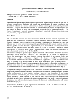
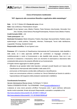
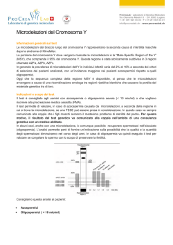
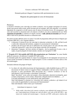
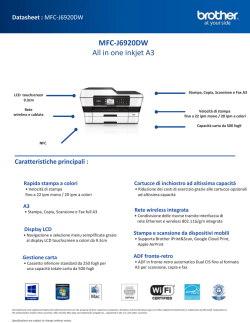
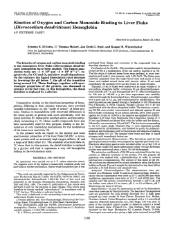
Downloaded from http://jmg.bmj.com/ on June 16, 2017 - Published by group.bmj.com
429
I Med Genet 1993; 30: 429-432
ICF syndrome with variable expression in sibs
G Gimelli, P Varone, Annalisa Pezzolo, Margherita Lerone, V Pistoia
Abstract
We describe a new familial case of ICF
syndrome (immunodeficiency, centromeric instability, facial anomalies) in a
woman of 29 years and in her brother of
30 years. The proband showed mental
retardation, facial anomalies, recurrent
respiratory infections, combined deficit
of IgM and IgE immunoglobulin classes,
and paracentromeric heterochromatin
instability of chromosomes 1, 9, and 16.
The brother had minor signs of the syndrome and had an apparently normal
phenotype. Their parents were healthy
and non-consanguineous. Chromosome
anomalies consisted of homologous and
non-homologous associations, chromatid
and isochromatid breaks, deletions of
whole arms, interchanges in the paracentromeric region, and multibranched configurations of chromosomes 1, 9, and
16. CD bands and fluorescence in situ
hybridisation with alphoid DNA sequence probes specific for the centromeres of chromosomes 1 and 16 showed
that the centromere was not directly
implicated in the formation of multibranched configurations. These cases indicate the autosomal recessive mode of
inheritance and the variable expressivity
of the ICF syndrome.
( Med Genet 1993;30:429-32)
Laboratorio di
Citogenetica, Istituto
G Gaslini, Largo
Gerolamo Gaslini 5,
I-16148 Genova, Italy.
G Gimelli
P Varone
A Pezzolo
Servizio di Genetica
Molecolare, Istituto G
Gaslini, I-16148
Genova, Italy.
M Lerone
Istituto di Oncologia
Clinica e
Sperimentale,
Universita di Genova,
Laboratorio di
Immunopatologia,
Istituto G Gaslini,
Centro
Interuniversitario per
la Ricerca sul Cancro,
I-16148 Genova, Italy.
V Pistoia
Correspondence to
Dr Gimelli.
Received 24 February 1992.
Revised version accepted
4 September 1992.
Case report
The proband, a female (fig 1), was the second
child born to healthy, non-consanguineous
parents. She was born in December 1960 and
came to our attention at the age of 29 because
of mental retardation and phenotypic malformations. She was the term product of a normal
pregnancy. Delivery was by caesarean section
because of podalic presentation. The fetal
movements were reported to be normal. Developmental milestones were retarded. The
child crawled at the age of 12 months, sat up at
the age of 14 months, and started to walk with
help at the age of 2 years. Initial speech development was delayed but improved with
therapy so that by 3 years she was putting two
or three words together although her speech
was indistinct and poorly formed.
At the age of 6 years she was admitted to a
'special school' and evaluation of her psychomotor development showed an IQ of 90 with a
primary lack of language. At the age of 2 years
she presented palsy of the right facial nerve.
From 4 years of age she suffered recurrent
respiratory tract infections and diarrhoea. On
clinical examination she had a peculiar face
characterised by roundness, hypertelorism
with epicanthic folds, an upturned, small nose
with a flat nasal bridge, micrognathia, protrusion of the tongue, and macroglossia. Dermatoglyphics were unremarkable.
The older brother of the proband, born in
1959, does not have the phenotypic characteristics of ICF syndrome, but turned out to have
the cytogenetic and immunological features.
A syndrome characterised by combined immunodeficiency, instability of paracentromeric
heterochromatin, and facial anomalies was
reported by Tiepolo et all and Hulten et al.2
Another eight cases have since been reported
by various authors.39 All these patients were
ascertained because of recurrent respiratory
infections and facial anomalies, and Maraschio
et at6 proposed the acronym ICF (immunodeficiency, centromeric instability, facial anomalies) (McKusick No 242860).
No consanguinity has been reported. Two
sibs with ICF syndrome were reported by
Fasth et al.9 In the two families reported by
Tiepolo et al'0 and Valkova et aF a brother of
the probands died after recurrent respiratory
infections and thus it was suggested that the
ICF syndrome could have an autosomal recessive mode of inheritance.
In this paper we report a familial occurrence
of ICF syndrome ascertained in a female and
her brother, both adult, with variable expressivity in the phenotypic, cytogenetic, and immunological features.
Figure 1 The proband at the age of 29 years.
Downloaded from http://jmg.bmj.com/ on June 16, 2017 - Published by group.bmj.com
430
Gimelli, Varone, Pezzolo, Lerone, Pistoia
Table I Surface marker analysis of peripheral blood
MNC from the patient and her brother.
Surface marker*
Proband
Brother
(% positive cells) (% positive cells)
CD2
CD3
CD4
CD8
CD20
CD16
CD56
CD11b
HLA-DR
CD4/CD8
75
67
23-5
38 1
10 5
13
15
25
21
0-61
87
79
27
53
3
5
5
11
8
0-5
*
As determined by flow cytometry on cells stained in indirect
immunofluorescence.
IMMUNOLOGICAL INVESTIGATIONS
Peripheral blood mononuclear cells (MNC)
were isolated on a Ficoll-Hypaque density
gradient, washed twice with PBS, and stained
with a battery of murine monoclonal antibodies (mabs), as previously reported.'" The antiT cell mabs OKT3-CD3, OKT4-CD4,
OKT8-CD8, and OKTl 1-CD2 were from
Ortho Pharmaceutical Co (Raritan, NJ). The
anti-B cell mab Bl-CD20 was from Coulter
(Hialeah, FL). An anti-HLA-DR mab,
OKIal, was from Ortho. A mab that reacts
with Fc receptors for IgG located on granulocytes and natural killer (NK) cells, LeulIbCD16, was purchased from B-D (Sunnyvale,
CA). Another NK cell specific mab, NKH-1CD56, was from Coulter. A mab that reacts
with monocytes-macrophages, NK cells, and a
subpopulation of T cells, OKM1-CD 1 lb, was
from Ortho. Mouse mabs were detected by
indirect immunofluorescence with fluorescein
conjugated goat anti-mouse immunoglobulin
(Ig) serum (polyvalent) (NEN, Florence,
Italy). Cell suspensions stained with fluorescein conjugated antibodies were fixed with 1 %
paraformaldehyde and analysed by flow cytometry using a FACS analyser (Facs-Star,
B-D). Control preparations consisted of
unstained cells, cells stained with the labelled
second reagent alone, and cells treated with
unrelated antibodies of the same isotype as test
mabs.
Serum IgG, IgA, and IgD were normal in
the proband, her brother, and mother. Serum
IgM and IgE were reduced in the proband, in
particular IgM 15 5 mg/dl (normal range 77 to
280 mg/dl) and IgE <2 kU/I (normal range
>9 kU/i). IgM and IgE were also reduced in
the brother, in particular IgM 46 mg/dl and
IgE < 2 kU/1. IgG subclasses were all normal.
The immunophenotype of the peripheral
Table 2 Frequency of cells with abnormalities.
Culture medium
Proband
Difco TC-chromosome medium
Difco + FUdR
Cells scored Cells with abnormalities
(No)
No of cells (%)
DifcQ + thymidine
Difco+5-azacytidine
157
71
139
140
26 (16-5)
12 (16-9)
Difco+thymidine
Difco+5-azacytidine
127
65
74
52
3
2
10
2
Brother
Difco TC-chromosome medium
Difco+FUdR
37 (26-6)
23 (16-4)
(2-4)
(3-1)
(13-5)
(3-8)
Stretching only
No of cells (%)
27 (17 2)
17 (24-0)
45 (62-5)
11 (15-4)
18
10
37
8
(14-0)
(15-4)
(50-0)
(15-4)
blood MNC from the proband and her brother
investigated (table 1). MNC from both the
proband and her brother were found to contain
a reduced proportion of CD4 + T cells, with a
CD4/CD8 ratio of 0 61 and 0 5, respectively;
this finding was confirmed in a subsequent test
performed after six months (not shown). Normal proportions of T, B, and NK cells with a
normal CD4/CD8 ratio were detected in the
peripheral blood of the mother.
Quantification of serum Ig at various intervals showed that both the proband and her
brother had a selective deficiency of IgM and
IgE, with normal concentrations of IgG, IgG
subclasses, and IgA. In contrast, normal levels
of all Ig classes and IgG subclasses were
detected in the mother's serum.
Taken together, these findings indicate that
the proband and her brother shared a number
of immunological abnormalities related to both
cell mediated and humoral responses.
was
CYTOGENETIC STUDIES
Chromosome analyses were performed on peripheral blood cultures by QFQ, GTG, and
CBG banding. The first culture showed associations and interchanges, breaks and multibranched configurations in the centromeric
regions of chromosomes 1, 16, and, to a lesser
extent, chromosome 9, suggesting the anomalies found in the ICF syndrome.
Six months later, a second sample of blood
from the proband and her brother was analysed and a lymphoblastoid cell line was
initiated from EBV transformed lymphocytes
of the proband. At the same time fibroblast
cultures were set up from a skin biopsy. Lymphocytes, fibroblasts, and lymphoblasts were
treated with FUdR, 5-azacytidine, and excess
of thymidine.
In lymphocytes we found that the overall
frequencies of the chromosome anomalies
ranged from 16 4% in untreated cultures to
26-6% in cultures treated with an excess of
thymidine in the proband, and from 2 36% to
13 5% in her brother (table 2). No anomalies
were detected in the mother and the father
refused investigation.
Chromosome abnormalities consisted, principally, of interchanges in the centromeric regions among homologous or non-homologous
chromosomes, deletions of whole arms,
chromatid and isochromatid breaks in the
centromeric regions, and multibranched
configurations formed by a variable number of
arms of the same or different chromosomes (fig
2).
Chromosome 1 frequently showed stretching of paracentromeric heterochromatin (fig
3A). CD bands showed the presence of an
active centromere on the short arm of multibranched chromosomes 1 and 16 (fig 3C,D).
The short arm of chromosome 1 and 16 were
never duplicated in the multibranched configurations.
In the EBV transformed lymphocytes only
two cells of the 100 analysed showed chromosome abnormalities, while skin fibroblasts
showed only stretching of the paracentromeric
Downloaded from http://jmg.bmj.com/ on June 16, 2017 - Published by group.bmj.com
ICF syndrome with variable expression in sibs
431
*
Figure 2 Chromosome abnormalities in ICF syndrome. (A) Multibranched
configuration of two chromosomes 16 (16ppqqqq). (B) Multibranched chromosome
(lpqq) and association of two chromosomes 16. (C) Multibranched chromosome 1
(lpqq) with a chromosome 16 associated or interchanged (QFQ banding).
A
B
7
:
W
1
-
for two minutes. Hybridisation mixture contained 65% formamide, 2 x SSC, 10% dextran
sulphate, sonicated salmon sperm DNA
(100 gg/ml), and 15 ng of probe per slide.
Hybridisation mixture was denatured at 70°C
for five minutes and applied to prewarmed
slides (37°C), sealed with a coverslip, and
incubated in a humidified box at 37°C for 12 to
16 hours. Coverslips were removed and the
slides washed in 65% formamide in 2 x SSC at
43°C for 20 minutes.
Detection of the biotinilated probes was
achieved by incubation with fluorescein isothiocianate (FITC) labelled avidin (5 gg/ml).
The fluorescent signals were amplified by
subsequent incubation with biotin linked
anti-avidin antibody followed by fluorescein
conjugated avidin. Chromosome preparations
were counterstained with propidium iodide
and DAPI, mounted in an antifade medium,
and observed under an epifluorescence microscope. The hybridisation signal was always
present, as a unique spot, in the centromeric
region of chromosomes 1 (fig 3B) and 16. The
duplicated long arms show no hybridisation
signal of the alphoid DNA probes.
"i..
.:.
.1
k...:
7'
r...*
A, .11
:-
L.
-
C
'IV
NV44,
sLj
>'e
\
Figure 3 Centromeric heterochromatin in ICF syndrome. (A) Stretching of
heterochromatin of a chromosome 1. (B) In situ fluorescence hybridisation ojf an
alphoid DNA sequence (pSD1-1); the signal (arrow) detects the centromerieon the
short arm of a multibranched chromosome I (1pqq). (C,D) CD bands show the
presence of an active centromere (arrow) on the short arm of multibranched
chromosomes 1 (lpqq) and 16 (16pqq). The large dot present on 16q is an a rtefact.
heterochromatin of chromosome 1 in the 42%
of the cells examined.
FLUORESCENCE IN SITU HYBRIDISATION
Probes pSD1-1 (D1Z5) and pSE16-2 (D 16Z2)
(Oncor) detect specifically alphoid I)NA sequences at the centromere of chrornosomes
1 and 16.1213 The probes were labellecI by nick
translation with biotin-16-dUTP (Boe-hringer,
Mannheim, FRG). In situ hybridisa tion and
fluorescence detection was performedlaccording to Pinkel et al.'4
Slides were treated with RNAse (i100 pg/ml
in 2 x SSC) for one hour at 37°C, w ashed in
2 x SSC, dehydrated in an ethanol seiries, and
air dried. Chromosomal DNA was tthen denatured in 70% formamide, 2 x SSC at 70°C
Discussion
Autosomal recessive inheritance of the ICF
syndrome has been postulated because parents
are normal, it is observed in both sexes, and
two sibs in two published families died of
recurrent respiratory infections. Recently two
sibs with ICF syndrome belonging to a heavily
inbred family were described by Fasth et al.9
In previous reports, patients were referred
to clinicians for recurrent infections, facial
anomalies, and mental retardation. Most of
them were observed early in their life and some
died in childhood.
Our proband was observed for the first time
at 29 and her brother at 30 years of age. Unlike
her brother, the proband suffered recurrent
infections particularly viral.
Mental retardation and facial anomalies
were present in the proband but not in her
brother and chromosomal anomalies were
more frequent in the proband than in her
brother. Thus, it can be postulated that there
is some correlation between the frequency of
chromosomal anomalies, different levels of immunoglobulins, and phenotypic manifestations of the ICF syndrome.
Immunological studies showed that the proband and her brother had reduced proportions
of circulating CD4 + T cells, with an inverted
CD4/CD8 ratio, and decreased serum levels of
IGM and IGE; these abnormalities were consistently detected on different occasions. In
contrast, no evidence for T cell subset or Ig
imbalances could be found in the mother. An
inverted CD4/CD8 ratio may be found in
certain lymphoproliferative diseases (Hodgkin's disease, T gamma lymphoproliferative
disease), in transplanted patients receiving immunosuppressive treatment, and during the
course of various viral infections, particularly
the acquired immunodeficiency syndrome
(AIDS). Our probands had no clinical or
Downloaded from http://jmg.bmj.com/ on June 16, 2017 - Published by group.bmj.com
432
Gimelli, Varone, Pezzolo, Lerone, Pistoia
laboratory evidence for any of the above diseases. It is tempting to speculate that the
chromosomal rearrangements typical of the
ICF syndrome affect predominantly or exclusively the CD4 + T cell subset, thus leading to
a selective depletion of this lymphocyte subpopulation and, eventually, to an increased risk
of infections caused by viruses and other intracellular pathogens. This issue is now being
investigated in our laboratories.
Hypogammaglobulinaemia appears to
represent a common feature of the ICF syndrome. The published cases indicate that
hypogammaglobulinaemia may affect the three
main classes (IgG, IgM, and IgA) or only
some of them; furthermore, an IgE deficit is
frequently observed. Both our proband and
her apparently healthy brother had a reduced
serum level of IgM and IgE.
In conclusion, the results of immunological
studies suggest that abnormalities of both cell
mediated and humoral responses may be
involved in the pathogenesis of the immunodeficiency occurring in the course of the ICF
syndrome. The reasons why the proband, but
not her brother, had a lifelong history of respiratory infections can be attributed to the
lower levels of immunoglobulins than her
brother and thus to a different expression of
the ICF syndrome. The molecular mechanisms underlying the immunodeficiency are so
far obscure.
In our first report of a case of ICF syndrome
we pointed out that chromosomal anomalies
could be correlated with the presence of a
fragile site in the centromeric heterochromatin
that causes cross links during DNA replication
or interchanges preventing the normal segregation of the chromatids.10 The more complex
figures involving two or more of chromosomes
1, 9, and 16 could be explained by the centromeric regions of these chromosomes being
characterised by the preferential location of
satellite II DNA and being likely to be associated in interphase.
Fryns et aP pointed out that centric fission
of chromosomes 1, 9, and 16 leads to the
formation of functional centromeres of the
short or long arms of these chromosomes and
through successive duplications they can give
rise to the observed multibranched configurations. In situ fluorescence hybridisation and
CD bands showed that the centromeres of
chromosomes 1 and 16, involved in multibranched configurations, were not split in two
or more parts, as hypothesised by Fryns et al.3
In fact, a single centromere was always present
on the short arm and absent on the long arm of
chromosomes 1 and 16, as shown in fig 3B-D.
Furthermore, in the present cases and in that
reported previously,'0 only the long arms of
chromosomes 1, 9, and 16, where the centromere was absent, were duplicated in the multibranched configurations.
These observations allow us to postulate
that the centromeres are not directly involved
in the origin of the multibranched chromosomes, but that paracentromeric heterochromatin of chromosomes 1, 9, and 16 was
involved. An autosomal recessive mutation of a
gene(s) that controls the normal process
of condensation of part of the centromeric
heterochromatin could be the cause of the
chromosomal anomalies found in this syndrome, as postulated by Maraschio et al.'
We thank Professors Marco Fraccaro and
Paola Maraschio for their collaboration in the
preparation of this paper.
1 Tiepolo L, Maraschio P, Gimelli G, Cuoco C, Gargani GF,
Romano C. Concurrent instability at specific sites of
chromosomes 1, 9, and 16 resulting in multibranched
structures. Clin Genet 1978;14:313-4.
2 Hulten M. Selective somatic pairing and fragility at 1q12 in
a boy with a common variable immunodeficiency. Clin
Genet 1978;14:294.
3 Fryns JP, Azou M, Jaeken J, Eggermont E, Pedersen JC,
Van den Berghe H. Centromeric instability of chromosomes 1, 9, and 16 associated with combined immunodeficiency. Hum Genet 1988;57:108-10.
4 Howard PJ, Lewis IJ, Harris F, Walker S. Centromeric
instability of chromosomes 1 and 16 with variable immunodeficiency: a new syndrome. Clin Genet 1985;
27:501-5.
5 Valkova G, Ghenev E, Tzancheva M. Centromeric instability of chromosomes 1, 9, and 16 with variable immunodeficiency. Support of a new syndrome. Clin Genet
1987;31:1 19-24.
6 Maraschio P, Zuffardi 0, Dalla Fior T, Tiepolo L. Immunodeficiency, centromeric heterochromatin instability
of chromosomes 1, 9, and 16, and facial anomalies: the
ICF syndrome. J Med Genet 1988;25:173-80.
7 Carpenter NJ, Filipovich A, Blaese RM, Carey TL, Berkel
AI. Variable immunodeficiency with abnormal condensation of heterochromatin of chromosomes 1, 9, and 16. J
Pediatr 1988;112:757-60.
8 Turleau C, Cabanis MO, Girault D, et al. Multibranched
chromosomes in the ICF syndrome: immunodeficiency,
centromeric instability, and facial anomalies. Am J7 Med
Genet 1989;32:420-4.
9 Fasth A, Forestier E, Holmberg E, et al. Fragility of the
centromeric region of chromosome 1 associated with
combined immunodeficiency in siblings: a recessively
inherited entity? Acta Paediatr Scand 1990;79:605-12.
10 Tiepolo L, Maraschio P, Gimelli G, Cuoco C, Gargani GF,
Romano C. Multibranched chromosomes 1, 9, and 16 in a
patient with combined IgA and IgE deficiency. Hum
Genet 1979;51:127-37.
11 Pistoia V, Prasthofer EF, Tilden AB, et al. Large granular
lymphocytes (LGL) from patients with expanded LGL
populations acquire cytotoxic functions and release lymphokines upon in vitro activation. Blood 1986;68:1095.
12 Waye JS, Durfy SJ, Pinkel D, Kenwrick S, Patterson M,
Davies KE, Willard HF. Chromosome-specific alpha
satellite DNA from human chromosome 1: hierarchical
structure and genomic organization of a polymorphic
domain spanning several hundred kilobase pairs of centromeric DNA. Genomics 1987;1:43-51.
13 Greig GM, England SB, Bedford HM, Willard HF. Chromosome specific alpha satellite DNA from the centromere
of human chromosome 16. AmJ Hum Genet 1989;45:86272.
14 Pinkel D, Straume T, Gray JW. Cytogenetic analysis using
quantitative, high-sensitivity, fluorescence hybridization.
Proc Natl Acad Sci USA 1986;83:2934-8.
15 Maraschio P, Tupler R, Dainotti E, Piantanida M, Cazzola
G, Tiepolo L. Differential expression of the ICF (imanmunodeficiency, centromeric heterochromatin, facial
omalies) mutation in lymphocytes and fibroblasts. 7 Med
Genet 1989;26:452-6.
Downloaded from http://jmg.bmj.com/ on June 16, 2017 - Published by group.bmj.com
ICF syndrome with variable expression in
sibs.
G Gimelli, P Varone, A Pezzolo, M Lerone and V Pistoia
J Med Genet 1993 30: 429-432
doi: 10.1136/jmg.30.5.429
Updated information and services can be found at:
http://jmg.bmj.com/content/30/5/429
These include:
Email alerting
service
Receive free email alerts when new articles cite this article. Sign up in the
box at the top right corner of the online article.
Notes
To request permissions go to:
http://group.bmj.com/group/rights-licensing/permissions
To order reprints go to:
http://journals.bmj.com/cgi/reprintform
To subscribe to BMJ go to:
http://group.bmj.com/subscribe/
© Copyright 2026 Paperzz