特 集
放射線診療の過去・現在・未来
造影剤
2.MRI造影剤の過去・現在・未来
谷本 伸弘,栗林 幸夫
慶應義塾大学医学部 放射線診断科
MR Contrast Agents: Past, Recent Trends, and Prospects
Akihiro Tanimoto, M.D. and Sachio Kuribayashi, M.D.
Summary
Non-specific extracellular gadolinium chelates have been widely used in contrastenhanced MR imaging. These agents distribute into the intravascular and interstitial spaces
and differentially alter tissue contrast through relatively selective enhancement of proNICHIDOKU-IHO
ton relaxation based on differences in compartmentation, distribution, and residence times
Vol. 50 No. 1 114–132 (2005)
of the pharmaceutical. They also allow the evaluation of physiological parameters, such
as the status or presence of the blood-brain-barrier or renal function.
Meanwhile, liver-specific MR contrast agents have been developed as the first tissue-specific pharmaceuticals to include
superparamagnetic iron oxide (SPIO) particles and hepatobiliary paramagnetic agents. SPIO particles are phagocytosed by
reticuloendothelial cells in the liver, resulting in negative enhancement of the liver parenchyma on T2- or T2*-weighted imaging. Hepatobiliary paramagnetic agents are partially taken up by hepatocytes, yielding positive sustained enhancement of the
liver parenchyma on T1-weighted imaging. These agents have been demonstrated to improve the detection and characterization of hepatic neoplasms, and are expected to simplify the existing multi-step diagnosis in liver imaging.
Numerous magneto-pharmaceuticals have been studied as potential tissue-specific MR contrast agents. A wide variety of
vector and carrier molecules, including antibodies, peptides, proteins, polysaccharides, liposomes, and cells, have been developed to deliver magnetic labels to specific sites. Macromolecular MR contrast agents (blood pool agents) will be beneficial for
improving the quality of MR angiography, for quantitating capillary permeability, and for myocardial perfusion imaging. The
permeability of macromolecular tracers may also provide tissue uptake information for both contrast and chemotherapeutic
drugs. Dedicated blood pool agents will be available within the next few years. Contrast-enhanced lymphography after interstitial or intravenous injection will be another major step forward in diagnostic imaging. Ultra small SPIO (USPIO) particles
have been evaluated in multicenter clinical trials for lymph node MR imaging, and the clinical impact is taken up for discussion. It is likely that USPIO and gadolinium chelates will augment the MRI of atherosclerotic plaques, a systemic inflammatory disease of the arterial wall. Thrombus-specific agents based on gadolinium-labeled peptides are on the doorstep. Gd-porphyrins also can be used as a dual probe for MRI contrast agents and as a radiation sensitizer for neutron capture therapy. This
may facilitate tumor detection and treatment planning and potentially improve the safety and efficacy of radiation therapy.
Expectations for these new tumor-, pathology-, and receptor-specific agents are high.
A series of recent studies indicate that MRI cell tracking has great potential for the evaluation and optimization of stem and
progenitor cell therapy. MR tracking of magnetically labeled cells following transplantation or transfusion could determine the
distribution of these cells and monitor therapeutic effect. Up to now, SPIO particles appear to be the contrast agent of choice to
label stem cells, owing to their biocompatibility and strong effects on proton T2* relaxation. Cell-tracking research has been performed in disease models of the central nervous system and the infarcted myocardium. With its excellent spatial resolution and
the ability to track labeled cells over prolonged periods of time, MRI cell tracking is likely to become an important technique.
Technical advances in MR imaging will further facilitate the exploitation of tissue-specific MR contrast agents. The past
history, recent trends, and prospects of MR contrast agent development are outlined in this review.
Department of Diagnostic Radiology,
Keio University School of Medicine
114(114)
日獨医報 第50巻 第 1 号 114–132(2005)
日獨医報 第50巻 第 1 号 2005
剤の造影効果の動態解析は、ある種の生理学的パラメー
はじめに
タを評価するのに有用であり、なかでも腫瘍組織のanMRI造影剤として最初に上市されたGd-DTPA
giogenesisやmicrovessel densityの定量化が報告されて
(Magnevist ;日本シエーリング)
をはじめとする非特異
いる6)。さらに躯幹部のMR angiography(MRA)への応
性
(細胞外液性)
ガドリニウム
(Gd)
造影剤の使用件数は、
用は呼吸停止下高速スキャンを用いたPrinceらによって
世界で数千万例に達している。また最初の臓器特異性造
開かれた7)。また急性心筋梗塞において、機能性合胞体
影剤である超常磁性酸化鉄(superparamagnetic iron
である心筋細胞の細胞膜破綻が、梗塞心筋の遅延造影効
oxide:SPIO)製剤(以下、SPIO)も臨床に使用されるよ
果に関与していることは興味深い8)。細胞外液性Gd造影
うになって 7 年余が経過し、その臨床的有用性も確立し
剤の開発は、類薬の 2 倍濃度の1.0M-gadobutrolにてひ
つつある。このほか、現在承認待ちの肝細胞特異性造影
とまず終了した。1.0M-gadobutrolはbolus投与により
剤Gd-EOB(ethoxybenzyl)-DTPAをはじめ、血液プー
T2*susceptibility perfusion imagingやMRAにおける造
ル造影剤やリンパ節特異性造影剤などが臨床応用に向け
影能の向上が期待されている 9、10)。
®
て研究開発が進んでいる。2004年 5 月に京都で開催さ
一方、細胞外液性造影剤とは異なるコンセプトの造影
れたISMRM(International Society for Magnetic Reso-
剤も、早くから開発が行われていた。特に肝特異性造影
nance in Medicine:国際磁気共鳴医学会)での造影剤関
剤の研究が進んでおり、magnetiteは1978年にはすでに
係のトピックスは、従来のMRIと一線を画する再生医療
MRI造影剤としての可能性が示されていた11)。肝網内系
に関する話題であり、いよいよMRIが単なるmorphol-
を標的としたSPIOとしての応用は、米国Harvard大学
ogyの壁を越えて、cell biologyやbiochemistryと融合し
Massachusetts General HospitalのStarkらが臨床治験の
て医学に貢献することが現実化しつつあるといえよう。
初期成績を報告したのが最初である12)。その後米国、欧
本稿ではMRI造影剤の歴史と開発状況、今後の可能性に
州、アジアで相次いで認可されたFeridex ®(栄研化学)
[Endorem TM(Advanced Magnetics)]は肝の画像診断に
ついて言及する。
おける有用性を確立したが、最近はbolus投与できる
MRI造影剤の歴史
Resovist ® Inj.(日本シエーリング)に置き換わりつつあ
る。常磁性体では肝細胞特異性造影剤Fe-EHPGの開発
最初のMRI造影剤である細胞外液性Gd錯体Gd-DTPA
が細胞特異性造影剤の最初の試みである 13)。
の人体への使用は、1984年のLaniadoらの健常ボランテ
組織特異性MRI造影剤の開発は1990年代に種々試み
ィアへの投与報告が最初とみられる1)。爾来、類薬のGd
られた領域である。薬剤を組織特異的に到達させる方法
錯体であるG d - H P - D O 3 A(P r o H a n c e ® ;エーザイ)、
論(targeting strategy)を、シンチグラフィ以上の空間分
®
Gd-DTPA-BMA(Omniscan ;第一製薬)、Gd-DOTA
®
( M A G N E S C O P E ; 栄 研 化 学 )、 g a d o b u t r o l
®
(Gadovist ;日本シエーリング)、gadoversetamide
®
(OptiMARK ;Mallinckrodt)の開発が進み、MRI装置
の進歩とともに急速に臨床使用が拡大した。浸透圧、粘
解能をもつMRIに適用すべく、組織特異性MRI造影剤
の研究が精力的になされた。しかしいずれも実用化には
至らず、PETの台頭と普及もあって一時研究が下火とな
ってしまったが、最近は再び精力的に研究が進められて
いる。
度などに造影剤間で相違はあるものの、体内動態、造影
効果、副作用頻度には大差がなく、本質的には同じ細胞
MRI造影剤開発の現況
(表 1)
外液性造影剤である。MR高速撮像法と細胞外液性Gd造
影剤を併用し経時的に病変の造影効果を観察するdynamic
MRIの臨床応用は、1986年にNiendorfらが脳腫瘍に、
Ohtomoらが肝腫瘍に用いた報告が始まりである
2、3)
。
またGdを用いたMR灌流画像(perfusion imaging)への
4)
可能性は早くから示され 、T2*effectによるcerebral
5)
blood flowの定量化が試みられた 。細胞外液性Gd造影
1.肝特異性造影剤
大きく網内系に貪食されるタイプと、肝細胞に取り込
まれるタイプがある。
1)
超常磁性酸化鉄SPIO
(superparamagnetic iron oxide)
製剤
肝網内系をターゲットとしたSPIOとしては、1997年
115(115)
日獨医報 第50巻 第 1 号 2005
にFeridex ® 、2001年にResovist ® Inj.が上市された。
表1
MRI造影剤の種類
おのおの、直径 5nm程度の酸化鉄coreがdextran、
carboxydextranで被覆され集簇した形状をもち、粒
子径はおのおの100〜250nm、57nmである。SPIO
細胞外液
Gd-DTPA (Magnevist)
Gd-DOTA (MAGNESCOPE)
Gd-DTPA-BMA (Omniscan)
Gd-HP-DO3A (ProHance)
Gadoversetamide(OptiMARK)
Gadobutrol (Gadovist)
A
A
A
A
C*1
B*2
A
A
A
A
A
A
肝網内系
Ferumoxides (Feridex, Endorem)
Ferucarbotran (Resovist Inj.)
A
A
A
A
肝細胞
Gd-BOPTA (MultiHance)
Gd-EOB-DTPA (Primovist)
Gd-DTPA-DeA
MnDPDP (mangafodipir)
AG-USPIO
MION-ASF
B*1
B
C*2
E
E
E
A
A
E
A
D
D
血液プ−ル MS-325 (Gadofosveset)
B22956/1
Gadomer-17
P-792 (Vistarem)
Ferumoxtran-10 (Combidex, Sinerem)
NC 100150 (Clariscan)
SH U 555C
VSOP
D
E
D
E
E
E
E
E
C
C
C
D
B
B
D
D
Plaque
Gadofluorines
Ferumoxtran-10 (Combidex, Sinerem)
E
E
D
B
リンパ節
Ferumoxtran-10 (Combidex, Sinerem)
Gadofluorines
Gd-DTPA-PGM
Gd-carrying liposome
E
E
E
E
B
D
D
D
腫瘍
Gd-porphyrins
MION-MoAb
Gd-DTPA-9.2.27
D
E
E
D
D
D
膵
MnDPDP
MION-CCK
E
E
A
D
心筋
MnDPDP
MION-antimyosin
E
E
A
D
炎症
Ferumoxtran-10 (Combidex, Sinerem)
MION-IgG
E
B
細胞追跡
Ferumoxides (Feridex)
Ferucarbotran (Resovist)
D
D
D
D
その他
Glucose感応薬
pH感応薬
遺伝子発現
D
D
D
D
D
D
消化管
Ferric ammonium citrate(FerriSeltz)
MnCl2 (LumenHance)
OMP
A
E
C*2
A
C
B
粒子は血中のopsoninと結合して、投与量の約80%
が肝網内系Kupffer細胞に貪食される。Kupffer細胞
内ではSPIOはlysosome顆粒に集積してclusterを形
成し、大きいclusterは局所磁場を擾乱してT2*を短
縮し、肝の信号強度を低下させる。また小さいclusterは水分子のmagnetic centerへの接近を容易とし、
T1およびT2を短縮する。T2*、T2短縮効果は非常
に強力で、通常はT2*、T2強調画像で使用して肝の
信号低下を得ることで腫瘍の検出能を向上させる。
SPIOはT1短縮効果もGd系造影剤より強く、短いTE
のパルス系列を用いれば肝の信号増強が得られる
14)
。肝の信号強度低下あるいは上昇が起こる結果、
腫瘍−肝コントラストを向上させ肝腫瘍の診断に寄
与する。SPIO造影MRIにおけるパルス系列の最適
化に関しては機種と磁場強度により多少の相違はあ
るが、基本的にはT2強調fast SE、T1強調gradient
echo(GRE)、T2*強調GREの 3 つの組み合わせが推
奨される。
SPIOの臨床的有用性については数多くの報告があ
る。転移性肝癌ではSPIO併用MRIは最も効果的な
非侵襲的診断法であり、造影剤としてSPIOは第一選
択といえよう(図 1)。肝転移の検出能において、
S P I O 造影M R I はd u a l p h a s e d y n a m i c C T より優
れ15)、ほぼCTAP(CT during arterial portography)
に匹敵する16)。手術を前提とした場合には、SPIO造
影MRIはCTAPの特異度(supecificity)の低さを補
う
17)
。当院手術例での検討では、SPIO造影MRIは
CTAP + CTHA(CT-hepatic arteriography)と比較
して感度(sensitivity)は匹敵し、特異度と正確度
(accuracy)はやや優れていた18)。このほか、胆道系
腫瘍も転移性肝癌と同様に網内系が欠如しているの
で、その広がり診断にもSPIO造影MRIが期待され
る。
肝細胞癌に関しては、小さい多血性肝細胞癌の検
出能はGd-dynamic MRIの方がSPIO造影MRIより優
薬剤
開発動向
国内 海外
標的
れるという報告が多い19、20)。治療を急ぐべき多血性
肝細胞癌を診断するという意味では、Gd-dynamic
MRIに先んじてSPIO造影MRIを選択するケースは
116(116)
A:発売中,B:治験終了,C:治験中,D:研究段階,
E:研究未施行
*1:承認申請後中止,*2:開発中止
日獨医報 第50巻 第 1 号 2005
A
B
図 1 SPIOによる微小肝転移の診断(75歳・男性,大腸癌肝転移)
CTAPにてS4に微小な灌流欠損が認められる(矢印).
SPIO造影T2*強調画像(FSPGR 130/9/60˚)にて,径 3mmの転移巣(矢印)が網内系欠損域として
描出されている.
少ない。肝硬変では網内系機能の低下によりSPIOの造
21)
影効果も低下する 。細胞外液性造影剤では、肝硬変の
A
B
強調画像の造影パターンをみることでAHと高分化肝細
胞癌の鑑別に役立つ可能性がある 27)。
有無に造影効果が影響されず、dynamic studyにて腫瘍
Resovist ® Inj.は急速静注が可能であり、市場でも点
血流情報が得られる点が有利である。しかしAP-shunt
滴投与のF e r i d e x ® から移行がほぼ完了した。T 1 強調
によるdynamic study上の偽病変の否定に、SPIOは有効
GREにてdynamic MRIを施行すると、perfusion phase
である
22)
。相補う情報を得るために、同一検査日にGd
の少なくとも30秒間は肝臓の信号増強が起こるが、時
。
間経過とともに肝は黒化に転ずる14)。肝細胞癌などの多
Angio-CT(CTAPおよびCTHA)では、sensitivityは高い
血性腫瘍がGd製剤のdynamic MRIと同様にSPIOの急速
が偽陽性も多く、specificity、positive predictive value
静注により高信号として描出されるかについては否定的
(PPV)が低いことが知られ、specificityの高いSPIO併
である。これに対し、Resovist ® Inj.静注によるEPI-per-
用MRIは病変の有無の確認に有用である。肝細胞癌の診
fusion imagingが報告されており、SPIOの弱点であっ
断におけるS P I O 併用M R I とA n g i o - C T の比較では、
た腫瘍の血流情報が得られる可能性が示されている28)。
SPIOはAngio-CTと同等のsensitivityと、より高い
このほか、SPIOは脾臓にも取り込まれその信号強度
とSPIOの両者を使用することも試みられている
specificityを示している
23 )
18、24)
。
を低下させるので、悪性リンパ腫29)、転移性脾腫瘍、脾
SPIOは多血性肝細胞癌においてはproblem-solving
過誤腫、副脾などの診断に有用性が期待される。また最
modality としての役割が大きい。このほか、SPIOは
近、SPIO(Feridex ® )をセンチネルリンパ節の診断に応
vascularityの高くない肝細胞癌の検出や、腺腫様過形成
用する試みがなされている 3 0 )。乳癌症例で乳房内に
(adenomatous hyperplasia:AH)・限局性結節性過形成
SPIOを局注すると、リンパ流に乗ったSPIOは腋窩リン
(focal nodular hyperplasia:FNH)など網内系を保持し
パ節のうちセンチネルリンパ節にまず流入する。SPIO
ている肝腫瘤の質的診断に有用である。SPIO造影T2*
が流入したリンパ節は磁力センサーないしMRIで同定可
強調画像はKupffer細胞機能の低下を鋭敏に反映するの
能である。また後述するように、SPIOでラベルした細
25)
で 、肝硬変ではT2強調画像よりT2*強調画像にて信号
胞の体内追跡など、多彩な用途に応用されている。
26)
低下が起きにくい 。同様の考え方を応用し、T2、T2*
117(117)
日獨医報 第50巻 第 1 号 2005
2)
USPIO(ultrasmall SPIO)
積したGd-BOPTAとGd-EOB-DTPAが毛細胆管へ輸送
USPIOは後述するMR lymphographyやMRAのみなら
される過程はATP(アデノシン三リン酸)
依存の能動輸送
ず、肝臓にも用途が期待される造影剤である。肝臓で
により起こり、cMOAT(canalicular membrane organic
は、粒子径が小さいために緩徐に肝網内系に貪食され
anion transporting polypeptide)の関与が考えられてい
る。 U S P I O では r 2 / r 1 比が1 . 6 〜1 . 7 と低 い( r 2 : 3 5 〜
る 40)。
53mM −1・sec −1 、r1:20〜24mM −1 ・sec −1)。Feridex ® や
このほか、本邦では臨床治験に至っていないが、ビタ
Resovist ® Inj.と比較してT2短縮効果がやや弱いためT2
ミ ン B 6 類 似 の 構 造 を し た M n D P D P( m a n g a f o d i p i r
強調画像での肝臓の黒化が軽度である一方、T1短縮効
trisodium)が欧米で発売されている41)。これはビタミン
果が強力となる。T1強調GREでは肝と肝内血管信号を
B6の触媒活性形である補酵素pyridoxal-5’-phosphateの
増強するなどの、T1短縮効果を狙いとした使用が容易
2 つの連結形を示すキレート剤DPDPを用いることで、
。肝血管腫では造影剤の血液プール効果に
肝細胞の膜輸送系に認識される 42)。MnDPDPは投与量
よりT1強調GREで著明な信号増強、T2強調画像で信号
が5애mol/kgと少量であるが、副作用の頻度がやや高いこ
低下を示し、転移性肝腫瘍や肝細胞癌との鑑別に有用で
と、点滴投与であることなど、臨床的耐容性の面でGd
である
ある
31、32)
33)
。米国での第 2 相用量設定試験では20애mol Fe/
kgが至適用量とされている
34)
ベースの肝細胞特異性造影剤より使いにくい点がある。
肝細胞特異性造影剤は、投与後早期相から平衡相にか
。
3)
常磁性肝細胞特異性造影剤
(paramagnetic hepatocyte-specific agent)
けては細胞外液性造影剤同様にdynamic studyによる血
流情報の評価が可能である。さらに肝胆道分布相
Gdベースの常磁性肝細胞特異性造影剤には、承認申
™
請中のGd-EOB-DTPA(Primovist ;Schering AG)、臨
(hepatobiliary phase)では正常肝実質に造影剤が取り込
まれ肝臓を高信号化することで、腫瘍−肝臓コントラス
床 治 験 が 終 了 し た G d - B O P T A( M u l t i H a n c e ;
トを高め診断能が向上する 43、44)。本邦での臨床治験の
Bracco)、Gd-DTPA-DeAがある。Gd-EOB-DTPA、
結果、至適臨床投与量はGd-BOPTAが0.1mmol/kg、
Gd-BOPTAは欧州で発売されているが、本邦で今後の
Gd-EOB-DTPAが0.025mmol/kgとなった。肝細胞特異
承認の可能性があるものは現時点でGd-EOB-DTPAの
性造影剤は蛋白結合率が高いために、血漿中ではGd-
みとなっている。これらはGd-DTPA 類似の基本骨格
DTPAよりr1(T1緩和度:T1 relaxivity)が高い。血漿中
に、ベンゼン環と脂溶性の基が付加された構造式を有し
r1はGd-DTPAで4.9mM −1 ・sec −1 に対しGd-BOPTAで
ている。脂溶性が高まることによって細胞膜通過性が亢
6.9mM −1・sec −1、Gd-EOB-DTPAで8.7mM −1・sec −1であ
進し、肝細胞内に取り込まれやすくなる。肝臓に取り込
る45、46)。Gd-BOPTAは臨床投与量が0.1mmol/kgと細胞
まれ胆汁排泄される比率はGd-EOB-DTPAが約50%35)、
外液性Gd系造影剤と同一であるから、dynamic MRIで
®
36)
、Gd-DTPA-DeAが約90%であ
は細胞外液性Gd系造影剤に同等以上の造影効果が期待
る 。肝細胞特異性造影剤は胆汁中と尿中に排泄される
できる。またGd-BOPTAは肝胆道分布相(投与後40〜
ので、高度の肝機能障害があっても尿中排泄される代償
120分)での撮像が付加的診断情報を与える47、48)。しか
が働く。sinusoidに到達した造影剤は濃度勾配に従った
し、治験機構当局にはGd-DTPAよりやや高い副作用発
受動拡散により血管内皮間を透過した後、抱合胆汁酸・
現率(約 6%)を凌駕する臨床的有用性と判断されず、
脂肪酸の能動輸送、あるいは濃度勾配に従う有機陽イオ
Gd-BOPTAの申請は中断している。
Gd-BOPTAが約 4%
37)
ン・アルブミン結合陰イオンなどの受動輸送により肝
細胞膜を通過して肝細胞に取り込まれる
38)
多血性肝細胞癌に対しては、dynamic studyが可能
。受動輸送
で、かつ肝胆道分布相での腫瘍検出の向上が期待される
の過程では、有機陰イオントランスポーターのひとつで
肝細胞特異性造影剤がSPIOより有用である可能性があ
ある多選択トランスポーターO A T P 1(o r g a n i c a n i o n
る。ただしGd-EOB-DTPAは投与量が少ないため、①
39)
。
dynamic MRI動脈相での造影効果がGd-DTPAより劣る
肝細胞内ではミクロソーム分画の薬物代謝酵素により水
可能性があること、② 血中半減期が短くなり、血管腫
酸化され、次いでグルクロン酸抱合・硫酸抱合をうけて
と他腫瘤との鑑別に重要な、遅延相における造影剤の血
適度な水溶性をもち胆汁中に排泄される。肝細胞内に集
管腫内poolingが弱い可能性があること、には注意を要
transporting polypeptide 1)の関与が疑われている
118(118)
日獨医報 第50巻 第 1 号 2005
する。また肝胆道分布相で低信号に描出される結節が高
大が起こる。
分化肝細胞癌であるとすれば、肝硬変患者の「癌の早期
現在、造影MRAでは細胞外液性Gd造影剤を急速静注
発見」に大変有用である。ただし高分化肝細胞癌では
し、bolus trackingあるいはtest injection法によりdy-
Gd-EOB-DTPA投与後の肝胆道分布相にて、周囲肝組
namic MRIを行う方法が最も普及し、アーチファクトが
49)
織より強く造影される現象が報告されている 。一方自
少なく美しい画像が提供されている。高い血中滞留性に
験例では、AHで周囲肝よりやや低信号を示した症例も
よりMRA撮像のimaging window(至適撮像時間の幅)を
経験している。良性腫瘍であるFNHもGd-EOB-
広げ、高い血中relaxivityにより細小血管の描出能を向
DTPA 44)やMnDPDP 50)を取り込み、周囲肝より強く造影
上させる。最近は機器の進歩に伴い、より高速により広
される。肝胆道分布相における腫瘍の造影様式と分化度
範囲のMRAが撮像可能となってimaging windowの問題
との間にはoverlapが多々みられ、発育様式やdoubling
はなくなり、高いrelaxivityによる細小血管の描出が血
timeなど生物学的悪性度との関連も含め、今後のさらな
液プール造影剤の主目的となっている。今後はparallel
る検討が必要である。肝細胞特異性造影剤は非特異的造
imagingの普及によりtime-resolved MRAのような時間
影効果を併せ持つので、間質を豊富に含む転移性肝癌で
分解能が向上したMRAへの応用も期待される。
は腫瘍内の造影効果が遷延する可能性がある。転移性肝
血液プール造影剤はMRAのほか、多彩な用途に応用
癌に対してはSPIO による診断がより単純かもしれな
が期待されている。高分子性を利用して組織のcapillary
い。包括医療時代を迎え、肝細胞特異性MRI造影剤の評
量と透過性の差をMRIで検出できる。Gd-DTPAのよう
価と肝画像診断の簡略化は今後の課題である。
な低分子造影剤では正常組織でも血管透過性が非常に高
肝 細胞特 異性造影剤は 肝機能(h e p a t i c f u n c t i o n a l
いため、正常−異常組織間での差を検出しにくい。これ
reserve)診断への応用が可能と期待されている。ラット
に対し、分子量数千〜数万程度の高分子造影剤は正常組
の急性、慢性肝障害におけるGd-EOB-DTPAの造影効
織では毛細管を透過しないが、異常組織ではある程度透
果の検討では、急性肝障害では正常群に比して早期の造
過性が亢進するため透過しうる。組織の造影効果を測定
影効果が有意に低く、慢性肝障害では早期造影効果が低
して組織のcapillary量と透過性を定量した結果、slow-
51)
いだけでなく後期に造影効果が遷延した 。肝葉を部分
growingの腫瘍はrapid-growingの腫瘍に比較して組織
的に虚血に至らせて肝代謝能を測定した実験的報告もあ
のcapillary量、透過性ともに有意に低いことが示された
り、肝切除を踏まえた部分肝機能診断が可能となれば臨
初期の報告がある56)。腫瘍の血管新生能、ひいては悪性
床的意義は大きい52)。またMR胆道造影として胆道シン
度を推定することが可能になると期待される。また放射
チグラフィやDICの代替となる可能性をもつ。
線療法は抗ガン剤の組織到達に影響を与えるが、血液プ
4)
肝細胞特異性USPIO
ール造影剤を用いてcapillary透過性を定量化することに
このほか研究中の肝細胞特異性造影剤に、SPIOにア
より、capillary透過性を亢進し組織内の抗ガン剤の濃度
シアロ糖蛋白を結合させて肝細胞膜に存在するgalac-
を最も高めるには放射線療法後何日目が適切か、を推定
tose receptor経由で肝細胞内に取り込まれるタイプの
可能という 57)。
造影剤があり、肝腫瘍の診断法として研究されてい
る
53、54)
99m
このほか、心筋虚血をみるためのperfusion studyと梗
Tc-GSA
塞心筋を観察するために遅延造影相で汎用されている細
(galactosyl human serum albumin)シンチグラフィと同
胞外液性Gd製剤に対し、血液プール造影剤を用いて、
。また肝予備能検査法である
様に、肝機能診断への可能性が示されている
54、55)
。
実験的に興味深い結果が示されている。正常心筋は血液
プール造影剤により投与早期に造影されるが、血流途絶
2.血液プール造影剤
した梗塞心筋は造影されない。再灌流(虚血)心筋は徐々
1)
血液プール造影剤とは
に造影され、wash-outも緩徐である58)。正常心筋はより
血液プール造影剤とは、血中滞留時間を長くして血管
早期に造影される59)。血液プール造影剤と再灌流心筋の
腔の描出能向上その他を目的として開発された造影剤群
造影パターンには造影剤の分子量による相違があるが、
である。血液プール造影剤は高分子化により、腎からの
血液プール造影剤の応用により虚血があっても透過性が
排泄遅延と、水分子との回転相関時間の延長によるr1増
亢進していない心筋、透過性が亢進した心筋、壊死心筋
119(119)
日獨医報 第50巻 第 1 号 2005
を鑑別できる可能性が示された。非侵襲的な心筋障害の
脈MRAへの応用が既に健常ボランティアに対し試みら
程度と範囲の診断は、急性心筋梗塞においてPCI
れ良好な描出能が示されている72)。また前述したように
(percutaneous coronary intervention)による再灌流治療
血液プール造影剤のこのほかの役割として、心筋虚血範
囲の評価や73)、腫瘍のcapillary透過性を評価することで
効果を判定するにあたり期待される。
2)
血液プール造影剤開発の現況
放射線療法の効果をモニタリングしたり、腫瘍と瘢痕組
血液プール造影剤は、血中で蛋白結合するタイプ、
織を鑑別する可能性が実験的に報告されている 74、75)。
Gd化合物の重合体(polymeric Gd-complex)、および
こ の ほ か 、 P - 7 9 2( V i s t a r e m ™ ; G u e r b e t )は 分 子 量
USPIOに分類される。
5,000Da以下と小さいが血中で集合体を形成する性質が
① 蛋白結合タイプ
あり、25mM −1 ・sec −1 以上と高いr1を示す76、77)。いまだ
この範疇の造影剤として最初に開発されたのはMS-
動物実験であるがP-792を心筋perfusion imagingに用い
325(Gadofosveset)で、血中アルブミンと可逆的結合を
ることにより、細胞外液性Gd造影剤より心筋虚血を描
して高いr1(30mM ・sec 以上)と長い血中半減期を示
出するimaging windowが延長し有用であった78)。また
す60)。MRAではbolus投与のfirst passと平衡状態での良
呼吸停止・呼吸同期プロトコルの冠状動脈MRAともに
−1
−1
好な造影効果が報告されている
61、62)
。一方では投与直
描出能が向上した79)。心臓MRIに有望な造影剤として期
後では74%が蛋白と結合していないfree型のMS-325で
待されている。
あるため造影効果が弱く、またfree型は血管外に漏出す
③ USPIO
るためbackgroundが造影されMRA造影能に不利に働
U S P I O は、肝を標的としたS P I O よりはr 2(T 2 緩和
く、という指摘もある 63)。このほか、Gd-BOPTAは蛋
度:T2 relaxivity)が小さいがr1が大きく、また粒子径
白結合能が高いために、血中でのr1がGd-DTPAより高
が小さいため血中滞留時間が長いので血液プール造影剤
いので、MRAに応用してより細部の描出能の向上が得
としての使用が可能である。USPIOとしてferumoxtran-
64)
。 ま た B 2 2 9 5 6 / 1( g a d o c o l e t i c a c i d
10( Sinerem ® 、Combidex ® ;Advanced Magnetics)、
trisodium)はGd-DTPAに胆汁酸(deoxycholic acid)を
N C 1 0 0 1 5 0 、 S H U 5 5 5 C 、 V S O P( v e r y s m a l l
結合させた誘導体で、高い蛋白結合能による高r1と血中
superparamagnetic iron oxide particle)などが知られて
滞留性により、45分の間冠状動脈の良好な描出が報告
いる 80– 83)。MRAでの造影効果はdoseに依存し、25〜
されている65)。今後は、black blood法やtrue-FISP法を
50애mol Fe/kg程度が必要とされる。bolus投与でのfirst
用いた冠状動脈MRAとの比較もなされるであろう。
pass MRAでは、細胞外液性Gd造影剤によるMRAと比
られている
② Gd重合体
肩する造影能が得られている 83)。またUSPIO投与後平
Gd重合体として最初に開発されたのは、Gd-DTPAが
衡状態でのMRAでは、健常ボランティアのnavigator
albuminと共有結合して高分子化したalbumin-Gd-
echo併用MRAで肺動脈末梢までの良好な描出能が報告
DTPAであったが、安定性と水溶性の面で劣り実用化に
されているが 80)、末梢動脈における50%以上狭窄の描
は至らなかった66)。その後dextranやpolylysineと結合し
出能は従来の細胞外液性Gd造影剤によるMRAを凌駕し
たGd重合体が開発されたが、合成過程にて分子量が一
ないという多施設臨床試験の報告もある81)。このほか、
定しない欠点と、排泄経路の問題があり、これも実用化
USPIOは腎動脈、冠状動脈MRAへの応用や84)、下肢深
しなかった 67、68)。新たに血液プール造影剤として開発
部静脈血栓症による肺塞栓症に対し、下肢静脈から肺動
されてきたのがdendrimerで、dendritic(樹枝状)なcas-
脈MRAの連続的撮像にUSPIOを応用したという報告が
cade polymerが多数結合した構造をしており、高分子
なされている 85)。
化、分子量の安定性、高い分子内Gd濃度、水溶性すべ
細胞外液性Gd造影剤、常磁性体の血液プール造影剤
てを満たしている69)。現在有望視されているdendrimer
と比較して、USPIOの特徴は決して血管外に漏出しな
70)
としてGadomer-17がある 。1 分子に24個のGdイオン
い点である。このことは特に腎・肝など実質臓器内分岐
を含み分子量が約35,000Daと、腎糸球体濾過を可能と
のMRAには有利に働くかもしれない。
しつつ高分子化による十分な血中滞留性と高いr1を達成
している。通常のMRAへの応用だけでなく
120(120)
71)
、冠状動
④ その他の血液プール造影剤
新しいタイプの造影剤として、ヒトの赤血球内にGd-
日獨医報 第50巻 第 1 号 2005
DTPAを導入したものの研究がされている86)。通常の赤
セルを形成する 91)。後述のUSPIOでは皮下注後12〜24
血球と比較して、電顕上は形態的にほとんど変化なく 5
時間で最大の造影がみられるのに対し、Gadofluorine 8
日以上も安定であり、またヘモグロビン濃度も変化しな
では短時間に造影検査が施行できる。Gd-DTPA-PGM
い。容易に作製可能で、従来の造影剤と比較しても抗原
はdextranとpoly-L-lysineの重合体であるPGMをGd-
性や網内系への取り込みがなく、動態が通常の赤血球と
DTPAでラベルした製剤で、皮下注および静注投与が試
同様であるため、極めて生理的に組織血流量を定量でき
みられている92、93)。PGMはラットにて静注投与量の63
る可能性をもつ。
%がリンパ節に集積するという 93)。Gd-carrying liposomeはgadobutrolをliposomeで包んだ平均径約180nm
3.リンパ節特異性造影剤
の造影剤で、組織内投与によるリンパ系の造影を目的と
1)
陽性造影剤
している94) 。また細胞外液性Gd造影剤Gd-DOTAを皮下
現在研究中のリンパ節を陽性造影する常磁性造影剤
注して、リンパ管とリンパ節を健常ボランティアで描出
Gadofluorine Mは、マクロ環構造をもつ分子量1,530の
した報告がなされている95)。この方法は非常に簡便で、
Gdキレート剤である87)。perfluorooctyl鎖を加えること
primary lymphedemaやlymphoceleなどリンパ管流の障
で水溶性と脂溶性を併せ持ち、溶液中でmicelles( ミセ
害の診断に役立つ可能性がある。これらinterstitial MR
87)
−1
ル)を形成する 。r1は水溶液・血液中ともに15mM ・
−1
sec 程度と差がない。静注後15分で正常リンパ節の造
lymphographyは、センチネルリンパ節の描出に有用と
思われ、今後の展開が期待される。
影が始まり、60〜90分で造影効果は頂点に達する。24
2)
陰性造影剤
時間で造影効果は大きく減じ、7 日以内に1/3は尿中、
USPIOによるMR lymphographyは、リンパ節内のマ
87)
。Gadofluorine Mの静注によ
クロファージによる貪食が造影機序の主体である。
るリンパ節の造影機序は 2 通り考えられている。ひとつ
USPIOの基本的構造はSPIOと同様である。酸化鉄粒子
は静注された造影剤が毛細血管内皮間隙からゆっくり間
(酸化第一鉄FeOと酸化第二鉄Fe 2 O 3 の混合物の結晶)か
質腔に漏出してリンパ管内に入り、その後は急速にリン
らなる径が 5 n m 程度のc o r e を、d e x t r a n やc a r b o x y -
2/3は便中に排泄される
パ節に到達して貪食される非特異的な経路で
88)
、
dextranなどの生分解性高分子で被包化した微小粒子が
GadofluorineやGd-DTPA-PGM(polyglucose-associated
多数集簇し結合して粒子化したもので、平均粒子径は
macrocomplex)が緩徐に取り込まれる部分である。第 2
30nm以下である。USPIOでは、肝のKupffer細胞によ
に、リンパ節内毛細血管内皮間隙から直接リンパ節の髄
る認識が弱く取り込みが少ないため、血中半減期が84
洞に漏出して取り込まれる経路で、これがUSPIOと同
〜200分と長くリンパ節や骨髄の細網内皮系の貪食細胞
様にGadofluorineにより早期からリンパ節が造影される
89)
あるいは機能組織球などに相対的に多く取り込まれる
主な機序となっている 。腫瘍リンパ節では早期からの
88)
造影効果はみられないが、24時間後にはおそらく血管
用した、血液プール造影剤としての応用については前述
透過性が亢進した腫瘍血管からの緩徐な漏出のために腫
した。
87)
。また、USPIOの比較的長い血中半減期と高いr1を利
。一方Gadomer-17の静注
既に海外では長期間にわたり U S P I O(S i n e r e m ® 、
では、リンパ節内での血液プール効果主体のためにリン
Combidex ® )の治験が行われてきており、頭頸部癌、肺
パ節造影効果が弱く、適さないことが判明している87)。
癌、前立腺癌などの転移リンパ節診断における有効性が
このほか、陽性造影剤を組織内に局所注入して所属リ
示されてきた96–99)。頭頸部など高次機能の集中している
ンパ管・リンパ節を造影するinterstitial MR lymphog-
領域では、不要なリンパ節廓清が回避できる可能性があ
raphyが試みられており、Gadomer-17やGadofluorine
る100)。骨盤臓器悪性腫瘍でもMR lymphographyは廓清
M、Gadofluorine 8、Gd-DTPA-PGM、Gd-carrying
の目安を与える可能性がある96)。また肺癌の縦隔リンパ
瘍や壊死が軽度造影される
liposomeなどが試みられている
90–94)
。Gadofluorine 8は
節転移の診断にUSPIOを応用してsensitivity 92%、
Gd がperfluorochemicalsと結合したマクロ環構造をも
specificity 80%を得ており、stagingにおいて付加的情
ち、水溶性と脂溶性を併せ持つG d 製剤で、分子量は
報が得られる可能性が示されている97)。
3+
1,180Daである。Gadofluorine Mと同様に液体中ではミ
最近、米国での多施設第 3 相臨床治験における、全身
121(121)
日獨医報 第50巻 第 1 号 2005
悪性腫瘍のリンパ節転移診断の詳細な報告がされた99)。
以上のsensitivity、specificity、accuracyを示したが、
リンパ節全体では、単純MRIでリンパ節短径が10mm超
肺・縦隔ではsensitivity 81%、specificity 63%、accu-
を転移と判断するsize criteriaにてsensitivity 54%、
racy 73%と他部位よりやや低い成績であった。患者単
specificity 82%、accuracy 68%で、size criteriaに読
位(患者が少なくとも 1 つの転移リンパ節をもつかどう
影者の主観を加えた判定ではsensitivity 91%、specific-
かの判定)では、specificityが単純MRIのsize criteriaで
ity 51%、accuracy 71%であり、読影者の主観的判定か
60%、読影者主観が39%に対しUSPIO造影が75%、ac-
らはより小さいリンパ節を転移と判定する傾向があっ
curacyがsize criteriaで69%、読影者主観で66%に対
た。一方USPIO造影MRI では、PPVが2 0 %改善し、
しUSPIO造影が80%で改善したが、sensitivityは単純
sensitivity 85%、specificity 85%、accuracy 85%と
M R I s i z e c r i t e r i a が7 7 %、読影者主観が9 0 %に対し
いずれも改善した。部位別では、頭頸部癌にて単純MRI
USPIOが84%で、改善しなかった。ちなみに本治験で
のsize criteriaがsensitivity 46%、 specificity 92%、
読影者の判定に用いられたガイドライン(表 2)は、かつ
accuracy 69%で、読影者主観を加えてsensitivity 84
て盛んに行われた足背からのリンパ管造影における良
%、 specificity 76%、accuracy 80%に対し、USPIO造
性・悪性リンパ節の造影パターンに類似しており興味深
影MRIでのsensitivity 96%、 specificity 87%、accuracy
い。MRIでも、いまだ熟練した読影医の形態学による診
93%と改善した。乳癌も頭頸部癌と同様の傾向であっ
断が重要であることの示唆に富む。USPIO静注後、リ
たが、肺・縦隔および腹部骨盤悪性腫瘍では、単純MRI
ンパ節群の間で造影程度に不均一があることも報告され
のsize criteriaおよび読影者主観が、高いsensitivityと
ており、全身のリンパ節検索についての問題点と思われ
低いspecificityを示した。USPIO造影MRIは腹部で80%
る96)。USPIOの皮下注も試されているが101)、造影され
表 2
No.
Post dose
USPIOによるリンパ節の造影パターン
Description
Diagnosis
#1
No blackening of node or node is
hyperintense to surrounding tissue;
heterogeneous or homogenous
architecture
Metastatic
#2
Node has central high signal with
darkening along the peripheral rim;
heterogeneous architecture
Metastatic
#3
Partial darkening whereby more than
50% of the node has area of high
signal intensity; heterogeneous
architecture
Metastatic
#4
Less than 50% of node has high signal
intensity; heterogeneous architecture
Possibly
Metastatic
Node having an overall dark signal other
than a central or hilar area of fat seen on
T1 sequence; heterogeneous architecture
Non-metastatic
#6
Node having an overall dark signal with
speckles of subtle granularities;
homogenous architecture
Non-metastatic
#7
Node having an overall dark signal
intensity; homogenous architecture
Non-metastatic
#5
fat
fat
リンパ節のMRI上の評価をシェーマ化したもの.かつての足背リンパ管造影の読影基準と類似している.
(Anzai Y, et al: Evaluation of neck and body metastases to nodes with ferumoxtran 10-enhanced MR imaging:
phase III safety and efficacy study. Radiology 228: 777–788, 2003より転載)
122(122)
日獨医報 第50巻 第 1 号 2005
ないリンパ節があること、局所に酸化鉄による色素沈着
発揮しマクロファージのapoptosisを引き起こし、内膜
が起こることが問題点である。またUSPIOは腫瘍性リ
平滑筋細胞によるcollagenの過剰産生と相まって、内皮
ンパ節には取り込まれず、炎症性リンパ節には取り込ま
に脂質coreとfibrous capを有するplaqueが完成する。
れることから、10mm以上の腫大リンパ節のcharacter-
plaque形成の過程において、vulnerable plaqueは多くの
102、103)
。ただし悪性リンパ腫のリン
マクロファージを含むので、USPIOを取り込む。比較
パ節と炎症との鑑別、またリンパ節内脂肪沈着と腫瘍と
的大量
(〜1mmol Fe/kg)
のUSPIOを静注すると数日後に
の鑑別が問題となる。リンパ節の微小転移発見には、体
plaqueの信号低下が起こることを利用して、plaqueの性
動・磁化率アーチファクト低減と空間分解能の向上が重
質を鑑別する試みがなされている109、110)。plaqueだけで
要である。
なく、炎症の存在する多発性硬化症plaqueや脳梗塞、脳
izationに期待される
欧州での頭頸部癌におけるUSPIOの第 3 相多施設臨
腫瘍にもUSPIOは取り込まれ、その造影範囲は細胞外
床試験では、短径10mm以上のリンパ節について、sen-
液性Gd造影剤とはやや異なるという110)。脳病変への集
sitivity 88%、specificity 77%以上であったという。偽
積の機序として、血管透過性の亢進によって間質腔へ放
陽性は炎症によるもの、偽陰性は微小転移が検出されな
出されたU S P I O がマクロファージに貪食される経路
かったことによる。造影前MRIが予想外に高いsensitiv-
と、血液中で単球に取り込まれたUSPIOが脳の病変部
ityとnegative predictive value( NPV)を示しており、
へ達する経路が推定される。
USPIOではこれらは改善しなかった
104)
。MR lymphog-
静注によるMR lymphographyに検討されている
raphyの臨床的意義と診断的限界を、対象部位ごとに把
Gadofluorine Mは、動脈硬化plaqueへの応用も試され
握することが必要であろう。リンパ節によるUSPIOの
ている(図 2)。特にlipid豊富なplaqueに親和性が高く、
貪食は非常に緩徐で、リンパ節の十分な低信号化を得る
静注後 1 時間でplaqueが造影されており、vulnerable
ためには24〜48時間後の撮像が必要である。また
plaque の早期発見に期待される 111)。USPIOとは異な
50애mol Fe/kgという比較的大量の鉄負荷も無視できな
り、Gadofluorine Mはplaque内膜下のマクロファージ
い。一方Gadofluorinでは、有効投与量がGdベースで
が豊富な部分には入らない。より深い層の泡沫細胞や
0.025mmol/kgと少量であり、造影効果も15分で得られ
debrisが多い部に入り、レーザー治療でのポルフィリン
ることが利点である。
体が親和性を示す部と類似性があるが、詳細は不明であ
る 112)。
4.動脈硬化プラーク(plaque)造影剤
MR plaque imagingにおいても、MR lymphography
動脈硬化plaqueのMRIは、double IR法によるblack
においても、陰性造影剤であるUSPIOとGadofluorine
blood imagingなどを用いて頸動脈や冠状動脈に応用さ
に代表される陽性造影剤とを比較していずれが優れてい
れてきている
105–107)
。plaqueは、器質化して潰瘍や剥離
るのか。陽性造影剤は静注後比較的短時間で撮像できる
を作りにくい安定plaque(stable plaque)と、柔らかく血
点が使いやすいが、安全性などクリアされなくてはなら
栓形成や剥離をきたしcardiovascular eventsを引き起こ
ない問題も多く、結論はまだ先である。
しやすい不安定plaque(vulnerable plaque)に大きく分類
できる。plaque量(負荷burden)、組成(lipidicかfibrous
5.腫瘍特異性造影剤
か)、炎症の程度を定量化できれば、vulnerable plaque
造影剤を腫瘍へ到達させる方法として、ポルフィリン
を同定し、plaqueへの治療効果のモニタリングに応用可
誘導体など腫瘍親和性物質とモノクローナル抗体の応用
能である。
が考えられる 113)。ポルフィリン誘導体は生体内にて高
Plaqueの成因としては、LDL cholesterolの内皮への
い水溶性と安定性を示し、光過敏症を抑えつつ腫瘍細胞
流入と、血管内皮の破綻からの血中単球の内皮への接
のミトコンドリアに高い親和性を示すことにより、造影
着・流入という大きく 2 つの機序が関与すると考えられ
剤として、また殺腫瘍効果を狙いとして開発されてき
ている
108)
。LDLは内皮内で酸化され、一方単球は内皮
た 。 M n - T P P S[ m a n g a n e s e m e s o - t e t r a k i s( 4 -
内でマクロファージとなって、酸化修飾されたLDLを
sulfonatophenyl porphyrin)]、Gd-hematoporphyrinや
貪食し、泡沫細胞となる。酸化修飾LDLは細胞毒性を
Gd-TCP(gadolinium-tetra-carboranylmethoxyphenyl123(123)
日獨医報 第50巻 第 1 号 2005
porphyrin)、gadolinium mesoporphyrinなどが研究さ
れている113–115)。特にGd-TCPは中性子捕獲療法におけ
る放射線増感剤としての用途も期待される。しかしポル
フィリンは壊死部にも取り込まれるため、拡張した間質
腔に漏出する機序も考えられ、真に腫瘍特異性があるか
どうかには疑問も投げかけられている115、116)。
モノクローナル抗体を結合した造影剤も長年研究され
てきている117–119)。腫瘍特異性という意味では最も選択
性が高いはずであるが、高い抗体量が必要とされ、実用
化へはいまだ遠い。最近では、黒色腫に対する抗体を
Gd-DTPAと結合させたGd-DTPA-9.2.27が高い腫瘍親
和性と造影効果を示している 1 1 3 )。腫瘍親和性造影剤
は、癌と周囲炎症の境界や癌周囲の小さな転移巣などが
発見できる可能性や、特定部位あるいは多チャネルコイ
ルによるwhole-body MRIと組み合わせ、全身の腫瘍ス
クリーニングに用いられる可能性がある。今後はPETや
拡散強調画像による悪性腫瘍の検出との比較検討もなさ
れなければならない。
図2
Gadofluorine Mによるplaque imaging(Schering AG
社提供)
ウサギにGadofluorine M(100애mol/kg)静注後24時間のIR画像.
plaqueが白く描出されている.
(Weinmann HJ, et al: Tissue-specific MR contrast agents. Eur
J Radiol 46: 33–44, 2003より転載)
6.その他の組織特異性造影剤
1)
膵imaging
肝細胞特異性造影剤MnDPDPが正常膵を強く造影す
る現象が知られている
120)
2+
。Mn イオンはミトコンドリ
アに親和性が強く、実験的にはミトコンドリアが豊富な
121、122)
肝、膵、心筋、消化管粘膜などに優位に分布する
またMION-antimyosinがSPECT以上の解像力で心筋梗
塞の範囲を描出できると報告されている 129)。
3)
副腎・その他のimaging
。
副腎ではMnDPDPによる腺腫と転移性腫瘍の鑑別能
膵の造影機序として、おそらく遊離したMn がミトコ
の向上が報告されている 1 3 0 )。このほか、G d - D O 3 A -
ンドリアの豊富な膵にも取り込まれることが推定され
cholesterolが副腎皮質を造影し、髄質腫瘍や転移性副腎
る。癌はほとんど造影されないので強いコントラストが
腫瘍との鑑別に有効と期待された 131)。炎症巣に対して
生じ、小膵癌の検出に期待される。ただし慢性膵炎で
はMION-IgGが研究された 132)。最近は滑膜の炎症巣の
も造影効果が低下し、炎症と癌との鑑別は容易でな
マクロファージを描出するのにUSPIOが応用されてい
2+
123)
。helical CTと比較してのMnDPDPの優位性は、
る 133 )。神経をターゲットにした造影剤[MION-WGA
主に微小肝転移の検出にある123、124)。一方、膵内分泌腫
(wheat germ agglutinin)]も研究され、早期の末梢神経
い
瘍に取り込まれ、微小なinsulinomaの局在診断には有用
伝達異常を検出できる可能性がある 134)。
と考えられる 125 )。このほかcholecystokininにMION
組織特異性MRI造影剤に必要な条件として、高い組織
[ monocrystalline iron oxide nanocompound
特異性(high tissue specificity)、高い組織内緩和度
(nanoparticles)]を結合させた膵特異性造影剤が研究さ
(high tissue relaxivity)、安全性(safety)などが挙げら
れたが、今後の発展は不明である 126)。
れる。核医学に比較して、MRIの優位性は高い画像コン
2)
心筋imaging
トラストと空間分解能にあったが、最近ではPETとCT
2+
心筋では、Mn イオンはタリウムイオンと同様にカ
との融合画像が普及しつつあり、PETの空間分解能の問
ルシウムチャネルを介して正常心筋細胞内へ能動的に取
題はかなり解決されている。組織特異性MRI造影剤開発
り込まれ、数時間心筋細胞内に残存する。MnDPDPが
の最大のネックは、核医学と比較して薬剤量あたりの検
心筋viabilityの評価への応用に期待されている
124(124)
127、128)
。
出感度が低いことである。診断に必要な画像コントラス
日獨医報 第50巻 第 1 号 2005
トを得るのに十分な量の造影剤がターゲットに集積する
必要があるが、組織特異性MRI造影剤により明瞭で客観
的な造影効果を得るのに必要なdoseは、mol数換算でシ
5
6
8.細胞内環境の画像化(chemical exchange dependent saturation transfer:CEST)
生体内環境や生体内物質を造影剤でprobeして画像化
ンチグラフィのおよそ10 〜10 倍である。したがって、
する試みが行われており、pHに感応するタイプの造影
MRI造影剤には高いreceptor親和性と造影剤 1 分子あた
剤、glucose濃度に感応する造影剤などが開発されてい
りの高いrelaxivity、および細胞内外での優れた生分解
る 142–144)。pH感応型造影剤は高分子にGdイオンが結合
性が要求される。高分子ポリマーにGdを多数個結合さ
した構造をもち、pHが低くなると陰電荷を示して分子
せ、1 分子あたりのr1を高めるか、SPIOなどhigh r2の
構築が変化し、水分子の結合が可能となって造影効果を
物質を用いることは、合目的であるといえる。例えば、
発揮する。pH = 7(生理的pH)では電荷が消失して水分
3+
94,200個のGd を273nmのperfluorocarbon emulsion
6
粒子に閉じ込め、粒子molあたり10 オーダーという極
135)
子が遊離し、造影効果が消失する。腫瘍組織は一般に周
囲健常部よりもpHが低いとされ、腫瘍特異的な造影効
。安全性
果が得られる可能性がある。glucose感応型造影剤も同
がクリアされれば、1.5テスラの商用機でも分子映像法
様にglucoseの存在下で構造が変化し、水分子へのアク
(molecular imaging)への応用を狙った十分な造影効果
セスが可能となって造影効果を発揮する。生体内分子と
めて高いr1、r2が実験的に達成されている
自由水との化学交換飽和移動(chemical exchange de-
が期待できよう。
pendent saturation transfer:CEST)を反映する造影剤
7.細胞追跡画像(cell tracking)
種々の組織に分化するpotentialをもつ幹細胞を、既存
®
®
のFeridex 、Resovist Inj.ないし新たに開発したSPIO
のコンセプトは、細胞内・生体内代謝と種々の病態を画
像化できる可能性をもつ 145)。造影剤の未来像のひとつ
かもしれない。
でラベルして、生体内での幹細胞の動態を追跡する方法
が数多く研究されている。再生医療のモニタリングに用
いるcell trackingとして期待されている。
9.Molecular imagingとのかかわり
細胞・分子レベルの生物学的過程を画像化する方法で
細胞のSPIOラベリングには種々の方法があり、細胞
ある。核医学の領域では、すでに種々のペプチドが細胞
とS P I O をi n c u b a t i o n して細胞貪食を利用する方法か
を標的としたimagingに用いられている。腫瘍細胞は細
ら、HIV由来tat-peptideを介する方法が報告されてい
胞接着因子である糖蛋白integrinをreceptorとして基底
る
136)
。SPIOでラベルした細胞を局所注入した後の信号
膜に接着する。alpha(v)beta3 integrinは腫瘍細胞の転
低下を経時的に観察する方法が、神経系137)、心筋138)で
移に必要な血管内皮基底膜への接着とangiogenesisに関
報告されている。またSPIOでラベルした細胞を静注し
与する。一方、細胞基底膜には細胞接着に関係する
た後の動態を肝臓と腎臓でモニタリングする方法も報告
fibronectinやvitronectin、collagenがあり、これら蛋白
139)
。ちなみに細胞 1 個をどの程度の鉄量で
に発現しているペプチドにRGD(tripeptide arginine-
ラベルすれば信号の低下としてMRI上で検出可能かを検
glycine-aspartic acid)がある。glycosylated RGD-con-
討した研究がされている。撮像条件により決定するSNR
taining peptide(RGD-peptide)を腫瘍のintegrinに対す
(signal-to-noise ratio)との関係で要求される鉄量は変
るPET tracerとして応用する試みがされている146)。また
化するが、FIESTA(fast imaging employing steady
血栓を標的としたimaging 147)、lymphomaで発現する
state acquisition)を使用した場合は 1 細胞あたりfmol
somatostatin receptorを標的としたimagingなどが報告
(femtomole:10〜15mole)のオーダーの鉄が必要であ
されている148)。これら小ペプチドに高いrelaxivityの造
る140)。一方、in vitroにて、Feridex ® によるラベリング
影剤を結合させることは十分可能であり、molecular
は間葉系幹細胞の骨・脂肪織への分化は抑制しないが軟
MR imagingへの可能性が考えられる。
されている
骨への分化を抑制するという報告があり、一連の研究
への一種の警告とも受け取れる
141)
。
画像化から一歩進んで、疾患の基礎概念を分子レベル
の変化として捉え、遺伝子治療のvectorや、遺伝子発現
(gene expression)の画像化へ応用することが期待され
ている。またtransferrinを結合させたMIONを担体とし
125(125)
日獨医報 第50巻 第 1 号 2005
て、腫瘍に発現するtransferrin receptorをMR画像化す
ることが実験的に示されている
149)
。
agentをはじめとする新しい造影剤をいくつか手にして
いるであろう。そのときにMRIはどのようなtoolとして
使用されているか、興味は尽きない。さらに何年か後に
10.消化管造影剤
は、多くの取捨選択を経て生き残ったある種の組織特異
消化管造影剤により、消化管とこれに接した臓器・病
性MRI造影剤が日常的に使用されているかもしれない。
変の区別が容易となる。現在臨床使用されている
ただし核医学に取って代わり使用されているMRI造影剤
( ferric ammonium citrate)は、
FerriSeltz ®(大塚製薬)
は数少なく、臨床的には相変わらず細胞外液性Gd造影
通常用量では常磁性を示す第二鉄イオンの働きで上部消
剤が汎用されていると予測する。現在まで数十年間も汎
化管内容をT1強調画像で高信号化する陽性造影剤であ
用されているX線ヨード造影剤の安全性・有用性・臨床
る。最近はもっぱら、2 倍程度の高用量で用いてheavy
的耐容性を鑑みるに、ヨード造影剤に比肩する以上の
T2強調画像にて消化管内容を低信号化させ、MR
Gd-DTPAがそれほど偉大な発明であったと思うからで
cholangiopanceatography(MRCP)の画質を改善する使
ある。
用法が一般的である
150)
。このほか、海外では塩化マン
ガン溶液がLumenHance ® の名で開発が進んでいる151)。
【参考文献】
本邦では開発が中断しているが、消化管内容を低信号
1)Laniado M, Weinmann HJ, Schorner W, et al: First use of
化する陰性経口造影剤として、高分子ポリマーに
GdDTPA/dimeglumine in man. Physiol Chem Phys Med
SPIOを吸着させたOMP(oral magnetic particle)があ
る 152、153)。水溶液中で高いT2緩和効果を発揮しその信
号強度を低下させる。消化管内容が黒化するために、病
変の描出率向上のみならず消化管内容からの蠕動による
アーチファクトが軽減することが期待される。注腸で使
用して、下部大腸癌の進達度診断を向上させ、またGd
造影による消化管壁の造影効果の明瞭化なども期待でき
る。経口造影剤はあくまで、診断の信頼度を増すことに
おける役割が大きい。
NMR 16: 157–165, 1984
2)Niendorf HP, Felix R, Laniado M, et al: Magnetic resonance
imaging of intracranial tumors using gadolinium-DTPA.
Initial experience with fast imaging. Acta Radiol(suppl)
369: 561–563, 1986
3)Ohtomo K, Yoshikawa K, Itai Y, et al: Dynamic MRI of
hepatic tumors using gadolinium-DTPA−preliminary results. Radiat Med 4: 51–53, 1986
4)Runge VM, Clanton JA, Price AC, et al: The use of Gd
DTPA as a perfusion agent and marker of blood-brain barrier
disruption. Magn Reson Imaging 3: 43–55, 1985
5)Villringer A, Rosen BR, Belliveau JW, et al: Dynamic
MRI造影剤の将来
imaging with lanthanide chelates in normal brain: contrast
due to magnetic susceptibility effects. Magn Reson Med
6: 164–174, 1988
MRI造影剤の開発は、化学・薬学的研究から始まり免
6)Hawighorst H, Knapstein PG, Knopp MV, et al: Cervical
疫学、分子生物学など多様な分野を包含した研究領域と
carcinoma: standard and pharmacokinetic analysis of time-
なってきている。またMRI機器の進歩も、造影剤の有用
intensity curves for assessment of tumor angiogenesis and
性と必要性を高め、その研究開発を促進する。核医学の
世界ではmolecular imagingという用語が用いられるよ
patient survival. MAGMA 8: 55–62, 1999
7)
Prince MR, Narasimham DL, Stanley JC, et al: Breath-hold
gadolinium-enhanced MR angiography of the abdominal
うになって久しいが、同義語としてはMRIでは実用化し
aorta and its major branches. Radiology 197: 785–792, 1995
ていない。ただ、肝特異性造影剤は網内系細胞のlyso-
8)Kim RJ, Fieno DS, Parrish TB, et al: Relationship of MRI
someや肝細胞のreceptorなど、細胞の特定のターゲット
delayed contrast enhancement to irreversible injury, inf-
を画像化している点では、一種のmolecular imagingと
いえるであろう。また核医学では既にgene expression
や細胞内代謝をprobeする試みがされているが、これら
がMRIの進むべき方向かどうかの見極めには、今しばら
くの基礎研究が必要である。
10年後には、おそらくわれわれはUSPIOやblood pool
126(126)
arct age, and contractile function. Circulation 100: 1992–
2002, 1999
9)Heiland S, Benner T, Reith W, et al: Perfusion-weighted
MRI using gadobutrol as a contrast agent in a rat stroke
model. J Magn Reson Imaging 7: 1109–1115, 1997
10)Goyen M, Herborn CU, Vogt FM, et al: Using a 1 M Gdchelate (gadobutrol) for total-body three-dimensional MR
日獨医報 第50巻 第 1 号 2005
angiography: preliminary experience. J Magn Reson Imaging 17: 565–571, 2003
11)Ohgushi M, Nagayama K, Wada A: dextran-magnetite: a
new relaxation reagent and its application to T2 measurements in gel system. J Magn Reson 29: 599–601, 1978
ing 22: 211–215, 1998
23)Ward J, Guthrie JA, Scott DJ, et al: Hepatocellular carcinoma in the cirrhotic liver: double-contrast MR imaging
for diagnosis. Radiology 216: 154–162, 2000
24)Choi D, Kim S, Lim J, et al: Preoperative detection of
12)Stark DD, Weissleder R, Elizondo G, et al: Superpar-
hepatocellular carcinoma: ferumoxides-enhanced MR im-
amagnetic iron oxide: clinical application as a contrast agent
aging versus combined helical CT during arterial
for MR imaging of the liver. Radiology 168: 297–301, 1988
portography and CT hepatic arteriography. AJR Am J
13)Lauffer RB, Greif WL, Stark DD, et al: Iron-EHPG as an
Roentgenol 176: 475–482, 2001
hepatobiliary MR contrast agent: initial imaging and
25)Tanimoto A, Oshio K, Suematsu M, et al: Relaxation ef-
biodistribution studies. J Comput Assist Tomogr 9: 431–
fects of clustered particles. J Magn Reson Imaging 14: 72–
438, 1985
77, 2001
14)Reimer P, Muller M, Marx C, et al: T1 effects of a bolus-
26)
Tanimoto A, Yuasa Y, Shinmoto H, et al. Superparamagnetic
injectable superparamagnetic iron oxide, SH U 555 A:
iron oxide- mediated hepatic signal intensity change in
dependence on field strength and plasma concentration−
patients with and without cirrhosis: pulse sequence effects
preliminary clinical experience with dynamic T1-weighted
MR imaging. Radiology 209: 831–836, 1998
and Kupffer cell function. Radiology 222: 661–666, 2002
27)谷本伸弘,山下智裕,倉田忠宜,ほか:超常磁性酸化鉄
15)
Ward J, Naik KS, Guthrie JA, et al: Hepatic lesion detection:
(SPIO)造影MRIによる高分化肝細胞癌
(w-HCC)と腺腫様
comparison of MR imaging after the administration of
過形成(AH)の鑑別診断.第28回日本磁気共鳴医学会大会
superparamagnetic iron oxide with dual-phase CT by using alternative-free response receiver operating characteristic analysis. Radiology 210: 459–466, 1999
抄録集 182:10,2000
28)Ichikawa T, Arbab AS, Araki T, et al: Perfusion MR imaging with a superparamagnetic iron oxide using T2-
16)Senéterre E, Taourel P, Bouvier Y, et al: Detection of he-
weighted and susceptibility-sensitive echoplanar sequences:
patic metastases: ferumoxides-enhanced MR imaging versus
evaluation of tumor vascularity in hepatocellular carcinoma.
unenhanced MR imaging and CT during arterial
portography. Radiology 200: 785–792, 1996
17)Strotzer M, Gmeinwieser J, Schmidt J, et al: Diagnosis of
liver metastases from colorectal adenocarcinoma. Compari-
AJR Am J Roentgenol 173: 207–213, 1999
29)Weissleder R, Stark DD, Rummeny EJ, et al: Splenic
lymphoma: ferrite-enhanced MR imaging in rats. Radiology 166: 423–430, 1988
son of spiral-CTAP combined with intravenous contrast-
30)
石山公一,佐志隆士,片寄喜久,ほか:Superparamagnetic
enhanced spiral-CT and SPIO-enhanced MR combined with
Iron Oxide(Ferumoxides)の乳房内投与下MR lymphogra-
plain MR imaging. Acta Radiol 38: 986–992, 1997
phyによる腋窩リンパ節描出の試み:MRIによるセンチネ
18)Tanimoto A, Wakabayashi G, Shinmoto H, et al: Superparamagnetic iron oxide-enhanced MR imaging for focal
ルリンパ節同定に向けての初期経験.日医放会誌 62:
744–746,2002
hepatic lesions: A comparison with CT-arterioportography
31)Saini S, Edelman RR, Sharma P, et al: Blood-pool MR
plus CT-hepatic arteriography. J Gatroenterol, 2005 (in
contrast material for detection and characterization of fo-
press)
cal hepatic lesions: initial clinical experience with ultrasmall
19)
Tang Y, Yamashita Y, Arakawa A, et al: Detection of hepatocellular carcinoma arising in cirrhotic livers: comparison of gadolinium- and ferumoxides-enhanced MR imaging. AJR Am J Roentgenol 172: 1547–1554, 1999
superparamagnetic iron oxide (AMI-227). AJR Am J
Roentgenol 164: 1147–1152, 1995
32)Mayo-Smith WW, Saini S, Slater G, et al: MR contrast
material for vascular enhancement: value of superparamag-
20)Pauleit D, Textor J, Bachmann R, et al: Hepatocellular
netic iron oxide. AJR Am J Roentgenol 166: 73–77, 1996
carcinoma: detection with gadolinium- and ferumoxides-
33)Harisinghani MG, Saini S, Weissleder R, et al: Differen-
enhanced MR imaging of the liver. Radiology 222: 73–80,
tiation of liver hemangiomas from metastases and hepato-
2002
cellular carcinoma at MR imaging enhanced with blood-
21)
Elizondo G, Weissleder R, Stark DD, et al: Hepatic cirrhosis
and hepatitis: MR imaging enhanced with superparamagnetic iron oxide. Radiology 174: 797–801, 1990
22)Beets-Tan RG, Van Engelshoven JM, Greve JW: Hepatic
adenoma and focal nodular hyperplasia: MR findings with
superparamagnetic iron oxide-enhanced MRI. Clin Imag-
pool contrast agent Code-7227. Radiology 202: 687–691,
1997
34)
Saini S, Sharma R, Baron RL, et al: Multicentre dose-ranging
study on the efficacy of USPIO ferumoxtran-10 for liver
MR imaging. Clin Radiol 55: 690–695, 2000
35)Hamm B, Staks T, Muhler A, et al: Phase I clinical evalu-
127(127)
日獨医報 第50巻 第 1 号 2005
ation of Gd-EOB-DTPA as a hepatobiliary MR contrast
sults from phase II and phase III clinical trials in Japan. J
agent: safety, pharmacokinetics, and MR imaging. Radi-
Magn Reson Imaging 10: 450–460, 1999
ology 195: 785–792, 1995
49)Ni Y, Marchal G, Yu J, et al: Prolonged positive contrast
36)Caudana R, Morana G, Pirovano GP, et al: Focal malig-
enhancement with Gd-EOB-DTPA in experimental liver
nant hepatic lesions: MR imaging enhanced with gadolinium
tumors: potential value in tissue characterization. J Magn
benzyloxypropionictetra-acetate (BOPTA) −preliminary
Reson Imaging 4: 355–363, 1994
results of phase II clinical application. Radiology 199: 513–
520, 1996
37)Tanimoto A, Kadoya M, Kawamura Y, et al: Safety and
50)Murakami T, Baron RL, Peterson MS, et al: Hepatocellular carcinoma: MR imaging with mangafodipir trisodium
(Mn-DPDP). Radiology 200: 69–77, 1996
efficacy of a novel hepatobiliary MR contrast agent GN-
51)Satoh Y, Tanimoto A, Izutsu M, et al: Evaluation of hepa-
1140 (Gd-DTPA-DeA): from the results of phase I and phase
tocyte-specific paramagnetic contrast agent [Gadolinium-
II clinical trials. Acad Radiol 9(suppl 2): S457–S459, 2002
ethoxybenzyl- DTPA] for MR imaging of acute and chronic
38)Cavagna F, Tirone P, Felder E, et al: Hepatobiliary contast
liver injury. 3rd SMR in Nice. Book of Abstracts: 1132,
agents for MRI. Liver imaging: Ferrucci JT and Stark DD
eds., Andover Medical Publishers, Boston, Massachusetts
(USA): 384–393, 1990
39)Pascolo L, Cupelli F, Anelli PL, et al: Molecular mecha-
1995
52)Shimizu J, Dono K, Gotoh M, et al: Evaluation of regional
liver function by gadolinium-EOB-DTPA-enhanced MR
imaging. Dig Dis Sci 44: 1330–1337, 1999
nisms for the hepatic uptake of magnetic resonance imag-
53)
Weissleder R, Reimer P, Lee AS, et al: MR receptor imaging:
ing contrast agents. Biochem Biophys Res Commun 257:
ultrasmall iron oxide particles targeted to asialoglycoprotein
746–752, 1999
receptors. AJR Am J Roentgenol 155: 1161–1167, 1990
40)Clement O, Muhler A, Vexler V, et al: Gadolinium-
54)
Schaffer BK, Linker C, Papisov M, et al: MION-ASF: bio-
ethoxybenzyl-DTPA, a new liver-specific magnetic reso-
kinetics of an MR receptor agent. Magn Reson Imaging 11:
nance contrast agent. Kinetic and enhancement patterns in
411–417, 1993
normal and cholestatic rats. Invest Radiol 27: 612–619, 1992
55)
Reimer P, Weissleder R, Lee AS, et al: Asialoglycoprotein
41)
Federle M, Chezmar J, Rubin DL, et al. Efficacy and safety
receptor function in benign liver disease: evaluation with
of mangafodipir trisodium (MnDPDP) injection for hepatic
MR imaging. Radiology 178: 769–774, 1991
MRI in adults: results of the U.S. Multicenter phase III
56)
van Dijke CF, Brasch RC, Roberts TP, et al: Mammary car-
clinical trials. Efficacy of early imaging. J Magn Reson
cinoma model: correlation of macromolecular contrast-
Imaging 12: 689–701, 2000
enhanced MR imaging characterizations of tumor microvas-
42)Elizondo G, Fretz CJ, Stark DD, et al: Preclinical evaluation of MnDPDP: new paramagnetic hepatobiliary contrast
agent for MR imaging. Radiology 178: 73–78, 1991
culature and histologic capillary density. Radiology 198:
813–818, 1996
57)Schwickert HC, Stiskal M, Roberts TP, et al: Contrast-en-
43)Reimer P, Rummeny EJ, Shamsi K, et al: Phase II clinical
hanced MR imaging assessment of tumor capillary
evaluation of Gd-EOB-DTPA: dose, safety aspects, and
permeability: effect of irradiation on delivery of chemo-
pulse sequence. Radiology 199: 177–183, 1996
therapy. Radiology 198: 893–898, 1996
44)Vogl TJ, Kümmel S, Hammerstingl R, et al. Liver tumors:
58)
Roberts HC, Saeed M, Roberts TP, et al: Assessment of acute
comparison of MR imaging with Gd-EOB-DTPA and Gd-
and reperfused myocardial ischemia using GdDTPA,
DTPA. Radiology 200: 59–67, 1996
GdDTPA-cascade-24- polymer and albumin-GdDTPA. Acad
45)
Schuhmann-Giampieri G: Liver contrast media for magnetic
resonance imaging. Interrelations between pharmacokinetics
and imaging. Invest Radiol 28: 753–761, 1993
46)Vittadini G, Felder E, Tirone P, et al: B-19036, a potential new hepatobiliary contrast agent for MR proton imaging. Invest Radiol 23 (suppl 1): S246–S248, 1988
Radiol 5 (suppl 1): S31–S33, 1998
59)
Lim TH, Lee DH, Kim YH, et al: Occlusive and reperfused
myocardial infarction: detection by using MR imaging with
gadolinium polylysine enhancement. Radiology 189: 765–
768, 1993
60)Caravan P, Cloutier NJ, Greenfield MT, et al: The inter-
47)Kuwatsuru R, Kadoya M, Ohtomo K, et al: Comparison of
action of MS-325 with human serum albumin and its ef-
gadobenate dimeglumine with gadopentetate dimeglumine
fect on proton relaxation rates. J Am Chem Soc 124: 3152–
for magnetic resonance imaging of liver tumors. Invest
Radiol 36: 632–641, 2001
48)Tanimoto A, Kuwatsuru R, Kadoya M, et al: Evaluation of
gadobenate dimeglumine in hepatocellular carcinoma: re-
128(128)
3162, 2002
61)Grist TM, Korosec FR, Peters DC, et al: Steady-state and
dynamic MR angiography with MS-325: initial experience
in humans. Radiology 207: 539–544, 1998
日獨医報 第50巻 第 1 号 2005
62)Bluemke DA, Stillman AE, Bis KG, et al: Carotid MR
75)
Lee JW, Moon WK, Weinmann HJ, et al: Contrast-enhanced
angiography: phase II study of safety and efficacy for MS-
MR imaging of postoperative scars and VX2 carcinoma in
325. Radiology 219: 114–122, 2001
rabbits: comparison of macromolecular contrast agent and
63)Corot C, Violas X, Robert P, et al: Comparison of differ-
gadopentetate dimeglumine. Radiology 229: 132–139, 2003
ent types of blood pool agents (P792, MS325, USPIO) in
76)Port M, Corot C, Rousseaux O, et al: P792: a rapid clear-
a rabbit MR angiography-like protocol. Invest Radiol 38:
ance blood pool agent for magnetic resonance imaging:
311–319, 2003
preliminary results. MAGMA 12: 121–127, 2001
64)
Goyen M, Herborn CU, Lauenstein TC, et al: Optimization
77)Gaillard S, Kubiak C, Stolz C, et al: Safety and pharma-
of contrast dosage for gadobenate dimeglumine-enhanced
cokinetics of p792, a new blood-pool agent: results of clini-
high-resolution whole-body 3D magnetic resonance angiog-
cal testing in nonpatient volunteers. Invest Radiol 37: 161–
raphy. Invest Radiol 37: 263–268, 2002
166, 2002
65)Paetsch I, Huber ME, Bornstedt A, et al: Improved three-
78)Dewey M, Kaufels N, Laule M, et al: Magnetic resonance
dimensional free-breathing coronary magnetic resonance
imaging of myocardial perfusion and viability using a blood
angiography using gadocoletic acid (B-22956) for intravas-
pool contrast agent. Invest Radiol 39: 498–505, 2004
cular contrast enhancement. J Magn Reson Imaging 20: 288–
79)Dirksen MS, Lamb HJ, Kunz P, et al: Improved MR coro-
293, 2004
nary angiography with use of a new rapid clearance blood
66)Schmiedl U, Ogan MD, Moseley ME, et al: Comparison of
pool contrast agent in pigs. Radiology 227: 802–808, 2003
the contrast-enhancing properties of albumin-(Gd-DTPA)
80)
Ahlstrom KH, Johansson LO, Rodenburg JB, et al: Pulmo-
and Gd-DTPA at 2.0 T: and experimental study in rats. AJR
nary MR angiography with ultrasmall superparamagnetic
Am J Roentgenol 147: 1263–1270, 1986
iron oxide particles as a blood pool agent and a navigator
67)Gibby WA, Billings J, Hall J, et al: Biodistribution and
magnetic resonance imaging of cross-linked DTPA polysaccharides. Invest Radiol 25: 164–172, 1990
echo for respiratory gating: pilot study. Radiology 211: 865–
869, 1999
81)Leiner T, Ho KY, Ho VB, et al: Multicenter phase-II trial
68)
Schuhmann-Giampieri G, Schmitt-Willich H, Frenzel T, et
of safety and efficacy of NC100150 for steady-state con-
al: In vivo and in vitro evaluation of Gd-DTPA-polylysine
trast-enhanced peripheral magnetic resonance angiography.
as a macromolecular contrast agent for magnetic resonance
Eur Radiol 13: 1620–1627, 2003
imaging. Invest Radiol 26: 969–974, 1991
82)Taupitz M, Schnorr J, Abramjuk C, et al: New generation
69)Wiener EC, Brechbiel MW, Brothers H, et al: Dendrimer-
of monomer-stabilized very small superparamagnetic iron
based metal chelates: a new class of magnetic resonance
oxide particles (VSOP) as contrast medium for MR
imaging contrast agents. Magn Reson Med 31: 1–8, 1994
angiography: preclinical results in rats and rabbits. J Magn
70)Misselwitz B, Schmitt-Willich H, Ebert W, et al: Pharmacokinetics of Gadomer-17, a new dendritic magnetic resonance contrast agent. MAGMA 12: 128–134, 2001
Reson Imaging 12: 905–911, 2000
83)
Tombach B, Reimer P, Bremer C, et al: First-pass and equilibrium-MRA of the aortoiliac region with a
71)Dong Q, Hurst DR, Weinmann HJ, et al: Magnetic reso-
superparamagnetic iron oxide blood pool MR contrast agent
nance angiography with gadomer-17. An animal study origi-
(SH U 555 C): results of a human pilot study. NMR Biomed
nal investigation. Invest Radiol 33: 699–708, 1998
17: 500–506, 2004
72)Herborn CU, Barkhausen J, Paetsch I, et al: Coronary
84)Stillman AE, Wilke N, Li D, et al: Ultrasmall superpar-
arteries: contrast-enhanced MR imaging with SH L 643A
amagnetic iron oxide to enhance MRA of the renal and
−experience in 12 volunteers. Radiology 229: 217–223,
coronary arteries: studies in human patients. J Comput Assist
2003
Tomogr 20: 51–55, 1996
73)
Gerber BL, Bluemke DA, Chin BB, et al: Single-vessel coro-
85)
Sandstede JJ, Krause U, Pabst T, et al: Deep venous throm-
nary artery stenosis: myocardial perfusion imaging with
bosis and consecutive pulmonary embolism as the first sign
Gadomer-17 first-pass MR imaging in a swine model of
of an ovarian cancer: MR angiography using an intravas-
comparison with gadopentetate dimeglumine. Radiology
cular contrast agent (CLARISCAN). J Magn Reson Imag-
225: 104–112, 2002
ing 12: 497–500, 2000
74)Kim YH, Choi BI, Cho WH, et al: Dynamic contrast-en-
86)
Johnson KM, Tao JZ, Kennan RP, et al: Gadolinium-bearing
hanced MR imaging of VX2 carcinomas after X-irradia-
red cells as blood pool MRI contrast agents. Magn Reson
tion in rabbits: comparison of gadopentetate dimeglumine
and a macromolecular contrast agent. Invest Radiol 38: 539–
549, 2003
Med 40: 133–142, 1998
87)
Misselwitz B, Platzek J, Weinmann HJ: Early MR lymphography with gadofluorine M in rabbits. Radiology 231: 682–
129(129)
日獨医報 第50巻 第 1 号 2005
688, 2004
model. Invest Radiol 30: 706–711, 1995
88)Weissleder R, Elizondo G, Wittenberg J, et al: Ultrasmall
102)Guimaraes R, Clement O, Bittoun J, et al: MR lymphog-
superparamagnetic iron oxide: characterization of a new
raphy with superparamagnetic iron nanoparticles in rats:
class of contrast agents for MR imaging. Radiology 175:
pathologic basis for contrast enhancement. AJR Am J
489–493, 1990
Roentgenol 162: 201–207,1994
89)
Anzai Y, Prince MR: Iron oxide-enhanced MR lymphography:
103)
Vassallo P, Matei C, Heston WD, et al: AMI-227-enhanced
the evaluation of cervical lymph node metastases in head
MR lymphography: usefulness for differentiating reactive
and neck cancer. J Magn Reson Imaging 7: 75–81, 1997
from tumor-bearing lymph nodes. Radiology 193: 501–
90)Misselwitz B, Schmitt-Willich H, Michaelis M, et al: In-
506, 1994
terstitial magnetic resonance lymphography using a poly-
104)
Sigal R, Vogl T, Casselman J, et al: Lymph node metastases
meric t1 contrast agent: initial experience with Gadomer-
from head and neck squamous cell carcinoma: MR im-
17. Invest Radiol 37: 146–151, 2002
aging with ultrasmall superparamagentic iron oxide par-
91)Misselwitz B, Platzek J, Raduchel B, et al: Gadofluorine
8: initial experience with a new contrast medium for in-
ticles (Sinerem MR) − results of a phase-III multicenter
clinical trial. Eur Radiol 12: 1104–1113, 2002
terstitial MR lymphography. MAGMA 8: 190–195, 1999
105)Fayad ZA, Fuster V, Fallon JT, et al: Noninvasive in vivo
92)Harika L, Weissleder R, Poss K, et al: MR lymphography
human coronary artery lumen and wall imaging using
with a lymphotropic T1-type MR contrast agent: Gd-DTPA-
black-blood magnetic resonance imaging. Circulation 102:
PGM. Magn Reson Med 33: 88–92, 1995
506–510, 2000
93)Harika L, Weissleder R, Poss K, et al: Macromolecular in-
106)Cai JM, Hatsukami TS, Ferguson MS, et al: Classifica-
travenous contrast agent for MR lymphography: charac-
tion of human carotid atherosclerotic lesions with in vivo
terization and efficacy studies. Radiology 198: 365–370,
multicontrast magnetic resonance imaging. Circulation
1996
106: 1368–1373, 2002
94)
Misselwitz B, Sachse A: Interstitial MR lymphography using
107)Helft G, Worthley SG, Fuster V, et al: Progression and
Gd-carrying liposomes. Acta Radiol Suppl 412: 51–55, 1997
regression of atherosclerotic lesions: monitoring with se-
95)Ruehm SG, Schroeder T, Debatin JF: Interstitial MR lym-
rial noninvasive magnetic resonance imaging. Circulation
phography with gadoterate meglumine: initial experience
in humans. Radiology 220: 816–821, 2001
96)
Bellin MF, Roy C, Kinkel K, et al: Lymph node metastases:
105: 993–998, 2002
108)Schwartz CJ, Valente AJ, Sprague EA: A modern view of
atherogenesis. Am J Cardiol 71: 9B–14B, 1993
safety and effectiveness of MR imaging with ultrasmall
109)Ruehm SG, Corot C, Vogt P, et al: Magnetic resonance
superparamagnetic iron oxide particles−initial clinical
imaging of atherosclerotic plaque with ultrasmall
experience. Radiology 207: 799–808, 1998
superparamagnetic particles of iron oxide in hyperlipi-
97)Nguyen BC, Stanford W, Thompson BH, et al: Multicenter
demic rabbits. Circulation 103: 415–422, 2001
clinical trial of ultrasmall superparamagnetic iron oxide in
110)Corot C, Petry KG, Trivedi R, et al: Macrophage imag-
the evaluation of mediastinal lymph nodes in patients with
ing in central nervous system and in carotid atheroscle-
primary lung carcinoma. J Magn Reson Imaging 10: 468–
rotic plaque using ultrasmall superparamagnetic iron oxide
473, 1999
in magnetic resonance imaging. Invest Radiol 39: 619–
98)Harisinghani MG, Barentsz J, Hahn PF, et al: Noninvasive
detection of clinically occult lymph-node metastases in
625, 2004
111)Sirol M, Itskovich VV, Mani V, et al: Lipid-rich athero-
prostate cancer. N Engl J Med 348: 2491–2499, 2003
sclerotic plaques detected by gadofluorine-enhanced in
99)Anzai Y, Piccoli CW, Outwater EK, et al: Evaluation of
vivo magnetic resonance imaging. Circulation 109: 2890–
neck and body metastases to nodes with ferumoxtran 10enhanced MR imaging: phase III safety and efficacy study.
Radiology 228: 777–788, 2003
100)
Anzai Y, Blackwell KE, Hirschowitz SL, et al: Initial clini-
2896, 2004
112)Woodburn KW, Fan Q, Kessel D, et al: Phototherapy of
cancer and atheromatous plaque with texaphyrins. J Clin
Laser Med Surg 14: 343–348, 1996
cal experience with dextran-coated superparamagnetic iron
113)Shahbazi-Gahrouei D, Williams M, Rizvi S, et al: In vivo
oxide for detection of lymph node metastases in patients
studies of Gd-DTPA-monoclonal antibody and gd-
with head and neck cancer. Radiology 192: 709–715, 1994
porphyrins: potential magnetic resonance imaging con-
101)Vassallo P, Matei C, Heston WD, et al: Characterization
trast agents for melanoma. J Magn Reson Imaging 14: 169–
of reactive versus tumor-bearing lymph nodes with interstitial magnetic resonance lymphography in an animal
130(130)
174, 2001
114)
Kim TK, Choi BI, Park SW, et al: Gadolinium mesoporphyrin
日獨医報 第50巻 第 1 号 2005
as an MR imaging contrast agent in the evaluation of
diphosphate (MnDPDP)-enhanced magnetic resonance
tumors: an experimental model of VX2 carcinoma in rab-
imaging of acute reperfused myocardial injury in a cat
bits. AJR Am J Roentgenol 175: 227–234, 2000
model: part II: comparison with cine magnetic resonance
115)Ikezaki K, Nomura T, Takahashi M, et al: Selective and
imaging. Invest Radiol 40: 56–61, 2005
prolonged MRI enhancement by Mn-TPPS in an experi-
129)Weissleder R, Lee AS, Khaw BA, et al: Antimyosin-la-
mental rat brain tumour with peripheral benzodiazepine
beled monocrystalline iron oxide allows detection of myo-
receptors. Neurol Res 16: 393–397, 1994
116)Ni Y, Petre C, Miao Y, et al: Magnetic resonance imag-
cardial infarct: MR antibody imaging. Radiology 182:
381–385, 1992
ing-histomorphologic correlation studies on paramagnetic
130)Mitchell DG, Outwater EK, Matteucci T, et al: Adrenal
metalloporphyrins in rat models of necrosis. Invest Radiol
gland enhancement at MR imaging with Mn-DPDP. Ra-
32: 770–779, 1997
diology 194: 783–787, 1995
117)
Unger EC, Totty WG, Neufeld DM, et al: Magnetic reso-
131)Mühler A, Platzek J, Raduchel B, et al: Characterization
nance imaging using gadolinium labeled monoclonal an-
of a gadolinium-labeled cholesterol derivative as an or-
tibody. Invest Radiol 20: 693–700, 1985
gan-specific contrast agent for adrenal MR imaging. J
118)
Gohr-Rosenthal S, Schmitt-Willich H, Ebert W, et al: The
Magn Reson Imaging 5: 7–10, 1995
demonstration of human tumors on nude mice using ga-
132)Weissleder R, Lee AS, Fischman AJ, et al: Polyclonal hu-
dolinium-labelled monoclonal antibodies for magnetic
man immunoglobulin G labeled with polymeric iron oxide:
resonance imaging. Invest Radiol 28: 789–795, 1993
antibody MR imaging. Radiology 181: 245–249, 1991
119)Suwa T, Ozawa S, Ueda M, et al: Magnetic resonance im-
133)Lutz AM, Seemayer C, Corot C, et al: Detection of syn-
aging of esophageal squamous cell carcinoma using mag-
ovial macrophages in an experimental rabbit model of an-
netite particles coated with anti-epidermal growth factor
tigen-induced arthritis: ultrasmall superparamagnetic iron
receptor antibody. Int J Cancer 75: 626–634, 1998
oxide-enhanced MR imaging. Radiology 233: 149–157,
120)
Gehl HB, Vorwerk D, Klose KC, et al: Pancreatic enhancement after low-dose infusion of Mn-DPDP. Radiology
180: 337–339, 1991
121)
Maynard LS, Cotzias GC: The partition of manganese among
organs and
intracellular organelles of the rat. J Biol Chem
214: 489–495, 1955
2004
134)Enochs WS, Weissleder R: MR imaging of the peripheral
nervous system. J Magn Reson Imaging 4: 251–257, 1994
135)Morawski AM, Winter PM, Crowder KC, et al: Targeted
nanoparticles for quantitative imaging of sparse molecular
epitopes with MRI. Magn Reson Med 51: 480–486, 2004
122)Baba Y, Lerch MM, Stark DD, et al: Time after excision
136)Josephson L, Tung CH, Moore A, et al: High-efficiency
and temperature alter ex vivo tissue relaxation time mea-
intracellular magnetic labeling with novel superparamag-
surements. J Magn Reson Imaging 4: 647–651, 1994
netic-Tat peptide conjugates. Bioconjug Chem 10: 186–
1 2 3 )Rieber A, Tomczak R, Nussle K, et al: MRI with
191, 1999
mangafodipir trisodium in the detection of pancreatic
137)Dunning MD, Lakatos A, Loizou L, et al: Superparamag-
tumours: comparison with helical CT. Br J Radiol 73:
netic iron oxide-labeled schwann cells and olfactory en-
1165–1169, 2000
sheathing cells can be traced in vivo by magnetic reso-
124)Schima W, Fugger R: Evaluation of focal pancreatic
nance imaging and retain functional properties after trans-
masses: comparison of mangafodipir-enhanced MR im-
plantation into the CNS. J Neurosci 24: 9799–9810, 2004
aging and contrast-enhanced helical CT. Eur Radiol 12:
138)Himes N, Min JY, Lee R, et al: In vivo MRI of embryonic
2998–3008, 2002
125)Hamoud AK, Khan MF, Aboalmaali N, et al: Mangan-en-
stem cells in a mouse model of myocardial infarction.
Magn Reson Med 52: 1214–1219, 2004
hanced MR imaging for the detection and localisation of
139)Bos C, Delmas Y, Desmouliere A, et al: In vivo MR im-
small pancreatic insulinoma. Eur Radiol 14: 923–925,
aging of intravascularly injected magnetically labeled mes-
2004
enchymal stem cells in rat kidney and liver. Radiology
126)
Reimer P, Weissleder R, Shen T, et al: Pancreatic receptors:
initial feasibility studies with a targeted contrast agent
for MR imaging. Radiology 193: 527–531, 1994
127)
Wendland MF, Saeed M, Bremerich J, et al: Thallium-like
test for myocardial viability with MnDPDP-enhanced MRI.
Acad Radiol 9(suppl 1): S82–S83, 2002
128)
Yang DH, Kim TH, Choi JW, et al: Manganese dipyridoxyl
233: 781–789, 2004
140)Heyn C, Bowen CV, Rutt BK, et al: Detection threshold
of single SPIO-labeled cells using FIESTA. Magn Reson
Med 53: 312–320, 2005
141)Kostura L, Kraitchman DL, Mackay AM, et al: Feridex
labeling of mesenchymal stem cells inhibits chondrogenesis but not adipogenesis or osteogenesis. NMR Biomed
131(131)
日獨医報 第50巻 第 1 号 2005
17: 513–517, 2004
142)Mikawa M, Miwa N, Akaike T, et al: Design of a pH-sen-
148)Cholewinski W, Kowalczyk JR, Stefaniak B, et al: Diagnosis and staging of children’s lymphoma using the tech99m
Tc-depreotide.
sitive contrast agent for functional MRI. 6th ISMRM in
netium-labelled somatostatin analogue,
Sydney. Book of abstracts: 210, 1998
Eur J Nucl Med Mol Imaging 31: 820–824, 2004
143)Mikawa M, Miwa N, Brautigam M, et al: Gd(3+)-loaded
149)Moore A, Josephson L, Bhorade RM, et al: Human trans-
polyion complex for pH depiction with magnetic resonance
ferrin receptor gene as a marker gene for MR imaging.
imaging. J Biomed Mater Res 49: 390–395, 2000
Radiology 221: 244–250, 2001
144)Zhang S, Trokowski R, Sherry AD: Paramagnetic CEST
150)高原太郎,佐伯光明,野坂俊介,ほか:陰性造影剤とし
agents: Imaging sugar via the bulk water. 12th ISMRM
ての高濃度フェリセルツを用いたMR cholangiographyの
in Kyoto. Book of abstracts, 2004
画質改善.日医放会誌 55:697–699,1995
145)Ward KM, Aletras AH, Balaban RS: A new class of con-
151)Small WC, DeSimone-Macchi D, Parker JR, et al: A
trast agents for MRI based on proton chemical exchange
multisite phase III study of the safety and efficacy of a
dependent saturation transfer (CEST). J Magn Reson 143:
new manganese chloride-based gastrointestinal contrast
79–87, 2000
agent for MRI of the abdomen and pelvis. J Magn Reson
146)Haubner R, Wester HJ, Burkhart F, et al: Glycosylated
Imaging 10: 15–24, 1999
RGD-containing peptides: tracer for tumor targeting and
152)Rubin DL, Muller HH, Young SW, et al: Optimization of
angiogenesis imaging with improved biokinetics. J Nucl
an oral magnetic particle formulation as a gastrointesti-
Med 42: 326–336, 2001
nal contrast agent for magnetic resonance imaging. In-
147)Pearson DA, Lister-James, McBride WJ, et al: Thrombus
vest Radiol 29: 81–86, 1994
imaging using technetium-99m-labeled high-potency
153)MacVicar D, Jacobsen TF, Guy R, et al: Phase III trial of
GPIIb/IIIa receptor antagonists. Chemistry and initial
oral magnetic particles in MRI of abdomen and pelvis.
biological studies. J Med Chem 39:1372–1382, 1996
Clin Radiol 47: 183–188, 1993
132(132)
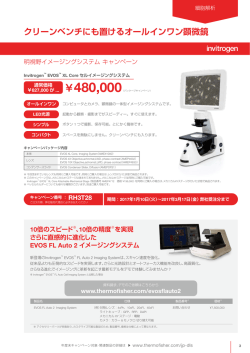
© Copyright 2026 Paperzz