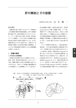
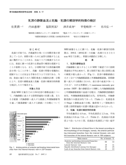
第回臨床解剖研究会記録 ..~ 肝臓の静脈供給における副肝静脈の意義 ―比較解剖の立場で― 宮 木 孝 昌 東京医科大学人体構造学講座・愛知医科大学解剖学講座・横浜市立大学医学部組織学講座 肝臓の静脈には,肝静脈 3 種類(右肝静脈,中肝 は尾端)で下大静脈に合流している.尾状葉には尾状 静脈,左肝静脈)のほかに,しばしば副肝静脈が現れ 突起と乳頭突起の 2 つがあり,両突起の位置は異な る.肝静脈 3 種類と副肝静脈の違いの 1 つは,下大 る.ヒトの尾状葉は乳頭突起のことで,その静脈は細 静脈への合流部である.肝静脈 3 種類は肝臓後面の くて,副肝静脈には含めていない.ヒト以外の哺乳類 大静脈溝の上端で下大静脈に合流しており,副肝静脈 では,尾状突起のほうが発達している. は大静脈溝の中間あるいは下端で下大静脈に合流して ヒトの副肝静脈に相当する静脈は,調査した哺乳類 いる.肝内静脈分布をみると,右肝静脈は右葉,中肝 では,独立した肝葉の尾状葉(尾状突起)肝静脈また 静脈は尾状葉と方形葉,左肝静脈は左葉をおもな領域 としている.副肝静脈の領域はおもに右葉(後区)の 一部であり,門脈右枝の一部の枝の領域になる. 副肝静脈の名称Elias and Sherrick (1969)によれ ば,下大静脈の中間部あるいは下端部に直接合する肝 静脈には,上・下尾状葉肝静脈,中間・下後肝静脈, 後外側肝静脈がある2).これらの肝静脈の領域は尾状 葉(乳頭突起)または右葉の一部である.独立した肝 葉には分布していない. すでに調査した哺乳類 12 科の動物(ウサギ類のナ キウサギ,メキシコウサギ,カイウサギ,偶蹄類のヤ ギ,ウシ,ブタ,奇蹄類のウマ,食肉類のツキノワグ マ,アシカ,アザラシ,ネコ,有袋類のカンガルー, 食虫類のスンクス,げっ歯類のラット)では,肝静脈 は大静脈溝の上端で下大静脈に合流するものと,大静 脈溝の中間あるいは下端で合流するものが存在してい る4~12).前者は,ヒトと同様に,3 種類の肝静脈(右 肝静脈,中肝静脈,左肝静脈)であり,後者は最右肝 静脈または尾状葉 (尾状突起) 肝静脈である.例えば, ナキウサギでは,門脈右枝が外側右葉の全域に分布し て,そこから流出する静脈を集める最右肝静脈(別 名,外側右葉肝静脈)が大静脈溝の下端(後端あるい 64 臨床解剖研究会記録 No. 14 2014. 2 Fig. 1 The posterior view of the human liver. Two large accessory hepatic veins (1, 1) empty the distal portion of the inferior vena cava (VC) in the groove for vena cava. C: caudate lobe, D: right lobe, P: portal vein, S: left lobe 脈から肝輸入静脈の門脈と輸出静脈の肝静脈が発生す る1).肝輸出静脈の肝静脈は,右,中,左肝静脈とな り,下大静脈の大静脈溝の上端部に合流する.下大静 脈の中間部または下端部に合する静脈は,尾状葉(乳 頭突起)肝静脈や細い静脈として残るが,時に右葉の 後部に広がる比較的大きな肝静脈として現れる.これ が副肝静脈である.ヒト以外の哺乳類の発生では,こ の下大静脈の部に直接合する静脈が見られる3).この 静脈が哺乳類の尾状葉(尾状突起)または外側右葉の 肝静脈として存続するものと推測される. Upper: the posterior view of a cast of the liver in a laboratory animal of musk shrew, Suncus. Lower: the visceral aspect of a cast of the liver in the cattle fetus, Bos taurus (Miyaki et al 2006). Fig. 2 Photographs of two casts of the venous and portal systems of the mammal liver. The color (blue) indicates more clearly the distribution of the hepatic venous pattern. DV: ductus venosus, P: portal vein, U: umbilical vein, V: hepatic vein, VC: vena cava はそれより広い流出領域を持つ最右肝静脈に相当する. 文 献 1) Corliss CE. 1976. Patten's Human Embryology, Elements of Clinical Development, McGraw-Hill, New York, pp403, 404 2) Elias H, Sherrick J G. 1969. Morphology of the Liver, Academic Press, New York, pp270, 271, 281 3) Hochstetter F. 1894. Entwicklung des Venensystems der Wirbeltiere. Anat Hefte 3: 460489 4) 宮木孝昌,坂井建雄.2001.ナキウサギ,メキシコウサギお よびカイウサギの肝臓の血管分布.形態科学 5: 1319 5 ) 宮木孝昌,坂井建雄. 2002 .ツキノワグマの肝臓の血管分 布.形態科学 6: 18 6 ) 宮木孝昌ほか. 2004a .ヤギの肝臓の門脈分布と肝静脈分 布.形態科学 7: 57 63 7 ) 宮木孝昌ほか. 2004b .ウマの肝臓の門脈分布と肝静脈分 布.形態科学 7: 65 71 8 ) 宮木孝昌ほか. 2005a .アシカ,アザラシ,カンガルーの肝 臓の門脈分布と肝静脈分布.形態科学 8: 93 99 9 ) 宮木孝昌ほか. 2005b.ブタとネコの肝臓の門脈分布と肝静 脈分布.形態科学 9: 1722 10 ) 宮木孝昌ほか. 2006a .ウシの肝臓の門脈分布と肝静脈分 布.形態科学 9: 67 72 11 ) 宮木孝昌ほか. 2006b .ラットの肝臓の門脈分布と肝静脈分 布.形態科学 10: 27 31 12) 宮木孝昌ほか.2009.スンクスの肝臓の血管分布.形態科学 13: 4952 副肝静脈の発生肝臓の血管の由来は,臍腸間膜静 Signiˆcance of the accessory hepatic vein in the venous drainage of the liver from the viewpoint of comparative anatomy Takayoshi MIYAKI Department of Anatomy, Tokyo Medical University, Aichi Medical University and Yokohama City University Graduate School of Medicine The large accessory hepatic vein often appears with existing three main hepatic veins in the human liver. The three main hepatic veins join the proximal portion of the inferior vena cava in the groove for the vena cava, but the large accessory hepatic vein empties the distal or intermediate portion of the inferior vena cava. In mammals of twelve families, the hepatic vein emptying the distal or intermediate portion of the inferior vena cava occupies one hepatic lobe. The large accessory hepatic vein appearing in the human corresponds to the hepatic vein of the lateral right lobe or the caudate lobe hepatic vein in the liver of the mammal. Key words: accessory hepatic vein, hepatic vein, liver, human, mammals 肝臓の静脈供給における副肝静脈の意義 65
© Copyright 2026 Paperzz