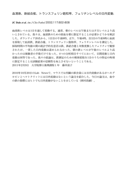
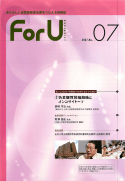
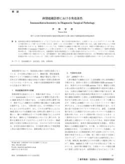
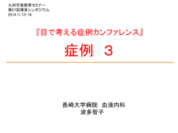
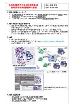
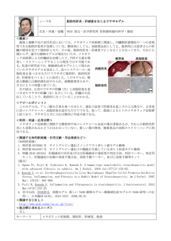
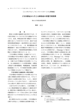
ただいま、ページを読み込み中です。5秒以上、このメッセージが表示されている場
合、Adobe® Reader®(もしくはAcrobat®)のAcrobat® JavaScriptを有効にしてください。
日皮会誌:112( 6 )
,837―843,2002(平14)
Adobe® Reader®のメニュー:「編集」→「環境設定」→「JavaScript」で設定できます。
「Acrobat JavaScriptを使用」にチェックを入れてください。
Angiomyofibroblastoma of the Vulva
なお、Adobe® Reader®以外でのPDFビューアで閲覧されている場合もこのメッセージが表示さ
れます。Adobe® Reader®で閲覧するようにしてください。
守屋美佳子
古田 淳一
梅林 芳弘
要
旨
腫瘤を認めた.腫瘤は,MRI の T1 強調画像では筋と
44 歳女性.右大陰唇皮下の 5×3cm 大の腫瘤を主訴
同程度の信号(図 2a)
,T2 強調画像では高信号(図 2
に受診.MRI で,大陰唇皮下に境界明瞭な腫瘤を認め,
b)
で, 造影剤により増強された. 周囲組織への浸潤,
T1 強調画像では筋と同程度の信号,T2 強調画像では
腟との連続性はなかった.
高信号であった.組織学的に,腫瘍は細胞成分が粗で
病理組織学的所見:腫瘍の境界は比較的明瞭であっ
粘液腫様の部分と,卵円型及び紡錘型の細胞が密に増
た.腫瘍は,細胞成分が粗であり粘液腫様の部分(図
殖している部分より構成されていた.また壁の薄い血
3)と,卵円型及び紡錘型の細胞が密に増殖している部
管が増生している像を認めた.免疫組織化学的に,腫
分(図 4a,b)より構成されていた.また壁の薄い血管
瘍 細 胞 は vimentin,desmin 陽 性,CD34,S-100,α-
が増生している像が認められた(図 5).周囲には軽度
smooth muscle actin,myoglobin は 陰 性,progester-
の炎症細胞の浸潤がみられた.alcian blue,PAS は陰性
one receptor は 70% の細胞で陽性,estrogen receptor
であった.
は 20% の細胞で陽性であった.臨床像,画像所見およ
免疫組織化学的所見:腫瘍細胞は,vimentin と des-
び 組 織 学 的 所 見 よ り angiomyofibroblastoma of the
min は陽性.UEA-I,第 VIII 因子関連抗原,CD34,
S-
vulva と診断した.
100,
α-smooth muscle actin,myoglobin は陰性であっ
た.progesterone receptor は 70% の細胞で陽性,es緒
言
trogen receptor は 20% の細胞で陽性を示した.
Angiomyofibroblastoma は女性の外陰部に好発する
治療および経過:臨床像,画像所見および組織学的
良性腫瘍である.局所浸潤性,再発性の強い aggres-
所 見 よ り angiomyofibroblastoma of the vulva と 診 断
sive angiomyxoma とは好発部位をともにするので鑑
した.治療は単純切除を行ったが,画像所見を参照し
別が重要であるが,本邦皮膚科領域よりの報告は多く
つつ腫瘍に切り込まないよう注意しながら摘出した.
1)
2)
ない
.われわれは最近本腫瘍の 1 例を経験したの
で,それを報告するとともに,aggressive angiomyxo-
現在術後 19 カ月経過しているが臨床所見および画像
上再発をみない.
ma との鑑別点について考察する.
症
例
患者:44 歳女性.
初診:2000 年 5 月 1 日.
家族歴・既往歴:特記すべきことなし.
現病歴:2 年前に右大陰唇皮下の腫瘤に気づいた.
放置していたものの徐々に増大してきたため,当院婦
人科を受診し,当科を紹介された.
現症:右大陰唇皮下に 5×3cm 大,弾性軟,境界比較
的明瞭,可動性良好な腫瘤を認めた.被覆表皮には著
変なし(図 1)
.
画像所見:MRI および X 線 CT で,大陰唇皮下に,
5.5×2.3×4.5cm 大のおたまじゃくし型で境界明瞭な
日立総合病院皮膚科(主任 梅林芳弘主任医長)
平成13年10月22日受付,平成14年 1 月10日掲載決定
別刷請求先:(〒317―0077)茨城県日立市城南町 2―
1―1 日立総合病院皮膚科 守屋美佳子
図1
手術時の臨床像
838
守屋美佳子ほか
a
b
図2
考
MRI 像:矢印で腫瘍を示す(a : T1 強調画像,b : T2 強調画像)
按
Aggressive angiomyxoma は , 1983 年に Steeper
3)
blastoma として分離した.その好発部位はやはり女性
の外陰部で,Fletcher らの原著では,angiomyofibro-
と Rosai により提唱された主として女性の会陰,骨盤
blastoma of the vulva として報告されている.なお,両
腔に生じる腫瘍で,緩徐に増殖し局所浸潤,局所再発
腫瘍ともに男性の鼠径部や陰!から発生したとの報告
率の高いことを特徴とす る.こ れ に 対 し 1992 年 に
がその後出た5)∼7)が,angiomyofibroblastoma の男性発
4)
Fletcher ら は,aggressive angiomyxoma と組織は類
生例は,angiomyofibroblastoma-like tumor と表現され
似するものの局所再発のない腫瘍を angiomyofibro-
ている7).
angiomyofibroblastoma
図3
839
組織像:細胞成分の疎な部分(HE 染色,×40)
Fletcher らによると,angiomyofibroblastoma は単
blastoma は desmin 陽性 , aggressive angiomyxoma
純切除でもまず再発しないが,aggressive angiomyxo-
は desmin 陰性で,両者の鑑別に有用とされているが,
ma は 70% 以上の症例で 2 年以内に再発するという.
その後の検討では desmin の染色態度に差はないとさ
従って両者の鑑別が重要になってくるが,鑑別のポイ
れている11)16).
ントは以下の通りである .まず臨床的に,angiomy-
一 方,angiomyofibroblastoma に お け る α-smooth
ofibroblastoma は aggressive angiomyxoma よ り 表 在
muscle actin の陽性例は 14%(6!
43),CD34 の陽性例
性で小型であり,多くは 5cm 未満である.組織学的に
は 15%(4!
27)と 低 い の に 対 し,aggressive angio-
4)
は angiomyofibroblastoma は境界明瞭であり,壁の薄
myxoma で は α-smooth muscle actin の 陽 性 例 88%
い血管が多数増生する.腫瘍細胞は卵円型に近く細胞
(36!
41),CD34 の陽性例 56%(10!
18)と,いずれも比
密度の高い部分と低い部分が混在するのが特徴とされ
較的染まる症例が多く差が見られるようである2).と
2)
る .これに対 し aggressive angiomyxoma は よ り 浸
はいえ,これらの染色性には症例によるバリエーショ
潤性であり,壁が肥厚した血管や硝子化を伴った血管,
ンがあり,免疫組織化学的に両者を鑑別することは困
異常な筋性動脈が増生する.腫瘍細胞は紡錘型から星
難とも思われる.中には両者の中間的な特徴を有する
芒状である.自験例は最大径が 5cm と両者の境界では
も の11)16)17)や,悪 性 化 を 思 わ せ る angiomyofibroblas-
あるが,病理組織学的に angiomyofibroblastoma の特
toma(angiomyofibrosarcoma とされている18))の報告
徴を有していた.なお,文献的には 12cm 大までの an-
もあることから,両者は同一疾患単位で,発生から診
giomyofibroblastoma が報告されており4),平均 4.5cm
断されるまでの期間や病変の大きさによって病理所見
8)
大とされる .
が異なるのではないかとの疑問も提出されている17).
免疫組織学的に両者を鑑別する試みはこれまでに
もし二つの疾患が同一スペクトラム上に配置しうるも
種々2)4)9)∼15)行われてきている.森本ら2)による報告例の
のであるなら,免疫組織化学的に両者を鑑別しようと
集計によれば,両者ともに vimentin は 100% 陽性,
いう試みは,免疫組織化学的に良悪性を判断しようと
desmin も 86% 以上の症例で陽性となっている.S-100
いう企てに等しくなり,かかる方法が他の腫瘍で確立
はい ず れ も 100% 陰 性 で あ る.従 っ て,vimentin,
していない以上,これらの腫瘍の鑑別において免疫組
desmin,S-100 の染色態度では両者の鑑別は不可能で
織化学の有用性が乏しいのも首肯できることになる.
ある.なお,Fletcher らの原著では,angiomyofibro-
Angiomyofibroblastoma という名は,本腫瘍を構成
840
守屋美佳子ほか
a
b
図4
組織像:細胞成分の密な部分(HE 染色,a:×40,
b:×200)
している血管成分と間質成分に由来する4).そして後
者は myofibroblast とみ な さ れ て い る 訳 で あ る が,
subtype のものが増殖しているということになる.
Aggressive angiomyxoma では,estrogen receptor,
Scalli ら20)によれば myofibroblast は免疫組織化学的に
progesterone receptor が 高 率 に 陽 性 と な る と さ れ
4 つの subtype に分けうるという.すなわち,! vi-
る21).一 方,angiomyofibroblastoma に お い て estro-
mentin のみを発現するもの," vimentin,desmin,α-
gen receptor,progesterone receptor の染色を検討し
smooth muscle actin のすべてを発現するもの,# vi-
ている報告は多くないが,Laskin ら10)によれば,estro-
mentin と α-smooth muscle actin を発現するもの,$
gen receptor は 6 例中 6 例,progesterone receptor
vimentin と desmin を発現するもの,である.自験例を
は 6 例中 5 例で陽性であったという.この所見から,
含む angiomyofibroblastoma の多くは,この 4 番目の
これらの腫瘍が女性の外陰部に存在する estrogen re-
angiomyofibroblastoma
図5
841
組織像:血管の増生(HE 染色,×40)
ceptor,progesterone receptor 陽性の特殊な間質細胞
の大きさの血管と紡錘型細胞の密な増殖が特徴であ
に由来し,性ホルモンに反応して増殖する可能性が示
る.細胞成分に富み粘液腫様の部分はない.superficial
唆されている21)22).
angiomyxoma は,体のどこにでも発生しうるポリー
両腫瘍で MRI を施行した報告も多くない.angio-
プ状の腫瘍である.組織学的には,真皮から皮下にか
myofibroblastoma の 1 例 で T1 強 調 画 像 で 筋 と 同 信
けて存在する多房性の粘液腫で血管の増生を欠く.自
号,T2 強調画像で高信号であったと報告されてお
験例においてはいずれも臨床像と組織所見から鑑別で
2)
り ,自験例の所見と一致する.一方,T1 強調画像で
きた.
低信号であったとする報告もある23).aggressive an-
angiomyofibroblastoma の 患 者 は そ の 好 発 部 位 か
giomyxoma では T1 強調画像で低信号,T2 強調画像
ら,まず婦人科を受診し,そこで治療されているのか
では高信号24),あるいは低信号領域と高信号領域が混
もしれない.しかし,皮膚外科をやっていると自験例
在して認められた25)26)とされている.これらの報告を
のように婦人科から紹介されることもありうるので,
見る限り,MRI の所見上,両者に明確な差はないよう
皮膚科医としては,本腫瘍を aggressive angiomyxo-
にも思われる.
ma をはじめとする鑑別疾患とともに知っておく必要
なお,女性外陰部に発生する軟部組織腫瘍としては,
があると考え,報告した.なお,本腫瘍の治療は単純
aggressive angiomyxoma 以 外 に も cellular angiofi-
切除で十分であるが,術前に MRI などの画像診断によ
broma と superficial angiomyxoma が挙 げ ら れ る27).
り腫瘍の拡がりを評価しておくことは重要と思われ
cellular angiofibroma は女性外陰部に好発する通常 3
る.
cm 以下の腫瘍で,硝子化を伴う壁の肥厚した中程度
文
1)磯田美登里,藤原 恵:Angiomyofibroblastoma,
西日皮膚,59 : 235―237, 1997.
2)森本謙一,古谷喜義,坪井賢朗,西阪 隆:Angiomyofibroblastoma of the vulva,西日皮膚,61 :
481―483, 1999.
3)Steeper TA, Rosai J : Aggressive angiomyxoma of
献
the female pelvis and perineum . Report of nine
cases of a distinctive type of gynecologic softtissue neoplasm , Am J Surg Pathol, 7 : 463 ― 475,
1983.
4)Fletcher CDM, Tsang WYW, Fisher C, Lee KC,
Chan JKC : Angiomyofibroblastoma of the vulva.
842
守屋美佳子ほか
A benign neoplasm distinct from aggressive angiomyxoma, Am J Surg Pathol , 16 : 373―382, 1992.
5) Tsang WYW, Chan JKC , Lee KC , Fisher C ,
Fletcher CD : Aggressive angiomyxoma. A report
of four cases occurring in men, Am J Surg Pathol ,
16 : 1059―1065, 1992.
6)Iezzoni JC, Fecher RE, Wong LS, Rosai J : Aggressive angiomyxoma in males . A report of four
cases, Am J Clin Pathol, 104 : 391―396, 1995.
7)Laskin WB, Fetsch JF, Mostofi FK : Angiomyofibroblastoma like tumor of the male genital tract.
Analysis of 11 cases with comparison to female
angiomyofibroblastoma and spindle cell lipoma ,
Am J Surg Patho, 22 : 6―16, 1998.
8)Nielsen GP, Young RH : Mesenchymal tumors and
tumor-like lesions of female genital tract. A selective review with emphasis on recently described
entities, Int J Gynecol Pathol , 20 : 105―127, 2001.
9)Fetsch JF, Laskin WB, Lefkowitz M, Kindblom
LG, Meis-Kindblom JM : Aggressive angiomyxoma . A clinicopathologic study of 29 female patients, Cancer, 78 : 79―90, 1996.
10)Laskin WB, Fetsch JF, Tavassoli FA : Angiomyofibroblastoma of the female genital tract. Analysis of 17 cases including a lipomatous variant ,
Hum Pathol , 28 : 1046―1055, 1997.
11)Granter SR, Nucci MR, Fletcher CDM : Aggressive angiomyxoma. Reappraisal of its relationship
to angiomyofibroblastoma in a series of 16 cases,
Histopathology, 30 : 3―10, 1997.
12)Silverman JS, Albukerk J, Tamsen A : Comparison of angiomyofibroblastoma and aggressive angiomyxoma in both sexes. Four cases composed
of bimodal CD 34 and factor XIIIa positive dendritic cell subsets, Pathol Res Pract , 193 : 673―682,
1997.
13)Ockner DM, Sayadi H, Swanson PE, Ritter JH ,
Wick MR : Genital angiomyofibroblastoma. Comparison with aggressive angiomyxoma and other
myxoid neoplasms of skin and soft tissue, Am J
Clin Pathol, 107 : 36―44, 1997.
14) 新宅雅幸 : Aggressive angiomyxoma と Angiomyofibroblastoma―病理組織学的検討―,癌の臨
床,43 : 523―529, 1997.
15)Bigotti G, Coli A, Gasbarri A, Castagnola D, Ma-
donna V , Bartolazzi A : Angiomyofibroblastoma
and aggressive angiomyxoma. Two benign mesenchymal neoplasms of the female genital tract.
An immunohistochemical study, Pathol Res Pract ,
195 : 39―44, 1999.
16)福永真治:主に成人に発生する良性筋線維芽細胞
性腫瘍,病理と臨床,18 : 102―109, 2000.
17)Takeshima Y, Shinkoh Y, Inai K : Angiomyofibroblastoma of the vulva. A mitotically active variant? Pathol Int, 48 : 292―296, 1998.
18)沼本 敏,宮内洋一,西 京子ほか:外陰部に発生
した angiomyofibroblastoma の一例,病理と臨床,
18 : 477―481, 2000.
19)Nielsen GP, Young RH, Dickersin GR, Rosenberg
AE : Angiomyofibroblastoma of the vulva with
sarcomatous transformation(“angiomyofibrosarcoma”
),Am J Surg Pathol , 21 : 1104―1108, 1997.
20)Skalli O, Schurch W, Seemayer T, et al : Myofibroblasts from diverse pathologic settings are heterogeneous in their content of actin isoforms and intermediate filament proteins, Lab Invest , 60 : 275―
285, 1989.
21)McCluggage WG, Patterson A, Maxwell P : Aggressive angiomyxoma of pelvic parts exhibits
estrogen and progesterone receptor positivity, J
Clin Pathol, 53 : 603―605, 2000.
22)McCluggage WG, White RG : Angiomyofibroblastoma of the vulva, J Clin Pathol , 53 : 803―806, 2000.
23) Mortele KJ , Lauwers GJ , Mergo PJ , Ros PR :
Perineal angiomyofibroblastoma, CT and MR findings with pathologic correlation, J Comput Assist
Tomogr, 23 : 687―689, 1999.
24)森田哲郎,樋口健史,酒井邦夫ほか:Aggressive
angiomyxoma の 1 例.MRI および CT 所見を中
心として,臨放線,41 : 475―478, 1996.
25)Kobayashi Y, Minami M, Ohtomo K, Matsuoka Y :
MR imaging and CT appearances of aggressive
angiomyxoma, AJR, 169 : 1752―1753, 1997.
26)一柳暢孝,山田拓己,鎌田成芳ほか:陰!内 Aggressive angiomyxoma の 1 例,泌尿紀要,45 : 69―
72, 1999.
27)Nucci MR, Fletcher : Vulvovaginal soft tissue tumours. update and review, Histopathology, 36 : 97―
108, 2000.
angiomyofibroblastoma
843
Angiomyofibroblastoma of the Vulva
Mikako Moriya, Junichi Furuta and Yoshihiro Umebayashi
Department of Dermatology, Hitachi General Hospital, Ibaraki, Japan
(Received October 22, 2001 ; accepted for publication January 10, 2002)
A 44-year-old woman developed an asymptomatic subcutaneous tumor measuring 3×5 cm in size on her
right labium majorum. CT and MRI showed a well-circumscribed lesion. On T1-weighted MR images, the tumor was isointense in relation to muscle, and, on T2-weighted images, it showed hyperintensity. Histopathologically, the tumor was composed of a hypocellular area in loosely edematous stroma and a hypercellular
area. Spindle― or oval― shaped tumor cells were arranged with numerous thin-walled vessels. Immunohistochemical examination revealed that the tumor cells were positive for vimentin and desmin and negative for
CD34, S-100, α-smooth muscle actin, and myoglobin. A part of the tumor expressed estrogen receptors and
progesterone receptors. We diagnosed the tumor as an angiomyofibroblastoma of the vulva.
(Jpn J Dermatol 112 : 837∼843, 2002)
Key words : angiomyofibroblastoma, aggressive angiomyxoma, vulva
© Copyright 2026 Paperzz