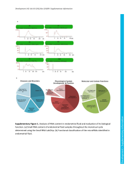
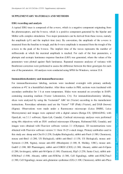
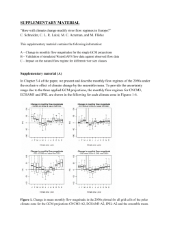
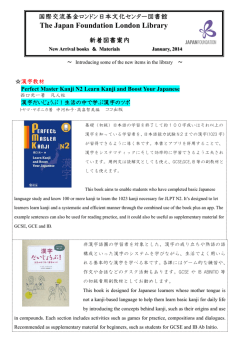
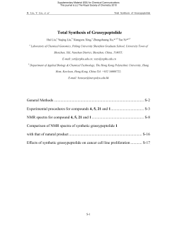
Development 143: doi:10.1242/dev.125989: Supplementary information
Supplementary Figure S1. Diagram of neurogenic regions of a 3-day pluteus larva
discussed in this work. Left, a view from the ventral (oral) side, with the APD at top. Right,
a lateral view. SynB neurons (yellow) differentiate in or adjacent to the APD (green) and
ciliary band (magenta), while serotonergic neurons (dark blue) are restricted to the apical
Development • Supplementary information
plate. Two additional neurons differentiated in the pharyngeal endoderm (brown).
Development 143: doi:10.1242/dev.125989: Supplementary information
SoxC protein and Z167 or Brn1/2/4 knockdown did not reduce SoxC expression. (A and
B) SoxC antibody immunostained (green) control (A) and SoxC morpholino-injected (B)
embryos. SoxC protein signal was eliminated in the morphant. Embryos were at 52 hours.
SoxC antibody courtesy of Dr. Robert Burke. (C and D) Control embryo (C) and Z167
morphant hybridized with probes for SoxC (green) and Z167 (red) transcripts. Z167
knockdown does not affect SoxC expression, but Z167 transcripts are down regulated,
indicating its probable negative autoregulation. Control (E) and Brn1/2/4 knockdown
embryos (F) hybridized in situ for SoxC (red) and SynB (green). Brn1/2/4 knockdown does
not affect expression of SoxC, but prevents formation of SynB-positive neurons. Embryos
were at 46 hours and are shown in lateral view with the APD at top. Nuclei were stained with
DAPI (blue). The white bar in A represents 20 micrometers.
Development • Supplementary information
Supplementary Figure S2. SoxC morpholino knockdown prevented accumulation of
Development 143: doi:10.1242/dev.125989: Supplementary information
Supplementary Figure S3. SoxC mRNA accumulates in two phases. (A) Temporal
expression pattern from a high-resolution transcription analysis (Materna et al., 2010). (B)
Development • Supplementary information
Temporal expression pattern from a microarray experiment (Wei et al., 2006).
Supplementary Figure S4. Further analysis of the Brn1/2/4 mis/overexpression
phenotype. (A and B) Control (A) and Brn1/2/4-overexpressing embryos (B)
immunostained for Hnf6 (cillary band and APD, green) and Spec1 (aboral ectoderm, red). (C
and D) Control (C) and Brn1/2/4-overexpressing (D) embryos immunostained for GSC (oral
ectoderm, red) and Endo1 (midgut and hindgut, green). (A”-D”) are the corresponding DIC
images and DAPI fluorescence (blue) is shown in (A-D). (E, F and G), Control (E) and
Brn1/2/4-overexpressing (F and G) embryos immunostained for SynB (red) and Nk2.1
(green), a transcription factor operating in a gene regulatory pathway separate from that for
serotonergic neurons, which leads to production of the ciliated apical tuft (Dunn et al., 2007).
SynB staining encompasses most of the ectoderm and Nk2.1 staining is radialized, lacking
dorsal-ventral polarity. Nuclei are stained with DAPI (blue). E and F are lateral views and G
is toward the APD. The white bar in A represents 20 micrometers.
Development • Supplementary information
Development 143: doi:10.1242/dev.125989: Supplementary information
© Copyright 2026 Paperzz