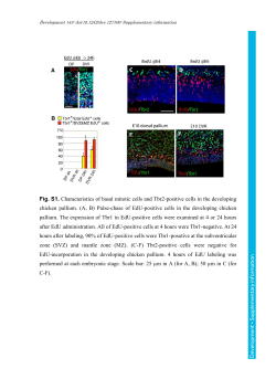
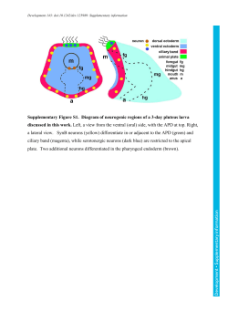
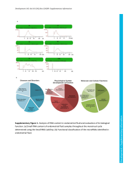
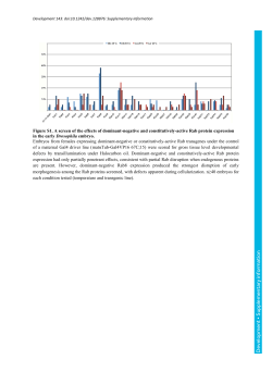
Development 142: doi:10.1242/dev.119446: Supplementary Material Fig. S1. Smad4 is not essential for the activation of nodal signaling. RT-qPCR analysis of the mRNA levels of the indicated genes in Smad4-mutant and control testes. The mean value of control testes was set as 1. Con, control; cKO, conditional knockout. Data show the mean ± sd. **P < 0.01. Statistical significance was assessed by Student’s t-test. Development | Supplementary Material Development 142: doi:10.1242/dev.119446: Supplementary Material Fig. S2. SMAD2, but not SMAD3, is required for the activation of nodal signaling. (A, B) RT-qPCR analysis of the mRNA levels of indicated genes in Smad2- (A) and Smad3 (B) -mutant and control testes. The mean value of control testes was set as 1. Data show the mean ± sd. **P < 0.01. Statistical significance was assessed by Student’s t-test. Development | Supplementary Material Development 142: doi:10.1242/dev.119446: Supplementary Material Fig. S3. Smad2 signaling is not involved in pluripotency gene expression. (A) RT-qPCR analysis of the mRNA levels of pluripotency genes in Smad2-mutant (Rosa) and control testes at E13.5. The mean value of control testes was set as 1. Data show the mean ± sd. **P < 0.01. Statistical significance was assessed by Student’s t-test. (B) Representative images of control and the Smad2-mutant (Rosa) testis sections stained for SOX2 and UTF1 at E13.5. The areas outlined in white are shown at higher magnification to the right. Scale bars: 50 m. Development | Supplementary Material Development 142: doi:10.1242/dev.119446: Supplementary Material Fig. S4. Deletion of Smad2 signaling does not affect sexual fate of somatic cells. (A) Representative images of control and the Smad2-mutant testis sections stained for SOX9 (red) and TRA98 (green) at E13.5. The areas outlined in white are shown at higher magnification to the right. (B-C) RT-qPCR analyses of the mRNA levels of male-specific (Sox9) and female-specific (Foxl2 and Bmp2) genes in Smad2-mutant and control gonads at E13.5. Con, control; cKO, conditional knockout. The mean value of control testes (B) or ovaries (C) was set as 1. Data show the mean ± sd. Scale bars: 50 m. Development | Supplementary Material Development 142: doi:10.1242/dev.119446: Supplementary Material Fig. S5. SMAD2 was deleted via germ cell specific recombination (related to Fig. 3). (A) Representative images of indicated testis sections stained for GFP and TRA98. The areas outlined in white are shown at higher magnification to the right. (B) An example showing that successful recognition of GFP-positive cells (yellow circle) by Development | Supplementary Material Development 142: doi:10.1242/dev.119446: Supplementary Material TissueQuest at E13.5. (C-D) Scatter plot of cells recognized by TissueQuest based on DAPI signals in the indicated testes sections. X-axis indicates value of GFP signal in cells and Y-axis indicates value of pSMAD2 in cells. Numbers presents percentage of indicated cell population among total cells. (E) Representative images of the indicated testis sections stained for pSMAD2 and GFP in indicated testes at E13.5. White arrowheads represent GFP/pSMAD2-double positive cells in control testes. Yellow arrowheads and blue arrowheads represent GFP-positive pSMAD2-negative and GPF/pSMAD2-double positive cells in mutant testes, respectively. Scale bars: 50 m. Development | Supplementary Material Development 142: doi:10.1242/dev.119446: Supplementary Material Fig. S6. The phenotype of Smad2-mutant mimics Nanos2 knockout mice. (A-B) Representative images of the Nanos2+/- or Nanos2-/- testis sections stained for SCP3/TRA98 (A) and DMC1/TRA98 (B) at E17.5. (C-D) Representative images of the Smad2-cKO testis sections stained for SCP3/TRA98 at E16.5 and DMC1/TRA98 at E17.5. The number indicates the percentage of SCP3/TRA98 or DMC1/TRA98 double positive cell in TRA98 positive cells. Scale bars: 50 m. Development | Supplementary Material Development 142: doi:10.1242/dev.119446: Supplementary Material Fig. S7 Meiotic entry of germ cells in Smad2flox/flox/Rosa-CreERT testes. (A-B) Representative images of control and Smad2-mutant testis sections stained for DMC1/TRA98 (A) and Cleaved Caspase-3/TRA98 (B). The numbers represent percentages of DMC1-positive germ cells. The areas outlined in white are shown at higher magnification to the right. White arrows indicate Cleaved Caspase3/TRA98-positive cells. (C) Percentage of Cleaved Caspase-3/TRA98 double positive cells. cKO, conditional knockout. Data show the mean ± sd. Statistical significance was assessed by Student’s t-test. Scale bars: 50 m. Development | Supplementary Material Development 142: doi:10.1242/dev.119446: Supplementary Material Fig. S8. Germ cell-specific expression of OTX2 in fetal testes. (A) Whole mount in situ hybridization with an Otx2 probe for E13.5 testis and ovary. (B) Indicated gonad sections were subjected to double immunostaining for OTX2 (green) and TRA98 (red). (C) RT-qPCR analysis of Otx1 and Otx3 mRNA levels in the E13.5 testes of Otx2-mutant mice. The mean value of control testes was set as 1. Data show the mean ± sd. Statistical significance was assessed by Student’s t-test. Scale bars: 50 m. Development | Supplementary Material Development 142: doi:10.1242/dev.119446: Supplementary Material Fig. S9. Expression patterns of pp38 in fetal testes. Double immunostaining images for pp38 (red) and MVH (green) of male gonads at E11.5, E12.5 and E13.5. Arrows represent somatic cells. Scale bar: 50 m. Development | Supplementary Material Development 142: doi:10.1242/dev.119446: Supplementary Material Fig. S10. Disruption of male differentiation after suppressing p38 signaling. Representative images of the indicated testis sections stained for NANOS2/TRA98 and DNMT3L/TRA98 (Related to Fig. 6E). Scale bars: 50 m. Development | Supplementary Material Development 142: doi:10.1242/dev.119446: Supplementary Material Table S1. Primer sets used for RT-qPCR Nanos2, forward (5′–3′) ACAGCAGTCAGCAGTCTC Nanos2, reverse (5′–3′) CCGAGAAGTCATCACCAG Nodal, forward (5′–3′) AGCCAAGAAGAGGATCTGGTATGG Nodal, reverse (5′–3′) GACCTGAGAAGGAATGACGGTGAA Lefty 1, forward (5′–3′) AGTCCTGGACAAGGCTGATGTG Lefty 1,reverse (5′–3′) CGAACACTAGCAGGTGAGTGGA Lefty 2, forward (5′–3′) ATCGACTCTAGGCTCGTGTCCATC Lefty 2,reverse (5′–3′) CACAATTGCCTTGAGCTCCGTAGTC Otx1, forward (5′–3′) AGGGCGGAAGCTATGGTCAGGGATAC Otx1, reverse (5′–3′) CGGGCTCCTTGTAATCCAAGCAATCGG Otx2, forward (5′–3′) TATGGACTTGCTGCATCCCTCCGTGGGCTA Otx2, and reverse (5′–3′) TGGCAGGCCTCACTTTGTTCTGACCTCCAT Otx3, forward (5′–3′) AACAACCTGATGCACTACTCGTCTT Otx3, reverse (5′–3′) CAATGCTCGTGGTTTTACTGTTCAGG Mvh, forward (5′–3′) GTTGAAGTATCTGGACATGATGCAC Mvh, reverse (5′–3′) CGAGTTGGTGCTACAATAATACACTC G3pdh, forward (5′–3′) ACCACAGTCCATGCCATCAC G3pdh,reverse(5–3′) TCCACCACCCTGTTGCTGTA Stra8, forward (5′–3′) CCTAAGGAAGGCAGTTTACTCCCAGTC Stra8, reverse (5′–3′) GCAGGTTGAAGGATGCTTTGAGC Uft1, forward (5′–3′) ATGTCCCGGTGACTACGTCTGATG Uft1,reverse (5′–3′) AGTCTCGGAGTTTGTCCTTGAGGAA Sox2, forward (5′–3′) GCGGAGTGGAAACTTTTGTCC Sox2,reverse (5′–3′) CGGGAAGCGTGTACTTATCCTT Oct4, forward (5′–3′) TCACCTTGGGGTACACCCAG Oct4,reverse (5′–3′) CATGTTCTTAAGGCTGAGCTGC Nanog, forward (5′–3′) CCGCTTGCACTTCATCCTTTG Nanog, reverse (5′–3′) CCTCAGCCTCCAGCAGATGC Cyp26b1, forward (5′–3′) TGGACTGTGTCATCAAGGAGGT Cyp26b1, reverse (5′–3′) GTCGTGAGTGTCTCGGATGCTA Sox9, forward AAGACCACCCCGATTACAAGTACCA (5′–3′) Sox9, reverse (5′–3′) TCAGATCAACTTTGCCAGCTTGCAC Bmp2, forward (5′–3′) GATACAGGAAGCTTTGGGAAACAGTAG Development | Supplementary Material Development 142: doi:10.1242/dev.119446: Supplementary Material Bmp2, reverse (5′–3′) CTGTGTTCATCTTGGTGCAAAGACC Foxl2, forward (5′–3′) GCCTCAACGAGTGCTTCATCAAGGT Foxl2, reverse (5′–3′) AGTTGTTGAGGAACCCCGATTGCAG Smad4, forward (5′–3′) CCTGTTGTGACTGTGGATGGCTATG Smad4,reverse (5′–3′) AGACCTTTATATACGCGCTTGGGTAGA Development | Supplementary Material
© Copyright 2026 Paperzz